predicts acute myeloid leukemia outcome
Houda Alachkar, … , Clara D. Bloomfield, Guido Marcucci
J Clin Invest. 2014;
124(4)
:1512-1524.
https://doi.org/10.1172/JCI70921
.
Aberrant expression of the secreted protein, acidic, cysteine-rich (osteonectin) (SPARC)
gene, which encodes a matricellular protein that participates in normal tissue remodeling, is
associated with a variety of diseases including cancer, but the contribution of SPARC to
malignant growth remains controversial. We previously reported that SPARC was among
the most upregulated genes in cytogenetically normal acute myeloid leukemia (CN-AML)
patients with gene-expression profiles predictive of unfavorable outcome, such as mutations
in isocitrate dehydrogenase 2 (IDH2-R172) and overexpression of the oncogenes brain and
acute leukemia, cytoplasmic (BAALC) and v-ets erythroblastosis virus E26 oncogene
homolog (ERG). In contrast, SPARC was downregulated in CN-AML patients harboring
mutations in nucleophosmin (NPM1) that are associated with favorable prognosis. Based
on these observations, we hypothesized that SPARC expression is clinically relevant in
AML. Here, we found that SPARC overexpression is associated with adverse outcome in
CN-AML patients and promotes aggressive leukemia growth in murine models of AML. In
leukemia cells, SPARC expression was mediated by the SP1/NF-
k
B transactivation
complex. Furthermore, secreted SPARC activated the integrin-linked kinase/AKT (ILK/AKT)
pathway, likely via integrin interaction, and subsequent
b
-catenin signaling, which is
involved in leukemia cell self-renewal. Pharmacologic inhibition of the SP1/NF-
k
B complex
resulted in SPARC downregulation and leukemia growth inhibition. Together, our data
indicate that evaluation of SPARC expression has prognosticative value and SPARC is a
potential therapeutic target […]
Research Article
Oncology
Find the latest version:
SPARC promotes leukemic cell growth and
predicts acute myeloid leukemia outcome
Houda Alachkar,1,2 Ramasamy Santhanam,1,2 Kati Maharry,2,3 Klaus H. Metzeler,2 Xiaomeng Huang,1,2 Jessica Kohlschmidt,2,3 Jason H. Mendler,1,2 Juliana M. Benito,4 Christopher Hickey,1,2 Paolo Neviani,2,5 Adrienne M. Dorrance,1,2 Mirela Anghelina,1,2 Jihane Khalife,1,2 Somayeh S. Tarighat,1,2 Stefano Volinia,5 Susan P. Whitman,2,5 Peter Paschka,2
Pia Hoellerbauer,1,2 Yue-Zhong Wu,1,2 Lina Han,4 Brad N. Bolon,6 William Blum,1,2
Krzysztof Mrózek,2 Andrew J. Carroll,7 Danilo Perrotti,2,5 Michael Andreeff,4 Michael A. Caligiuri,1,2 Marina Konopleva,4 Ramiro Garzon,1,2 Clara D. Bloomfield,1,2 and Guido Marcucci1,2,5
1Division of Hematology, Department of Medicine, and 2Comprehensive Cancer Center, The Ohio State University (OSU), Columbus, Ohio, USA. 3Alliance for Clinical Trials in Oncology Statistics and Data Center, Mayo Clinic, Rochester, Minnesota, USA. 4Department of Leukemia,
The University of Texas MD Anderson Cancer Center, Houston, Texas, USA. 5Department of Molecular Virology, Immunology and Cancer Genetics,
and 6Comparative Pathology and Mouse Phenotyping Shared Resource, Comprehensive Cancer Center, OSU, Columbus, Ohio, USA. 7Department of Genetics, University of Alabama at Birmingham, Birmingham, Alabama, USA.
Aberrant expression of the secreted protein, acidic, cysteine-rich (osteonectin) (SPARC) gene, which encodes
a matricellular protein that participates in normal tissue remodeling, is associated with a variety of diseases
including cancer, but the contribution of SPARC to malignant growth remains controversial. We previously
reported that SPARC was among the most upregulated genes in cytogenetically normal acute myeloid
leuke-mia (CN-AML) patients with gene-expression profiles predictive of unfavorable outcome, such as mutations
in isocitrate dehydrogenase 2 (IDH2-R172) and overexpression of the oncogenes brain and acute leukemia,
cytoplasmic (BAALC) and v-ets erythroblastosis virus E26 oncogene homolog (ERG). In contrast, SPARC was
downregulated in CN-AML patients harboring mutations in nucleophosmin (NPM1) that are associated with
favorable prognosis. Based on these observations, we hypothesized that SPARC expression is clinically relevant
in AML. Here, we found that SPARC overexpression is associated with adverse outcome in CN-AML patients
and promotes aggressive leukemia growth in murine models of AML. In leukemia cells, SPARC expression was
mediated by the SP1/NF-
κ
B transactivation complex. Furthermore, secreted SPARC activated the
integrin-linked kinase/AKT (ILK/AKT) pathway, likely via integrin interaction, and subsequent
β
-catenin signaling,
which is involved in leukemia cell self-renewal. Pharmacologic inhibition of the SP1/NF-
κ
B complex resulted
in SPARC downregulation and leukemia growth inhibition. Together, our data indicate that evaluation of
SPARC expression has prognosticative value and SPARC is a potential therapeutic target for AML.
Introduction
The secreted protein, acidic, cysteine-rich (SPARC) gene, located at chromosome bands 5q31–q33 (1), is highly conserved in verte-brates and encodes a 32-kDa matricellular protein also known as osteonectin or BM-40 (2, 3) that regulates cell-matrix interactions and tissue remodeling through the binding of collagen and other extracellular matrix proteins and activation of matrix metallopro-teinases (2). SPARC also interacts with and regulates several growth factors, including TGF-β, FGF, VEGF, and PDGF (4–7). This pleiotropic activity suggests that the SPARC protein is likely to be expressed when tissue undergoes normal or pathologic remodeling.
SPARC expression has been shown to be altered in a variety of human conditions (e.g., diabetes, obesity) and in several types of cancer. However, the biologic and clinical significance of this gene is controversial, and conflicting reports have classified SPARC as either a tumor suppressor gene or an oncogene. The difficulty in assigning a specific function to the SPARC protein is related to the diverse roles that it can play both intracellularly in
malig-nant cells and extracellularly in the surrounding microenviron-ment (8). Low expression levels of SPARC were found in ovarian (9), colorectal (10, 11), and pancreatic cancer (12), whereas high
SPARC expression was reported in breast cancer (13, 14), mela-noma (15, 16), and glioblastoma (17). Stromal SPARC expression was associated with poor prognosis in non–small cell lung can-cer (18) and with disease recurrence in breast ductal carcinoma in situ (19), whereas low stromal expression of SPARC predicted poor prognosis in colon cancer (20).
In hematologic malignancies, the role of SPARC is equally controversial. It was found to be downregulated at diagnosis in patients with del(5q) myelodysplastic syndromes (MDS) and upregulated following treatment with lenalidomide (21–23).
SPARC was also found to be downregulated in acute myeloid leu-kemia (AML) with MLL rearrangements, usually associated with unfavorable prognosis, and upregulated in AML with t(8;21) or inv (16), which is usually associated with favorable prognosis, although no correlation of SPARC expression with outcome was reported (24). In chronic myelogenous leukemia, the accumula-tion of intracellular SPARC mediated by the Fyn/ERK signaling pathway seemingly contributed to imatinib resistance (25).
Recently, we observed that SPARC was upregulated in gene expression profiles (GEPs) associated with prognostically
unfa-Authorship note: Clara D. Bloomfield and Guido Marcucci are co–senior authors and contributed equally to this work.
Conflict of interest: The authors have declared that no conflict of interest exists.
vorable gene mutations (i.e., those in isocitrate dehydrogenase 2 [IDH2-R172]; ref. 26) or overexpressed genes (i.e., brain and acute leukemia, cytoplasmic [BAALC] and v-ets erythroblastosis virus E26 oncogene homolog [ERG]; ref. 27), and downregulated in GEP associated with the prognostically favorable nucleophosmin (NPM1) mutation (28) in cytogenetically normal AML (CN-AML). This led us to hypothesize that SPARC overexpression contributes to a more aggressive phenotype in AML. Thus, we dissected the clinical significance of SPARC overexpression in AML, the mecha-nisms by which this gene is deregulated, and the downstream effects of this aberrantly expressed gene. We show that SPARC
overexpression independently predicts adverse outcome in CN-AML patients, thus representing what we believe to be a novel prognostic marker in AML. Consistent with these findings, we also demonstrate that SPARC contributes to leukemia growth in vitro and aggressive disease in vivo. SPARC overexpression activates integrin-linked kinase/AKT (ILK/AKT) and in turn β-catenin and could be targeted by modulating the SP1/NF-κB/miR-29b network, thereby also representing a potential therapeutic target in AML.
Results
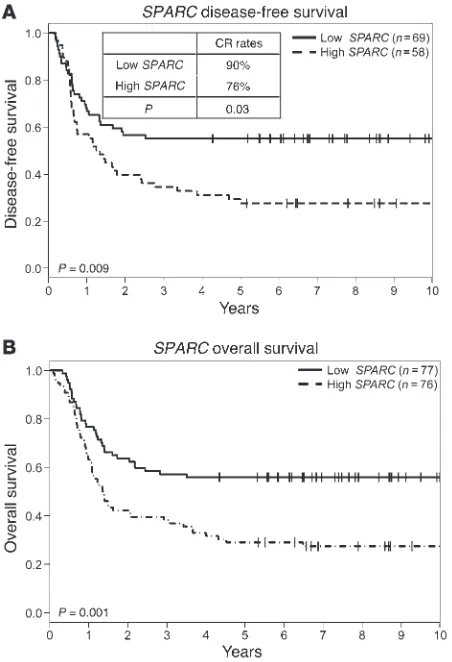
SPARC overexpression is associated with adverse clinical outcome in CN-AML. SPARC expression was analyzed by nCounter assays (NanoString Technologies) (29) in 153 younger (age range, 18–59 years) adults with primary CN-AML treated with cytarabine- daunorubicin–based regimens; clinical and molecular character-istics are shown in Supplemental Table 1 (supplemental mate-rial available online with this article; doi:10.1172/JCI70921DS1).
Patients were dichotomized into higher and lower SPARC express-ers using the median value cut-off. With a median follow-up of 8.7 years, higher SPARC expressers had lower odds of achieving a complete remission (CR) (P = 0.03) and shorter disease-free survival (DFS) (P = 0.009; 5-year DFS 28% vs. 55%) and overall survival (OS) (P = 0.001; 5-year OS 29% vs. 56%) than lower expressers (Figure 1, A and B). In multivariable analyses, higher SPARC expression was independently associated with lower odds of CR (P = 0.007), once adjusting for white blood count (WBC) (P = 0.003), and shorter OS (P = 0.03), once adjusting for FLT3 internal tandem duplication (FLT3-ITD) (P < 0.001), WT1 (P = 0.003), and RUNX1 (P = 0.006) mutations and WBC (P < 0.001). There was also a trend for shorter DFS (P = 0.08) once adjusting for FLT3-ITD (P < 0.001) and WT1
mutation (P = 0.004). These data support the notion that SPARC
is differentially expressed across AML patients and that the differ-ence in the SPARC expression in this patient population has poten-tially biologic and clinical relevance.
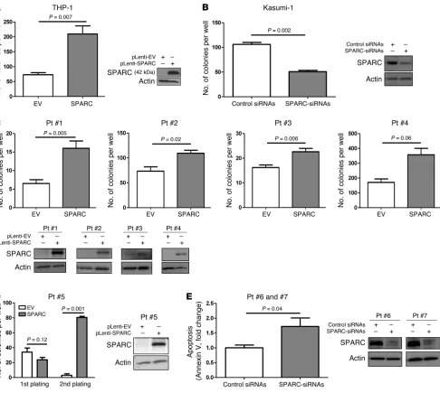
SPARC promotes leukemia growth in vitro and an aggressive disease in vivo. Having shown that higher SPARC expression was associated with adverse outcome, next we interrogated the SPARC -depen-dent mechanisms mediating the aggressive phenotype. THP-1 cells express a relatively lower endogenous level of SPARC mRNA (Supplemental Figure 1) and therefore represent a suitable in vitro model for gain-of-function experiments. THP-1 cells were stably infected with pLenti-SPARC (THP-1/SPARC) or empty vector as a control (THP-1/EV). Ectopic expression of SPARC was confirmed by Western blotting (Figure 2A). A significant increase in the ability of colony formation was observed in THP-1/SPARC cells compared with the THP-1/EV cells (mean, 209 vs. 72; P = 0.007; Figure 2A). Likewise, when we knocked down SPARC (Figure 2B) in Kasumi-1 cells that express a relatively higher endogenous lev-els of the gene (Supplemental Figure 1), we observed a decrease in the colony-forming ability in SPARC siRNA–transfected cells compared with the control siRNA–transfected cells (mean, 106 vs. 50; P = 0.002; Figure 2B). Similar experiments were also conducted in primary blasts from 5 AML patients (numbers 1–5) express-ing a relatively lower level of endogenous SPARC and 2 patients (numbers 6, 7) expressing a relatively higher level of endogenous
SPARC (see patients’ cytogenetic and molecular features in Supple-mental Table 2). For patients 1–5; blasts were infected with either the pLenti-SPARC or the pLenti-EV vector. A significant increase in colony forming ability was observed in SPARC-infected blasts compared with EV controls for patients 1–4 (means, 16 vs. 7;
P = 0.005; 109 vs. 73; P = 0.02; 23 vs. 16; P = 0.006 and 357 vs. 170;
P = 0.06) (Figure 2C). In blasts from a patient 5, ectopic SPARC expression did not significantly increase the colony-forming abil-ity after first plating, but a significant increase in the number of colonies was observed after the secondary plating (mean, 80 vs. 3;
[image:3.585.53.279.83.414.2]P = 0.001; Figure 2D). For patients 6 and 7, we observed an increase in spontaneous apoptosis in AML blasts when SPARC endogenous expression was knocked down by siRNAs (Figure 2E), suggesting that SPARC promotes both growth and survival of leukemia cells.
Figure 1
SPARC overexpression is associated with adverse clinical outcome in CN-AML. Outcome of CN-AML patients (n = 153) according to SPARC
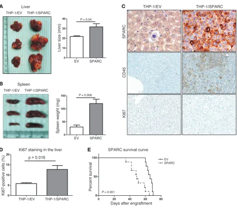
To examine the contribution of SPARC to leukemia growth in vivo, NOD/SCID/gamma (NGS) mice were injected with THP-1/ SPARC cells or THP-1/EV cells via tail vein. Eight weeks later, 3 mice in each group were sacrificed. Bone marrow engraftment of the leukemia cells was confirmed by CD45 antibody staining. The THP-1/SPARC mice had significantly larger livers (P = 0.04; Figure 3A) and spleens (P = 0.008; Figure 3B) than THP-1/EV con-trols. THP-1/SPARC cells formed coalescing masses obliterating large expanses of the liver parenchyma (Supplemental Figure 2), while THP-1/EV cells were observed either alone or in small clus-ters within hepatic sinusoids without effacing the hepatic cords.
[image:4.585.53.539.81.516.2]The THP-1/SPARC cells had a monomorphic phenotype and a seemingly higher proliferative capacity compared with the more pleomorphic character and increased apoptotic tendency of the THP-1/EV cells (see immunohistochemistry (IHC) staining for SPARC, CD45, and Ki67 in Figure 3C and quantification of Ki67 in Figure 3D). Histopathology of spleen sections showed increased infiltration of leukemic cells and destruction of splenic architec-ture in tissues obtained from THP-1/SPARC mice compared with tissues obtained from THP-1/EV mice (Supplemental Figure 2). The THP-1/SPARC mice (n = 9) also had a significantly shorter survival than the THP-1/EV mice (n = 9) (2 × 10⁶ cells were injected
Figure 2
per mouse; median survival, 49 vs. 66 days; P = 0.001; Figure 3E). Similar results were obtained in a repeated experiment where a higher number of cells (5 × 10⁶) were injected into each mouse
(Supplemental Figure 3). Consistent with these results, mice inject-ed with the SPARC-shRNA–infectinject-ed CG-SH cells, which other-wise express high levels of endogenous SPARC, showed a trend for smaller spleens than mice injected with control shRNA–infected cells (Supplemental Figure 4).
SPARC activates ILK/AKT/β-catenin signaling pathways. Next, we asked how SPARC contributes to the mechanisms of leukemia growth. Several cell-surface receptors have been identified as interacting with the secreted SPARC protein, including some of the membrane integrins (30, 31). This interaction activates ILK, a multifunctional cytoplasmic serine/threonine kinase (32–34) that phosphorylates and activates AKT and phosphorylates and
inactivates glycogen synthase kinase 3β (GSK3β; Ser9). GSK3β
promotes β-catenin degradation, but once phosphorylated, its activity is suppressed, and β-catenin stabilizes and translocates into the nucleus (35). Activated AKT phosphorylates and stabi-lizes β-catenin at residue Ser552 (36). Thus, the net result of the SPARC/integrin/ILK interplay is enhanced β-catenin. The latter is required for self-renewal of leukemia stem cells (LSCs) (37–41). Thus, we hypothesized that SPARC overexpression leads to aber-rant β-catenin activation in AML.
SPARC is a secreted protein (2, 3). We found significantly higher levels of SPARC in the media collected from THP-1/SPARC cell cultures than in the media collected from THP-1/EV cell cultures (Figure 4A). We incubated THP-1 cells with conditioned media from THP-1/SPARC cells and THP-1/EV cells and performed colony assay 48 hours after incubation. We observed higher
num-Figure 3
[image:5.585.51.515.77.485.2]ber of colonies in THP-1/SPARC media incubated THP-1 cells compared with THP-1/EV media incubated cells (Supplemental Figure 5), suggesting that the conditioned media from SPARC transfected cells induce cell growth in SPARC-nonexpressing cells. Coimmunoprecipitation assays with SPARC and surface inte-grins showed binding of the protein at least to inteinte-grins αv, β3,
and α5 (Figure 4B) and lower or no binding to integrins β1, α4,
and β4 (data not shown). Kinase activity assay of anti-ILK
immu-noprecipitate from THP-1/SPARC cells incubated with recombi-nant GSK3 showed increased P-GSK3β(Ser9) (mean 2.4 ± 0.68 of 3 different experiments) as compared with the immunoprecipitate from THP-1/EV cells, thereby reflecting higher ILK activity in the presence of SPARC (Figure 4C). Furthermore, increased endoge-nous P-AKT(Ser473) and P-GSK3β(Ser9) were detected in THP-1/ SPARC cells compared with THP-1/EV cells (Figure 4D, lines 1 and 2). Knocking down ILK (Figure 4D, lines 3 and 4) decreased AKT and GSK3β phosphorylation in THP-1/SPARC cells. Consistently, functional inhibition of ILK activity using the T315 (ILK inhibi-tor) (42) abolished SPARC-induced cell growth as assessed by MTS and colony-forming assay (Supplemental Figure 6).
ILK activates AKT through PI3K activation (43), suggesting that pharmacologic blockade of PI3K signaling should result in abrogat-ing the SPARC effect on AKT and GSK3β phosphorylation. Indeed, THP-1/SPARC cells treated with 50 nM PI3K inhibitor LY294002 showed decreased AKT(Ser473) and P-GSK3β(Ser9) compared with untreated THP-1/SPARC cells (Supplemental Figure 7).
Next, we tested whether blocking integrin receptors would decrease SPARC function. GSK3β phosphorylation in THP-1/ SPARC cells was reduced in THP-1/SPARC cells incubated with antibodies against αv, β3, and β1 (Figure 4E); indeed, blocking αvβ3
was sufficient to decrease SPARC-dependent GSK3β phosphoryla-tion (Supplemental Figure 8). Consistent with these findings, we observed higher levels of P–β-catenin(Ser552) (Figure 4F) and an
increased β-catenin nuclear translocation (Figure 4G) in THP-1/ SPARC compared with THP-1/EV cells.
Once in the nucleus, β-catenin interacts with TCF/LEF tran-scription factors and promotes expression of genes supporting cell growth and proliferation (e.g., MYC and CCND1). Ectopic SPARC
expression resulted in an approximately 4-fold increase in TCF/LEF reporter activity, as measured by luciferase assay in SPARC-trans-fected 293T cells compared with EV-transSPARC-trans-fected controls (P < 0.001; Figure 4H). The SPARC-induced TCF/LEF activity decreased upon ILK siRNA knockdown (Figure 4I). SPARC protein levels in 293T cells assessed by Western blot are shown in Supplemental Figure 9. Increase in SPARC-induced β-catenin–TCF/LEF activity was also supported by upregulation of the TCF/LEF target gene MYC (Fig-ure 4J). We also observed consistent results in other cell lines (e.g., MV4-11 cells) forced to express SPARC (Supplemental Figure 10).
Finally, we validated our findings in blasts from AML patients. Forced expression of SPARC increased P–β-catenin (Ser552) (Fig-ure 5A; patient 2), while siRNA SPARC knockdown decreased P–β-catenin(Ser552) (Figure 5B; patient 6) and total β-catenin (Figure 5C; patient 8). SPARC overexpression and knockdown in primary blasts resulted, respectively, in significant increase and decrease of MYC mRNA expression (Figure 5, D and E). To ensure that increased TCF/LEF target gene expression was not due to other leukemogenic mechanisms operative in AML cells, we also showed that forced SPARC could increase MYC and CCND1
expression in CD34+ cells from cord blood cells of normal donors
(Supplemental Figure 11).
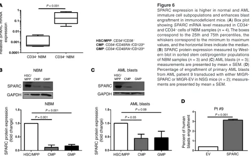
It has been reported that β-catenin is expressed and activated in LSCs and that it is required for self-renewal (37, 38). Thus, we postulated that SPARC expression may be higher in less differenti-ated hematopoietic cell subpopulations. To prove this, we showed that SPARC expression was significantly higher in the CD34+
compared with CD34– cells in normal bone marrow cells (NBM)
(n = 4; P < 0.001; Figure 6A), and in CD34+/CD38–
HSCs/multipo-tent progenitors (HSC/MPP) compared with CD34+/CD38+ cells
(common-myeloid progenitors [CMP] and granulocyte-monocyte progenitors [GMP]) both in NBM (n = 3) and AML blasts (n = 3; patients n. 1,5 and 9) (Figure 6, B and C). Furthermore, we showed that SPARC-transduced primary AML blasts (patient 9) indeed exhibited a higher percentage of engraftment in NSG mice than those transduced with GFP/EV control (3.1% vs 0.4% circulating blasts 12 weeks after blast i.v. injection; Figure 6D).
SP1, NF-κB, and miR-29b modulate SPARC expression in AML. Next, we asked which mechanisms lead to aberrant SPARC expression in AML. We identified binding sites for SP1, NF-κB, and RUNX1 in the (–651/1) promoter region (Transcriptional Regulatory Ele-ment Database [TRED] website; http://rulai.cshl.edu/cgi-bin/ TRED/tred.cgi?process=home) and binding sites for miR-29 in the 3′ UTR region of the human SPARC gene. Indeed, SPARC has been reported to be a miR-29 target (44). Notably, we have recently shown that activation and overexpression of oncogenes involved in AML (i.e., KIT, FLT3, and DNMTs) require interaction and trans-activation of the transcription factors SP1 and NF-κB and inhibi-tion of miR-29b (45). Therefore, we postulated that a similar mech-anism could be responsible for SPARC overexpression.
To test this hypothesis, we first performed luciferase gene report-er assays by cloning the SPARC promoter region spanning the SP1-, NF-κB–, and RUNX1-binding sites into the pGL4.11 luciferase vector. Then, 293T cells were cotransfected with the reporter gene and vectors expressing SP1 and p65 (NF-κB subunit). A
signifi-Figure 4
SPARC activates ILK/AKT/β-catenin signaling pathways in THP-1 cells and enhances β-catenin nuclear translocation and transcrip-tional activity. THP-1 cells were transfected with EV or pLenti-SPARC vectors. (A) Western blot measuring secreted SPARC protein levels in media collected from cell culture of THP-1 cells infected with pLenti-SPARC vector compared with cells infected with pLenti-EV. (B) SPARC and integrin coimmunoprecipitation assay. (C) ILK kinase activity assay. Lysates from THP-1/SPARC cells and THP-1/EV cells were immunoprecipitated with anti-ILK antibody and assayed utilizing GSK3 fusion protein (ILK substrate). Numbers represent ratio of band intensity to control band. (D) Western blot analysis of P-AKT(Ser473) and P-GSK3β(Ser9) levels in THP-1/SPARC cells compared with THP-1/ EV in the presence of endogenous ILK or following ILK knockdown or (E) following blocking integrin receptors with integrin antibodies. Lanes separated by a line were run on the same gel but were noncontigu-ous. (F) Western blot analysis of P–β-catenin (Ser552) in THP-1/SPARC cells compared with THP-1/EV cells. (G) Immunofluorescence confocal microscopy images showing β-catenin (green) and cell nuclei (blue) in EV- or SPARC-transfected THP-1 cells stained with β-catenin antibody and Draq5 antibody (nuclear staining); arrows indicate cells exhibiting
cant increase in luciferase activity was found in cells transfected with SP1- or p65-expressing vectors compared with EV controls (P < 0.001 for both; Figure 7A). Ectopic expression of SP1 and p65 also resulted in significant increases in endogenous SPARC
expression in THP-1 cells (Figure 7, B and C), whereas SP1 and p65 siRNA knockdown resulted in significant decrease of SPARC
expression in Kasumi-1 cells (Figure 7, D and E).
A significant decrease of SPARC promoter luciferase activity was found in 293T cells transfected with RUNX1 expression vector (P < 0.001; Figure 7F). In contrast, RUNX1-WT knockdown resulted in SPARC upregulation in MV4-11 cells (Figure 7G), while knock-down of RUNX1-RUNX1T1 translocation by siRNAs resulted in a significant SPARC downregulation in RUNX1-RUNX1T1–positive Kasumi-1 cells (Figure 7H).
Notably, ectopic expression of p65 and SP1 increased their respective enrichment on the SPARC promoter (Figure 7, I and J), and forced RUNX1-WT expression decreased SP1, but not p65, enrichment as measured by ChIP in THP-1 cells (Figure 7K).
These results are also consistent with the finding that RUNX1 -mutated CN-AML patients exhibit higher SPARC expression when compared with the RUNX1-WT patients (Supplemental Fig-ure 12). Together, these results indicate that SP1, p65 and RUNX1
loss-of function promoted SPARC expression, while RUNX1-WT inhibited SPARC expression.
As SPARC was found to be a target for miR-29a and miR-29c (44) and the SPARC 3′ UTR region was predicted to have binding sites for miR-29b (46, 47), we cloned the SPARC 3′ UTR region in lucif-erase reporter vector (pGL3) and cotransfected this construct with the synthetic microRNA (miR-29b and miR-9*) or scramble oligos in 293T cells. We found a 60% decrease of SPARC 3′ UTR–lucifer-ase activity in cells cotransfected with miR-29b compared with the activity in cells cotransfected with scramble (P < 0.001) or an unre-lated microRNA: miR-9* (Figure 8A). Kasumi-1 cells transfected with miR-29b showed significant decreases in endogenous SPARC
[image:8.585.43.338.81.516.2](Figure 8B). AML blasts transfected with miR-29b also showed sig-nificant decreases in SPARC levels at 24 hours (Figure 8C).
Figure 5
We have previously shown that miR-29b is downregulated in AML via a SP1/NF-κB/HDAC inhibitory complex and that pharmaco-logic disruption of this complex results in miR-29b upregulation and downregulation of direct (i.e., SP1, DNMT3A, DNMT3B) and indirect (i.e., DNMT1, FLT3, KIT) miR-29b targets (45, 48, 49). Consistent with these reports, we showed that bortezomib at con-centrations achievable in vivo (45, 50) significantly increased miR-29b expression (Figure 8D) and in turn decreased SP1 and SPARC
mRNA and protein levels in Kasumi-1 cells (Figure 8, E and F) and in blasts from 3 different patients with primary AML (numbers 6, 10, and 7) (Figure 8, G and H). Consistent with the in vitro results, we found SPARC expression was significantly reduced in blood and spleen from CG-SH cell–engrafted NSG mice treated with bortezomib (1 mg/kg) twice a week for 4 weeks compared with vehicle-treated mice (Supplemental Figure 13).
Discussion
Although altered SPARC expression has been observed in cancer, the clinical significance of both upregulation and downregula-tion of the gene and the encoded protein remain to be fully eluci-dated (15, 17, 51–58). In contrast with solid tumors, the number of reports investigating this gene in hematologic malignancies is relatively small. In MDS, 5q deletion results in allelic loss and significant downregulation of SPARC expression in the progeni-tor compartment (22, 23), and a dramatic upregulation of SPARC
occurs in response to lenalidomide (23). Although this suggests that SPARC expression is a treatment-response predictor and per-haps plays a role in normal hematopoiesis, to date, no biological evidence supporting this notion has been reported. Indeed, SPARC was found to be dispensable for murine hematopoiesis (59).
Nev-ertheless, the complexity of the hematopoietic role of SPARC is illustrated by a recent report (60). These authors showed in a myeloproliferative murine model that increased SPARC expression in the bone marrow stroma favored fibrotic changes, while loss of
SPARC expression resulted in a defective stromal niche (60). Here, we sought to dissect the role of SPARC in AML, starting from the observation that this gene was found overexpressed in GEPs associated with distinct molecular subsets of patients har-boring prognostically unfavorable mutations or overexpressed genes. Supporting the hypothesis that SPARC overexpression likely contributes to aggressive myeloid leukemogenesis, we showed that high SPARC expression was associated with adverse outcomes in CN-AML patients. Although SPARC overexpression was associ-ated with other unfavorable molecular markers at diagnosis, in multivariable models for outcome, the impact of SPARC remained significant even after adjusting for other molecular prognostica-tors, supporting an independent role of this gene in determin-ing an aggressive clinical phenotype. To our knowledge, this is the first study reporting the clinical relevance of SPARC expres-sion in AML. We validated these findings through mechanistic experiments using AML preclinical models and primary blasts. We showed that SPARC upregulation promoted an aggressive phenotype in AML cells. Mice engrafted with AML cells forced to express SPARC had increased leukemia burden and shorter sur-vival compared with controls.
[image:9.585.49.548.78.392.2]The mechanisms leading to SPARC upregulation in AML blasts are multifactorial and likely converge to NF-κB–dependent path-ways, which have been previously shown to be constitutively acti-vated in AML LSCs (61). We found that SPARC may function through an autocrine mechanism; once secreted by AML cells,
Figure 6
SPARC expression is higher in normal and AML immature cell subpopulations and enhances blast engraft ment in immunodeficient mice. (A) Box plot showing SPARC mRNA level measured in CD34+
and CD34– cells of NBM samples (n = 4). The boxes
the protein interacted with the leukemia cell membrane integrins and activated ILK/AKT/β-catenin signaling, thereby promoting cell engraftment, growth, and survival. Pharmacologic disruption of the SP1/NF-κB transactivation complex (45, 62) resulted in SPARC downregulation, thereby representing a potentially novel therapeutic strategy for SPARC-dependent AML.
[image:10.585.50.536.79.527.2]A finding in our study was that SPARC enhanced β-catenin activity in AML cells, known to be required for leukemia growth and stemness. The increase in β-catenin activity was seemingly enhanced via the SPARC-integrin-ILK axis. ILK is a kinase that links the cell adhesion receptors, integrins, and growth factors with the downstream signaling pathways involving AKT and
Figure 7
SP1, NF-κB, and miR-29b modulate SPARC expression in AML. (A) Luciferase activities measured in 293T cells cotransfected with PGL4.11 luciferase vector containing the SPARC promoter region (PGL4.11/SPARC promoter) and either SP1-expressing (left panel) or p65-expressing (right panel) vectors; measurements were normalized to that obtained in 293T cells cotransfected with PGL4.11/SPARC-promoter vector or EV. (B) Western blot analysis for SPARC protein expression measured in THP-1 cells transfected with SP1-expressing vector or (C) p65-expressing vector. (D) SPARC mRNA expression measured in Kasumi-1 cells transfected with SP1 siRNAs or (E) p65 siRNAs. (F) Luciferase activities measured in 293T cells cotransfected with a PGL4.11 luciferase vector containing the SPARC promoter region (PGL4.11/SPARC-promoter) and a RUNX1-expressing vector; measurements were normalized to that in 293T cells cotransfected with PGL4.11/SPARC-promoter vector and EV. (G) Western blot analysis showing SPARC protein levels in MV4-11 cells transfected with RUNX1 siRNAs or (H) Kasumi-1 cells transfected with RUNX1-RUNX1T1 siRNA. (I) ChIP showed p65 enrichment on SPARC promoter in THP-1 cells transfected with p65-expressing vector. (J) ChIP showed SP1 enrichment on SPARC promoter in THP-1 cells transfected with SP1-expressing vector. (K) Change in SP1 enrichment on SPARC
oncogenes, constitutive SPARC expression was dependent on a previously reported regulatory molecular network that involves SP1/NF-κB and miR-29b (45). The leukemogenic role of this net-work that can be targeted pharmacologically has been extensively studied by our group not only in AML (45) but also in other types of leukemia (45, 62). Our data support the view that SPARC over-expression is a potentially actionable therapeutic target via phar-macological disruption of the SP1/NF-κB/miR-29b network by, for example, bortezomib, used here as a proof of concept. Other compounds, including inhibitors of HDAC (e.g., vorinostat, AR-42) or SP1 (e.g., mithramycin A) or synthetic miR-29b mim-ics may also interfere with the SP1/NF-κB/miR-29b network and downregulate SPARC (45, 50, 65, 66).
GSK3β (43). This ultimately results in stabilization and nuclear localization of β-catenin and activation of genes promoting cell-cycle progression and proliferation (63). Consistent with these results, we observed that SPARC levels were higher in both normal and immature AML cell subpopulations that indeed displayed a higher engraftment potential in immunodeficient mice. While SPARC/ILK/AKT signaling was found involved in glioma cell invasion and survival (64), to our knowledge, the relevance of this mechanism in AML has not been previously reported.
[image:11.585.49.521.76.510.2]Having observed that SPARC overexpression contributes to more aggressive phenotype in AML patients, we asked whether it is possible to target SPARC expression and demonstrate the clini-cal relevance of our findings. We showed that, similarly to other
Figure 8
and 5 μl propidium iodide (PI) (eBioscience). After 20 minutes incubation, fluorescence was quantified by flow cytometry on a FACSCalibur instrument.
Immunofluorescent staining, cell sorting, flow cytometry and confocal microscopy. Cells were washed and stained with CD34, CD38, CD45RA, and CD123 antibodies (eBioscience) and sorted into hematopoietic stem cells and HSC/ MPP (CD34+/CD38–), CMP (CD34+/CD45RA–/CD123lo) and GMP (CD34+/ CD45RA+/CD123lo) using BD FACSAriaII cell sorter (BD Biosciences).
For confocal microscopy analysis, 1 × 10⁶ cells were processed as detailed in Supplemental Experimental Procedures.
In vivo experiments. Four- to six-week-old NOD/SCID mice (The Jackson Laboratory) were i.v. injected via tail vein with 5 × 10⁶ THP-1 cells infected with plenti-EV or plenti-SPARC. Eight weeks later, mice (n = 6) were euthanized; spleens, livers, and sternums were isolated. Xenograft transplantations were performed as detailed in Supplemental Experimental Procedures.
Patients. We studied pretreatment bone marrow and blood samples with 20% or more blasts from 153 patients with primary CN-AML (age range, 18–59 years) who were treated with cytarabine-daunorubicin–based regi-mens on Cancer and Leukemia Group B (CALGB) 9621 or 19808 protocols (71). Per protocol, no patient included in our analysis received allogeneic transplantation in first CR. The median follow-up was 8.7 years.
Cytogenetic and mutational analyses. The diagnosis of CN-AML was based on the analysis of 20 or more metaphases in pretreatment bone marrow speci-mens subjected to short-term cultures and confirmed by central karyotype review (72). Patients were also characterized centrally for FLT3-ITD (73), FLT3
tyrosine kinase domain (TKD) mutations (74), MLL partial tandem duplica-tion (75, 76), NPM1 (28, 77), WT1 (78), CEBPA (79), IDH1, and IDH2 (26) TET2
(80), ASXL1 (81), and DNMT3A (71) mutations as previously reported.
Statistics. Statistical analyses relative to clinical outcome were performed by the Alliance Statistics and Data Center. Definitions of clinical end points — CR, DFS, and OS — are as reported previously (26). The differences in base-line clinical and molecular features between higher and lower SPARC express-ers were tested using the Fisher’s exact and Wilcoxon rank sum tests for cat-egorical and continuous variables, respectively. Estimated probabilities of DFS and OS were calculated using the Kaplan-Meier method, and the log-rank test evaluated differences between survival distributions. Mechanistic and biological experiments were analyzed with paired and unpaired 2-tailed
t tests as required, and a P value of less than 0.05 was considered significant. Data are presented as mean ± SEM in all figures where error bars are shown.
Study approval. Informed consent to use patient samples for investiga-tional studies was obtained from each patient according to OSU institu-tional guidelines and in accordance with the Declaration of Helsinki, and protocols were approved by the IRB at OSU and each center involved in the CALGB trials. All animal studies were performed in accordance with OSU institutional guidelines for animal care and under protocols approved by the OSU Institutional Animal Care and Use Committee.
Acknowledgments
This work was supported in part by grants from the National Can-cer Institute (Bethesda, Maryland, USA) (CA101140, CA016058, CA77658, CA55164, CA100632, and CA140158), the Coleman Leukemia Research Foundation, and the Conquer Cancer Foun-dation (to J.H. Mendler).
Received for publication May 14, 2013, and accepted in revised form January 2, 2014.
Address correspondence to: Guido Marcucci, The Ohio State Univer-sity Comprehensive Cancer Center, Biomedical Research Tower, 460 W. 12th Ave, Columbus, Ohio 43210, USA. Phone: 614.366.2261; Fax: 614.688.4028; E-mail: guido.marcucci@osumc.edu.
While our findings emphasize a role of SPARC in the homeo-stasis of myeloblasts, several other reports have underscored the interplay of SPARC protein with the microenvironment to cre-ate a niche favorable for cancer growth (8). Thus, future stud-ies of SPARC in AML need to address the dual intracellular and extracellular role of this protein.
In conclusion, SPARC deregulation is clinically relevant in AML because overexpression of this gene independently predicts adverse outcome in subsets of AML patients, contributes to aggressive AML growth likely via β-catenin activation, and is an actionable therapeutic target. This suggests that future clinical studies target-ing the activity of SPARC protein are warranted in AML.
Methods
Plasmids and reagents. SPARC clone was purchased from Invitrogen and inserted in pLenti6.2/V5-DEST (Invitrogen) and MIGR (Addgene). pCMV-p65, pSUPER-SP1, and pCMV-RUNX1 expression vectors were also used (45). OFF-TARGET control and ON-TARGET plus siRNA-SMARTpool reagents against SPARC, SP1, p65, and RUNX1 (each contains a mix of at least 4 different sequences) were purchased from Dharmacon. siRNA against RUNX1-RUNX1T1 was custom designed (Dharmacon). SPARC shRNA (a set of 6 different sequences) was purchased from Thermo Fisher Scientific. Bortezomib is commercially available (Millennium Pharmaceuticals).
Cell lines and primary blasts. THP-1, MV4-11, Kasumi-1, and CG-SH cells were cultured in RPMI medium supplemented with 10%–20% FBS. Blasts from AML patients were maintained in RPMI medium supplemented with 20% FBS and 1× StemSpan CC100 (StemCell Technologies). AML blasts used in the experiments were obtained from apheresis blood samples collected from patients treated at OSU and stored in the OSU Leukemia Tissue Bank.
Transient transfection, RNA interference, and viral induction. Transient transfec-tion of cells was performed utilizing 1 to 2 μg of plasmid or 1 nmol of siRNA per reaction and Nucleofector Kit (Amaxa) according to the manufacturer’s instructions (67). Retroviral and lentiviral infection were performed with an MOI of 3 and greater than 10, respectively, to obtain efficiencies that approx-imately reached 40%, as previously reported (68, 69). Cells were selected by antibiotic selection with blasticidin or sorting for GFP-positive cells.
RNA extraction and RNA expression quantification. Total RNA was extracted using Trizol reagent (Invitrogen). SPARC expression in CN-AML patients was
measured by Nano-String nCounter system according to the manufacturer’s instructions (Nano-String). Gene cDNA was synthesized using SuperScript III reagents (Invitrogen) and the TaqMan MicroRNA Reverse Transcrip-tion Kit (Applied Biosystems) according to the manufacturer’s instrucTranscrip-tions. Quantitative real-time RT-PCR (qRT-PCR) was performed using commer-cially available TaqMan Gene Expression Assay primers and probes and the 7900HT Fast Real-Time PCR System (Applied Biosystems). The expression levels were normalized to 18S for gene or U44 for microRNA expression.
Immunoprecipitation, Western blot analysis, ChIP, and antibodies. Immuno-precipitation and Western blot were performed as previously described (70). ChIP assays were performed using the EZ ChIP Kit (Millipore) accord-ing to the manufacturer’s protocol. DNA was quantified usaccord-ing qRT-PCR with SYBR green incorporation (Applied Biosystems) and primers specific for SPARC promoter and normalized to their input. Antibodies and prim-ers are listed in Supplemental Experimental Procedures.
Clonogenic and viability analyses. Methylcellulose clonogenic assays were carried out by plating 1 × 10³ cells of different cell lines or 2 × 10⁴ primary blasts in 0.9% MethoCult (StemCell Technologies) (68). Colony numbers were scored 10 days later.
1. Swaroop A, Hogan BL, Francke U. Molecular analysis of the cDNA for human SPARC/osteonec-tin/BM-40: sequence, expression, and localization of the gene to chromosome 5q31-q33. Genomics. 1988;2(1):37–47.
2. Lane TF, Sage EH. The biology of SPARC, a protein that modulates cell-matrix interactions. FASEB J. 1994;8(2):163–173.
3. Motamed K. SPARC (osteonectin/BM-40). Int J Bio-chem Cell Biol. 1999;31(12):1363–1366.
4. Kupprion C, Motamed K, Sage EH. SPARC (BM-40, osteonectin) inhibits the mitogenic effect of vascular endothelial growth factor on micro-vascular endothelial cells. J Biol Chem. 1998; 273(45):29635–29640.
5. Phan E, Ahluwalia A, Tarnawski AS. Role of SPARC — matricellular protein in pathophysiology and tissue injury healing. Implications for gastritis and gastric ulcers. Med Sci Monit. 2007;13(2):RA25–RA30. 6. Raines EW, Lane TF, Iruela-Arispe ML, Ross R, Sage
EH. The extracellular glycoprotein SPARC interacts with platelet-derived growth factor (PDGF)-AB and -BB and inhibits the binding of PDGF to its recep-tors. Proc Natl Acad Sci U S A. 1992;89(4):1281–1285. 7. Schiemann BJ, Neil JR, Schiemann WP. SPARC
inhibits epithelial cell proliferation in part through stimulation of the transforming growth factor-beta-signaling system. Mol Biol Cell. 2003; 14(10):3977–3988.
8. Podhajcer OL, Benedetti LG, Girotti MR, Prada F, Salvatierra E, Llera AS. The role of the matricel-lular protein SPARC in the dynamic interaction between the tumor and the host. Cancer Metastasis Rev. 2008;27(4):691–705.
9. Mok SC, Chan WY, Wong KK, Muto MG, Berkow-itz RS. SPARC, an extracellular matrix protein with tumor-suppressing activity in human ovarian epi-thelial cells. Oncogene. 1996;12(9):1895–1901. 10. Cheetham S, Tang MJ, Mesak F, Kennecke H, Owen
D, Tai IT. SPARC promoter hypermethylation in colorectal cancers can be reversed by 5-Aza-2′deoxycytidine to increase SPARC expression and improve therapy response. Br J Cancer. 2008; 98(11):1810–1819.
11. Yang E, Kang HJ, Koh KH, Rhee H, Kim NK, Kim H. Frequent inactivation of SPARC by promoter hypermethylation in colon cancers. Int J Cancer. 2007;121(3):567–575.
12. Sato N, et al. SPARC/osteonectin is a frequent tar-get for aberrant methylation in pancreatic adeno-carcinoma and a mediator of tumor-stromal inter-actions. Oncogene. 2003;22(32):5021–5030. 13. Watkins G, Douglas-Jones A, Bryce R, Mansel RE,
Jiang WG. Increased levels of SPARC (osteonectin) in human breast cancer tissues and its association with clinical outcomes. Prostaglandins Leukot Essent Fatty Acids. 2005;72(4):267–272.
14. Gilles C, Bassuk JA, Pulyaeva H, Sage EH, Foidart JM, Thompson EW. SPARC/osteonectin induces matrix metalloproteinase 2 activation in human breast can-cer cell lines. Cancan-cer Res. 1998;58(23):5529–5536. 15. Ledda F, Bravo AI, Adris S, Bover L, Mordoh J,
Pod-hajcer OL. The expression of the secreted protein acidic and rich in cysteine (SPARC) is associated with the neoplastic progression of human mela-noma. J Invest Dermatol. 1997;108(2):210–214. 16. Kato Y, et al. High production of
SPARC/osteonec-tin/BM-40 in mouse metastatic B16 melanoma cell lines. Pathol Oncol Res. 2000;6(1):24–26.
17. Golembieski WA, Ge S, Nelson K, Mikkelsen T, Rempel SA. Increased SPARC expression promotes U87 glioblastoma invasion in vitro. Int J Dev Neuro-sci. 1999;17(5–6):463–472.
18. Koukourakis MI, et al. Enhanced expression of SPARC/osteonectin in the tumor-associated stro-ma of non-sstro-mall cell lung cancer is correlated with markers of hypoxia/acidity and with poor progno-sis of patients. Cancer Res. 2003;63(17):5376–5380.
19. Witkiewicz AK, et al. Stromal CD10 and SPARC expression in ductal carcinoma in situ (DCIS) patients predicts disease recurrence. Cancer Biol Ther. 2010;10(4):391–396.
20. Liang JF, et al. Relationship and prognostic signifi-cance of SPARC and VEGF protein expression in colon cancer. J Exp Clin Cancer Res. 2010;29:71. 21. Fidler C, Strickson A, Boultwood J, Waincoat
JS. Mutation analysis of the SPARC gene in the 5q-syndrome. Am J Hematol. 2000;64(4):324. 22. Lehmann S, O’Kelly J, Raynaud S, Funk SE, Sage
EH, Koeffler HP. Common deleted genes in the 5q- syndrome: thrombocytopenia and reduced erythroid colony formation in SPARC null mice. Leukemia. 2007;21(9):1931–1936.
23. Pellagatti A, et al. Lenalidomide inhibits the malig-nant clone and up-regulates the SPARC gene mapping to the commonly deleted region in 5q- syndrome patients. Proc Natl Acad Sci U S A. 2007; 104(27):11406–11411.
24. DiMartino JF, et al. Low or absent SPARC expres-sion in acute myeloid leukemia with MLL rear-rangements is associated with sensitivity to growth inhibition by exogenous SPARC protein. Leukemia. 2006;20(3):426–432.
25. Fenouille N, et al. Persistent activation of the Fyn/ERK kinase signaling axis mediates imatinib resistance in chronic myelogenous leukemia cells through upregulation of intracellular SPARC. Can-cer Res. 2010;70(23):9659–9670.
26. Marcucci G, et al. IDH1 and IDH2 gene mutations identify novel molecular subsets within de novo cytogenetically normal acute myeloid leukemia: a Cancer and Leukemia Group B study. J Clin Oncol. 2010;28(14):2348–2355.
27. Schwind S, et al. BAALC and ERG expression levels are associated with outcome and distinct gene and microRNA expression profiles in older patients with de novo cytogenetically normal acute myeloid leukemia: a Cancer and Leukemia Group B study. Blood. 2010;116(25):5660–5669.
28. Becker H, et al. Favorable prognostic impact of NPM1 mutations in older patients with cytogeneti-cally normal de novo acute myeloid leukemia and associated gene- and microRNA-expression signa-tures: a Cancer and Leukemia Group B study. J Clin Oncol. 2010;28(4):596–604.
29. Payton JE, et al. High throughput digital quanti-fication of mRNA abundance in primary human acute myeloid leukemia samples. J Clin Invest 2009; 119(6):1714–1726.
30. Sturm RA, et al. Osteonectin/SPARC induction by ectopic beta(3) integrin in human radial growth phase primary melanoma cells. Cancer Res. 2002; 62(1):226–232.
31. Pavasant P, Yongchaitrakul T. Secreted protein acidic, rich in cysteine induces pulp cell migration via αvβ3 integrin and extracellular signal-regulated kinase. Oral Dis. 2008;14(4):335–340.
32. McDonald PC, Fielding AB, Dedhar S. Integrin-linked kinase — essential roles in physiology and cancer biology. J Cell Sci. 2008;121(pt 19):3121–3132. 33. Barker TH, et al. SPARC regulates extracellular
matrix organization through its modulation of integrin-linked kinase activity. J Biol Chem. 2005; 280(43):36483–36493.
34. Weaver MS, Workman G, Sage EH. The copper binding domain of SPARC mediates cell survival in vitro via interaction with integrin beta1 and acti-vation of integrin-linked kinase. J Biol Chem 2008; 283(33):22826–22837.
35. Novak A, Dedhar S. Signaling through beta-catenin and Lef/Tcf. Cell Mol Life Sci. 1999;56(5–6):523–537. 36. He XC, et al. PTEN-deficient intestinal stem cells
initiate intestinal polyposis. Nat Genet. 2007; 39(2):189–198.
37. Siapati EK, et al. Proliferation and bone mar-row engraftment of AML blasts is dependent
on beta-catenin signalling. Br J Haematol. 2011; 152(2):164–174.
38. Wang Y, et al. The Wnt/beta-catenin pathway is required for the development of leukemia stem cells in AML. Science. 2010;327(5973):1650–1653. 39. Shi Q, et al. Secreted protein acidic, rich in
cyste-ine (SPARC), mediates cellular survival of glio-mas through AKT activation. J Biol Chem. 2004; 279(50):52200–52209.
40. Nie J, Sage EH. SPARC inhibits adipogenesis by its enhancement of beta-catenin signaling. J Biol Chem. 2009;284(2):1279–1290.
41. Chang W, Wei K, Jacobs SS, Upadhyay D, Weill D, Rosen GD. SPARC suppresses apoptosis of idio-pathic pulmonary fibrosis fibroblasts through constitutive activation of β-catenin. J Biol Chem. 2010;285(11):8196–8206.
42. Lee SL, et al. Functional role of mTORC2 versus integrin-linked kinase in mediating Ser473-Akt phosphorylation in PTEN-negative prostate and breast cancer cell lines. PLoS One. 2013;8(6):e67149. 43. Tabe Y, et al. Activation of integrin-linked kinase is a critical prosurvival pathway induced in leukemic cells by bone marrow-derived stromal cells. Cancer Res. 2007;67(2):684–694.
44. Zhu XC, et al. microRNA-29a suppresses cell pro-liferation by targeting SPARC in hepatocellular carcinoma. Int J Mol Med. 2012;30(6):1321–1326. 45. Liu S, et al. Sp1/NFκB/HDAC/miR-29b regulatory
network in KIT-driven myeloid leukemia. Cancer Cell. 2010;17(4):333–347.
46. Luna C, Li G, Qiu J, Epstein DL, Gonzalez P. Role of miR-29b on the regulation of the extracellular matrix in human trabecular meshwork cells under chronic oxidative stress. Mol Vis. 2009;15:2488–2497. 47. Kapinas K, Kessler CB, Delany AM. miR-29 sup-pression of osteonectin in osteoblasts: regulation during differentiation and by canonical Wnt sig-naling. J Cell Biochem. 2009;108(1):216–224. 48. Garzon R, et al. MicroRNA-29b induces global
DNA hypomethylation and tumor suppressor gene reexpression in acute myeloid leukemia by targeting directly DNMT3A and 3B and indirectly DNMT1. Blood. 2009;113(25):6411–6418. 49. Bernot KM, et al. Eradicating acute myeloid
leuke-mia in a MllPTD/wt:Flt3ITD/wt murine model: a path to novel therapeutic approaches for human disease. Blood. 2013;122(23):3778–3783. 50. Blum W, et al. Clinical and pharmacodynamic
activity of bortezomib and decitabine in acute myeloid leukemia. Blood. 2012;119(25):6025–6031. 51. Porter PL, Sage EH, Lane TF, Funk SE, Gown
AM. Distribution of SPARC in normal and neo-plastic human tissue. J Histochem Cytochem. 1995; 43(8):791–800.
52. Massi D, Franchi A, Borgognoni L, Reali UM, San-tucci M. Osteonectin expression correlates with clinical outcome in thin cutaneous malignant melanomas. Hum Pathol 1999;30(3):339–344. 53. McClung HM, Golembieski WA, Schultz CR,
Jankowski M, Schultz LR, Rempel SA. Deletion of the SPARC acidic domain or EGF-like module reduces SPARC-induced migration and signaling through p38 MAPK/HSP27 in glioma. Carcinogen-esis. 2012;33(2):275–284.
54. Golembieski WA, et al. HSP27 mediates SPARC-induced changes in glioma morphology, migra-tion, and invasion. Glia. 2008;56(10):1061–1075. 55. Yunker CK, et al. SPARC-induced increase in
glioma matrix and decrease in vascularity are asso-ciated with reduced VEGF expression and secre-tion. Int J Cancer. 2008;122(12):2735–2743. 56. Ledda MF, et al. Suppression of SPARC expression
58. Aoi W, et al. A novel myokine, secreted protein acidic and rich in cysteine (SPARC), suppresses colon tumorigenesis via regular exercise. Gut. 2012; 62(6):882–889.
59. Siva K, Jaako P, Miharada K. SPARC is dispensable for murine hematopoiesis, despite its suspected pathophysiological role in 5q- myelodysplastic syndrome. Leukemia. 2012;26(11):2416–2419. 60. Tripodo C, et al. Stromal SPARC contributes to the
detrimental fibrotic changes associated with myelo-proliferation while its deficiency favors myeloid cell expansion. Blood. 2012;120(17):3541–3554. 61. Guzman ML, et al. Preferential induction of
apop-tosis for primary human leukemic stem cells. Proc Natl Acad Sci U S A. 2002;99(25):16220–16225. 62. Mishra A, et al. Aberrant overexpression of IL-15
initiates large granular lymphocyte leukemia through chromosomal instability and DNA hyper-methylation. Cancer Cell. 2012;22(5):645–655. 63. Hannigan G, Troussard AA, Dedhar S.
Integrin-linked kinase: a cancer therapeutic target unique among its ILK. Nat Rev Cancer. 2005;5(1):51–63. 64. Shi Q, et al. Targeting SPARC expression decreases
glioma cellular survival and invasion associated with reduced activities of FAK and ILK kinases. Oncogene. 2007;26(28):4084–4094.
65. Mims A, et al. Increased anti-leukemic activity of decitabine via AR-42-induced upregulation of miR-29b: a novel epigenetic-targeting approach in acute myeloid leukemia. Leukemia. 2013;27(4):871–878. 66. Huang X, et al. Targeted delivery of microRNA-29b by
transferrin-conjugated anionic lipopolyplex nanopar-ticles: a novel therapeutic strategy in acute myeloid leukemia. Clin Cancer Res. 2013;19(9):2355–2367.
67. Hickey CJ, et al. Lenalidomide-mediated enhanced translation of C/EBPα-p30 protein up-regulates expression of the antileukemic microRNA-181a in acute myeloid leukemia. Blood. 2013;121(1):159–169. 68. Eiring AM, et al. miR-328 functions as an RNA decoy to modulate hnRNP E2 regulation of mRNA trans-lation in leukemic blasts. Cell. 2010;140(5):652–665. 69. Dorrance AM, et al. The Rac GTPase effector p21 activated kinase is essential for hematopoietic stem/progenitor cell migration and engraftment. Blood. 2013;121(13):2474–2482.
70. Liu S, et al. Bortezomib induces DNA hypometh-ylation and silenced gene transcription by interfer-ing with Sp1/NF-κB-dependent DNA methyltrans-ferase activity in acute myeloid leukemia. Blood. 2008;111(4):2364–2373.
71. Marcucci G, et al. Age-related prognostic impact of different types of DNMT3A mutations in adults with primary cytogenetically normal acute myeloid leukemia. J Clin Oncol. 2012;30(7):742–750. 72. Mrozek K, et al. Central review of cytogenetics is
necessary for cooperative group correlative and clinical studies of adult acute leukemia: the Can-cer and Leukemia Group B experience. Int J Oncol. 2008;33(2):239–244.
73. Thiede C, et al. Analysis of FLT3-activating muta-tions in 979 patients with acute myelogenous leu-kemia: association with FAB subtypes and identi-fication of subgroups with poor prognosis. Blood. 2002;99(12):4326–4335.
74. Whitman SP, et al. FLT3 D835/I836 mutations are associated with poor disease-free survival and a distinct gene-expression signature among younger adults with de novo cytogenetically normal acute
myeloid leukemia lacking FLT3 internal tandem duplications. Blood. 2008;111(3):1552–1559. 75. Whitman SP, et al. Long-term disease-free
survi-vors with cytogenetically normal acute myeloid leukemia and MLL partial tandem duplication: a Cancer and Leukemia Group B study. Blood. 2007; 109(12):5164–5167.
76. Caligiuri MA, et al. Partial tandem duplication of ALL1 as a recurrent molecular defect in acute myeloid leukemia with trisomy 11. Cancer Res. 1996; 56(6):1418–1425.
77. Dohner K, et al. Mutant nucleophosmin (NPM1) predicts favorable prognosis in younger adults with acute myeloid leukemia and normal cyto-genetics: interaction with other gene mutations. Blood. 2005;106(12):3740–3746.
78. Becker H, et al. Mutations of the Wilms tumor 1 gene (WT1) in older patients with primary cytogenetically normal acute myeloid leukemia: a Cancer and Leu-kemia Group B study. Blood. 2010;116(5):788–792. 79. Marcucci G, et al. Prognostic significance of, and
gene and microRNA expression signatures asso-ciated with, CEBPA mutations in cytogenetically normal acute myeloid leukemia with high-risk molecular features: a Cancer and Leukemia Group B Study. J Clin Oncol. 2008;26(31):5078–5087. 80. Metzeler KH, et al. TET2 mutations improve the