The Effect of Electrical Stimulation on Neuronal Outgrowth and the Development of a New Method for
Chronic Long-Term Stimulation and Recording from Groups of Neurons in Culture.
Thesis by Tina Kramer Garyantes
In Partial Fulfillment of the Requirements for the Degree of
Doctor of Philosophy
California Institute of Technology Pasadena, California
1992
Acknow ledgments
First, I must thank my thesis advisor, Jerry Pine, whose support has been instrumental in shaping both this thesis and, I suspect, my career. Henry Lester has provided support throughout my tenure as a graduate student: all of Jerry's graduate students have benefited from his efforts to include us in the lab. I would also like to thank Wade Regehr for teaching me to use the IC fabrication lab and collaborating with me on the calcium measuring experiments. Thanks also go to David Tank for allowing me to perform these experiments at AT&T. Chi-Bin Chien and John Gilbert were invaluable for helping me learn neurophysiology and electronics and mostly for supporting me through some rather anxious times. I would not have done this without them. Becky Tanamachi and Annie Gouin also deserve thanks for teaching and helping me with cell culture.
I also would like to thank my earlier scientific advisors; Howard Berg, Mel Simon, John Balderschweiler, Scott Emr, Ken Foster, Anne Marie Weber, Don Schultz, and Frank Kelly; who have all been most supportive and encouraged me by word and example to continue in science. They will probably never know what a great influence they have been. In these labs I have had the privilege of working with some wonderful fellow scientists, notably, Dave Blair, Steve Block, Pat Connelly, Joe Falke, Jeff Gelles, Tracy Handel, Kathy Kanes, Graham Lowe, Markus Meister, Paul Meyer, Joel Morgan, Kenji Oosawa, Jeff Segall, Paula Watnick, and Paul West. Thanks also to my committee members, Henry Lester, Carver Mead,Paul Patterson, Jerry Pine, and David Van Essen.
Just as important to me, are the friends I have made throughout my time at Caltech. Prufrock has been a constant support throughout the last eight years and I would like to thank its members through the years, they were really the ones that initiated me into
graduate life. I would also like to thank my friends, only some of whom I can possibly list here. Thank-you, Brenda Baker, Jim Barry, Rosie Benzanelli, Chi-Bin Chien, Sam Coker, Lane Dailey, Pam Derry, Elaine Gese, Hardin Gilbert, John Gilbert, Danny Jones, Bill Healy, Bill and Beth Katz, Sue Larson, Kath Mixter-Mayne, Joel Morgan, Mark Muldoon, Nalini and Larry Murdter, Margaret Rankin, Pat Renfranz, Joe Rushanan, Roy Smith, Steve Warwick, Paula Watnick, Elizabeth Williams, and Lei Yu.
Most importantly, I want to thank my parents family for everything they have done for me since my birth. They are extremely important to me and I hope to be closer in the near future. I am extremely fortunately to also have a loving family in-law, who are grateful that I'm finally out of school but could not have been more supportive while I was still there, and two of the best friends in the world, Bev and Greg Matthews. Thank-you for your support so far. I hope I will always have you around me.
iii
Abstract
In this dissertation, I shall examine the response of neurite outgrowth from cultured rat superior cervical ganglion (SCG) neurons to electrical stimulation and to changes in cytoplasmic calcium. Previous studies have shown that suprathreshold electrical stimulation arrests axonal growth from mouse dorsal root ganglion (DRG) and Helisoma neurons (Fields et al., 1990; Cohan and Kater, 1986). Cohan and collaborators (1987) have attributed the arrest of neurite outgrowth from Helisoma neurons to a rise in the growth-cone calcium concentration, [Ca]gc. In the experiments presented in this dissertation, neurite outgrowth from neonatal rat SCG neurons continued unabated during continuous suprathreshold electrical stimulation at 10 Hz for up to one hour. As in previous studies, the internal calcium concentration rose during stimulation. Fura-2 measurements showed that growth cone calcium levels rose from about 100 nM to greater than 500 nM, before settling at about 350 nM during stimulation. Despite this increase, neurite outgrowth continued. My results suggest that electrical activity is not a universal signal for neurons to stop growing and that a rise in internal calcium does not always arrest the migration of growth cones.
References
Cohan C.S., Connor I.A., and Kater S.B. (1987). Electrically and chemically mediated increases in intracellular calcium in neuronal growth cones. I. Neurosci., 7:3588-3599.
Cohan C.S. and Kater S.B. (1986). Suppression of neurite elongation and growth cone motility by electrical activity. Science 232:1638-1640.
Fields R.D., Neale E.A., and Nelson P.G. (1990). Effects of patterned electrical activity
Acknowledgements Abstract
Table of Contents
Part A:Tbe Activity Dependance of Neuronal Quterowtb
ii
111
v 1
Chapter 1: The Effects of Electrical Activity on Cell Growth
2
1.1 Evidence that Electrical Activity Affects Neuronal Development 2 1.1.1 Synapse Regulation by Electrical Activity 3 1.1.2 Electrical Activity Regulates Process Segregation and Pruning 4 1.2 Evidence that Electrical Activity Regulates Neuronal Morphology 7 1.2.1 Assessing Neurite Outgrowth During Electrical Stimulation 7 1.2.2 Neurotransmitter Application Affects Neurite Outgrowth 8 1.2.3 Changes in Membrane Polarization May Regulate Neurite Outgrowth 11
1.3 Summary 12
References 13
Chapter 2: The Effects of Intracellular Calcium on Outgrowth
182.1 Stimulation, [Cali, and Neuron Outgrowth 18
2.2 Calcium Alone May Regulate Neurite Outgrowth 22
2.3 Correlation of Growth Cone Calcium Concentrations with Growth 24
2.4 Summary 26
References 27
Chapter 3: Electrical Stimulation and Changes in [Cali Affect
31Neurite Elongation from Sympathetic Neurons I
3.1 The Choice of Neonatal Rat SCG Neurons 32
3.2 Methods 34
3.2.1 Cell Culture 34
3.2.2 Media 34
3.2.3 Substrate 35
3.2.4 Stimulation 35
3.2.5 Outgrowth Experiments 37
3.2.6 Calcium Measurements 38
3.3 Reliability of Measurements 43
3.3.1 Reliability of Outgrowth Measurements 43
3.3.2 Reliability of Calcium Measurements 46
Chapter 4: Effects of Electrical Stimulation and Changes in [Cali
on Outgrowth from SCG Neurons II 51
4.1 Results 51
4.1.1 Possible Reasons Why Neurite Outgrowth Slows 58
4.1.2 Stimulation 61
4.1.3 Outgrowth During Stimulation 62
4.1.4 Growth-Cone Calcium and Neurite Outgrowth 67
4.2 Discussion 71
4.2.1 Electrical Activity and Neurite Outgrowth? 71
4.2.2 [Cali and Outgrowth 73
4.2.3 Different Responses from SCG Neurons 75
4.2.4 in Vitro Results Relate to Growth in Vivo 76
4.2.5 Conclusions 77
References 77
Part II: Wells: Deyices for Chronic Recordine and Stimulation of 82 Cultured Neurons
Chapter 5: Chronic Electrode to Neuron Interfaces 83
5.1 Introduction 83
5.2 Comparison of Recording and Stimulation Techniques 84
5.2.1 Glass-Pipette Electrodes 85
5.2.2 Multi-Electrode Culture-Dish Arrays 87
5.2.3 Diving Boards 89
5.2.4 Well Eectrodes 90
5.3 Stimulation and Recording with Well Electrodes: The Theory 91
5.3.1 Stimulation 92
5.3.2 Recording 94
5.3.2a Noise 95
5.3.2b Interference 97
5.4 Design Criterion for the Well Electrodes 97
References 100
Chapter 6: Fabrication of the Well Electrodes 101
6.1 Fabrication: Introduction 101
6.2 Fabrication 105
6.3 Fabrication Techniques Developed Specifically for the Wells 114
6.4 Preparation of the Wells for Experiments 116
6.5 Platinization 118
6.6 Protein Coating 120
6.7 Plating Cells 121
6.8 Moving Cells 121
Chapter 7: Experimental Results 126 7.1 Stimulation and Recording with Wells: the Electronics 126 7.2 Stimulation and recording with wells: success 130
7.2.1 Stimulation 130
7.2.2 Recording 130
7.2.3 Simultaneous Stimulation and Recording 135
7.3 Solutions 136
7.3.1 Zero Residual Charge 136
7.3.2 Impractical Methods to Achieve Simultaneous Stimulation and 138 Recording
7.3.3 Digital Subtraction :the Method of Choice 139 7.4 Summary of Attempt to Record an Action Potential Elicited with a Well 139
Electrode.
References 140
Chapter 8: The Future for Well Electrodes 141
8.1 Solutions: Easy Loading Designs 142
8.1.1 Two-Piece Wells 142
8.1.2 Ratcheted Wells 144
8.2 Neural Probes 145
8.3 Summary 146
References 146
Part A
The Activity Dependence of Neuronal Outgrowth
The mammalian nervous system develops from billions of individual neurons. In the first part of this dissertation, I shall examine the possibility that electrical activity or changes in intracellular-calcium concentration ([CaJi), regulate a portion of this
development-neurite elongation. Specifically, I will report the result of experiments that investigate the effect of electrical activity on neurite elongation from cultured neurons of the neonatal rat superior cervical ganglion (SCG).
2
Chapter 1
The Effects of Electrical Activity on Cell Growth
In an attempt to understand the roles that electrical activity or changes in [Cali may play in process outgrowth and elongation from neurons, I will review effects that these stimuli have on neuronal morphological development in general. I present evidence suggesting that electrical activity affects the development of some parts of the nervous system, emphasizing data that relate to the experiments presented in Chapters 3 and 4, which are studies of the effect of electrical activity and the accompanying increase in [Cali on process elongation from SCG neurons. In Section 1.1, I discuss evidence that electrical activity regulates neuronal morphology in vivo. I then review, in Section 1.2, evidence that electrical activity regulates neuronal morphology in cultUre. In Section 1.3, I summarize these findings and relate them to the experiments described in the subsequent chapters.
1.1 Evidence that Electrical Activity Affects Neuronal Development
Electrical activity may regulate many developmental decisions. It is easy to imagine that electrically active cells such as neurons could receive developmental instructions from pre- or postsynaptic activity. These instructions might regulate synapse formation and efficacy, or neurite outgrowth and structure. In vivo studies provide evidence that electrical activity regulates both synapse formation and strength, and neurite segregation and
1.1.1 Synapse Regulation by Electrical Activity
Electrical activity influences the strength and number of synapses in both the central
nervous system (eNS) and the neuromuscular junction (NMJ). Nelson and associates (1989), have shown, for instance, that in cultures of mouse CNS neurons both the number
and the efficacy of synaptic connections increase with electrical stimulation. When two
cultured populations of dorsal root-ganglion (DRG) neurons synapse on neurons from the
ventral horn, those synapses from the population stimulated with chronic phasic stimulation
increased in number and efficacy whereas those from the unstimulated population remained
unaffected (Nelson et aI., 1989; 1990). In contrast, the rate of growth of the stimulated
population toward the ventral horn is indistinguishable from that of the unstimulated
population. This finding suggests that stimulation regulates synapse formation in, but not
outgrowth from, at least some CNS neurons.
Electrical activity has also been implicated in the regulation of synapse formation
and elimination at the neuromuscular junction (NMJ). Muscle fibers initially receive
innervation from many neurons, but activity-dependent competition among the convergent
neurons normally leads to the elimination of all synapses except those from a single afferent
neuron (for reviews, see Jansen and Fladby, 1990; Van Essen et aI., 1990; Thompson,
1985). In studies designed to evaluate the effect of electrical activity on synapse
elimination, muscle cells preferentially retained their innervation from inactive neurons
during the period of synapse elimination (Callaway et al., 1987; 1989; Callaway and Van
Essen, 1989). In contrast, active motor neurons had a net advantage in the number of
synapses formed and retained during reinnervation experiments (Ribchester and Taxt,
1983; 1984; Ribchester et al., 1988). We can explain this discrepancy by postulating that,
although inactive synapses are retained preferentially, inactivity reduces the capacity of
neurons to form new synapses (Callaway et al., 1989). Inactivity may also reduce the rate
4
As in the DRG neurons discussed previously, motoneuron outgrowth continues during,
and may even be enhanced by, normal electrical activity (Ribchester and Taxt, 1983; 1984;
Ribchester, 1988).
Dahm and Landmesser (1988; 1991) studied the effect of electrical activity on the
development of the chick lumbsacral neuromuscular junction. As in the DRG and NMJ
systems described in the last paragraph, electrical activity appears to regulate motoneuron
development in the chick. Chronic neuromuscular blockade prevented cell death,
apparently by increasing synapse formation. The blockade also increased nerve
fasciculation and the peak rate of synapse formation.
Changes in synaptic morphology have also been found following activity-induced
learning. In sensitized Aplysia sensory neurons, brief trains of electrical activity paired
with the stimuli potentiate the inhibition of activity caused by sensitization (Small et al.,
1989). The application of serotonin mimics this effect (Glanzman et al., 1990).
Anatomical changes occur at the synapse in Aplysia neurons following sensitization or
habituation (Bailey and Chen, 1983; 1988). There is an increase in number, size, and
vesicle complement of the active zones and synaptic varicosities followed long-term
sensitization in vivo. A complementary decrease follows long-term habituation. The time
course of these synaptic changes correlates with the time course of memory formation,
suggesting that the alterations may contribute to long-term sensitization (Bailey and Chen,
1989). Learning has also been correlated with synaptic changes in vertebrates (see Chang
and Greenough, 1984, Burd and Nottebohm, 1985 and Greenough 1984).
1.1.2 Electrical Activity Regulates Process Segregation and Pruning
Evidence from the visual system also supports the hypothesis that electrical activity
5
segregation and pruning, though not neurite elongation. During retinal ganglion cell outgrowth to the amphibian tectum, activity at least partially regulates axonal segregation, since blockade of normal electrical activity results in improper tectal map formation. For example, the implantation of a third eye into frog embryos can lead to a dually innervated tectum. Normally, the retinal projections initially cover the tectum in a continuum. Later they segregate into eye-specific stripes. TIX-induced blockade of retinal ganglion cell activity can prevent this segregation (Reh and Constantine-Paton, 1985).
Electrical activity is also important for the proper retinal-tectal map formation in goldfish (Schmidt and Edwards, 1983). Blockade of electrical activity with TIX impairs the proper segregation of retinal-ganglion cell growth cones and neurites once they have reached the goldfish tectum (Schmidt and Edwards, 1983). Yet, bulk growth is seemingly unaffected since the receptive field of individual ganglion cells is not enlarged, and the cells' ability to grow to the tectum is not impaired. Schmidt (1989) showed that activity sharpened the retinal-tectal map during the regeneration of rctinotectal projections. What seems to be most important is the lack of synchrony of signals from both eyes, because raising fish under a stroboscopic light that synchronizes the activity of the retinal ganglion cells from both eyes effectively prevents the reordering of the initially disorganized retinal tectal map (Schmidt and Eisele, 1985). From these studies one can conclude that proper segregation and pruning, but not growth, of the retinal-ganglion cell processes to the tectum requires electrical activity.
6
Pruning by cell death has also been shown to be at least partially regulated by
activity in studies of the rat retinal-colliculur system. Rat retinal-ganglion cell axons
initially innervate both the ipsilateral and the contralateral superior colliculus. Later, an
invened map of the retina fonns by the elimination of the ipsilateral connections and the
refinement of the contralateral projections. TfX blockade of one eye during the period of
nonnal retinal-ganglion cell death results in the persistence of an extensive ipsilateral
projection from the other active eye (Fawcett et aI., 1984).
Activity probably also regulates axonal pruning in the primary visual conex. Ocular
dominance columns or strips that receive input from only one eye divide Layer IV of the primary visual conex. As in the tectum, LGN, and colliculus, initially the inputs to the two
eyes intenningle. TfX activity blockade of both eyes of a cat (Sretavan et aI., 1988) or
darkness induced activity reduction in kittens (Swindale, 1981) prevents or delays the
segregation of the inputs into ocular dominance columns. Likewise, when two nerves fire
synchronously the inputs remain overlapping whereas when the stimulation is
asynchronous the input segregates into columns (Stryker and Strickland, 1984).
Evidence from the visual system thus demonstrates that electrical activity plays a
role in regulating both axonal segregation and pruning. In contrast, there is no evidence
that activity inhibits or stimulates neurite outgrowth or elongation. This result seems to
hold across species and areas of the visual system.
1.2 Evidence that Electrical Activity Regulates Neuronal Morphology
The best evidence that electrical activity affects neurite elongation comes from
culture experiments. I shall discuss the most direct assessment of the effect of electrical
activity on growth from two cell types in Section 1.2.1. I shall discuss less direct studies
in depolarizing or activity inhibiting media in Sections 1.2.2 and 1.2.3, respectively. All
these experiments suggest that electrical activity regulates neurite outgrowth. I shall
discuss the stimulation experiments first because they address the question of whether
electrical activity regulates neuronal outgrowth most directly.
1.2.1 Assessing Neurite Outgrowth During Electrical Stimulation.
Electrical activity has been shown to impair neurite outgrowth from mouse DRG
neurons (Fields et al. 1990) and Helisoma buccal-ganglion neurons (Cohan and Kater,
1986) in culture. Fields and associates (1990) have shown that DRG neurons reversibly
stop growing when stimulated above threshold by a strong electrical field. Stimulation was
achieved by the application of an electrical potential between two chambers of a culture
dish: one chamber contained the cell bodies and proximal processes and the other contained
the distal processes. A third unstimulated chamber that contained distal processes acted as
a control. The magnitude of the response varied with the frequency of the stimulation. The
experiments employed four stimulus paradigms: phasic! stimulation and chronic
stimulation at 2.5, 5, and 10 Hz. All four stimulation paradigms caused the DRG neurons
to retract their filopodia and lamellipodia, but only phasic stimulation caused essentially all
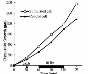
of the neurites to retract. In response to 10 Hz chronic stimulation, for instance, only 75%
of the neurites retracted. Interestingly, after 24 hours of stimulation numerous neurons
apparently accommodated to the stimulus and resumed outgrowth. Furthermore, after 72
hours of continuous stimulation there was no significant difference between the number of
neurons growing in the stimulated and non stimulated compartments.
8
Growth cones from identified neurons in the Helisoma buccal-ganglion also stop
growing when electrically stimulated. Electrical stimulation of the soma of B 19 and B5 neurons, using cell-attached patches, causes the growth cones to reversibly stop advancing (Cohan and Kater, 1986). Chronic stimulation at four hertz inhibited outgrowth from 92% of B 19 growth cones; 50% stopped growing completely or retracted.
1.2.2 Neurotransmitter Application Affects Neurite Outgrowth
Another way to investigate if activity affects neurite outgrowth is to stimulate neurons by adding excitatory neurotransmitters. These experiments assume that since neurotransmitters open ion channels to depolarize the membrane, the neurotransmitters stimulate the cell above threshold. However, the stimulation is seldom measured directly. The addition of neurotransmitters does, however, often affect neurite outgrowth. If mechanisms common to electrical stimulation mediate the effects of neurotransmitter application, then the response of neurons to neurotransmitter application will mimic their response to electrical stimulation.
9
stimulates dendritic outgrowth (Mattson et al., 1988b). The inhibitory effect of glutamate
is thought to be due to the opening of voltage-dependent calcium channels (Mattson et aI.,
1988a), as would occur with elecoical stimulation. These results are consistent with
elecoical activity and glutamate application affecting outgrowth by a common mechanism.
In all three cell types, neurite outgrowth responds to glutamate application the same way as
outgrowth responds to electrical stimulation, regardless of whether stimulation induces or
inhibits outgrowth.
Glutamate also induces filopodial extensions of several microns from hippocampal
neurons (S. Smith and C. Jahr, personal communication). These extensions are dependent
upon the continued presence of glutamate source, retracting upon removal of the
glutamate-filled pipette.
The addition of serotonin to the culture medium stopped outgrowth from the
serotonin-sensitive B 19 neurons but not the serotonin-insensitive He/isoma B5 neurons
(Haydon et aI., 1987; Haydon et aI., 1984). Murrain et aI., (1990) extended these findings
to show that serotonin inhibits outgrowth from Helisoma B 19 but not B 15 neurons in the
dissected intact ganglia. Both dopamine and serotonin inhibit growth cone motility from
adult Helisoma neurons, most likely by regulating membrane voltage (McCobb and Kater,
1988). There are three types of evidence that support this hypothesis. First, neurons that
stop growing in response to serotonin or dopamine also show evidence of sustained
electrical excitation. Second, the magnitude of elecoical response and growth inhibition
correlate. Lastly, the injection of a hyperpolarizing current pulse prevents the transmitters
from both inhibiting outgrowth and electrically exciting the cells (McCobb and Kater,
1988).
Acetylcholine also inhibits outgrowth from certain neurons, including in cultures rat
retinal ganglion cells that endogenously produce acetylcholine (Lipton et al., 1988). The
I 0
depolarization or acetylcholine-induced calcium entry into the cells. Dopamine inhibits outgrowth from chick retinal neurons (Lankford et aI., 1987) although, because forskolin mimics this effect, the outgrowth inhibition is probably cAMP-dependent, not calcium-dependent.
This behavior appears to be somewhat age dependent since, in cultures of
embryonic Helisoma neurons, serotonin can both stop growing neurons and initiate growth for others (Goldberg et aI., 1991). Goldberg and Kater, (1989), have also looked at the effect of serotonin in vivo where transient perturbations in the endogenous serotonin levels during embryogensis alters neuronal morphology, dye coupling, and the strength of synaptic connections. These phenomena are not necessarily seen in culture.
Glutamate, acetylcholine, serotonin and dopamine; all appear to regulate the rate of neurite outgrowth from cultured neurons. The direction of the induced change is generally the same as that induced by electrical stimulation. This suggests that neurotransmitter application and electrical activity are affecting outgrowth by a common mechanism.
1.2.3 Changes in Membrane Polarization May Regulate Neurite Outgrowth
Another method to infer how electrical activity may affect neuronal outgrowth, is to grow cells in depolarizing media. Elevated extracellular potassium concentrations
depolarize cells relative to the surrounding media. We expect cells grown in a depolarizing medium to become more electrically active although without direct electrophysiological evidence this supposition is tenuous. Nonetheless, because of this anticipated
hypersensitivity, one method for mimicking some of the effects of electrical stimulation on neurite outgrowth may be to grow the cells in depolarizing medium.
these cells would increase their rate of elongation with electrical stimulation. In contrast, growth in depolarizing media impedes the rate of neurite outgrowth from mouse DRG neurons. Since the addition of veratrine, bradykinin, or potassium to the media all cause the cells to stop elongating this effect appears to be independent of the source of the depolarization (Robson and Burgoyne, 1989). This independence suggests, as Fields and collaborators (1990) have shown, that neurite outgrowth from DRG neurons is inhibited by electrical stimulation.
Depolarizing media also inhibit neurite outgrowth from other sensory, spinal cord, and sympathetic neurons. S ussdorf and Campenot (1986) have found that these neurons are all capable of growing in both media containing 5 mM and media containing 20 mM potassium but sensory and spinal neurons would not grow from 5 mM into 20 mM potassium. The threshold for this inhibition is cell-type dependent. Sympathetic neurons from the superior cervical ganglion (SCG) required a greater potassium concentration (50 mM versus 20 mM) to inhibit outgrowth than the other two neuronal cell types.
Interestingly, elevated potassium ion concentrations appears to inhibit axonal outgrowth from regenerating sympathetic neurons more than from other cell types.
Electrical activity is also likely to partially inhibit neurite outgrowth from Xenopus spinal cord neurons. When Bixby and Spitzer (1984) grew Xenopus neurons in medium that prevents electrical activity (Ca++ free medium with or without TIX), neurites extend more rapidly than those grown in control cultures.
1 2
1.3 Summary
The literature to date clearly demonstrates that electrical activity is capable of regulating synapse elimination and process segregation and pruning. Yet the evidence that electrical activity, independent of neurotransmitter application, regulates neurite elongation from vertebrate cells is less compelling. There are no examples of activity regulating neurite outgrowth in vivo. In fact there is evidence to the contrary: neurite elongation is unaffected by activity in the retino-tectal system (Sretavan et al., 1988) or during reinnervation of the mouse sternomastoid muscle (Rich and Lichtman, 1989).
In contrast, there is evidence from in vitro studies that electrical stimulation inhibits outgrowth. Work with the invertebrate Helisoma clearly shows that electrical stimulation inhibits neurite outgrowth (Cohan and Kater, 1986). Unfortunately, in studies with vertebrate neurons such as DRG neurons, large imposed electric field rather than
depolarization of a portion of the membrane stimulated the cells (Fields et al., 1990). This difference makes these results suspect since the imposed electric field, rather than the stimulation (stimulation was confirmed with intracellular recordings from model cells), may be responsible for the observed inhibition of outgrowth.
Other studies have used neurotransmitters or depolarizing media to both increase and decrease neurite outgrowth in vitro. Because these agents stimulate the cells
Although it has been suggested repeatedly in the literature that electrical activity
probably inhibits neurite elongation from many cell types, only one cell type clearly stops
growing in response to electrical activity. In Chapters 3 and 4, I shall show evidence that
at least for neonatal rat SCG neurons in our cultures outgrowth is independent of electrical
stimulation.
References
Anglister, L., Farber, I.e., Shaher, A. and Grinvald, A. (1982). Localization of voltage-sensitive calcium channels along developing neurites: their possible role in regulating neurite elongation. Dev. BioI. 94:351-365.
Bailey, e.R. and Chen, M. (1983) Morphological basis of long-term habituation and sensitization in Ap/ysia. Science 220:91-93.
Bailey, e.H. and Chen, M. (1988) Long-term memory in Ap/ysia modulates the total number of varicosities of single identified sensory neurons. PNAS, USA. 85: 2373-2377.
Bailey, e.H. and Chen, M. (1989) Time course of structural changes at identified sensory neuron synapses during long-term sensitization in Ap/ysia. J. Neurosci. 9: 1774-1780.
Bixby, J.L. and Spitzer, N.C. (1984) Early differentiation of vertebrate spinal neurons in the absence of voltage-dependent Ca2+ and Na+ Influx. Dev. BioI. 106:89-96.
Burd, G.D. and Nottebohm, F. (1985) Ultrastructural characterization of synaptic
terminals formed by newly generated neurons in a song control nucleus of the adult canary forebrain. J. Compo Neurol. 240: 143-152.
Callaway, E.M., Soha, 1.M. and Van Essen, D.e. (1987) Competition favouring inactive over active motor neurons during synapse elimination. Nature 328:422-426.
Callaway, E.M., Soha, 1.M. and Van Essen, D.e. (1989) Differential loss of neuromuscular connections according to activity level and spinal position of neonatal rabbit soleus motor neurons. 1. Neurosci. 9: 1806-1824.
Callaway, E.M. and Van Essen, D.e. (1989). Slowing of synapse elimination by
14
Chang F-L.F. and Greenough, W.T. (1984) Transient and enduring morphological correlates of synaptic activity and efficacy change in the rat hippocampal slice.
Brain Research 309:35-46.
Cohan, C.S. and Kater, S.B. (1986). Suppression of neurite elongation and growth cone motility by electrical activity. Science 232:1638-1640.
Dahm, L.M., Landmesser, L.T. (1988) The regulation of intramuscular nerve branching during normal development and following activity blockade. Dev. BioI. 130:621-644.
Dahm, L.M., Landmesser, L.T. (1991) The regulation of synaptogenesis during normal development and following activity blockade. I. Neurosci. 11 :238-255.
Fawcett, I.W., O'Leary, D.D.M. and Cowan, W.M. (1984) Activity and the control of ganglion cell death in the rat retina. PNAS 81 :5589-5593.
Fields, R.D., Neale, E.A. anJ Nelson, P.G. (1990). Effects of patterned electrical activity on neurite outgrowth from mouse neurons. 1. Neurosci. 10:2950-2964.
Glanzman, D.L., Kandel, E.R. and Schacher, S. (1990) Target-dependent structural changes accompanying long-term synaptic facilitation in Aplysia neurons. Science
249:799-802.
Goldberg, 1.1., Kater, S.B. (1989) Expression and function of the neurotransmitter serotonin during development of the Helisoma nervous system. Dev. BioI.
131 :483-495.
Goldberg, 1.1., Mills, L.R., and Kater, S.B. (1991) Novel effects of serotonin on neurite outgrowth in neurons cultured from embryos of Helisoma trivolvis. J. Neurobiol. 22: 182-194.
Greenough, W.T. (1984) Structural correlates of information storage in the mammalian brain: a review and hypothesis. TINS 7:229-233.
Haydon, P.G., McCobb, D.P. and Kater, S.B. (1984) Serotonin selectively inhibits growth cone motility and synaptogenesis of specific identified neurons. Science 226:561-564.
Haydon, P.G., McCobb, D.P. and Kater, S.B. (1987). The regulation of neurite outgrowth, growth cone motility, and electrical synaptogenesis by serotonin. I.
Neurobio. 18:197-215.
Jones, P.G., Rosser, S.J. and Bulloch, A.G.M. (1986) Glutamate enhancement of neurite outgrowth in Helisoma neurons. Soc. for Neurosci. Abst. 139.3.
Lankford, K., De Mello, F.G. and Klein, W.L. (1987) A transient embryonic dopamine receptor inhibits growth cone motility and neurite outgrowth in a subset of avian retina neurons. Neurosci. Letts. 75: 169-174.
Lipton, S.A., Frosch, M.P., Phillips, M.D., Tauck, D.L. and Aizenman, E. (1988). Nicotinic antagonists enhance process outgrowth and rat retinal ganglion cells in culture. Science 239: 1293-1296.
Mattson, M.P., Dou, P. and Kater, S.B. (1988a). Outgrowth-Regulating Actions of Glutamate in Isolated Hippocampal Pyramidal Neurons. 1. Neurosci. 8:2087-2100. Mattson, M.P., Lee, R.E., Adams, M.E., Gutherie, P.B., and Kater, S.B. (1988b)
Interactions between entorhinal axons and target hippocampal-neurons: A role for glutamate in the development of hippocampal circuitry. Neuron 1 :865-876. McCobb, D.P. and Kater, S.B. (1988) Membrane voltage and neurotransmitter regulation
of neuronal growth cone motility. Dev. BioI. 130:599-609.
Murrain, M., Murphy, A.D., Mills, L.R. and Kater, S.B. (1990). Neuron-specific modulation by serotonin of regenerative outgrowth and intracellular calcium within the CNS of Helisoma trivolvis. J. Neurobio. 21 :611-618.
Nelson, P.G., Yu,
c.,
Fields, R.D. and Neale, E.A. (1989) Synaptic connections in vitro: Modulation of number and efficacy by electrical activity. Science 244:585-587. Nelson, P.G., Fields, R.D., Yu, C. and Neale, E.A. (1990) Mechanisms involved inactivity-dependent synapse formation in mammalian central nervous system cell cultures. J. NeurobioI. 21: 138-156.
Pearce, LA., Cambray-Deakin, M.A. and Burgoyne, R.D. (1987) Glutamate acting on NMDA receptors stimulates neurite outgrowth from cerebellar granule cells. FEBS Letters 223:143-147.
Reh, T.A. and Constantine-Paton, M. (1985). Eye-specific segregation requires neural activity in three-eyed Ranapipiens.
J
.
Neurosci. 5:1132-1143.Ribchester, R.R. (1988) Activity-dependent and -independent synaptic interactions during reinnervation of partially denervated rat muscle. 1. Physiol. (Lond.) 401:53-75. Ribchester, R.R. and Taxt, T. (1983) Motor unit size and synaptic competition in rat
1 6
Ribchester, R.R. and Taxt, T. (1984) Repression of inactive motor nerve terminals in partially denervated rat muscle after regeneration of active motor axons. J. Physiol. (Lond.) 347:497-511.
Rich, M.M. and Lichtman, J.W. (1989) In vivo visualization of pre- and postsynaptic changes during synapse elimination in reinnervated mouse muscle. J. Neurosci. 9:1781-1805.
Robson, SJ. and Burgoyne, R.D. (1989). L-type calcium channels in the regulation of neurite outgrowth from rat dorsal toot ganglion neurons in culture. Neurosci. Lett. 104: 110-114.
Schmidt, IT. and Edwards, D.L. (1983) Activity Sharpens the Map during the
regeneration of the retinotectal projection in goldfish. Brain Research 269:29-39 Schmidt, IT. and Eisele, L.E. (1985) Stroboscopic illumination and dark rearing block the
sharpening of the regenerated retinotectal map in goldfish. Neurosci. Letts. 14:535-546.
Schmidt, l. T. (1989) Long-term potentiation and activity dependent retinotopic sharpening in the regenerating retinotectal projection of goldfish: common sensitive period and sensitivity to NMDA blockers. 1. Neurosci. 10:233-246.
Small, S.A., Kandel, E.R. and Hawkins, R.D. (1989) Activity-dependent enhancement of presynaptic inhibition in Aplysia sensory neurons. Science 243: 1603-1606.
Smith, S.l and lahr, C. personal communication.
Sretavan, D.W., Shatz, C.l. and Stryker, M.P. (1988). Modification of retinal ganglion cell axon morphology by prenatal infusion of tetrodotoxin. Nature 336:468-471. Stryker, M.P. and Strickland, S.L. (1984) Physiological segregation of ocular dominance
columns depends on the pattern of afferent electrical activity. Invest. Ophthalmol. Vis. Sci. 25:278.
Sussdorf, W.S. and Campenot, R.B. (1986). Influence of the extracellular potassium environment on neurite growth in sensory neurons, spinal cord neurons and sympathetic neurons. Dev. Brain Res. 25:43-52.
Swindale, N.V. (1981) Absence of ocular dominance patches in dark-reared cats. Nature 290:332-333.)
Thompson, W., Kuffler, D.P. and Jansen, J.K.S. (1979). The effect of prolonged,
reversible block of nerve impulses on the elimination of polyneuronal innervation of new-born rat skeletal muscle fibers. Neuroscience 4:271-281.
Chapter 2
The Effects of Intracellular Calcium on Outgrowth
If electrical activity affects neurite outgrowth, what is the mechanism of the regulation? Kater and his collaborators (1988; 1991) have proposed one possible mechanism. They suggest that the increase in [Cali that accompanies suprathreshold electrical activity is the causative agent in regulating outgrowth. This hypothesis is plausible because calcium enters the growth cone during stimulation and regulates many growth cone associated functions, such as the exocytosis of neurotransmitters (Smith and Augustine, 1988) and actin-based motility (Forscher, 1989; Smith, 1988).
In this chapter, I shall review the relationship between [Cali and neurite outgrowth, starting with experiments where electrical activity or stimuli that mimic electrical activity increase [Cali (Section 2.1). Studies that decouple the effects of calcium and activity will follow (Section 2.2). In Section 2.3, I shall discuss the correlation between growth cone calcium concentrations, [Ca]gc and neurite outgrowth rates. Finally, in Section 2.4, I shall summarize the evidence that [Cali can regulate outgrowth.
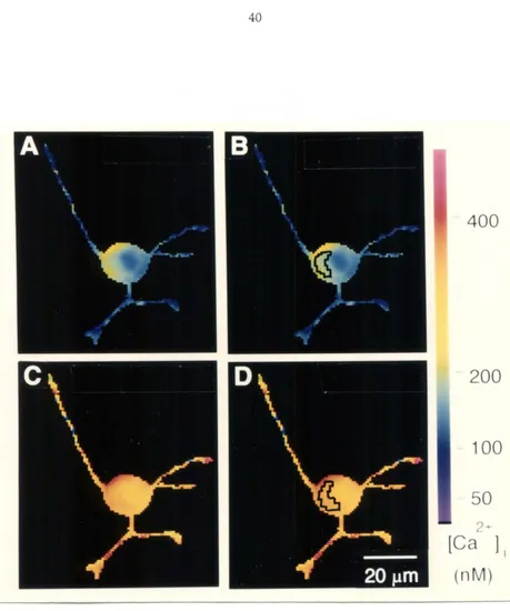
2.1 Stimulation, [Cali, and Neuron Outgrowth
Kater and collaborators (1988; 1991) have proposed that environmental stimuli that influence neurite outgrowth and synaptogenesis act through a common integrator, calcium. Such stimuli include neurotransmitters, electrical stimulation, electrical depolarization, and ion channel blockers. They suggest, on the basis of studies of mammalian (Connor, 1986)
to 300 nM (Kater et aI., 1988). The authors postulate that increasing the [Ca]gc above the levels pennissive for outgrowth would stop the cells from growing.
In support of the hypothesis described in the preceding paragraph, electrical
stimulation increases [Cali and [Ca]gc and inhibits neurite outgrowth from Helisoma buccal ganglion neurons (Cohan et aI., 1987). [Ca]gc rises transiently in Helisoma B5 neurons from 100 nM to 300 to 400 nM, before plateauing at 225 to 325 nM in response to stimulation at 3 Hz (Cohan et aI., 1987); stimulation at 4 Hz stops outgrowth from these same cells (Chan and Kater, 1986). Likewise, at serotonin concentrations that presumably stimulate Helisoma B 19 neurons above threshold, serotonin application stops outgrowth (Haydon et aI., 1987) and causes calcium to rise from 125 nM to 330 nM (Cohan et aI., 1987).
Evidence from studies of mouse DRG neurons also supports the hypothesis proposed by Kater and collaborators. Calcium concentrations in mouse ORG neurons increase when the neurons are stimulated at frequencies that arrest outgrowth (Fields et al., 1990a; 1 990b). As in Helisoma neurons, [Cali and [Ca]gc transiently increase well above final plateau levels. The magnitude of the calcium increase that accompanied the inhibition of growth was equal to or slightly less than that found with Helisoma neurons.
Surprisingly, after 24 hours of stimulation, some DRG neurons resumed growth,
apparently adapting to the electrical stimulation (Fields et aI., 1990b). The [Ca]gc does not differ significantly in neurons that apparently adapted to electrical stimulation and in
neurons that remained immobile (personal communication, Fields, 1990).
Electrical activity has other potentially calcium-dependent effects. Chronic
depolarization and electrical activity decrease the response of SCG neurons to a cholinergic factor. This decreased response allows adrenergic differentiation (Walicke et aI., 1977).
differentiation, and the addition of extracellular calcium or barium enhances adrenergic differentiation. Barium can prevent cholinergic differentiation in nondepolarizing
conditions, further suggesting that the effect is calcium dependent (Walicke and Patterson,
1981).
Application of an appropriate neurotransmitter should stimulate neurons electrically as well as change [Cali. Many researchers have correlated these neurotransmitter-induced changes in [Cali with the arrest of outgrowth from certain neurons. Murrain and
collaborators (1990), have shown that serotonin application transiently increases [Cali in only those Helisoma neurons whose growth stops in response to serotonin application, and not in those neurons whose growth is insensitive to serotonin. The serotonin-sensitive neurons normally have serotonin as a presynaptic agonist, not surprisingly. Serotonin application to serotonin-sensitive Helisoma neurons causes [Cali to increase from 130 nM to 290 nM. The [Cali returns to prestimulus levels within 30 minutes. Serotonin
application inhibits outgrowth and increases [Cali; surprisingly however, the growth inhibition remains even after the [Cali returns to prestimulus levels. The continued inhibition of growth, in spite of the measured decrease in [Cali. is inconsistent with the hypothesis that calcium directly regulates neurite outgrowth.
In contrast, glutamate limits dendritic outgrowth from hippocampal pyramidal cells grown in co-culture with entorhinal cortex explants, that spontaneously release glutamate. The application of glutamate antagonists reverses the glutamate-induced growth inhibition, the application of tetrodotoxin (TfX), or a reduction in the extracellular calcium
concentration (Mattson et al., 1988). The increase in [Cali probably mediates these responses since, in addition to the previously mentioned responses, calcium-channel blockers prevent the glutamate-induced inhibition of dendritic growth and calcium
ionophores or chronic depolarization inhibit neurite outgrowth. All this evidence suggests that the opening of voltage-dependent calcium channels, which results in an increase in calcium influx, leads to the observed alteration in dendritic outgrowth (Mattson et al., 1988).
Robson and Burgoyne (1989), suggest that L-type calcium channels regulate neurite outgrowth in rat DRG cultures. Neurons grown in depolarizing conditions, such as high potassium, veratridine, or bradykinin, show up to a 60 percent decrease in the number of neurons with neurites. The addition of nifedipine, an L-type calcium-channel blocker, selectively reversed the inhibitory effects of the depolarizing conditions. Calcium channels also contribute to neuronal plasticity associated with learning behaviors in Aplysia
(Edmonds et al., 1990).
Growth from neuroblastoma cells also is apparently calcium dependent.
Depolarizing medium containing 30 mM potassium causes an increase in growth-cone area and an increase in neurite elongation from neuroblastoma cells. Cadmium, a calcium-channel blocker, blocks these changes. Similar changes occur during incubation with 2 11M bromo-A23187, a calcium ionophore (Anglister et al., 1982). These results suggest that calcium entry might trigger neurite elongation.
22
DRG, Helisoma, and hippocampal neurons; and regulate neurotransmitter changeover from SCG neurons.
2.2 Calcium Alone May Regulate Neurite Outgrowth
The stimuli used to change [CaJi in the experiments just described (electrical stimulation, neurotransmitter application or depolarizing media) also affect the membrane
potential and are likely to trigger associated second messenger systems. To test whether [Cali can independently regulate neurite outgrowth, an experimenter must manipulate [Cali without simultaneously altering the membrane potential. These experiments differentiate
between the effects of changing [Cali and the effects of changing the membrane depolarization or electrical stimulation of the cell. Such treatments include the use of calcium channel blockers or calcium ionophores without electrical activity or
neurotransmitter application.
Many studies that correlate [Cali and growth do not directly measure the [Cali in
response to the imposed stimuli. Results are interpreted nonetheless with the assumption
that calcium concentrations do change. This assumption may not be warranted. For
example, as I shall discuss in chapters 3 and 4, bathing SCG neurons in low concentrations
of some ionophores does not allow the [Cali and extracellular calcium concentrations to
equilibrate. Because [Cali was not measured and is thus unknown, the actual [Cali may
be quite different from those expected, and may not be in a range that is physiologically relevant. Nonphysiological changes in [Cali are of little interest in determining whether
calcium normally regulates neurite development in vivo. It is nevertheless, instructive to
review evidence that changes in [Cali affect neurite outgrowth rate.
through channels and neurite outgrowth (Cooke et ai., 1988). In addition, Goldberg (1988) found that application of calcium-channel blocker or changes in extracellular calcium concentrations affect veil extension and growth from Ap/ysia neurons. Presumably, either the [Cali changed with the addition of calcium channel blockers or with the change in extracellular calcium concentration. A decrease in the extracellular calcium concentration from 11 mM to 1.3 mM stopped growth. The addition of barium or the calcium ionophore, bromo-A23187, which artificially raised the internal divalent ion concentration allowed the growth to resume.
Mattson and collaborators, (1988b), have shown that increases in [Cali caused by the addition of the calcium ionophore bromo-A23187 or decreases in [Cali caused by the addition of calcium channel blockers, inhibit outgrowth from cultured hippocampal neurons. Lanthium, presumably acting as a calcium-channel blocker and thus decreasing the rise of [Cali nonnally caused by serotonin, blocks the serotonin-induced inhibition of outgrowth (Mattson and Kater, 1987).
Suarez-Isla and collaborators (1984) have also used calcium-channel blockers in cultures of chick retinal neurons to show that a voltage-dependent calcium influx was necessary for both neurite extension and synapse fonnation between retinal neurons and muscle cells in culture.
Although the studies described in Sections 2.1 and 2.2 suggest that calcium
found that growth cones continue to grow, albeit more slowly, in calcium-free media, as long as calcium remains in the medium at the cell body.
2.3 Correlation of Growth Cone Calcium Concentrations with Growth Neurite outgrowth rate has been correlated with the [C,]j levels or with calcium-channel conductivity in a number of systems. Correlations between outgrowth rate and [Cali level seem to be cell-type dependent. In these studies, some of the variability may be due to inadvertent averaging of large variations in the local [Cali during the measurements
(Lipscombe et al.; 1988; Connor et al.; 1990; Silver et aI., 1990).
Some researchers, nonetheless, have correlated neurite outgrowth rate with [Cali.
Connor (1986) show that diencephalon cells from the rat central nervous system have outgrowth rates that correlate with increased [Cali. Calcium levels in active growth cones are over 200 nM whereas the levels in the soma are 60 to 80 nM. Cells without active
growth cones, in comparison, have uniform calcium concentrations of 30 to 70 nM. In
Helisoma buccal ganglion neurons, the internal free calcium concentrations are three times
higher in motile than in non-motile growth cones (Cohan et aI., 1987). Interestingly, the
calcium sensitivity of neurite elongation is different from that of growth cone movement (Mattson and Kater, 1987).
In contrast to the correlations developed in Helisoma neurons, Silver and
collaborators (1989) found that [Cali in motile advancing growth cones was low (60 nM), and was equal to [Cali in quiescent growth cones (55 nM). Motile growth cones that are
not advancing actively had slightly higher values of [Cali, (90 nM), possibly suggesting
that a small elevation of [Cali inhibits neurite extension. A further rise of [Cali (225 nM) appears to inhibit motility and to cause retraction of the growth cone back toward the cell
activation of quiescent growth cones; however, the data suggests that, in active growth cones, [Cali can regulate morphology and behavior.
If there is a variation in the calcium concentration in active and quiescent growth cones, it may arise from differences in channel activity. Lipton (1987) showed that in regenerating rat retinal-ganglion cells the calcium channels displayed bursting behavior, whereas, channels in quiescent cells did not. Likewise, Helisoma growth cones have 70 pS calcium channels that are active in growing growth cones and inactive in stable growth cones (Cohan et al., 1985). The gating of these channels in Helisoma is calcium
independent. Freeman and collaborators (1985) have shown that steady calcium currents are present only in actively growing filopodia tips. In the growing tip of lamprey giant axons, likewise, there is an additional spike found only in regenerating axons. The spike is due to an increase in a voltage-dependent calcium conductance in the growing axon
(MacVicar and Llinas, 1985). Finally, O'Lague and collaborators have shown that there are calcium channels in the growth cones of NGF-induced, polyethylene glycol-fused, PC-12 cells.
potentials. These changes probably arise from clustering of voltage-gated calcium channels in the membrane.
Silver and collaborators (1989) correlated small spatial gradients of [Cali within the neuroblastoma growth cone with outgrowth rate. The gradients although small, are
statistically significant; [Cali is lower by 5 to 10 nM in motile regions. The next year, (Silver et aI., 1990), these authors showed that directly below membrane regions where calcium channels cluster, the local calcium concentration could be elevated into the rnicromolar range. Such areas may reflect the immobile regions observed in the earlier study.
Polak: and collaborators (1991) showed recently that the serotonin-mediated inhibition of outgrowth from Helisoma neurons involves calmodulin: a calmodulin antagonist,
cas
9343B, blocks the serotonin-induced inhibition of neurite outgrowth. Because calmodulin has a relatively low binding affinity for calcium (Manalan and Klee,1984), calcium may increase only enough to activate calmodulin dependent pathways in localized regions of the cell.
2.4 Summary
Many experiments correlate changes in the [Cali with changes in neurite outgrowth rates. Both increases and decreases in [Cali from normal physiological levels (generally about 100 nM) inhibit neurite outgrowth in some circumstances. In the majority of cases increases in [Cali correlate with an inhibition of outgrowth, although some studies
changes as small as 125 to 330 nM in [Cali have been correlated with the arrest of outgrowth (Haydon et at., 1987; Cohan et at., 1987).
Many of the studies discussed in this chapter correlate elecuical activity with an increase in [Cali and a reduced rate of neurite outgrowth, but none show that these increases in [Ca]i cause the interruption of neurite outgrowth observed with elecuical stimulation. Furthermore, some experiments are inconsistent with the latter hypothesis. For instance, after Murrain and collaborators (1990) stimulated Helisoma neurons for 30
minutes the [Ca]i had returned to prestimulus levels but neurite outgrowth remained inhibited. Likewise, some DRG neurons resume outgrowth after 24 hours of continuous stimulation, but their average [Cali, is not significantly different from that of the other neurons in the culture that do not resume outgrowth (personnel communication, Fields,
1990). The experiments presented in chapters 3 and 4 of this dissertation will present evidence that elecuical stimulation and the concomitant increase in [Ca]i do not inhibit neurite outgrowth from one cell type - namely SCG n,,;-lrons.
References
Anglister, L., Farber, I.C., Shaher, A. and Grinvald, A. (1982). Localization of voltage-sensitive calcium channels along developing neurites: their possible role in regulating neurite elongation. Dev Bio 94:351-365.
Campenot, R.B. and Draker, D.O. (1989). Growth of sympathetic nerve fibers in culture does not require extracellular calcium. Neuron 3:733-743.
Cohan, C.S., Haydon, P.G. and Kater, S.B. (1985) Single channel activity differs in growing and non growing growth cones of isolated identified neurons of Helisoma.
J. Neurosci. Res. 13:285-300.
Cohan, C.S., Connor, lA.. and Kater, S.B. (1987). Elecuically and chemically mediated increases in intracellular calcium in neuronal growth cones. 1 Neurosci., 7: 3588-3599.
28
Connor, J.A. (1986). Digital imaging of free calcium changes and of spatial gradients in growing processes in single, mammalian central nervous system cells. P.N.A.S. 83:6179-6183.
Connor, J.A., Kater, S.B., Cohan, C. and Fink, L. (1990) Ca2+ dynamics in neuronal growth cones: regulation and changing patterns of Ca2+ entry. Cell Calcium
11 :233-239.
Cooke, I., Graf, R., Grau, S. and Meyers, D. (1988). Crustacean peptidergic secretory neurons in culture: outgrowth, secretory competence, and Ca++ currents. J. of Gen. Physiology 92:3a-4a.
Edmonds, B., Klein, M., Dale, N. and Kandel, E.R. (1990) Contributions of two types of calcium channels to synaptic transmission and plasticity. Science 250: 1142-1147. Fields, R.D., Gutherie, P.B. Nelson, P.G. and Kater, S.B. (1990b). Calcium homeostatic
capacity is regulated by patterned electrical activity in growth cones of mouse DRG neurons. 16th Soc. of Neurosci. Abst. 197.2: 457.
Fields, R.D., Neale, E.A. and Nelson, P.G. (1990a). Effects of patterned electrical activity on neurite outgrowth from mouse neurons. J. Neurosci. 10:2950-2964. Forscher, P. (1989) Calcium and polyphosphoinositide control of cytoskeletal dynamics.
TINS 12:468-474.
Freeman, J.A., Manis, P.B., Snipes, G.J., Mayes, B.N., Samson, P.c., Wikswo, Jr., J.P., and Freeman, D.B. (1985) Steady growth cone currents revealed by a novel circularly vibrating probe: A possible mechanism underlying neurite growth. J. Neurosci. Res. 13:257-283.
Goldberg, D.J. (1988). Local role of Ca2+ in fonnation of veils in growth cones. J. Neurosci. 8:2596-2605.
Haydon, P.G., McCobb, D.P. and Kater, S.B. (1987). The regulation of neurite outgrowth, growth cone motility, and electrical synaptogenesis by serotonin. J. Neurobio. 18: 197-215.
Kater, S.B., Mattson, M.P., Cohan, C. and Connor, J. (1988). Calcium regulation of the neuronal growth cone. Trends. in Neurosci. 11:315-321.
Kater, S.B. and Mills, L.R. (1991) Regulation of Growth Cone Behavior by Calcium J. Neurosci. 11 :891-899.
Lipton, S.A (1987) Bursting of calcium-activated cation-selective channels is associated with neurite regeneration in a mammalian central neuron. Neurosci Letts 82:21-28. MacVicar, B.A and Llinas, R.R. (1985) Barium Action Potentials in Regenerating Axons
of the Lamprey Spinal Cord. l. Neurosci. Res. 13:323-335.
Manalan, AS. and Klee, C.B. (1984) Calmodulin in: Advances in cyclic nucleotide and protein phosphorylation research, Vol 18 (Greengard P, Robinson GA, eds), pp 227-278. New York: Raven.
Mattson, M.P., Dou, P. and Kater, S.B. (1988). Outgrowth-regulating actions of glutamate in isolated hippocampal pyramidal neurons. l. Neurosci. 8:2087-2100. Mattson, M.P. and Kater, S.B. (1987) Calcium regulation of neurite elongation and
growth cone motility. 1 Neurosci 7:4034-4043.
Mattson, M.P., Lee, R.E., Adams, M.E., Gutherie, P.B. and Kater, S.B. (1988) Interactions between entorhino axons and target hippocampal-neurons: a role for glutamate in the development of hippocampal circuitry. Neuron 1 :865-876. Murrain, M., Murphy, AD., Mills, L.R. and Kater, S.B. (1990). Neuron-specific
modulation by serotonin of regenerative outgrowth and intracellular calcium within the CNS of Helisoma trivolvis. 1. Neurobio. 21:611-618.
O'Lague, P.H., Huttner, S.L., Vandenberg, C.A., Morrison-Graham, K. and Horn, R. (1985) Morphological properties and membrane channels of the growth cones induced in PC12 cells by nerve growth factor. l. Neurosci. Res. 13:301-321. Pearce, LA, Cambray-Deakin, M.A. and Burgoyne, R.D. (1987) Glutamate acting on
NMDA receptors stimulates neurite outgrowth from cerebellar granule cells. FEBS Letters 223:143-147.
Polak, K.A., Edelman, AM., Wasley, l.W.F. and Cohan, C.S. (1991) A novel
calmodulin antagonist, CGS 9343B, modulates calcium-dependant changes in neurite outgrowth and growth cone movements. l. Neurosci. 11 :534-542. Robson, S.l. and Burgoyne, R.D. (1989). L-type calcium channels in the regulation of
neurite outgrowth from rat dorsal root ganglion neurons in culture. Neurosci. Lett. 104: 110-114.
Silver, R. A., Lamb, A. G. and Bolsover, S. R. (1990) Calcium hotspots caused by L-channel clustering promote morphological changes in neuronal growth cones. Nature 343:751-754.
Smith, S.l. (1988) Neuronal cytomechanics: the actin-based motility of growth cones. Science 242:708-715.
Smith, S.1. and Augustine, G.1. (1988) Calcium ions, active zones and synaptic transmitter release. TINS 11 :458-464.
Suarez-Isla, B.A., Pelto, 0.1., Thompson, 1.M. and Rapoport, S.I. (1984). Blockers of calcium permeability inhibit neurite extension and formation of neuromuscular synapses in cell culture. Dev. Brain Res. 14:263-270.
Walicke, P.A., Campenot, R. and Patterson, P.H.(1977) Determination of transmitter function by neuronal activity. PNAS, USA 74:5767-5771.
Chapter 3
Electrical Stimulation and Changes in [Cali Do Not
Affect Neurite Elongation from Sympathetic
Neurons
Both electrical activity and the accompanying changes in [Cali have been proposed
as common regulatory mechanisms for neuronal outgrowth. In particular, experiments
suggest that electrical activity inhibits neurite outgrowth from Helisoma (Cohan and Kater,
1986) and mouse DRG (Fields et al., 1990) neurons in culture. To investigate the
generality of this control mechanism, I stimulated neurons from the neonatal rat SCG while
measuring the neurite outgrowth rate.
[Cali increases during stimulation of both Helisoma (Cohan et aI., 1987) and DRG
(Fields et aI., 1990a) neurons. This increase may be the mechanism whereby stimulation
inhibits outgrowth. Since electrical stimulation does not appear to inhibit outgrowth from
SCG neurons, we postulated that the [Ca]gc may not be increasing to the same extent in
SCG neurons as in the other cell types that response to electrical stimulation. To test this
hypothesis, I collaborated with Dr. Wade Regehr in Dr. David Tank's laboratory at Bell Laboratories, Murray Hill, NJ, to measure changes in the [Cali in both the soma and the
growth cones of SCG neurons during electrical stimulation. To funher test if [Cali
regulates outgrowth in general, we attempted (unsuccessfully) to elevate [Ca]gc
pharmacologically to greater than 500 nM for prolonged periods. A level of 500 nM is
significantly above the concentration postulated to inhibit neurite outgrowth by Kater and
collaborators (1988). I shall discuss the results of these experiments in Chapter 4.
In this chapter, I shall discuss why I chose to study the effect of electrical activity
32
stimulation and calcium experiments (Section 3.2). In Section 3.3, I shall discuss the reliability of these measurements.
3.1 The Choice of Neonatal Rat SCG Neurons
I chose to use neonatal rat
sea
neurons for these studies for four reasons. First, there have been extensive studies of neurite outgrowth from culturedsea
neurons.sea
neurite outgrowth rates in culture are both rapid and steady (Bray, 1973), although the absolute growth rate depends on the age of the ganglion at dissection (Argiro et al., 1984). The average outgrowth rates were 8 to 22 Ilm/hour for embryonicsea
neurons, 14 to 29 Ilm/hour for perinatalsea
neurons, and 4 to l3 Ilrn/hour for adultsea
neurons (Argiro et al. 1984) for collagen grown neurons. In our culture system, the cells extend many processes simultaneously. 1 We thought that the continuous nature and speed of neuritegrowth from
sea
neurons would simplify the outgrowth measurements.Second,
sea
neurons have, at the time of dissection, the minimum requirement for electrical activity regulated outgrowth: The neurons already receive presynaptic inputs (Rubin, 1985a), and these presynaptic inputs are electrically competent although they are not necessarily active (Rubin, 1985b). The axons are growing actively toward their targets at this time (Rubin, 1985b).sea
neurons continue to grow into adulthood (Voyvodic, 1987; Purves et aI., 1986), and that growth is apparently largely independent ofpreganglionic innervation (Voyvodic, 1987), unlike growth from the primary sensory systems (see Berry et al., 1978 for a review).
A third reason to study
sea
neurons is that results from studies on the effects of depolarizing media suggest thatsea
neurons might differ from other neurons in theirresponse to electrical stimulation. High potassium medium, a depolarizing medium that mimics some of the effects of electrical stimulation, does not appear to impede outgrowth from SCG neurons (Campenot, 1984; 1986; and Sussdorf and Campenot, 1986). SCG neurons, in contrast to sensory and spinal-cord neurons, continue to grow from medium with 5 mM potassium into medium with 20 mM potassium, and are impeded only when they try to grow into medium with 50 mM potassium. SCG neurons are able to grow in 50 mM potassium (Sussdorf and Campenot, 1986), whereas DRG neurons cannot do so
(Robson and Burgoyne, 1989). Neurite regeneration from SCG neurons, on the other
hand, is relatively more sensitive to elevated potassium concentrations than is similar growth from either spinal or sensory neurons (Sussdorf and Campenot, 1986).
A fourth reason to study SCG neurons is that outgrowth continues even if there is no extracellular calcium at the growth cones (Campenot and Draker, 1989)2. This
continued growth suggests that outgrowth is not dependent on local calcium influx. The
fact that growth continues when there is no external calcium at the growth cone suggests that calcium does not regulate outgrowth. Furthermore, this calcium independence
suggests that if electrical activity regulates outgrowth it does so in a calcium independent fashion.
3.2 Methods
In this section, I shall describe how I did the experiments described in Chapter 4. Specifically, I shall discuss how I prepared and stimulated the cells and how I measured the calcium concentrations.
3.2.1 Cell Culture
I anesthetized rat pups (0 to 3 days old) with Halothane and decapitated them, before removing the superior cervical ganglia. After transferring the ganglia into 2 mI of
Hank's balanced salts solution (HBSS) with penicillin/streptomycin and no divalent cations, I cleaned and incubated them at 37°C in HBSS with 0.125 percent trypsin for 30
minutes in a 5 percent C02 environment. I inactivated the trypsin after the incubation by soaking the ganglia in modified Leibovitz's L-15 media (L15) with 32.5 percent horse
serum for 10 minutes. Finally, I transferred the ganglia to LI5, shredded, triturated and plated them on previously prepared polylysine-Iaminin coated 35 mm dishes at 3000
neurons/dish. I allowed the cells to settle for 30 minutes to 3 hours before experiments began.
3.2.2 Media
I made the modified Ll5 by supplementing L15 as described in Mains and
Patterson, (1973), except I omitted the bovine serum albumin and methyl cellulose and substituted 10 percent horse serum for rat serum and of 4 ng/ml 2.5 S NGF (Boehringer
Mannheim) for 0.5 Ilg/ml 7 S NGF. The medium used during all outgrowth experiments was L15air. I prepared the LI5air by omitting sodium bicarbonate and serum from the
LI5, adjusting the pH to 7.2 (in air) with NaOH and adjusting the NGF concentration to 2
used in these experiments. In preliminary experiments, where I observed outgrowth for up to two weeks, there was no detectable decrease in SCG outgrowth at NGF concentration from I to 20 ng/ml. For experiments in high K+ LIS, I decreased the concentration of NaCI from 137 mM to 92 mM and increased the concentration of KCI from 5.6 mM to 50 mM. To make high K+/high Ca++ LIS, I also increased the concentration of CaCl2 from
1.26 mM to 5 mM: I did not compensate for this adjustment by adjusting the concentration of other divalent cations. For experiments with the calcium ionophore, bromo-A23187, I
added different amounts of a 20~M stock solution (minimum dilution was 1/50) of the ionophore in DMSO to the perfusing medium.
3.2.3 Substrate
I grew the cells on glass coverslips which I had glued under a 6 mm hole in the bottom of 35 mm petri dishes. I had previously soaked the dishes for 30 minutes in fuming nitric acid, rinsed them three times with distilled water, and soaked them overnight in Dulbecco's phosphate buffered saline (OPBS) with 100 ~g/ml polylysine, (Sigma
P8905). In the morning, I had rinsed, dried and stored the dishes at 4 °C until use. Just
before plating, I soaked them in a 20 ~g/ml solution of laminin (Sigma, L8263) in DPBS
for 45 minutes, and rinsed them three times with DPBS.
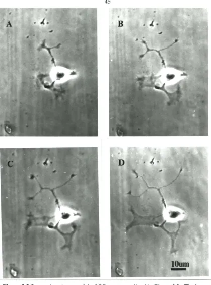
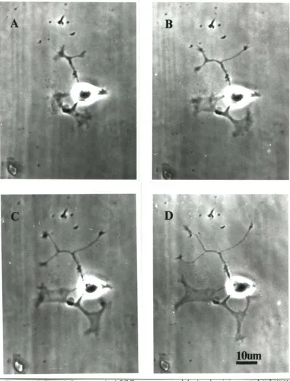
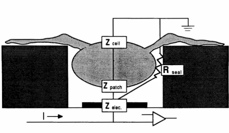
3.2.4 Stimulation
It is essential to have a reliable stimulation method if we are to study the affects of activity on neurite outgrowth. In preliminary experiments, I found that intracellular and
whole-cell electrode techniques stopped neurite outgrowth. Therefore, I used cell-attached
36
with cell-attached patch pipettes (KG-33 glass, Garner Glass Co.). Tip diameters were about 111m and electrode impedances were 5-15 MQ. I used a high-potassium, low-sodium saline in the pipette to depolarize the cell without breaking down the membrane. To make this saline, I modified the recipe from Marty and Neher, (1983); 140 roM KCl, 2 mM MgCI2, 11 mM EGTA-KOH (pH 7.3),1 roM CaCI2, and 10 roM HEPES, adjusted to pH
7.2 with KOH and to 320 mOs with sucrose. With this solution in the pipette, I apparently depolarized the portion of the membrane inside the pipette and caused the continuous opening of non inactivating channels, because the pipette potential approached the cell's
resting potential, -70 mV relative to the bath, after I obtained a seal. The open channels reduced the impedance of the patch and allowed the passage of enough current to stimulate the cell without damaging the membrane (see Regehr et aI., 1989 for a detailed
explanation). The reduced patch impedance also increased the signal-to-noise ratio for
recording evoked action potentials.
I positioned the electrodes while monitoring the voltage drop produced by a hyperpolarizing current pulse, (50 pA, 2 Hz). After the pipette had touched the cell membrane, indicated by an increase in the measured impedance of the pipette, I applied
light suction with a mouth pipette. I stopped the test pulses as soon as a seal had formed. I
observed each cell for 30 minutes without stimulation to check for pipette induced damage.
I did stimulation experiments only on cells that were undamaged by patching, i.e.,
continued to grow during this control period. I also rejected cells if I was unable to evoke