RESEARCH ARTICLE
Ihog and Boi elicit Hh signaling via Ptc but do not aid Ptc in
sequestering the Hh ligand
Darius Camp1,2,3, Billy Haitian He1, Sally Li1, Irene W. Althaus4, Alexander M. Holtz4,5,6, Benjamin L. Allen4,*, Frédéric Charron2,3,7,8,9,10,11,* and Donald J. van Meyel1,3,7,11,*
ABSTRACT
Hedgehog (Hh) proteins are secreted molecules essential for tissue development in vertebrates and invertebrates. Hh reception via the 12-pass transmembrane protein Patched (Ptc) elicits intracellular signaling through Smoothened (Smo). Hh binding to Ptc is also proposed to sequester the ligand, limiting its spatial range of activity. InDrosophila, Interference hedgehog (Ihog) and Brother of ihog (Boi) are two conserved and redundant transmembrane proteins that are essential for Hh pathway activation. How Ihog and Boi activate signaling in response to Hh remains unknown; each can bind both Hh and Ptc and so it has been proposed that they are essential for both Hh reception and sequestration. Using genetic epistasis we established here that Ihog and Boi, and their orthologs in mice, act upstream or at the level of Ptc to allow Hh signal transduction. In the
Drosophila developing wing model we found that it is through Hh pathway activation that Ihog and Boi maintain the boundary between the anterior and posterior compartments. We dissociated the contributions of Ptc from those of Ihog/Boi and, surprisingly, found that cells expressing Ptc can retain and sequester the Hh ligand without Ihog and Boi, but that Ihog and Boi cannot do so without Ptc. Together, these results reinforce the central role for Ptc in Hh binding
in vivoand demonstrate that, although Ihog and Boi are dispensable for Hh sequestration, they are essential for pathway activation because they allow Hh to inhibit Ptc and thereby relieve its repression of Smo.
KEY WORDS: Pattern formation, Morphogenesis, Signaling, Drosophila
INTRODUCTION
Hedgehog (Hh) proteins are secreted morphogens that are essential for many events in tissue development and cellular physiology. Despite significant progress, gaps remain in the characterization of the molecular mechanisms underlying Hh signal transduction. Key points
of control include the reception of the Hh signal at the cell surface and the distribution of Hh within tissues, both of which involve Patched (Ptc), a conserved 12-pass transmembrane protein. In humans, mutations of the Ptc ortholog PTCH1 cause Gorlin syndrome, holoprosencephaly, basal cell carcinoma and medulloblastoma. Therefore, understanding exactly how Ptc participates in Hh reception and distribution is essential to distinguish between normal Hh pathway function in development and aberrant Hh signaling in disease (Briscoe and Therond, 2013).
In the absence of Hh, Ptc maintains the 7-pass transmembrane protein Smoothened (Smo) in a repressed state by unknown means. Under these conditions, the Cubitus interruptus (Ci) transcription factor is proteolytically cleaved and acts as a transcriptional repressor. As part of the canonical signaling pathway that is activated in the presence of Hh, Ptc-mediated repression of Smo is relieved, leading to Smo accumulation at the cell surface and stabilization of full-length Ci (Ci155), which activates transcription of Hh target genes. The current view holds that Ptc is involved in sensing the extracellular Hh concentration (Briscoe and Therond, 2013), and that target genes respond in relation to the degree of induced pathway activation. One direct transcriptional target of Hh signaling is Ptc itself; Ptc is upregulated in response to high levels of Hh, and is required for the sequestration of Hh, which limits the distribution of Hh and its range of action within tissues via a poorly understood mechanism.
Experiments have yet to demonstrate unequivocally that Hh binds directly to Ptc in Drosophila; therefore, both Hh reception and sequestration have been proposed to involve Interference hedgehog (Ihog) and Brother of ihog (Boi), two closely related type I transmembrane proteins of the immunoglobulin superfamily (Zheng et al., 2010). Ihog and Boi both have an ectodomain consisting of four immunoglobulin-like (Ig) domains and two fibronectin type III (FN3) repeats, a transmembrane domain and a cytoplasmic tail (Kang et al., 2002; Lum et al., 2003; Yao et al., 2006). Their orthologs in vertebrates are Cell adhesion molecule downregulated by oncogenes (Cdon) and Brother of cdon (Boc). Intriguingly, the first FN3 domain of Ihog binds directly to Hh (McLellan et al., 2006; Yao et al., 2006), whereas the second FN3 domain interacts with Ptc (Zheng et al., 2010). This raises the possibility that Ihog and Boi are cofactors for Ptc in a multiprotein complex serving as the functional Hh receptor. This view is supported by phenotypes caused by loss-of-function mutations of theihogandboigenes inDrosophila, which mimic those ofsmo mutations (Camp et al., 2010; Zheng et al., 2010). For example, we and others have found thatihogandboiare essential for Hh pathway activation in developing wing imaginal disks, where Hh produced and secreted by cells in the posterior compartment is received and sequestered by cells in the anterior compartment. Anterior cells respond by adopting fates according to their degree of Hh exposure
1
McGill Centre for Research in Neuroscience and the McGill University Health Centre Research Institute, 1650 Cedar Avenue, Montreal, Quebec, Canada H3G 1A4.
2
Molecular Biology of Neural Development, Institut de Recherches Cliniques de Montréal (IRCM), Montreal, Quebec, Canada H2W 1R7.3Division of Experimental Medicine, McGill University, Montreal, Quebec, Canada H3A 1A3.4Department of Cell and Developmental Biology, University of Michigan, Ann Arbor, MI 48109, USA.
5
Cellular and Molecular Biology Program, University of Michigan, Ann Arbor, MI 48109, USA.6Medical Scientist Training Program, University of Michigan, Ann Arbor, MI 48109, USA.7Department of Neurology and Neurosurgery, McGill University, Montreal, Quebec, Canada H3A 2B4.8Program in Neuroengineering, McGill University, Montreal, Quebec, Canada H3A 2K6.9Department of Medicine, University of Montreal, Montreal, Quebec, Canada H3T 1J4.10Department of Anatomy and Cell Biology, McGill University, Montreal, Quebec, Canada H3A 0C7.
11
Department of Biology, McGill University, Montreal, Quebec, Canada H3A 1B1.
*Authors for correspondence (benallen@umich.edu; Frederic.Charron@ircm.qc.ca; don.vanmeyel@mcgill.ca)
O
posterior compartment. Evidence indicates that Ihog and Boi are functionally redundant with one another (Camp et al., 2010; Yan et al., 2010; Zheng et al., 2010), and that they act upstream of Smo (Camp et al., 2010; Zheng et al., 2010).
Although it is clear that Ihog and Boi are crucial for Hh pathway activityin vivo, their mechanism of action is poorly understood and could be illuminated by genetic approaches that clearly establish their position in the pathway relative to Ptc. A previous experiment to establish the epistatic relationship of Ihog and Ptc was confounded by the lack of a strong and penetrant ihog mutant phenotype (Yao et al., 2006), later found to be due in part to functional redundancy with Boi (Camp et al., 2010; Zheng et al., 2010). Using genetic epistasis experiments in flies and mice, we here directly demonstrate that Ihog and Boi (or Cdon and Boc in mice) act upstream or at the level of Ptc to allow Hh pathway activation. To better understand the interplay of Ihog and Boi with Ptc, we also used genetic approaches inDrosophilato dissociate the contributions of Ihog and Boi from functions attributed to Ptc within developing wing disks. Like Ptc, we find that Ihog and Boi allow transduction of the Hh signal, and that through their essential role in Hh pathway activation they maintain the anterior-posterior compartment boundary. Surprisingly, we find that, unlike Ptc, Ihog and Boi are dispensable for Hh retention and sequestration by responding cells.
RESULTS
Ihog and Boi function upstream or at the level of Ptc for Hh signal transduction
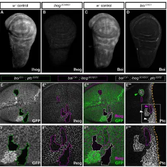
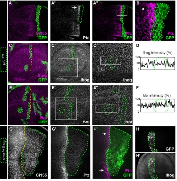
To examine Ihog and Boi expression in wing imaginal disks of third instar larvae (L3), we performed immunohistochemistry for Ihog and Boi with newly developed antisera. Each was expressed broadly
in both the anterior and posterior compartments (Fig. 1A,C), and overall these patterns of endogenous Ihog and Boi expression are consistent with previously published results (Yan et al., 2010; Zheng et al., 2010; Bilioni et al., 2013). We confirmed the specificity of these patterns by their absence inihogDC1 orboiC1
mutants (Fig. 1B,D), which allowed the use of these antisera to monitor Ihog and Boi levels in clones generated in mosaic animals. Expression of Ihog and Boi in the anterior compartment is consistent with their essential role for pathway activation in Hh-responding cells (Fig. 1A-D). Cells adjacent to the compartment boundary respond to high levels of Hh by upregulating the expression of Ptc, a high-threshold target of the canonical signaling pathway. Cells positioned more anteriorly are further away from the Hh source and respond to lower-threshold levels of Hh marked by increased accumulation of Ci155. According to these high- and low-threshold reporters of Hh signaling activity,boi;ihog double mutant clones lack Hh pathway activation altogether (Camp et al., 2010; Zheng et al., 2010). By contrast,ptcmutations fully activate the pathway (Phillips et al., 1990; Ingham et al., 1991). These opposing effects allowed us to unequivocally address the epistatic relationship between Ihog, Boi and Ptc by examining pathway activation in cells that were triple mutants for all three genes. We reasoned that if triple mutant cells were to mimic ptc mutants and fully activate the pathway, it would indicate that Ihog and Boi act upstream or at the same level as Ptc, but not downstream. For this we studied theboiC1and ihogDC1mutations that we had
reported previously (Camp et al., 2010), and the ptcS2 missense
[image:2.612.50.380.407.737.2]mutation, which is indistinguishable from null alleles, but gives rise to a dysfunctional Ptc protein that can be detected with anti-Ptc (Phillips et al., 1990; Jiang and Struhl, 1995; Li et al., 1995; Chen and Struhl, 1996). As Ptc is an Hh pathway target, activation of the
Fig. 1. Ihog and Boi function upstream or at the level of Ptc.(A-D) Immunohistochemistry for Ihog or Boi inw1118(w−) controls and in homozygous mutants forihogorboi. (E-E‴) Homozygous mutant clones generated in aboiC1/−mutant background:ptc
clones,ihogclones orihog;ptcdouble mutant clones. (E)ptcS2/S2clones (GFP-negative cells outlined in green) and (E′)ihogDC1/DC1clones (Ihog-negative
cells outlined in magenta). (E″) Merged image, in which clones mutant for bothptcS2andihogDC1lack both GFP and Ihog (outlined in white), and are triple mutants forboi,ihogandptc. (E‴) In the two triple mutant clones shown (white arrowheads) Ptc is expressed highly, similar toptcS2mutant cells (green arrowhead) or to control anterior cells adjacent to the compartment boundary. Cells lacking bothihogand
boi(magenta arrowhead) lacked Hh pathway activation and did not express Ptc. (F-F‴) Higher magnification views of the areas in E-E‴.
DEVEL
O
pathway (as in ptcS2 mutants) can be detected readily as strong
upregulation of Ptc expression and immunoreactivity. We studied mosaic wing disks, in which the MARCM system was used to generateptcS2and/orihogDC1mutant clones inboiC1hemizygotes.
Because all cells in these animals were mutant for boi, clones generated by FLP-mediated mitotic recombination in the wing disk were either (1) double mutant forboi;ptc, (2) double mutant for boi;ihogor (3) triple mutant forboi;ptc;ihog.As expected, double mutantboi;ptcclones within the anterior compartment activated the pathway even at a distance from the compartment boundary (i.e. in the absence of Hh ligand) (Fig. 1E,E″and green arrowhead in E‴), whereas double mutant boi;ihog clones abolished pathway activation even when close to the boundary (i.e. near the source of Hh ligand) (Fig. 1E′,E″ and magenta arrowhead in E‴). Like double mutant boi;ptcclones (green arrowhead in Fig. 1E‴), all cells within triple mutant boi;ptc;ihog clones fully activated the pathway (white arrowheads in Fig. 1E‴), as indicated by Ptc expression levels comparable to fully activated, wild-type cells nearest to the boundary (Fig. 1F-F‴). Therefore,ptcis epistatic to ihogandboi in vivo, which positions Ihog and Boi upstream or at the level of Ptc for Hh signal transduction.
Cdon and Boc act upstream or at the level of Ptch1 in vertebrate Hh signal transduction
We wondered whether the epistatic relationship ofptctoihogand boi is conserved across species, in particular with regard to the
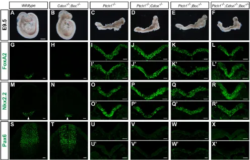
mouse orthologsPtch1,CdonandBoc. To test this, we generated mice lacking Ptch1, Cdon and Boc and examined their gross anatomy relative to controls (Fig. 2). Compared with wild-type embryos (Fig. 2A) at E9.5, Cdon;Boc double mutants develop normally for the most part (Fig. 2B), but have forebrain defects indicative of holoprosencephaly as described previously (Zhang et al., 2011). By contrast,Ptch1 mutant mice display significant defects at E9.5, including the failure to complete the turning process, exencephaly, neural tube closure defects and abnormal heart development (Fig. 2C) (Goodrich et al., 1997). Notably, Ptch1;Cdon and Ptch1;Boc double mutants, and in particular Ptch1;Cdon;Boctriple mutants, grossly phenocopyPtch1mutants (Fig. 2D-F). This suggests that loss ofCdonandBocis insufficient to reduce Hh signaling in the absence ofPtch1.
To precisely assess Hh pathway activity in these embryos, we performed a neural patterning analysis using a panel of Sonic hedgehog (Shh)-dependent markers of neural progenitors. InCdon; Boc double mutant embryos, we confirmed previous results demonstrating reduced Hh signaling (Allen et al., 2011), as indicated by reduced Foxa2 expression (Fig. 2G,H) and the ventral shift of Nkx2.2 (Fig. 2M,N) and Pax6 expression (Fig. 2S,T). Conversely, Ptch1−/− embryos have increased Hh signaling, as reflected by expanded expression of Foxa2 and Nkx2.2, and the loss of Pax6 throughout the neural tube (Fig. 2I,I′,O,O′,U,U′). Like Ptch1−/− mutants, double mutants (Ptch1−/−;Cdon−/− or
[image:3.612.58.554.356.674.2]Ptch1−/−;Boc−/−) and triple mutants (Ptch1−/−;Cdon−/−;Boc−/−)
Fig. 2.Ptch1is epistatic toCdonandBocduring Shh-dependent ventral neural tube patterning.(A-F) Lateral view of E9.5 mouse embryos. Wild-type (A) andCdon−/−;Boc−/−(B) embryos have completed the turning process and display closed neural tubes, whereasPtch1−/−(C),Ptch1−/−;Cdon−/−(D),Ptch1−/−; Boc−/−(E) andPtch1−/−;Cdon−/−;Boc−/−(F) embryos fail to turn and exhibit open neural tubes including excencephaly. Scale bar: 500 µm. (G-X′) Antibody detection of Foxa2 (G-L′), Nkx2.2 (M-R′) and Pax6 (S-X′) in transverse sections from E9.5 mouse embryos. Genotypes are indicated at the top of each column.
−/− −/−
O
exhibited complete activation of the Hh pathway in the neural tube (Fig. 2J-L′,P-R′,V-X′), demonstrating Ptch1 to be epistatic to Cdon and Boc. These experiments indicate that Cdon and Boc act upstream or at the same level as Ptch1, and confirm that their relationship within the Hh pathway is conserved between flies and mice.
Hh can be retained and sequestered by cells lacking Ihog and Boi
If Ihog and Boi and their orthologs in mice act upstream or at the level of Ptc, how might they function in pathway activation? To address this, we returned to experiments inDrosophila, for which it has been proposed that Ihog and Boi are essential for Hh signaling because without them Ptc cannot bind Hh with high affinity (Zheng et al., 2010). We focused on sequestration of the Hh signal by clones of cells near the anterior-posterior compartment boundary of developing wing disks, as sequestration must involve Hh binding at some level. Indeed, we and others have previously shown thatboi; ihogdouble mutant cells cannot sequester Hh, and instead permit ectopic pathway activation in more anterior cells that would normally be too distant from the morphogen source (Camp et al., 2010; Zheng et al., 2010). The requirement for Ihog and Boi in Hh sequestration could be explained in two ways: they could bind Hh
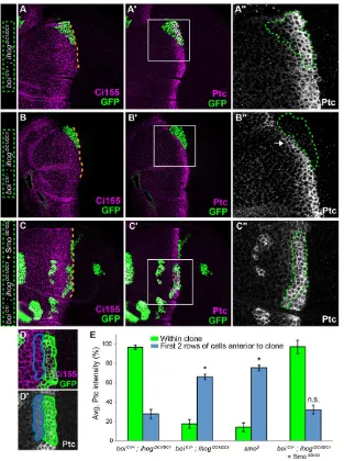
directly themselves, and/or they could act indirectly through pathway activation to influence another Hh-binding factor. To explore this, we used the MARCM technique to test whether Hh sequestration could be restored to boi;ihog double mutant cells simply by activating the pathway through expression of a constitutively active form of Smo (SmoSD123) (Jia et al., 2004). To assess sequestration, we identified anterior clones spanning the usual domain of Hh-induced pathway activation (as revealed by upregulation of Ptc), and then examined pathway activation in cells immediately anterior to these clones. In cells just anterior to control clones that sequester Hh (boiC1/+;ihogDC1/DC1), pathway activation
was normal, as assessed by Ptc expression levels (Fig. 3A-A″,D,E). By contrast, Ptc was upregulated in cells anterior to double mutant clones (boiC1/−;ihogDC1/DC1) (Fig. 3B-B″,E), as we have shown
previously (Camp et al., 2010). This indicates that cells lacking Ihog and Boi cannot activate the pathway and cannot sequester Hh. Similarly,smo3mutant clones also fail to sequester Hh (Chen and
[image:4.612.48.361.319.739.2]Struhl, 1996) (Fig. 3E), indicating that Ihog and Boi are insufficient to sequester Hh without pathway activation. Remarkably, we found that the ability ofboi;ihogdouble mutant clones to sequester Hh was fully restored by expressing SmoSD123to rescue pathway activation (Fig. 3C-C″,E). As theboiC1allele is a nonsense mutation at Trp626
Fig. 3. Ihog and Boi are dispensable for Hh
sequestration.(A-A″) In a controlboiC1/+heterozygote, the
MARCM technique was used to generate a GFP-positive
ihogDC1/DC1clone adjacent to the anterior-posterior compartment boundary (green outline in A′,A″). (A) Ci155 (magenta) accumulates in response to Hh signaling and marks the anterior-posterior boundary. (A′,A″) The high-threshold Hh target Ptc is expressed normally within this control clone, and there is no ectopic Ptc expression in cells just anterior to the clone: this indicates correct sequestration of the Hh signal by cells within the clone. (A″) Higher magnification view of boxed area in A′. (B-B″) AnihogDC1/DC1
clone (green outlines in B′,B″) in aboiC1/−mutant. (B″) Higher magnification view of boxed area in B′. Clones lacking bothihogandboidid not express Ptc (GFP-positive, green outline), but were identifiable as anterior in origin, as they express basal levels of Ci155. Failure to sequester Hh is indicated by high levels of Ptc in cells immediately anterior to the clone (arrow in B″). (C-C″) Aboi;ihogdouble mutant clone adjacent to the anterior-posterior compartment boundary that is expressingUAS-smoSD123(green outline in C′,C″). (C″) Higher magnification view of boxed area in C′. Expression ofsmoSD123in cells lackingboiandihogrescues pathway activation (Ptc expression within clone, C″) and is sufficient to sequester Hh. Orange dashed lines (A,B,C) mark the normal position of the anterior-posterior boundary. (D,D′) Quantification example. Average Ptc intensity within the clone (within green outline in D and green shaded region in D′) is measured within the usual high-Ptc expression zone. Average Ptc intensity anterior to the clone (within magenta outline in D and magenta shaded region in D′) consists of the first two rows of cells anterior to area measured in green. (E) Quantification of average Ptc intensity within (green bars) and anterior to (blue bars) the observed clones. Clones lacking Ihog and Boi but expressing SmoSD123(n=11) show similar patterns of Ptc intensity than control clones (n=5). By contrast, clones lacking Ihog and Boi (n=8), or the pathway activator Smo (n=9), displayed low Ptc within the clone and high Ptc anterior to it (ANOVA, F3,29=9.017,P=0.0002). All measurements were normalized to the average levels of Ptc expression that were observed in areas of equivalent size outside the clones but near the compartment boundary.
DEVEL
O
(Camp et al., 2010), we considered that it might encode an intact first FN3 domain that could theoretically bind Hh in this experiment. To test this, we replaced boiC1 with boiKO2-1, an
allele designed by gene targeting to delete the translation initiation codon, the signal sequence and the four Ig domains from the coding sequence of Boi (Zheng et al., 2010). In results that are identical to those forboiC1, we restored Hh sequestration toboi;ihogdouble
mutant cells by activating the pathway (supplementary material Fig. S1). This confirms that Ihog and Boi are dispensable for sequestration if Hh signaling is activated, and implicates an Hh-binding factor other than Ihog or Boi in this process.
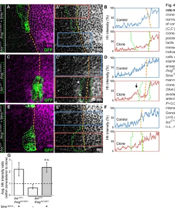
To investigate how sequestration of the Hh signal (as measured by Ptc levels) relates to retention of the Hh protein, we measured the labeling intensity of anti-Hh immunoreactivity within MARCM clones, and in cells just anterior to them. As cells of the anterior compartment do not produce Hh, we interpret Hh immunoreactivity within anterior compartment clones as an indication of their capacity to bind Hh produced and secreted by cells of the posterior compartment. In control animals heterozygous for boi, we noted that SmoSD123expression inihogclones (genotype:boiC1/+;ihogDC1/ DC1) did not influence Hh immunoreactivity compared with normal
levels seen just outside the clone (genotype: boiC1/+;ihogDC1/+)
staining (Fig. 4C-D), suggesting that Hh secreted from cells in the posterior compartment was not retained by cells within the clone. Instead, Hh accumulated immediately anterior to them (Fig. 4C-D,G), as if the movement of Hh from the posterior compartment was unimpeded byboi;ihogdouble mutant cells. Importantly, Hh staining was restored toboi;ihogdouble mutant cells that expressed SmoSD123, and the graded distribution of Hh within these clones was identical to nearby control cells outside the clones (Fig. 4E-F,G). Thus, pathway activation due to SmoSD123renders Ihog and Boi dispensable for cells to retain and sequester Hh.
Studies of cultured cells transfected with Ptc indicate that its localization at the plasma membrane is improved by co-expression of Ihog (Zheng et al., 2010). If Ihog/Boi were required for Ptc trafficking to the plasma membranein vivo, we anticipated that the subcellular distribution of Ptc might be altered in clones of wing disk epithelial cells lacking Ihog and Boi. However, we found no evidence that the distribution of Ptc was influenced by the presence or absence of Ihog/Boi (supplementary material Fig. S2).
Ptc is essential for Hh sequestrationin vivo, also in the absence of Ihog and Boi
[image:5.612.54.411.51.474.2]If cells lacking Ihog and Boi can sequester Hh once the pathway has Fig. 4. Ihog and Boi are dispensable for Hh retention.(A,A′) In aboiC1/+control, anihogDC1/DC1
clone (green outline) expressing SmoSD123displays a normal Hh gradient. (B) Average Hh staining intensity of control (blue) and clone (red) areas boxed in A′. (C,C′) In aboiC1/−mutant, anihogDC1/DC1MARCM
clone (green outline) is shown to cross the anterior-posterior boundary (orange dashed line). The clone lacks Hh staining, which instead accumulates immediately anterior to it (arrow in C′and D), indicating the inability of theboi;ihogdouble mutant cells within the clone to retain Hh. (D) Average Hh staining intensity of control (blue) and clone (red) areas boxed in C′. (E,E′) In aboiC1/−mutant, an ihogDC1/DC1clone (green outline) expressing SmoSD123shows that pathway activation in this manner rescues the normal Hh gradient within the clone. (F) Average Hh staining intensity of control (blue) and clone (red) areas boxed in E′. (G) Ratio of average Hh intensity within clones versus cells just anterior to the clones (ANOVA, F2,14=6.369,
P=0.0108). All measurements were normalized to Hh intensity in the posterior compartment of the wing. GenotypesboiC1/+;ihogDC1/DC1;UAS-smoSD123
(n=5 clones),boiC1/−;ihogDC1/DC1(n=7 clones) and
boiC1/−;ihogDC1/DC1;UAS-smoSD123(n=6 clones).
n.s., nonsignificant.
O
principal role in restricting the diffusion of Hh and thereby limiting the zone of pathway activation. Although it is controversial whether Ptc binds to Hh directly inDrosophila, Ptc is a prime candidate due to its upregulation following pathway activation. Inboi;ihogdouble mutant cells, perhaps Ptc levels are simply insufficient to sequester Hh because Ptc is not upregulated, due to a lack of signaling through the Hh pathway. We made several observations in support of a primary role for Ptc in Hh sequestration. First, cells that are mutant for the null allele ptc16 activate the pathway fully but do not
sequester Hh, allowing ectopic pathway activation in cells just anterior toptc16clones (Chen and Struhl, 1996) (Fig. 5A-A″and
arrow in B). In addition, neither Ihog nor Boi protein levels are affected within ptc16 clones compared with neighboring cells
(Fig. 5C-F), and so a lack of Hh sequestration inptc16clones cannot
be readily explained by loss of Ihog and Boi. Furthermore, we found that overexpression of Ihog was insufficient to sequester Hh insmo3
mutant clones, in which Ptc expression is not elevated (Fig. 5G-H). Finally, to determine whether Ptc can sequester Hh in cells lacking Ihog and Boi, we studied ptcS2, a missense mutation in the
intracellular sterol-sensing domain that, likeptc16, cannot repress
Smo and results in full pathway activation. Unlikeptc16cells, cells
homozygous forptcS2retain the ability to sequester Hh (Chen and
Struhl, 1996). As expected of the controls,boi;ptcS2double mutant
cells did sequester Hh, as there was no ectopic Ptc just anterior to these clones (Fig. 6A-A″), whereasboi;ihogdouble mutant cells did not sequester Hh (supplementary material Fig. S3). Triple mutant boi;ptcS2;ihog clones spanning the anterior-posterior boundary
were rare, but we found five examples, two of which are shown (Fig. 6B-B‴, supplementary material Fig. S3). In every case they fully sequestered the Hh signal, confirming that Ptc is essential for Hh sequestrationin vivo, also in the absence of Ihog and Boi.
Ihog and Boi act via Hh pathway activation to control compartment-specific cell affinity
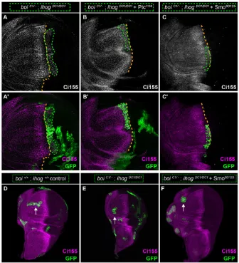
[image:6.612.50.410.373.741.2]Ihog and Boi are essential for activation of Hh signaling, which results in upregulation of Ptc, a high-threshold target that is at least partially responsible for Hh sequestration, independently of Ihog and Boi. If Ihog and Boi are not directly required to sequester the Hh signal, we wondered whether they might also be dispensable for the process by which cells of the anterior and posterior compartments are segregated from one another. Anterior cells adjacent to the boundary require Hh signaling to remain within the anterior compartment, as cells mutant forsmoorcisegregate from wild-type anterior cells, form tight borders with both anterior and posterior cells and fail to integrate with either compartment (Blair and Ralston, 1997; Rodriguez and Basler, 1997; Dahmann and Basler, 2000). Likewise,boi;ihog double mutant cells also segregate away from the anterior compartment (Fig. 7A,A′) (Camp et al., 2010), phenocopyingsmomutations (Rodriguez and Basler, 1997). This could reflect the requirement for Ihog and Boi in Hh signal transduction. However, as the boundary is thought to be implemented by mechanisms controlling compartment-specific cell affinity and adhesion (Dahmann and Basler, 1999), it could also reflect a direct role for Ihog and Boi in cell adhesion via their
Fig. 5. Ihog and Boi are unable to sequester Hh in cells lacking Ptc.
(A) A GFP-positive MARCM clone homozygous for theptc16null mutation
(green outline) is situated within the anterior compartment, adjacent to the anterior-posterior boundary (orange dashed line), as assessed by Ci155 immunostaining (magenta). (A′-B) Theptc16/16null clone does not express Ptc and fails to sequester Hh, as indicated by ectopic activation (Ptc expression) in cells anterior to the clone (arrows in A′and B). (B) Higher magnification view of boxed area in A″. (C-F) GFP-positive
ptc16/16MARCM clones stained for Ihog
(C-C″) or Boi (E-E″), focusing on clones of the anterior compartment (green outlines). (C″,E″) Higher magnification view of boxed area in C′and E′, respectively. (D,F) Average staining intensity of boxed areas in C″and E″, respectively. Gray line indicates total average intensity. Note that Ihog and Boi levels are unchanged inptc16/16clones compared with neighboring cells. (G-G″) A GFP-positive MARCM clone homozygous for thesmo3null mutation and overexpressing Ihog (green outline) of anterior origin is situated within the posterior compartment, adjacent to the anterior-posterior boundary (orange dashed line), as assessed by Ci155 (G) and Ptc immunostaining (G′). Despite the presence of Ihog, the clone is unable to sequester Hh as indicated by the upregulation of Ptc anterior to the clone (arrows in G″). (H,H′) Higher levels of Ihog (H′) are visible in asmo3clone overexpressing Ihog (green outline).
DEVEL
O
Ig and FN3-containing ectodomains. To address this, we tested whether segregation of double mutant cells (ihogDC1/DC1clones
inboiC1/− hemizygotes) could be rescued by pathway activation
within those cells through selective expression of SmoSD123 or Ptc1130, a dominant-negative form of Ptc that cannot repress Smo. In either instance, pathway activation in every double mutant clone examined (n>10 for each genotype) was sufficient to prevent
[image:7.612.49.368.58.261.2]mis-segregation of anterior cells into the posterior compartment (Fig. 7B-C′). These results indicate thatboiandihogare dispensable for the increased cell affinity and adhesion that accompanies compartmentalization of the developing wing disk. Reinforcing this interpretation was our examination of clones in the most anterior regions of the anterior compartment, where cells are exposed to little or no Hh. Control clones andboi;ihogdouble mutant clones in this
Fig. 6. Hh is sequestered by triple mutant cells for Ihog, Boi and PtcS2.(A-A″) In aboiC1/−mutant, a large
GFP-negative clone homozygous for theptcS2/S2
missense mutation is situated within the anterior compartment, adjacent to the anterior-posterior boundary (orange dashed line). (A′,A″) Ptc is highly expressed within the clone because PtcS2protein can be detected with anti-Ptc antibody, and because the Hh signaling pathway is highly active due to this mutation. Lack of ectopic Ptc staining just anterior to the clone indicates full Hh sequestration within the clone. (B-B‴) Two clones mutant for bothptcandihogare indicated by white outlines in B‴. These are triple mutants, as they are homozygous forptcS2/S2(GFP-negative, marked by green
outlines in B),ihogDC1/DC1(Ihog-negative, marked by magenta outlines in B′) and occur in aboiC1/−wing disk. (B‴) A triple mutant clone (white outline and arrowhead) near the compartment boundary can sequester Hh, as indicated by the absence of Ptc just anterior to the clone (white asterisk).
Fig. 7. Ihog and Boi act via the Hh pathway to maintain the anterior-posterior boundary.
(A,A′) In aboiC1/−mutant, a largeihogDC1/DC1clone (green outline) is shown to segregate away from the anterior compartment toward posterior territory (boundary marked by orange dashed line). These
boi;ihogdouble mutant cells are known to be of anterior origin because they all express basal levels of Ci155. (B-C′) By contrast,boi;ihogdouble mutants segregate normally and remain in anterior territory when Hh signaling is activated, using either Ptc1130 (B,B′) or SmoSD123(C,C′). (D) In anterior clones that are so far removed from the boundary that they cannot receive the Hh signal, control MARCM clones, in which both theboiandihoggenes are intact and homozygous (genotype:boi+/+;ihog+/+), have irregular shapes (green outlines of clones marked by white arrows). (E) Likewise, similarly positioned MARCM clones that are double mutant forboi;ihogalso have irregular shapes. (F) By contrast, activation of Hh signaling inboi;ihog
double mutant cells with SmoSD123causes such
clones to appear spheroid (green outlines of clones marked by white arrows), probably reflecting that pathway activation causes cells within the clone to acquire higher affinity for one another than for cells outside the clone, even in the absence of Ihog and Boi.
O
[image:7.612.47.383.368.737.2]region were randomly shaped, reflecting that cells within these clones had equal affinity for each other as they did with surrounding cells outside the clone (arrows in Fig. 7D,E). By contrast,boi;ihog double mutant clones that were activated by SmoSD123 were spheroid and formed smooth boundaries with neighboring cells, revealing stronger affinity of cells within the clone for one another (arrow in Fig. 7F). We conclude that Ihog and Boi are required for compartment segregation because they are necessary to activate Hh signaling. Any potential role they might have in cell adhesion in this context is dispensable upon Hh pathway activation. Interestingly, upregulation of Ptc in response to pathway activation also appears dispensable, asptcmutant clones respect the compartment boundary (Fig. 5A) (Rodriguez and Basler, 1997). This is strikingly distinct from Hh sequestration, for which our data suggest that Ptc upregulation is required.
DISCUSSION
We and others showed previously that Ihog and Boi are absolutely essential within Hh-responding cells for activation of the Hh signaling pathway, acting upstream of Smo (Camp et al., 2010; Zheng et al., 2010). In the experiments described here, we advance our understanding of Ihog and Boi function by drawing three major conclusions:
First, in genetic epistasis experiments we found that Ihog and Boi also act upstream or at the level of Ptc, supporting the idea that they function through Ptc to relieve suppression of Smo. This epistatic relationship appears conserved in evolution, as we found that Cdon and Boc also function upstream or at the level of Ptch1 for Hh signal transduction in mice. These genetic findings establish this relationship unequivocally, and so have profound implications for future studies to further clarify how these co-receptors participate in Hh reception and pathway activation.
Second, based on our experiments to dissect the relative contributions of Ihog and Boi in processes involving Ptc, we conclude that it is through their essential roles in Hh signal transduction that Ihog, Boi and Ptc contribute to anterior-posterior compartment segregation: once the pathway is activated, all three proteins are dispensable for maintenance of the compartment boundary, implicating other, yet unidentified, cell surface recognition molecules in compartment-specific cell affinity and adhesion.
Our third conclusion is that Ihog and Boi, unlike Ptc, are completely dispensable for the sequestration and retention of Hh. We show that cells lacking Ihog and Boi can sequester and retain the Hh signal if the pathway is activated and Ptc is upregulated. They do so via physiological levels of endogenous Ptc induced either by pathway activation inptcS2mutants or by expression of SmoSD123. Incidentally, we also found that Hh sequestration was rescued inboi;ihogdouble mutant clones overexpressing Ptc1130(Fig. 7B), a dominant-negative that fully activates the Hh pathway and upregulates endogenous, wild-type Ptc. This third conclusion is not consistent with the view that Ihog and Boi aid in addressing Ptc to the cell surface and that, once there, they are required for Ptc to bind and sequester Hh (Zheng et al., 2010). This view is based primarily on Ptc and Ihog overexpression in cultured cells, and on an experiment that failed to restore Hh sequestration toboi;ihogdouble mutant cells with mutation of cAMP-dependent protein kinase 1 (Pka-C1), which upregulates Ptc and other target genes because loss of Pka-C1 disinhibits the activity of the transcription factor Ci (Ohlmeyer and Kalderon, 1997; Chen et al., 1998; Price and Kalderon, 1999; Wang et al., 1999). It is unclear why Ptc upregulation in Pka-C1 mutants was unable to rescue Hh sequestration inboi;ihogdouble mutants, whereas Ptc upregulation
in our experiments was able to do so. In cells lacking Ihog and Boi, perhaps the level to which Ptc is upregulated inPka-C1mutants is inadequate. Regardless, our data indicate that Ptc has a central role in the binding and sequestration of Hh, whereas Ihog and Boi are dispensable, despite their requirement for Hh signal transduction.
Our results are consistent with vertebrate systems, in which current models strongly favor direct contacts between Hh and Ptc, primarily because: (1) expression of the Ptc ortholog Ptch1 promotes binding of Shh to transfected cells (Marigo et al., 1996; Stone et al., 1996), (2) radiolabeled Shh can be chemically cross-linked to Ptch1 expressed on the cell surface (Fuse et al., 1999) and (3) Ptch1 can reach the cell surface in the absence of the Ihog/Boi-related proteins Cdon and Boc (Izzi et al., 2011). Whether Ptc is sufficient on its own to bind Hh remains an important question that awaits technically challenging studies using purified proteins. An alternative possibility is that Hh could have additional receptor(s), with candidates including the proteoglycans Dally and Dally-like (Dlp), and Shifted, a secreted protein of the Wnt inhibitory factor 1 (WIF1) family (Han et al., 2004; Yan et al., 2010; Ayers et al., 2012; Avanesov and Blair, 2013).
Our results clearly distinguish a role for Ptc that relies on Ihog/Boi (Hh reception/signal transduction) from one that does not (Hh sequestration), and so they contribute to an emerging view of the function of Ihog, Boi and related proteins. In non-responding cells, others have shown that Ihog and Boi are involved in restricting the movement of Hh (Hartman et al., 2010; Bilioni et al., 2013), and so may contribute to its overall distribution. Within Hh-responding cells, where Ptc is co-expressed with Ihog and Boi, we find that Ihog and Boi are essential for Hh signal transduction, but not Hh sequestration and retention. As Ihog and Boi act upstream or at the level of Ptc, they must mediate a crucial, rate-limiting step in the inhibition of Ptc in response to Hh. However, as they are not essential for Ptc to bind Hh, how they affect Ptc function remains to be elucidated. Whereas the precise molecular mechanism remains elusive, several lines of evidence provide important clues. First, an Ihog variant lacking the cytoplasmic tail can rescueboi;ihogdouble mutants (Zheng et al., 2010; our results, data not shown). Second, the second Fn3 domain of Ihog or Boi interacts physically with Ptc and is quite distinct from the first Fn3 domain that harbors the Hh-interacting surface (McLellan et al., 2006; Zheng et al., 2010). Third, the presence of Ihog or Boi potentiates co-immunoprecipitation of Hh and Ptc (Zheng et al., 2010). Together, these results suggest that the primary role of Ihog and Boi in Hh signaling involves the ability of their ectodomains to form favorable protein complexes with Ptc or Hh, or with both simultaneously. Although our data indicate that Ptc does not need Ihog and Boi to bind Hh in vivo, we surmise that it is through these multimolecular complexes that Ihog and Boi allow Hh to inhibit Ptc and thereby relieve its suppression of Smo and the Hh signaling cascade.
MATERIALS AND METHODS
Fly stocks
Mutant and transgenic strains are described in the following references: boiC1and ihogDC1 (Camp et al., 2010),ptc16(Strutt et al., 2001),ptcS2 (Martin et al., 2001),smo3(Chen and Struhl, 1998),boiKO2-1(Zheng et al., 2010), UAS-SmoSD123-CFP (Jia et al., 2004),UAS-Ptc (Johnson et al., 1995) andUAS-ptc1130(Johnson et al., 2000).
Mosaic analysis
Mitotic recombination was used to generate clones of mutant cells within the developing wing disk that could be scored by either the absence or presence of green fluorescent protein (GFP). Clones marked by the absence of GFP were generated by crossingboiC1/C1,hs-FLP;FRT40A,FRT42B/+ females
toihogDC1,FRT40A,FRT42B,ptcS2,Ubi-GFP/+ males. In this way, three
DEVEL
O
types of clones could be generated with a standard heat-shock protocol: (1) ihogDC1/DC1, (2)ptcS2/S2and (3)ihogDC1/DC1,ptcS2/S2. For controls, these clones were generated in boiC1/+ heterozygotes (female progeny), with which clones were compared generated in boiC1/Y hemizygotes (male progeny) that are denotedboiC1/−in this paper for simplicity.
Clones marked by the presence of GFP were generated using a mosaic analysis of a repressible cell marker (MARCM) (Lee and Luo, 2001). MARCM clones were obtained by crossingboiC1/C1,hs-FLP;FRT40A,
tubP-GAL80; actP-GAL4, UAS-mCD8GFP/TM6B,Tb females to ihogDC1,
FRT40A; UAS-transgene-of-interest/TM6B,Tb males. In this way, our
controls for these experiments wereihogDC1/DC1clones generated inboiC1/+ heterozygotes (female progeny), with which were comparedihogDC1/DC1 clones generated in boiC1/− hemizygotes (male progeny). Within these MARCM clones, the induced GFP expression could be accompanied by induced expression of an additional transgene (UAS-SmoSD123-CFPor
UAS-ptc1130). In an experiment testing whetherboiKO2-1gave identical results
toboiC1, we crossed boiKO2-1, hs-FLP, UAS-mCD8GFP; FRT40A,
tubP-GAL80/In(2LR)Gla, wg[Gla-1] Bc[1];actP-GAL4/TM6B,Tbfemales to either
ihogDC1, FRT40A/CyO,P[Dfd-GMR-nvYFP]2 males (controls) orihogDC1,
FRT40A/CyO,P[Dfd-GMR-nvYFP]2;UAS-SmoSD123-CFP/TM6B,Tbmales.
Antibody generation and immunohistochemistry forDrosophila
Wandering L3 larvae were dissected, fixed in 2% formaldehyde and stained according to standard immunohistochemistry procedures. Rabbit or guinea pig polyclonal antisera were generated against Ihog-GST and Boi-GST fusion proteins, respectively. For Ihog, the fusion protein consisted of amino acids 736-886 of the cytoplasmic tail of Ihog-PA (FlyBase), and for Boi it comprised amino acids 874-998 of the cytoplasmic tail of Boi-PB (FlyBase). Anti-Ihog was used at a dilution of 1:10,000, and anti-Boi at 1:500. Monoclonal antibodies obtained from the Developmental Studies Hybridoma Bank (DSHB) included: mouse anti-Ptc (Apa 1, 1:50) and rat anti-Ci155 (2A1, 1:2000). Rabbit anti-Hh (1:200) was a gift from Philip Ingham (Institute of Molecular and Cell Biology, Singapore). Secondary antibodies were: Alexa Fluor 488-conjugated goat anti-rabbit (A-11034, 1:500); Alexa Fluor 647-conjugated goat anti-mouse (A-21236, 1:500); and Alexa Fluor 568-conjugated goat anti-rat (A-11077, 1:500), all from Molecular Probes, and DyLight 549-conjugated anti-guinea pig (106-505-003, 1:500) from Jackson Laboratories. Confocal images were collected on an Olympus FV1000 confocal microscope. All images show a single focal plane through the basolateral region of the wing disk epithelium, with anterior to the left and dorsal to the bottom. Measurements of fluorescence intensity were performed in ImageJ (NIH). Average Ptc and Hh intensity within clones was measured in the first two (Hh) or three (Ptc) rows of cells just anterior to the compartment boundary. Average Ptc and Hh intensity anterior to the clone was measured in the first two rows of cells just anterior to the clone. Background levels of fluorescence were subtracted prior to all measurements.
Mice and analysis of spinal cord tissues by immunofluorescence
CdoLacZ-2(Cole and Krauss, 2003), BocAP-2 (Zhang et al., 2011) and
Ptch1LacZ(Goodrich et al., 1997) mice have been described previously.
This study was performed in strict accordance with the recommendations in the Guide for the Care and Use of Laboratory Animals of the National Institutes of Health. All of the animals were handled according to approved institutional animal care and use committee (IACUC) protocols of the University of Michigan. The protocol was approved by the University Committee on Use and Care of Animals (UCUCA) at the University of Michigan (PRO00004017).Cdonmice were maintained on a C57BL/6 background; Boc mice were maintained on a mixed C57BL6;129S6/ SvEvTac background;Ptch1LacZmice were maintained on a mixed Swiss Webster;129S4/SvJaeJ;C57BL/6 background. Noon of the day on which a vaginal plug was detected was considered as E0.5. For all analyses, a minimum of three embryos of each genotype was examined; representative images are shown. Immunofluorescent analyses of E10.5 mouse neural tubes were performed essentially as described previously (Jeong and
anti-Nkx2-2 (74.5A5) and mouse IgG1 anti-Pax6 (PAX6). Secondary antibodies were used at a dilution of 1:500 (as above). Primary antibodies were incubated overnight at 4°C. DAPI (D1306, Molecular Probes) was used at a dilution of 1:30,000. A Leica SP5X confocal microscope was used to visualize fluorescence. For each genotype a minimum of three embryos were analyzed. Whole mouse embryos were imaged with a Nikon SMZ1500 stereomicroscope.
Graphs and statistics
All bar graphs express data as mean±s.e.m. Statistical analyses were carried out in Prism (GraphPad). All data were analyzed for statistical significance using a one-way ANOVA. Statistically significant differences in ANOVA (P<0.05) were followed by Dunnett’s post-hoc tests, and asterisks in graphs denote significance ofP-values comparing indicated group with controls (* forP<0.05).
Acknowledgements
We thank Tiago Ferreira, Emilie Peco and Katarina Stojkovic for technical assistance; Yong Rao and David Hipfner for advice; and Matthew Scott, Alana O’Reilly, Jin Jiang, Philip Ingham, Philip Beachy, Xiaoyan Zheng, Gary Struhl and the Bloomington Stock Center for fly stocks and reagents. Several antibodies were obtained from the DSHB at The University of Iowa, USA. Confocal microscopy of mouse neural patterning was performed in the Microscopy and Image Analysis Laboratory at the University of Michigan, USA.
Competing interests
The authors declare no competing financial interests.
Author contributions
D.C. conceived and performed experiments and wrote the manuscript; B.H.H. and S.L. performedDrosophilaexperiments; I.W.A. and A.M.H. performed the mouse neural patterning analyses; B.L.A. conceived the mouse experiments and edited the manuscript; F.C. and D.J.v.M. conceived experiments and wrote the manuscript. F.C. and D.J.v.M. contributed equally to this work.
Funding
This work was supported by grants from the Canadian Institutes of Health Research (CIHR) to F.C. [MOP74700, MOP119384, MOP123367] and to D.J.v.M. [MOP93767], the Natural Sciences and Engineering Research Council of Canada (NSERC) to D.J.v.M. [RGPIN/346134] and the Canada Foundation for Innovation (CFI) to F.C. and to D.J.v.M. F.C. is a Chercheur-Boursier Senior of the Fonds de Recherche du Québec-Santé(FRQ-S). A.M.H. is supported by an NIH predoctoral fellowship [F31 NS081806]. B.L.A. is supported by The University of Michigan Center for Organogenesis Research Team Grant, an American Heart Association scientist development grant [11SDG6380000] and by an NIH grant [R21 CA167122]. Deposited in PMC for release after 12 months.
Supplementary material
Supplementary material available online at
http://dev.biologists.org/lookup/suppl/doi:10.1242/dev.103564/-/DC1
References
Allen, B. L., Song, J. Y., Izzi, L., Althaus, I. W., Kang, J.-S., Charron, F., Krauss, R. S. and McMahon, A. P.(2011). Overlapping roles and collective requirement for the coreceptors GAS1, CDO, and BOC in SHH pathway function.Dev. Cell20, 775-787.
Avanesov, A. and Blair, S. S.(2013). The Drosophila WIF1 homolog Shifted maintains glypican-independent Hedgehog signaling and interacts with the Hedgehog co-receptors Ihog and Boi.Development140, 107-116.
Ayers, K. L., Mteirek, R., Cervantes, A., Lavenant-Staccini, L., Therond, P. P. and Gallet, A.(2012). Dally and Notum regulate the switch between low and high level Hedgehog pathway signalling.Development139, 3168-3179.
Bilioni, A., Sánchez-Hernández, D., Callejo, A., Gradilla, A.-C., Ibáñez, C., Mollica, E., Carmen Rodrıguez-Navas, M., Simon, E. and Guerrero, I.́ (2013). Balancing Hedgehog, a retention and release equilibrium given by Dally, Ihog, Boi and shifted/DmWif.Dev. Biol.376, 198-212.
Blair, S. S. and Ralston, A.(1997). Smoothened-mediated Hedgehog signalling is required for the maintenance of the anterior-posterior lineage restriction in the developing wing of Drosophila.Development124, 4053-4063.
Briscoe, J. and Therond, P. P.(2013). The mechanisms of Hedgehog signalling and its roles in development and disease.Nat. Rev. Mol. Cell Biol.14, 416-429.
O
Chen, Y. and Struhl, G.(1996). Dual roles for patched in sequestering and transducing Hedgehog.Cell87, 553-563.
Chen, Y. and Struhl, G.(1998). In vivo evidence that Patched and Smoothened constitute distinct binding and transducing components of a Hedgehog receptor complex.Development125, 4943-4948.
Chen, Y., Gallaher, N., Goodman, R. H. and Smolik, S. M.(1998). Protein kinase A directly regulates the activity and proteolysis of cubitus interruptus.Proc. Natl.
Acad. Sci. USA95, 2349-2354.
Cole, F. and Krauss, R. S.(2003). Microform holoprosencephaly in mice that lack the Ig superfamily member Cdon.Curr. Biol.13, 411-415.
Dahmann, C. and Basler, K.(1999). Compartment boundaries: at the edge of development.Trends Genet.15, 320-326.
Dahmann, C. and Basler, K.(2000). Opposing transcriptional outputs of Hedgehog signaling and engrailed control compartmental cell sorting at the Drosophila A/P boundary.Cell100, 411-422.
Fuse, N., Maiti, T., Wang, B., Porter, J. A., Hall, T. M. T., Leahy, D. J. and Beachy, P. A.(1999). Sonic hedgehog protein signals not as a hydrolytic enzyme but as an apparent ligand for patched.Proc. Natl. Acad. Sci. USA96, 10992-10999.
Goodrich, L. V., Milenković, L., Higgins, K. M. and Scott, M. P.(1997). Altered neural cell fates and medulloblastoma in mouse patched mutants.Science277, 1109-1113.
Han, C., Belenkaya, T. Y., Wang, B. and Lin, X.(2004). Drosophila glypicans control the cell-to-cell movement of Hedgehog by a dynamin-independent process.Development131, 601-611.
Hartman, T. R., Zinshteyn, D., Schofield, H. K., Nicolas, E., Okada, A. and O’Reilly, A. M.(2010). Drosophila Boi limits Hedgehog levels to suppress follicle stem cell proliferation.J. Cell Biol.191, 943-952.
Ingham, P. W., Taylor, A. M. and Nakano, Y.(1991). Role of the Drosophila patched gene in positional signalling.Nature353, 184-187.
Izzi, L., Lévesque, M., Morin, S., Laniel, D., Wilkes, B. C., Mille, F., Krauss, R. S., McMahon, A. P., Allen, B. L. and Charron, F.(2011). Boc and Gas1 each form distinct Shh receptor complexes with Ptch1 and are required for Shh-mediated cell proliferation.Dev. Cell20, 788-801.
Jeong, J. and McMahon, A. P.(2005). Growth and pattern of the mammalian neural tube are governed by partially overlapping feedback activities of the hedgehog antagonists patched 1 and Hhip1.Development132, 143-154.
Jia, J., Tong, C., Wang, B., Luo, L. and Jiang, J.(2004). Hedgehog signalling activity of Smoothened requires phosphorylation by protein kinase A and casein kinase I.Nature432, 1045-1050.
Jiang, J. and Struhl, G.(1995). Protein kinase A and hedgehog signaling in Drosophila limb development.Cell80, 563-572.
Johnson, R. L., Grenier, J. K. and Scott, M. P.(1995). patched overexpression alters wing disc size and pattern: transcriptional and post-transcriptional effects on hedgehog targets.Development121, 4161-4170.
Johnson, R. L., Milenkovic, L. and Scott, M. P.(2000). In vivo functions of the patched protein: requirement of the C terminus for target gene inactivation but not Hedgehog sequestration.Mol. Cell6, 467-478.
Kang, J.-S., Mulieri, P. J., Hu, Y., Taliana, L. and Krauss, R. S.(2002). BOC, an Ig superfamily member, associates with CDO to positively regulate myogenic differentiation.EMBO J.21, 114-124.
Lee, T. and Luo, L.(2001). Mosaic analysis with a repressible cell marker (MARCM) for Drosophila neural development.Trends Neurosci.24, 251-254.
Li, W., Ohlmeyer, J. T., Lane, M. E. and Kalderon, D.(1995). Function of protein kinase A in hedgehog signal transduction and Drosophila imaginal disc development.Cell80, 553-562.
Lum, L., Yao, S., Mozer, B., Rovescalli, A., Von Kessler, D., Nirenberg, M. and Beachy, P. A.(2003). Identification of Hedgehog pathway components by RNAi in Drosophila cultured cells.Science299, 2039-2045.
Marigo, V., Davey, R. A., Zuo, Y., Cunningham, J. M. and Tabin, C. J.(1996). Biochemical evidence that patched is the Hedgehog receptor.Nature 384, 176-179.
Martı́n, V., Carrillo, G., Torroja, C. and Guerrero, I.(2001). The sterol-sensing domain of Patched protein seems to control Smoothened activity through Patched vesicular trafficking.Curr. Biol.11, 601-607.
McLellan, J. S., Yao, S., Zheng, X., Geisbrecht, B. V., Ghirlando, R., Beachy, P. A. and Leahy, D. J.(2006). Structure of a heparin-dependent complex of Hedgehog and Ihog.Proc. Natl. Acad. Sci. USA103, 17208-17213.
Ohlmeyer, J. T. and Kalderon, D.(1997). Dual pathways for induction of wingless expression by protein kinase A and Hedgehog in Drosophila embryos.Genes Dev.11, 2250-2258.
Phillips, R. G., Roberts, I. J., Ingham, P. W. and Whittle, J. R.(1990). The Drosophila segment polarity gene patched is involved in a position-signalling mechanism in imaginal discs.Development110, 105-114.
Price, M. A. and Kalderon, D. (1999). Proteolysis of cubitus interruptus in Drosophila requires phosphorylation by protein kinase A.Development 126, 4331-4339.
Rodriguez, I. and Basler, K.(1997). Control of compartmental affinity boundaries by hedgehog.Nature389, 614-618.
Stone, D. M., Hynes, M., Armanini, M., Swanson, T. A., Gu, Q., Johnson, R. L., Scott, M. P., Pennica, D., Goddard, A., Phillips, H. et al.(1996). The tumour-suppressor gene patched encodes a candidate receptor for Sonic hedgehog.
Nature384, 129-134.
Strutt, H., Thomas, C., Nakano, Y., Stark, D., Neave, B., Taylor, A. M. and Ingham, P. W.(2001). Mutations in the sterol-sensing domain of Patched suggest a role for vesicular trafficking in Smoothened regulation.Curr. Biol.11, 608-613.
Wang, G., Wang, B. and Jiang, J.(1999). Protein kinase A antagonizes Hedgehog signaling by regulating both the activator and repressor forms of Cubitus interruptus.Genes Dev.13, 2828-2837.
Yan, D., Wu, Y., Yang, Y., Belenkaya, T. Y., Tang, X. and Lin, X.(2010). The cell-surface proteins Dally-like and Ihog differentially regulate Hedgehog signaling strength and range during development.Development137, 2033-2044.
Yao, S., Lum, L. and Beachy, P.(2006). The ihog cell-surface proteins bind Hedgehog and mediate pathway activation.Cell125, 343-357.
Zhang, W., Hong, M., Bae, G.-U., Kang, J.-S. and Krauss, R. S.(2011). Boc modifies the holoprosencephaly spectrum of Cdo mutant mice.Dis. Model. Mech. 4, 368-380.
Zheng, X., Mann, R. K., Sever, N. and Beachy, P. A. (2010). Genetic and biochemical definition of the Hedgehog receptor.Genes Dev.24, 57-71.