ORIGINAL RESEARCH ARTICLE
EFFECT OF MONITORED PHYSICAL ACTIVITY IN CHAGAS DISEASE PATIENTS WITH
BLENDED SENSOR PACEMAKER – A RANDOMIZED CONTROLLED TRIAL
1
Antônio da Silva Menezes Júnior,
1Rodrigo Silveira Rocha,
1Izabela Carneiro Zago Gouvêa,
and
2Edésio Martins
1
Pontifical Catholic University of Goiás - Medical, Pharmaceutical and Biomedical School
2Federal University of Goiás
ARTICLE INFO ABSTRACT
The study analyzed the effect of regular and monitored physical activity in patients (P) with Chagas disease (CD) and chronotropic incompetence using a blended sensor pacemaker. This was an open-label, longitudinal, prospective and randomized clinical trial. The 43 out of 50 selected P were evaluated in pre-ambulatory and 120 days after the randomization. 24 Pwere allocated to group G1 (with exercise activity - walking for 50 minutes at least 3 times per week) or G2 (without exercise activity; 19 P). The parameters recorded by the devices in percentages (%) were: "atrial sensing" (AS); atrial pacing (AP); time the patient remained at maximum sensor rate during physical activity (MSR), time the sensor detected mild to moderate physical activity (AT), time the sensor detected no physical activity (NAT - No activity), and the mean sensor rate (HR). The mean age was 65.58 ± 10.57 years, (58.14%) were women. After 120 days, MSR and NAT between G1 and G2 were statistically significant (p<0,05) The G1 presented better quality of life, according to AQUAREL and SF-36v2®.An improvement in telemetric data and quality of life was observed in chagasic P with a blended sensor who performed exercise compared with P who did not exercise.
Copyright © 2018,Antônio da Silva Menezes Júnior et al. This is an open access article distributed under the Creative Commons Attribution License, which permits unrestricted use, distribution, and reproduction in any medium, provided the original work is properly cited.
INTRODUCTION
Chagas Disease: According to the World Health Organization, Chagas disease (CD) affects an estimated 8 million people worldwide, and 10,000 people die of Chagas-related complications each year (World Health Organization, 2017). Despite being considered a neglected pathology, more recent data show that the total number of cases is declining, including those in the endemic South American regions (Coura & Dias, 2009).It has proven difficult to establish the exact prevalence of CD in the Americas, since there is some disagreement on prevalence in trends between different studies. Despite this, it is indisputable that the number of people infected is still significant in the context of infectious-parasitic diseases,
requiring attention from the nations involved (Strasen et al.,
2014).
*Corresponding author: Antônio da Silva Menezes Júnior
Pontifical Catholic University of Goiás - Medical, Pharmaceutical and Biomedical School
Migration is probably the biggest concern in non-endemic Chagas disease spread, however, it is worthy to mention vertical transmission and growing networks of organ donation
as additional causes (Manne-Goehler et al., 2016; Basile et al.,
2011; Cambronero-Cortinas, 2016). Twenty to thirty percent of patients with chronic CD will develop the cardiac form, which is the most serious, however, 60-80% of patients with chronic Chagas disease will never develop any symptoms and will have indeterminate Chagas disease – so this would be the
most frequent form (Rassi A Jr et al., 2012; Campiolo, 2016).
Treatment of Sinus Node Disease: The current devices indicated for the treatment of SND can modify the heart rate according to the metabolic rate required by the organism, presenting a superior benefit than that with fixed frequency
devices (Gillis et al., 2012; Bunch et al., 2009). The most
commonly sensors used are the respiratory volume and the tidal volume, known as the volume-minute sensor (Committee
ISSN: 2230-9926
International Journal of Development Research
Vol. 08, Issue, 06, pp.21213-21218, June, 2018
Article History:
Received 18th March, 2018
Received in revised form 07th April, 2018
Accepted 20th May, 2018
Published online 30th June, 2018
Key Words:
Chagas disease; Blended sensor; Pacemaker; Quality of life; Physical activity.
Citation: Antônio da Silva Menezes Júnior, Rodrigo Silveira Rocha, Izabela Carneiro Zago Gouvêa and Edésio Martins. 2018. “Effect of monitored physical activity in chagas disease patients with blended sensor pacemaker – a randomized controlled trial”, International Journal of Development Research, 8, (06), 21213-21218.
on Perioperative Evaluation (CAPO) Brazilian Society of
Cardiology, 2007; Epstein et al., 2013; Brandt et al., 2017;
Gillis et al., 2012; Bunch et al., 2009). There are recently
developed sensors that analyze the metabolic demand in an indirect way, taking two conditioning factors into account: the variation in respiratory rate associated with the body movement, and denominated double sensors or "blended
sensors" (Menezes et al., 2015; Pachón, 2015; Coman et al.,
2008). Several studies have demonstrated the benefit of physical exercise in patients with dual sensor pacemakers
(Newman et al., 2003; Baranchuk et al., 2007; Mendes et al.,
2016), and this recommendation has been gaining prominence in recent years. It has been widely recommended by different cardiology societies for the management of patients with
chronic cardiomyopathy (Committee on Perioperative
Evaluation (CAPO) Brazilian Society of Cardiology, 2007;
Epstein et al., 2013; Gillis et al., 2012). However, clinical
studies involving physical activity usually do not involve patients with Chagas disease, and therefore can be inaccurate
for this population (Barros et al., 2014). In this context, the
present study investigated the effect of regular and monitored physical activity in patients with Chagas disease and chronotropic incompetence using a blended sensor pacemaker.
MATERIALS AND METHODS
This was an open-label, longitudinal, prospective and randomized clinical trial. This study included patients (1) with Chagas disease and proven chronotropic incompetence; (2) implanted double-sensor-type pacemakers; (3) with congestive heart failure class I according to the New York Heart Association criteria; and (4) patients enrolled in the previous
study (Menezes et al., 2015). The patients had to fulfill all
inclusion criteria to be included in the study. The previous study was a prospective, observational, randomized and cross-sectional with the objective to analyze the response of the accelerometer with respect to the blended sensor to exercise in chagasic patients undergoing cardiopulmonary exercise test. Sample was comprised of 44 patients, with a mean age of 66±10.4 years, 58% were female, 54% as first implant, in 74% were functional class I and 26% were functional class II, left
ventricular ejection fraction was 58±7 (Menezes et al., 2015).
Patients who were under 18 years of age, or who were pregnant, had functional II, III or IV NYHA class, chronic obstructive pulmonary disease, acute cardiovascular disease in the previous 6 months, severe liver and/or renal disease, and patients with sustained ventricular tachyarrhythmia were excluded. Also excluded from the study were individuals unable to maintain clinical follow-up, and patients which were unable to provide consent.
A total of 43 of the 44 patients who participated in the
previous study (Menezes et al., 2015) were assessed. The
entire group signed a Free and Informed Consent Form was obtained before participation and the research was approved by the Research Ethics Committee of PUC Goiás, with CAAE authorization 08665012.40000.0037 dated 03/03/2013 and IRB approval is given. The patients were analyzed by telemetric collecting data and Quality of life (QoL) questionnaires application in pre-ambulatory and after 120 days follow-up, period of the study inclusion in the study. To start the monitored exercise activity, they underwent a new reassessment in the outpatient setting, with maximum heart rate (HRmax) of the pacemaker adjusted according to age (HRmax = 220 - age). The parameters recorded by the devices
were: percentage of the time the atrial sensor registered "atrial sensing" (AS); percentage of time it registered atrial pacing (AP); percentage of time the patient was and remained at maximum sensor rate during physical activity (MSR), percentage of time the sensor detected mild to moderate physical activity (AT), percentage of time the sensor detected no physical activity (NAT - No activity), and the mean sensor rate during the monitoring period (HR). The patients were randomized into two groups, according to a simple and computer-based randomization: the physically active patients in G1, patients who would not perform physical activity in G2. G1 received guidance for aerobic activities with a duration of 30 to 50 minutes and a frequency of at least 3 times per week 120 days. G2 was advised not to perform any physical activity for 90 to 120 days, and if they did, to inform the researchers. In addition, all patients were instructed to record and report any intercurrence (clinical disturbances) during the study period. At the end of the period, quality of life questionnaires were applied, reviewing the epidemiological data and reassessing the telemetric parameters. The SF-36v2® questionnaire deals with a generic questionnaire that assesses functionality and well-being from the patient’s point of view, composed of 36 questions divided into 8 domains: functional capacity, physical aspects, pain, general health status, vitality,
social aspects, emotional aspects and mental health (Oliveira et
al., 2008). The score was calculated following the model
performed by Oliveira et al. (2008). The AQUAREL
(Assessment of Quality of Life and Related Events) questionnaire is specific for assessing quality of life in patients with pacemakers, composed of 20 questions divided into three domains: chest discomfort (questions 1 to 6, 11 and 12), arrhythmia (questions 13 to 17) and dyspnea on exercise (questions 7 to 10, 18, 19 and 20). Each question presents five categories of response, with values from 1 to 5. The equivalence between the letters of the answers of each of the items and the Likert Scale of five points was performed: a) = 5; b) = 4; c) = 3; d) = 2; e) = 1. AQUAREL questionnaire was used as an extension of the SF-36v2® questionnaire, as suggested by the literature. Both questionnaires had already
been validated and translated into Portuguese (Barros et al.,
2014; Oliveira et al., 2008). The primary outcomes were the
significant increase of MHR (Maximum Heart Rate) and AT (the percentage of time the sensor interpreted as mild to moderate physical activity. The second outcome was the improvement in quality of life measured by SF36 and AQUAREL questionnaires. The categorical variables were expressed as mean and standard deviation. The continuous variables (telemetric parameters) were statistically analyzed by the Student's t-test. Mann-Whitney test for unpaired and Wilcoxon test for paired data. The Post-hoc Power was 98,1%. SPSS 21- software (SPSS Inc., Chicago, Illinois, USA) was used for Windows® to aid in this analysis. A P value <0.05 was considered statistically significant.
RESULTS
Transthoracic echocardiography performed before the randomization study determined mean ejection fraction was 54 ± 7%. Table 2 shows the telemetric parameters of the groups, before the intervention. In this period, it is observed that in both Group 1 and Group 2 a high percentage of AP (93.42 ± 3.67) and 92.95 ± 4.09, respectively) and low percentage of AS (6.58 ± 3.67 and 7.05 ± 4.09, respectively) was observed, indicating the presence of chronotropic incompetence. Statistical analysis does not show a significant difference between the groups. The results obtained after the intervention period are shown in Table 3. The devices in G1 recorded the following values: AS = 7.04 ± 2.49%, AP = 92.96 ± 2.49%, MSR = 16.00 ± 4.33%, AT = 20.71 ± 8.06%, NAT = 62.63 ± 10.54% and HR = 80.58 ± 9.36 bpm. The mean values recorded for G2 were: AS = 7.05 ± 3.31%, AP = 92.84 ± 3.17%, MSR = 8.37 ± 7.27%, AT = 14.21 ± 10.89%, NAT = 77.26 ± 17.35% and HR = 73.53 ± 13.79 bpm. The mean HR for G1 was 23.25% higher than prior to the study, as shown in Table 2. For G2, there was a 12.67% increase in HR. Statistical analysis showed a significant difference between the two groups in relation to MSR and NAT. Patients in G1 maintained a maximal HR for 16 ± 4.33% of the period, whereas patients in G2 had a mean value of only 8.37 ± 7.27%, presenting a statistically significant association (p <0.001;). The mean NAT value was 62.63 ± 10.54% for G1 and 77.26 ± 17.35% for G2, with a statistically significant difference (p = 0.001; Student –t- test: -3.42; 95% CI: -5.99 to 23.29).
A significant difference was not observed for AS (p = 0.99, Student’s t -test -0.01, 95% CI: -1.8 to 1.77), AP (p = 0.893, Test t: 0.14, 95% CI: -1.63 to 1.86) 0.03, 95% CI: 0.66 to 12.33), or HR (p = 0.053, Test t: 2.0, 95% CI: -0.08 to 14.2), when the groups were compared after the period of physical activity performed by G1. Table 4 shows the values obtained by the groups in each domain of the AQUAREL and
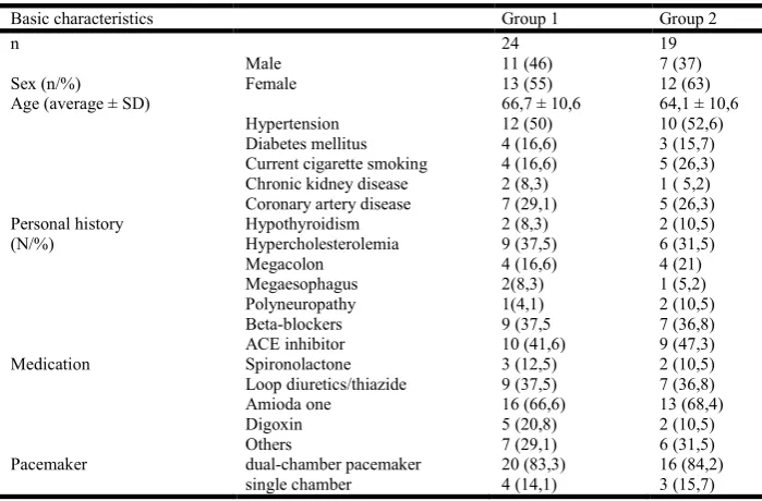
SF-36v2® questionnaires. Regarding the AQUAREL
[image:3.595.126.476.81.311.2]questionnaire, the domains that presented a statistically significant association were chest discomfort (p = 0.001, Test t: 3.64, 95% CI: 10.97 to 38.34) and exercise dyspnea (p <0.001, t test: 4.34, 95%: 16.38 to 44.96). At chest discomfort area, G1 group had a mean score of 88.15, representing a higher level of quality of life than G2 group, with a mean score of 63.50. The same occurred for the dyspnea domain during exercise, with a mean score of 89.88 and 59.21 for groups G1 and G2, respectively. There was a statistical difference in the arrhythmia domain of the AQUAREL questionnaire (p = 0.043; Test t: 4.34 95% CI: 16.38 to 23.22). In the domains of the SF-36v2® questionnaire, 5 were those that presented a statistically significant difference between the two groups: physical aspects (p <0.001); (p <0.001), emotional aspects (p <0.001), and mental health (p <0.001). The scores obtained by the G1 group in these domains were higher than those in G2 group, which shows better quality of life for the patients who performed physical activity. In relation to the functional capacity domains (p = 0.114, Test t: 1.62, 95% CI: -0.78 to 7.04), pain (p = 0.004, Test t: 3.06, 95% CI: 0.91 to 4.46) and social aspects (p = 0.023 T = 2.37, 95% CI: 0.20 to 2.51),
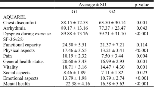
Table 1. Baseline characteristics of the study population
Basic characteristics Group 1 Group 2
n 24 19
Male 11 (46) 7 (37)
Sex (n/%) Female 13 (55) 12 (63)
Age (average ± SD) 66,7 ± 10,6 64,1 ± 10,6
Hypertension 12 (50) 10 (52,6)
Diabetes mellitus 4 (16,6) 3 (15,7) Current cigarette smoking 4 (16,6) 5 (26,3) Chronic kidney disease 2 (8,3) 1 ( 5,2) Coronary artery disease 7 (29,1) 5 (26,3)
Personal history Hypothyroidism 2 (8,3) 2 (10,5)
(N/%) Hypercholesterolemia 9 (37,5) 6 (31,5)
Megacolon 4 (16,6) 4 (21)
Megaesophagus 2(8,3) 1 (5,2)
Polyneuropathy 1(4,1) 2 (10,5)
Beta-blockers 9 (37,5 7 (36,8)
ACE inhibitor 10 (41,6) 9 (47,3)
Medication Spironolactone 3 (12,5) 2 (10,5)
Loop diuretics/thiazide 9 (37,5) 7 (36,8)
Amioda one 16 (66,6) 13 (68,4)
Digoxin 5 (20,8) 2 (10,5)
Others 7 (29,1) 6 (31,5)
Pacemaker dual-chamber pacemaker 20 (83,3) 16 (84,2)
[image:3.595.194.408.342.420.2]single chamber 4 (14,1) 3 (15,7)
Table 2. Telemetric parameters of the study groups before the intervention
Average ± SD p-value Group 1 Group 2
AS 6.58 ± 3.67 7.05 ± 4.09 0.69 AP 93.42 ± 3.67 92.95 ± 4.09 0.69 MSR 5.54 ± 3.82 4.95 ± 3.46 0.60 AT 12.83 ± 6.93 11.21 ± 7.32 0.46 NAT 81.67 ± 9.57 83.84 ± 10.16 0.47 HR 65.38 ± 3.65 65.26 ± 5.06 0.47
mean scores obtained by group G1 were also higher than those of group G2, but no statistical difference was observed between groups. During the study, no adverse events were identified. Participation in the study did not present additional risks to the patients, since they already had the device implanted after a documented clinical indication.
Table 3. Association between the telemetric parameters of the study groups after intervention
Average ± SD p-value
Group 1 Group 2
AS 7.04 ± 2.49 7.05 ± 3.31 0.990 AP 92.96 ± 2.49 92.84 ± 3.17 0.893 MHR 16.00 ± 4.33 8.37 ± 7.27 < 0.001 AT 20.71 ± 8.06 14.21 ± 10.89 0.030 NAT 62.63 ± 10.54 77.26 ± 17.35 0.001 HR 81± 9.36 73.53 ± 13.79 0.053
AP: atrial pacing; AS: atrial sensing; MSR: percentage of time the patient maintained maximum sensor rate during physical activity, AT: percentage of time the sensor interpreted as mild to moderate physical activity (activity time); G1: group submitted to monitored physical activity; G2: control group; HR: mean heart rate in the period; NAT: percentage of the time the sensor interpreted as out of physical activity (non-activity time); SD: standard deviation;
Table 4. Association between the study groups in the analysis of the quality of life, through the AQUAREL and SF-36v2®
questionnaires, after intervention
Average ± SD p-value
G1 G2
AQUAREL
Chest discomfort 88.15 ± 12.53 63.50 ± 30.14 0.001 Arrhythmia 89.17 ± 13.16 77.37 ± 23.47 0.043 Dyspnea during exercise 89.88 ± 13.76 59.21 ± 31.10 <0.001 SF-36v2®
Functional capacity 24.50 ± 5.51 21.37 ± 7.21 0.114 Physical aspects 17.46 ± 3.55 13.21 ± 3.41 <0.001
Pain 10.19 ± 2.32 7.50 ± 3.44 0.004
General health status 20.60 ± 3.43 16.99 ± 2.93 0.001 Vitality 18.71 ± 3.16 14.47 ± 4.30 0.001 Social aspects 8.46 ± 1.89 7.11 ± 1.82 0.023 Emotional aspects 13.79 ± 1.98 10.79 ± 2.74 <0.001 Mental health 22.38 ± 4.16 16.58 ± 5.63 <0.001 SD: standard deviation; G1: group submitted to monitored physical activity; G2: control group; AQUAREL: Assessment of Quality of Life and Related Events; SF-36v2®: Short Form Health Survey 36 version 2.
DISCUSSION
This study investigated the influence of physical activity on the chronotropic incompetence of patients with Chagas disease using a dual sensor pacemaker. The demographic findings point to an involvement of elderly patients, with an age range close to 65 years. No patient had a reduction in ejection fraction, with an overall mean of 54% and a slight variation between the groups (54.59% for the G1 group and 54.33% for the G2 group). These findings negate the possibility of significant heart failure influencing the data obtained, knowing that there was no significant structural heart disease in the group. This work results from a more extensive research initiated in 2013 with the initial objective of determining the dual sensor response in patients with Chagas disease,
especially those with chronotropic incompetence (Menezes et
al., 2015). This previous study showed a limitation in reaching the maximum proposed HR for the patients, due to non-adaptation to physical effort, considering that the difference between the mean HR with the double sensor and that with the accelerometer would have been statistically significant. In the present study, after monitoring one group with physical
activity compared to another without, the group with physical activity reached a range of responses with a higher mean HR due to a longer time of physical activity. Pachón (2015) points out that the dual sensor, which allows more tuning and a higher physiological heart rate, is highly desirable for chagasic cardiopathy, being of fundamental importance the realization of a detailed sensor program, through exercise and/or Holter monitoring, to achieve maximum suppression of arrhythmia with adequate adjustment of heart rate response. This higher physiological adjustment and heart rate during the off-peak period was observed in our study by means of a more appropriate response in the 120 days group with supervised physical activity training.
In the DUSISLOG (comparative single sensor or double sensor) study cited by Pachón (2015), demonstrated that patients with double sensor have significant increase when observed the distance covered in the 6-min WT, quality of life, and percentage of atrial pacing above 70 ppm for a single sensor, only in patients with greater atrial chronotropic disease defined as the absence of intrinsic atrial activity above 60 bpm (0% AS%), which represented 17% of the enrolled population. Patients with moderate congestive heart failure (0-10% AS) showed only a tendency for the symptomatic benefit of speed-sensitive pacing, whereas those with mild or no chronotropic incompetence (> 10% AS%) had less or no benefit. Based on these data, the author concludes that a speed-sensitive pacemaker with at least one sensor is highly desirable for patients with CD and the dual sensor is highly recommended for cases of severe chronotropic insufficiency. The findings of our study are in agreement with our research, taking into account the percentage of AS was 7.04 ± 2.49 in Group 1 (active) and 7.05 ± 3.31 in Group 2 (non-active), which reinforces the benefit in patients with significant chronotropic incompetence. Among the telemetric variables analyzed, the most expressive data was the MSR elevation in patients in G1 from 5.54% ± 3.82% to 16.00% ± 4.33%. It was observed that there was also an increase in G2, but it was lower than G1. This fact suggests that the pacemaker adapted better to the clinical conditions in G1, a fact reinforced by the association found in NAT. specifically, it was observed that the patients had their maximum heart rate almost exclusively during physical exercise. The devices in the G1 group distinguished moments of physical activity more accurately, reducing episodes of erroneous deflagration of stimuli by the pacemaker. These findings indicate a good response in chronotropic incompetence through regular physical activity. However, as our sample was limited, more studies should be conducted in search of more consistent results so that there may be a formal recommendation on regular physical activity for patients with CD and chronotropic incompetence using a dual sensor pacemaker.
[image:4.595.30.288.366.510.2]the randomization was efficient, and this reduces the chances of bias significantly. Our study did not investigate these factors. What we can affirm is that patients who performed regular physical activity had a better quality of life, whether it was a consequence of or only a finding that was not directly related to the intervention. As the literature is still very limited in this sense, further studies should be conducted to prove the beneficial effect of physical activity on the population in question. We identified significant improvement in telemetric data and quality of life parameters in chagasic patients with chronotropic incompetence with a blended sensor pacemaker who performed regular monitored exercise compared with patients who did not exercise. The study had some limitations, mainly the relatively small sample size and study results should not be generalized to other populations.
Acknowledgements
We would like to acknowledge Prof. Edésio Martins for statistical analysis and Editage for technical assistance.
REFERENCES
Baranchuk, A., Healey, J.S., Thorpe, K.E., Morillo, C.A., Nair, G., Crystal, E., Kerr, C.R., Connolly, S.J.,and CTOPP Investigators. 2007. The effect of atrial-based pacing on exercise capacity as measured by the 6-minute walk test: A substudy of the Canadian Trial of Physiological Pacing
(CTOPP). Heart Rhythm. 4, pp.1024-1028.
Barros, R.T., Carvalho, S.M., Silva, M.A., and Borges, J.B. 2014. Evaluation of patients’ quality of life aspects after cardiac pacemaker implantation. Rev Bras Cir Cardiovasc. 29, pp.37-44. doi/10.5935/1678-9741.20140009.
Basile, L., Jansa, J.M., Carlier, Y., Salamanca, D.D., Angheben, A., Bartoloni, A., Seixas, J., Van Gool, T., Canavate, C., Flores-Chavez, M., Jackson, Y., Chiodini, P.L., Albajar-Vinas, P., and Working Group on Chagas Disease. 2011. Chagas disease in European countries: the
challenge of a surveillance system. Euro Surveill. 16,
pp.19968.
Brandt, N.H., Kirkfeldt, R.E., Nielsen, J.C., Mortensen, L.S., Jensen, G.V.H., Johansen, J.B., and Haugan K. 2017. Single lead atrial vs. dual chamber pacing in sick sinus syndrome: extended register-based follow-up in the DANPACE trial. Europace. 19, pp.1981-1987. doi/10. 1093/europace/euw364
Bunch, T.J., Hayes, D.L., and Friedman, P.A. 2009. Clinically Relevant Basics of Pacing and Defibrillation. In: Cardiac Pacing, Defibrillation and Resynchronization [online]. Wiley-Blackwell,Oxford, UK. p. 1–42. doi.wiley.com/ 10.1002/9781444300659.ch1
Cambronero-Cortinas, E. 2016. Epidemiology of Chagas
Disease in Non-Endemic European Countries.
International Cardiovascular Forum Journal. 7, pp.11-14. doi/10.17987/icfj.v7i0.353
Campiolo, D.J. 2016. Correlation between clinical forms of
Chagas disease and metabolism bioenergetic of
Trypanosoma cruzi. [online] Universidade Estadual de Campinas. Available online at http://repositorio. unicamp. br/jspui/handle/REPOSIP/325036
Coman, J., Freedman, R., Koplan, B.A., Reeves, R., Santucci, P., Stolen, K.Q., Kraus, S.M., Meyer, T.E.,and LIFE Study Results. 2008. A blended sensor restores
chronotropic response more favorably than an
accelerometer alone in pacemaker patients: The LIFE
study results. Pacing Clin Electrophysiol. 31, pp.1433-1442. doi.wiley.com/10.1111/j.1540-8159.2008. 01207.x Committee on Perioperative Evaluation (CAPO) Brazilian
Society of Cardiology. 2007. I guidelines for perioperative evaluation. Arq Bras Cardiol. 89, pp.210-237.
Coura, J.R., and Dias, J.C.P. 2009. Epidemiology, control and surveillance of Chagas disease: 100 years after its discovery. Mem Inst Oswaldo Cruz. 104, pp.31-40. Epstein, A.E., DiMarco, J.P., Ellenbogen, K.A., Estes, N.A.M.,
Freedman, R.A., Gettes, L.S., Gillinov, A.M.,Gregoratos, G., Hammill, S.C., Hayes, D.L.,Hlatky, M.A., Newby, L.K., Page, R.L., Schoenfeld, M.H., Silka, M.J., Stevenson, L.W., and Sweeney, M.O. 2013. 2012 ACCF/AHA/HRS focused update incorporated into the ACCF/AHA/HRS 2008 guidelines for device-based therapy of cardiac rhythm abnormalities: a report of the American College of Cardiology Foundation/American Heart Association Task Force on Practice Guidelines and the Heart Rhythm Society. Circulation. 127, pp. e283-352. doi/10.1161/CIR.0b013e318276ce9b
Gillis AM, Russo AM, Ellenbogen KA, Swerdlow CD, Olshansky B, Al-Khatib SM, Beshai, J.F.,McComb, J.M., Nielsen, J.C., Philpott, J.M., and Win-Kuang Shen. 2012. HRS/ACCF Expert Consensus Statement on Pacemaker Device and Mode Selection. J Am Coll Cardiol. 60, pp.682-703.
Manne-Goehler, J., Umeh, C.A., Montgomery, S.P., and Wirtz, V.J. 2016. Estimating the burden of Chagas disease in the United States. PLoS Negl Trop Dis. 10, pp.e0005033. doi: 10.1371/journal.pntd.0005033.
Mendes, F. de S., Sousa, A.S., Souza, F.C., Pinto, V.L., Silva, P.S., Saraiva, R.M., Xavier, S.S., Veloso, H.H., Holanda, M.T., Costa, A.R., Carneiro, F.M., Silva, G.M., Borges, J.P., Tibirica, E., Pinheiro, R.O., Lara, F.A., Hasslocher-Moreno, A.M., Brasil, P.E., and Mediano, M.F. 2016. Effect of physical exercise training in patients with Chagas heart disease: study protocol for a randomized controlled trial (PEACH study). Trials. 17, pp.433. doi: 10.1186/s13063-016-1553-4
Menezes, J.A.S., Silva, A.P., Profahl, G.G., Ottobeli, C., and Louzeiro, J.F. 2015. Chronotropic incompetence in Chagas disease: effectiveness of blended sensor (volume/minute and accelerometer). Rev Bras Cir
Cardiovasc. 30, pp.311-315.
doi/10.5935/1678-9741.20150035
Newman, D., Lau, C., Tang, A.S.L., Irvine, J., Paquette, M., Woodend, K., Dorian, P., Gent, M., Kerr, C., Connolly, S.J., and CTOPP Investigators. 2003. Effect of pacing mode on health-related quality of life in the Canadian Trial of Physiologic Pacing. Am Heart J. 145, pp.430-437. Oliveira, B.G., Velasquez Melendez, G., Rincón, L.G., Ciconelli, R.M., Sousa, L.A. and Ribeiro, A.L. 2008. Health Related Quality of Life in Brazilian Pacemaker Patients. Pacing Clin Electrophysiol. 31, pp.1178-1183. doi:10.1111/j.1540-8159.2008.01159.x
Pachón, J.C. 2015. Chronotropic incompetence in Chagas disease: usefulness of dual sensor pacemaker based on
volume minute and accelerometer. RevBras Cir
Cardiovasc. 30, pp. III-VI.
doi/10.5935/1678-9741.20150011
Rassi, A. Jr., Rassi, A., and Rezende M, J. 2012. American Trypanosomiasis (Chagas Disease). Infect Dis Clin North Am. 26, pp.275-291. doi: 10.1016/j.idc.2012.03.002 Semelka, M., Gera, J., and Usman, S. 2013. Sick sinus
Strasen, J., Williams, T., Ertl, G., Zoller, T., Stich, A., and Ritter, O. 2014. Epidemiology of Chagas disease in Europe: many calculations, little knowledge. Clin Res Cardiol. 103, pp.1-10.
World Health Organization. 2017. Chagas disease (American
trypanosomiasis) [online]. Available online at
http://www.who.int/chagas/en/