INTRODUCTION
Ground state pluripotency is the hallmark of embryonic stem cells (ESCs), defining cells with the potential to produce any somatic cell type or primordial germ cells (PGCs), which establish the germ line. During mouse development, expression of the transcription factor Nanogis essential to the establishment of pluripotency in the primitive ectoderm (epiblast) (Mitsui et al., 2003; Silva et al., 2009). Nanog was discovered on the basis of its ability to rescue ESC self-renewal in the absence of leukemia inhibitory factor (LIF) (Chambers et al., 2003), an activity that is strictly dependent on Nanog homodimers, the formation of which is mediated by the tryptophan repeat (WR) domain (Mullin et al., 2008; Wang et al., 2008). Nanoghas recently been shown to act as a master regulator of pluripotency and its activation marks a final step of pluripotency acquisition during development, or in the reprogramming of somatic nuclei (Silva et al., 2009). Nanog orthologs exist in chick (Lavial et al., 2007) and Anolis (representing reptiles); however, recent sequencing of Xenopus tropicalisdemonstrates that the frog genome does not contain a Nanogortholog (Hellsten et al., 2010), raising the question of how pluripotency evolved in amniotes.
Amphibians are subdivided into two major lineages, the anurans (frogs) and urodeles (salamanders), which diverged from a urodele-like ancestor over 250 million years ago (Anderson et al., 2008; Rage and Rocek, 1989). Embryological evidence indicates that the
amphibian ancestor of amniotes was urodele-like (Bachvarova et al., 2009a; Bachvarova et al., 2009b) and recent work demonstrates that the molecular mechanisms governing mesoderm specification are conserved from urodeles to mammals (Swiers et al., 2010). Most importantly, Nieuwkoop demonstrated that the primitive ectoderm (animal cap) of embryos from axolotls can be induced to form PGCs, as well as somatic cells, in response to inducing signals (Nieuwkoop, 1969; Sutasurya and Nieuwkoop, 1974), suggesting that pluripotency is conserved between urodeles and mammals (Johnson et al., 2001; Johnson et al., 2003a; Johnson et al., 2003b). Here we report a Nanogortholog from axolotls and verify its identity by comparative mapping, structural analysis, and functional studies in ESCs. Our results confirm that mechanisms governing pluripotency are conserved between urodele amphibians and mammals.
MATERIALS AND METHODS
Gene expression analysis in axolotl embryos
Axolotl Nanogwas amplified using degenerate primers and Smart RACE (Clontech). Sequences were deposited in NCBI GenBank with accession number 290886079. Intron/exon boundaries were predicted by homology and cloned from genomic DNA. RNA probes were labeled with digoxigenin-labeled UTP. For hemi-sectioning, embryos fixed in 4% paraformaldehyde were embedded in low-melting-point agarose and bisected before whole-mount in situ hybridization.
Dimerization analysis by protein complementation assay (PCA) NanogcDNAs were inserted into a modified pEGFP-C1 (Clontech) termed pATG. The mWR, ZIP or FKBPv (Ariad) domains were fused into a synthetic KpnI site. PCA fusions were created by cloning inserts 5⬘of the hGL1 and hGL2 fragments bridged by a flexible linker (GlyGlyGlyGlySer)2. The linker-hGL1 and -hGL2 cDNAs were then
inserted into pATG between HindIII-BamHI sites. PCA fusion vectors were co-transfected at a 1:1 ratio with the transfection control pGL3-enhancer vector (Promega). For inducible dimerization, AP20187 (Ariad) was added after transfection. Forty-eight hours after transfection, cells were lysed in 1⫻Passive Lysis Buffer (Promega) and luminescence measured on a glow-max 96-well luminometer.
Development 137, 2973-2980 (2010) doi:10.1242/dev.049262 © 2010. Published by The Company of Biologists Ltd
1Institute of Genetics, School of Biology, University of Nottingham, Queens Medical Centre, Nottingham NG2 2UH, UK. 2School of Veterinary Medicine and Science, University of Nottingham, Sutton Bonington Campus, LE12 5RD, UK. 3Department of Biology, University of Kentucky, Lexington, KY 40506, USA. 4Wolfson Centre for Stem Cells, Tissue Engineering and Modelling, University of Nottingham,
Nottingham NG7 2RD, UK. 5School of Biosciences, University of Nottingham, Sutton Bonington Campus, LE12 5RD, UK.
*Author for correspondence (Andrew.D.Johnson@nottingham.ac.uk) Accepted 23 June 2010
SUMMARY
Cells in the pluripotent ground state can give rise to somatic cells and germ cells, and the acquisition of pluripotency is
dependent on the expression of Nanog. Pluripotency is conserved in the primitive ectoderm of embryos from mammals and
urodele amphibians, and here we report the isolation of a Nanogortholog from axolotls (axNanog). axNanogdoes not contain a
tryptophan repeat domain and is expressed as a monomer in the axolotl animal cap. The monomeric form is sufficient to regulate pluripotency in mouse embryonic stem cells, but axNanog dimers are required to rescue LIF-independent self-renewal. Our results show that protein interactions mediated by Nanog dimerization promote proliferation. More importantly, they demonstrate that the mechanisms governing pluripotency are conserved from urodele amphibians to mammals.
KEY WORDS: Pluripotency, Nanog, Axolotl, Xenopus
Axolotl
Nanog
activity in mouse embryonic stem cells
demonstrates that ground state pluripotency is conserved
from urodele amphibians to mammals
James E. Dixon1, Cinzia Allegrucci1,2, Catherine Redwood1, Kevin Kump3, Yuhong Bian1, Jodie Chatfield1, Yi-Hsien Chen1, Virginie Sottile4, S. Randal Voss3, Ramiro Alberio1,5and Andrew D. Johnson1,*
D
E
V
E
LO
P
M
E
N
2974
Transcriptional response analyses
Cells were co-transfected with firefly reporter vectors (0.25 mg/well), the transfection control pRL-TK vector (0.05 mg/well), and overexpression vectors. Promoter fusions were created from pGL3-basic. Luciferase assays were performed as for PCA analyses.
Cell cultures
Lentivirus-transduced cells were maintained under standard conditions with puromycin (1 mg/ml). To produce Nanog-overexpressing cell lines, NanogcDNAs were subcloned into a modified pSIN-IRES-Puro vector (Addgene) between SpeI and HindIII sites and fused to a C-terminal myc tag. Lentivirus was produced in HEK 293T cells as described (Yu et al., 2007). For LIF withdrawal, mouse ESCs were cultured without LIF; for LIF blocking, mouse ESCs were cultured with 100 ng/ml LIF-blocking antibody (R&D Systems). Cells (200,000 cells/well of a 6-well plate) were passaged after 72 hours, counted, and one-tenth of the cells replated. This was repeated at days 7 and 10. The ratio of cells in the presence versus absence of LIF was recorded. Neural stem cells (NSCs) were isolated from fetal Oct4-GFP mice and were cultured as described (Conti et al., 2005). For gene expression, RNA was extracted with DNase treatment, and cDNA prepared using Superscript III reverse transcriptase (Invitrogen). Relative expression levels were determined by quantitative (Q) PCR using TaqMan Gene Expression Master Mix and TaqMan Gene Expression Assays (Applied Biosystems).
Fusion-mediated reprogramming
ESC⫻NSC fusions were carried out as described (Silva et al., 2006). After fusion, cells were plated on gelatinised 10-cm dishes in ESC media and 24 hours later puromycin and G418 were added for 2 weeks.
Foci assays
For foci assays, NIH 3T3 cells were plated in dishes with 2% FCS-containing medium. Media were replaced every 2 days and foci formation was analyzed after 2 weeks.
Chromatin immunoprecipitation (ChIP)
ChIP was performed using the Magna ChIP A Kit (Millipore); 1⫻106cells
were used per group with a rabbit anti-myc antibody (3 mg, Sigma C3956) or IgG from rabbit serum (3 mg, Sigma I5006). QPCR analyses used primers described by Liang et al. (Liang et al., 2008).
Statistical analyses
Statistical analyses were performed by one-way ANOVA with Tukey’s post-hoc test with P<0.05.
RESULTS AND DISCUSSION
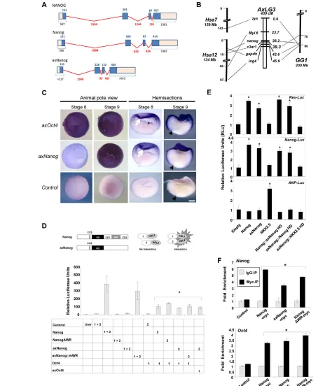
axNanogis expressed in animal caps with axOct4 RNA from blastula animal caps was used to isolate an axolotl Nanog ortholog (axNanog) (see Fig. S1 in the supplementary material). Homology with other Nanog sequences is evident only in the homeodomain (HD) and caspase cleavage site (CCS) (Fujita et al., 2008). axNanog is encoded by four exons, with an intron/exon structure conserved in mammalian Nanoggenes (Fig. 1A). Significantly, axNanogdoes not contain a recognizable WR domain.
AxNanog orthology was established by comparative mapping (Fig. 1B). Partial sequences were amplified and sequenced to identify a single-nucleotide polymorphism (SNP) that allowed us to differentiate Nanogalleles inA. mexicanum and A. t. tigrinum. Individuals from an interspecific A. mexicanum⫻A. t. tigrinum mapping cross were genotyped for Nanog alleles (Smith et al., 2005), and these data were used to locate the position of Nanog relative to genes (zyx,c3ar1,gapdh) flanking Nanogon human chromosome 12 and chicken chromosome 1. All three targeted loci mapped to the position of Nanog, with c3ar1and gapdh showing especially tight linkage (<5 cM). The results identify Nanog, c3ar1,
zyx and gapdh as marking a conserved syntenic chromosomal region, establishing the orthology of these loci among axolotl, human and chicken.
Whole-mount in situ hybridization (WISH) showed axNanog expression commencing at stage 9 in animal caps, following axOct4 activation at the midblastula transition (stage 8; Fig. 1C). Expression of Oct4(Pou5f1) before Nanogin the pluripotent domain resembles the sequence of events in mouse development (Chambers et al., 2003). AxNanogexpression peaked at mid-gastrula (stage 10.5), but was undetectable once gastrulation was completed (not shown); therefore, axNanogand axOct4are co-expressed in animal cap cells during the interval in which they are pluripotent.
axNanog is a monomer and physically interacts with Oct4
To identify conserved Nanog functions, we focused on the biochemical properties of axNanog. Nanog functions as a dimer in ESCs (Wang et al., 2008). We used a protein complementation assay (PCA) system (Remy and Michnick, 2006) to test whether axNanog forms homodimers and associates with other proteins. We tested the system by fusing Nanogto domains 1 and 2 of humanized Gaussia luciferase (hGL1 and hGL2; Fig. 1D, top), and co-expressed these constructs in HEK 293T (293T) cells. Luminescence was only detected from constructs containing the WR domain (see Fig. S2 in the supplementary material), confirming that this domain as necessary and sufficient for Nanog homodimerization (Mullin et al., 2008; Wang et al., 2008).
Co-expression of both axNanog::hGL fusions (1 and 2) did not reconstitute luciferase activity (Fig. 1D, bottom), indicating function as a monomer. We fused the mouse WR (mWR) domain to axNanog (axNanog::mWR); co-expression of these fusions produced a significant luciferase signal. Nanog physically interacts with Oct4 in ESCs (Wang et al., 2006; Liang et al., 2008), so we assessed axNanog and Nanog interaction with Oct4 by PCA. Monomers and homodimers of both proteins interacted with Oct4. We also tested for association of axNanog with axOct4 (Bachvarova et al., 2004), and it interacted efficiently, indicating that this complex is conserved in pluripotent cells.
Nanog monomers bind and activate Nanog targets We asked whether axNanog could drive transcription from mammalian promoters. Rex1(Zfp42) and Nanogpromoters were fused to a luciferase reporter and transfected into 293T cells. Transfection of axNanogand Nanogactivated expression to similar levels, but did not activate the atrial natriuretic peptide (ANP; NPPA) gene promoter, which is a target of NKX2.5, a homeobox transcription factor that is closely related to Nanog (Lyons et al., 1995). NKX2.5 showed the reciprocal pattern (Fig. 1E). Transcriptional activation of target genes is driven by the homeodomain of these molecules, as demonstrated by homeodomain swapping experiments (Fig. 1E).
We asked whether axNanog could bind native targets in the mouse genome. Myc-tagged fusions to Nanog, axNanog and NanogDWR (from which the WR domain is deleted) were expressed in ESCs and used for ChIP. There was target sequence enrichment (Liang et al., 2008) from the promoters of Nanogand Oct4(Fig. 1F). These experiments demonstrate that dimerization is not required for Nanog target recognition and that DNA binding specificity is conserved between Nanog and axNanog homeodomains.
RESEARCH REPORT Development 137 (18)
Fig. 1. Conservation of Nanoggenomic structure, expression profile and transcriptional activity.(A) Intron/exon structures of NANOG
(human), Nanog(mouse) and axNanog(axolotl) are aligned. Blue boxes denote protein coding regions. Numbers in blue represent amino acids. Numbers in red are intron lengths; ND, not determined. Black numbers indicate combined length of protein coding region with 5⬘and 3⬘
untranslated regions. (B) Conserved synteny of Nanoggenes. Genes linked to axNanogon linkage group 3 (AxLG3) map to human chromosomes 7 and 12 (Hsa7, Hsa12) and chicken chromosome 1 (GG1). (C) Whole-mount in situ hybridization (WISH) showing co-expression of axNanogand
axOct4in the animal cap of blastulae (arrow). Arrowhead points to blastopore. (D) Protein complementation assay (PCA) analyses of axNanog protein-protein interaction. (Top) Vectors express the interactors (cDNA1 and cDNA2) fused to hGL1 and hGL2. (Bottom) PCA shows that
axNanog::WR fusions form homodimers, and axNanog monomers interact with axOct4. *P<0.05. (E) Reporter expression from Nanog targets after co-transfection with the indicated constructs (:: indicating substitution with homeodomain, HD). *P<0.05. (F) QPCR quantification of Nanogand
Oct4promoters after immunoprecipitation by anti-myc antibody (mean±s.d.; n=3). Results show fold enrichment of precipitated DNA relative to a
1/100 dilution of input chromatin. *P<0.05.
D
E
V
E
LO
P
M
E
N
2976
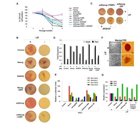
Dimerization of axNanog is necessary and sufficient for rescue of LIF-independent self-renewal
The absence of a WR domain in axNanog prompted two questions. First, can axNanog homodimers rescue self-renewal after LIF withdrawal? Second, can WR function be replaced by unrelated dimerization domains?
We replaced the WR domain of Nanog and axNanog::mWR with heterologous dimerization domains (see Fig. S3A in the supplementary material): Nanog::ZIP and axNanog::ZIP contain the GCN4 leucine zipper (ZIP), which induces dimerization. We also created molecules fused to an FKBP domain (FKBPv), which dimerize subject to induction: FKBP domains form a low level of dimers, which is enhanced by the drug AP20187. We demonstrated that each of these molecules dimerizes by PCA (see Fig. S3B,C in the supplementary material).
Puromycin (Puro)-resistant ESC lines overexpressing axNanog transgenes (~10- to 20-fold over background; see Fig. S3D in the supplementary material) were then tested for self-renewal after LIF withdrawal. By passage 4 (P4) after LIF withdrawal (LIF–), cell numbers in control cultures were only 1% (Fig. 2A) of those in cultures containing LIF (LIF+). Surviving cells exhibited low levels of alkaline phosphatase (AP) activity and appeared to be differentiated. Cell numbers in Nanog-overexpressing lines were ~88% of those in LIF+controls and these cells maintained high levels of AP with normal ESC morphology (Fig. 2B). However, lines expressing Nanog variants without the WR domain (axNanogand NanogDWR) behaved like LIF–cultures. Against this background, rescue of the axNanog::mWR line was clear. After four passages, ~30% of cells in LIF+cultures were retained and these were undifferentiated with high AP levels. Importantly, rescue by axNanog::mWR is equivalent to rescue by human NANOG, which is less efficient than mouse Nanogin this assay (Chambers et al., 2003). Furthermore, because endogenous Nanog RNA levels are not elevated by transgene expression (see Fig. S3D in the supplementary material), we attribute rescue to elevated Nanog activity resulting from transfected axNanog variants.
We found that axNanog-ZIPrescued self-renewal, with cultures showing ~20-40% of the cell numbers in LIF+controls after P4 (Fig. 2A). Again, rescued cells retained normal morphology and AP activity (Fig. 2C). Parallel cultures of Nanog::FKBPv and axNanog::FKBPv showed similar results to each other (cell numbers were ~27% and ~16% of those of control cells at P4, respectively), but the level of rescue increased ~2- to 3-fold (~47% and ~29% at P4, respectively) after supplementation with AP20187, confirming the importance of dimerization. Gene expression analysis after three passages showed that axNanog::mWRlines grown without LIF maintained Oct4and Tert expression (Fig. 2D) at levels equivalent to cells expressing NANOG.
Nanog homodimers associate with other transcription factors in protein complexes required for self-renewal (Wang et al., 2006; Liang et al., 2008). Fusions of hGL2 with Nac1 (Nacc1), Sall4, Dax1 (Nr0b1) and Hdac1 were tested for binding to axNanog homodimers. These factors interacted with Nanog and axNanog homodimers (Fig. 2E), although Nac1 interacted only with molecules containing the WR, as expected (Ma et al., 2009). By contrast, Hdac1 interaction does not require dimerization; it interacted equally well with Nanog or axNanog monomers. Therefore, conserved Nanog monomers interact with a subset of the factors that have been identified in the extended pluripotency network as required to mediate ESC self-renewal (Kim et al., 2008).
Rescue by axNanog::WR is less efficient than by Nanog. To test whether axNanoghas a more limited ability to sustain self-renewal, we passaged long-term cultures without LIF. Control ESCs could not survive beyond 6-7 passages (see Table S1 in the supplementary material). Cultures expressing axNanog and NanogDWRcould only be maintained for 7-8 passages. However, cultures expressing any dimerized form of axNanog could be cultured long term without LIF (for more than 25 passages and 60 days). All cell lines retained AP activity, although at lower intensity. They also adopted a more flattened morphology than cells maintained in LIF, resembling cells with epiblast-like identity (Guo et al., 2009). Furthermore, these cultures were indistinguishable from lines expressing elevated Nanog levels (Fig. 2F). QPCR at P25 confirmed the epiblast-like character of rescued cells, showing reduced Oct4and Rex1expression, with higher levels of Fgf5 (Fig. 2G). These data show that cells rescued by dimerized axNanog are equivalent to those overexpressing Nanog; thus, Nanogactivity is conserved between amphibians and mammals.
Nanog monomers regulate pluripotency
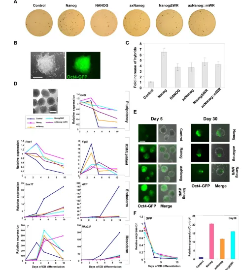
Nanog overexpression in ESCs enhances the transfer of pluripotency to neural stem cells (NSCs) after fusion (Silva et al., 2006; Silva et al., 2009). Assuming programming of pluripotency is a conserved Nanog function, we reasoned that monomers would enhance reprogramming. To test this, we prepared fetal NSCs from mouse embryos carrying an Oct4-GFPtransgene, and then made these neomycin (Neo) resistant. NSCs were fused to our ESC lines overexpressing monomers or homodimers of Nanog, NANOG or axNanog. Reprogramming efficiency was assessed by the number of GFP+PuroRNeoRhybrid colonies after 2 weeks of selection, followed by AP staining (Fig. 3A,B). Hybrid colonies appeared 5-7 days post-fusion and were expanded under standard conditions.
NSC reprogramming was enhanced 5- to 7-fold by Nanog overexpression (Fig. 3C), which is comparable to the findings of previous work (Wong et al., 2008). AxNanog, NANOG, NanogDWR and axNanog::mWR enhanced reprogramming at similar levels (3- to 5-fold), indicating that monomers and dimers function similarly. Furthermore, axNanog participates with factors in ESCs to program pluripotency in mammalian cells.
We next tested whether axNanog could repress differentiation in embryoid bodies (EBs) (Darr et al., 2006; Hamazaki et al., 2004). EBs were produced from Nanog-expressing ESCs and gene expression was assayed at various time points (Fig. 3D). In empty vector control EBs, Oct4 expression decreased to background levels within ~5 days. By day 3, Rex1levels decreased and Fgf5 increased, suggesting conversion to epiblast-like cells. Fgf5was extinguished between days 3 and 5, concomitant with specification to endoderm (Sox17, Afp) and mesoderm (T, Nkx2.5). By contrast, cells expressing Nanog variants delayed these effects. Downregulation of Oct4and Rex1was delayed in each line, and low Fgf5levels were maintained until at least day 5. In addition, mesoderm and endoderm markers were inhibited equally by monomers or dimers of Nanog or axNanog, indicating suppression of lineage commitment.
EBs from hybrid cells of NSC:ESC fusions were assessed for GFP expression after long-term culture. All cells were GFP positive at day 0, and at day 5 GFP was barely detectable from control EBs (Fig. 3E,F); however, EBs overexpressing Nanog, axNanogor NanogDWRretained a significant fraction of GFP-positive cells. By day 30, no GFP-expressing cells were detected in control EBs, whereas GFP-positive patches were apparent in a significant proportion of EBs overexpressing each Nanogconstruct.
RESEARCH REPORT Development 137 (18)
These data show that Nanogand axNanoghave similar abilities to prevent differentiation in EBs and to promote retention of pluripotent cells, demonstrating that the regulation of pluripotency is conserved in monomers. However, EBs expressing Nanogwere larger than those expressing monomers at day 30, but not day 5 (not shown), suggesting that homodimers enhance cell proliferation.
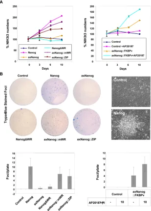
Nanog dimers transform somatic cells, promoting rapid proliferation
Accelerated proliferation and a transformed phenotype result from Nanog expression in NIH 3T3 (3T3) fibroblasts (Piestun et al., 2006). Based on our results with EBs, we asked whether this was due to dimerization. We produced 3T3 lines overexpressing the Nanog transgenes and confirmed that Nanog increases cell proliferation. Similar results were obtained from cells expressing
axNanog::mWR, axNanog::ZIPor axNanog::FKBPv, and effects of axNanog::FKBPvwere enhanced by AP20187. By contrast, lines expressing NanogDWR or axNanog monomers grew at rates similar to controls (Fig. 4A). Therefore, enhanced growth rates induced by Nanogin somatic cells result from dimerization. To test whether dimerization per se causes transformation of 3T3 cells, we performed foci formation assays (Piestun et al., 2006). Cells expressing Nanogformed small compacted foci after 2 weeks. Foci were also formed by cells expressing other dimerized Nanog molecules, with enhanced foci formation by axNanog::FKBPv after AP20187 addition (Fig. 4B). Cells expressing monomers, however, did not form foci, indicating that transformation is induced by dimerization.
[image:5.612.53.509.55.459.2]Together, our results indicate that the role of the WR domain is to promote proliferation, which occurs as a result of novel protein interactions specific to Nanog homodimers; pluripotency, Fig. 2. AxNanog dimerization is required to rescue ESC self-renewal.(A) ESC counts of cultures transfected with Nanogvariants grown for four passages with or without LIF. (B) Representative plates showing alkaline phosphatase (AP) expression (red) of ESC cultures expressing Nanog or axNanog monomers or homodimers with and without LIF. (C) Representative cultures of Nanog::ZIP-andFKBPv-expressing ESCs cultured with and without LIF or AP20187. (D) QPCR analysis ofOct4and Tert expression in Nanogvariant-expressing ESC cultures with or without LIF (analyzed at P3). Results show fold increase relative to control calibrator sample (mean±s.d.; n=3). (E) PCA showing interaction of axNanog with Nac1, Dax1, Sall4 and Hdac1 (mean±s.d.; n=3). (F) Representative images of cultures and colonies showing reduced AP activity (red) and flattened morphology of Nanog-expressing ESCs cultured for 25 passages without LIF. Scale bar: 200mm. (G) QPCR showing levels of Oct4, Rex1and Fgf5in cultures rescued by Nanog or axNanog dimers (mean±s.d.; n=3).
D
E
V
E
LO
P
M
E
N
2978
however, is regulated by the complexes that associate with monomers. Among the factors identified (Fig. 2E) that are exclusively associated with Nanog dimers, Nac1, Sall4 and Dax1 are known to enhance proliferation in other cell types
(Garcia-Aragoncillo et al., 2008; Ma et al., 2009; Yuri et al., 2009), and we presume that their association with Nanog in ESCs plays a similar role. Interestingly, the WR domain is exclusive to eutherian mammals (see Fig. S1 in the supplementary material)
[image:6.612.50.541.65.614.2]RESEARCH REPORT Development 137 (18)
Fig. 3.AxNanogenhances fusion-based reprogramming and prevents embryoid body (EB) differentiation. (A) Hybrid colonies from ESC fusions (expressing Nanogand puromycin resistance) with NSCs (expressing Oct4-GFPand Neo). (B) Reprogrammed hybrid colony. (C) Quantification of hybrids colonies (mean±s.d.; n=3). (D) Representative image of EB differentiation assay. QPCR analyses of EBs expressing axNanogat 3, 5 or 10 days of differentiation (mean±s.d; n=3). (E) Oct4-GFP fluorescence is maintained in EBs from ESC:NSC hybrids expressing axNanog. (F) QPCR analyses of Oct4-GFPexpression during hybrid EB differentiation (mean±s.d.; n=3). Scale bars: 200mm in B; 500mm in D,E.
D
E
V
E
LO
P
M
E
N
and our data suggest that its evolution is associated with the rapid expansion of the epiblast during development. In vitro, cell proliferation promoted by the WR domain is integral to the isolation of self-renewing pluripotent stem cells from early eutherian embryos (Alberio et al., 2010; Brons et al., 2007; Buehr et al., 2008; Evans and Kaufman, 1981; Honda et al., 2009; Schneider et al., 2007; Tesar et al., 2007; Thomson et al., 1998).
Conclusions
The identification of a Nanog ortholog in axolotls confirms that the pluripotent ground state is conserved in vertebrates. We propose that deletion of Nanog from the frog genome was enabled by the evolution of germ plasm in frogs, which repositioned PGCs to the vegetal hemisphere, and frog animal caps do not therefore initiate development from a pluripotent ground state. In addition to the absence of Nanog, the genome sequence of X. tropicalis also reveals a lineage-specific multiplication of many developmental regulatory genes, notably the Mixand Nodalgene families, which are represented by one and two genes, respectively, in axolotls (Swiers et al., 2010).
Importantly, the axolotl complement of Nodal and Mixgenes represents the predicted state of the tetrapod ancestor (Hellsten et al., 2010), and is consistent with the hypothesis that the molecular mechanisms governing early development are conserved from primitive chordates to mammals, but only in those organisms that must pass through ground state pluripotency to establish the germ line (Bachvarova et al., 2009a; Bachvarova et al., 2009b; Johnson et al., 2001; Johnson et al., 2003a; Johnson et al., 2003b). We view the identification of the conserved pluripotency network, which interacts with Nanog monomers, as a valuable tool for understanding how to direct the development of ESCs and for the reprogramming of somatic nuclei to pluripotency.
Acknowledgements
[image:7.612.52.361.62.492.2]We thank R. Yeomans, C. De Sousa, C. Calladine and L. Worrall for their contributions; A. Surani for Oct4-GFPtransgenic mice; S. Michnick for PCA constructs; P. Shaw, G. Morgan and F. Sablitzky for improvement of the manuscript; and Matt Loose for many discussions and for important help with genomics. Dimerization reagents were provided by Ariad, USA. This work was funded by EvoCell, Nottingham (UK), and the Medical Research Council (UK). Deposited in PMC for release after 6 months.
Fig. 4. Transformation and proliferation are enhanced in somatic cells by Nanog
homodimers.(A) Enhanced proliferation of 3T3 lines expressing Nanog or axNanog dimers. Shown are cell counts of triplicate experiments presented as a percentage of cell numbers from control cell lines (mean±s.d.; n=3). (B) Foci are induced by dimers of Nanog or axNanog. Representative foci (top) and number of foci (bottom) in cultures (mean±s.d; n=3). Scale bars: 100mm.
D
E
V
E
LO
P
M
E
N
2980
Competing interests statement
The authors declare no competing financial interests.
Supplementary material
Supplementary material for this article is available at
http://dev.biologists.org/lookup/suppl/doi:10.1242/dev.049262/-/DC1
References
Alberio, R., Croxall, N. and Allegrucci, C. (2010). Pig epiblast stem cells depend on activin/nodal signaling for pluripotency and self renewal. Stem Cells Dev. (in press).
Anderson, J. S., Reisz, R. R., Scott, D., Frobisch, N. B. and Sumida, S. S.
(2008). A stem batrachian from the early Permian of Texas and the origin of frogs and salamanders. Nature453, 515-518.
Bachvarova, R. F., Masi, T., Drum, M., Parker, N., Mason, K., Patient, R. and Johnson, A. D.(2004). Gene expression in the axolotl germ line: Axdazl, Axvh, Axoct-4, and Axkit. Dev. Dyn.231, 871-880.
Bachvarova, R. F., Crother, B. I. and Johnson, A. D.(2009a). Evolution of germ cell development in tetrapods: comparison of urodeles and amniotes. Evol. Dev.
11, 603-609.
Bachvarova, R. F., Crother, B. I., Manova, K., Chatfield, J., Shoemaker, C. M., Crews, D. P. and Johnson, A. D.(2009b). Expression of Dazl and Vasa in turtle embryos and ovaries: evidence for inductive specification of germ cells. Evol. Dev.11, 525-534.
Brons, I. G., Smithers, L. E., Trotter, M. W., Rugg-Gunn, P., Sun, B., Chuva de Sousa Lopes, S. M., Howlett, S. K., Clarkson, A., Ahrlund-Richter, L., Pedersen, R. A. et al.(2007). Derivation of pluripotent epiblast stem cells from mammalian embryos. Nature448, 191-195.
Buehr, M., Meek, S., Blair, K., Yang, J., Ure, J., Silva, J., McLay, R., Hall, J., Ying, Q. L. and Smith, A.(2008). Capture of authentic embryonic stem cells from rat blastocysts. Cell135, 1287-1298.
Chambers, I., Colby, D., Robertson, M., Nichols, J., Lee, S., Tweedie, S. and Smith, A.(2003). Functional expression cloning of Nanog, a pluripotency sustaining factor in embryonic stem cells. Cell113, 643-655.
Conti, L., Pollard, S. M., Gorba, T., Reitano, E., Toselli, M., Biella, G., Sun, Y., Sanzone, S., Ying, Q. L., Cattaneo, E. et al.(2005). Niche-independent symmetrical self-renewal of a mammalian tissue stem cell. PLoS Biol. 3, e283.
Darr, H., Mayshar, Y. and Benvenisty, N.(2006). Overexpression of NANOG in human ES cells enables feeder-free growth while inducing primitive ectoderm features. Development133, 1193-1201.
Evans, M. J. and Kaufman, M. H.(1981). Establishment in culture of pluripotential cells from mouse embryos. Nature292, 154-156.
Fujita, J., Crane, A. M., Souza, M. K., Dejosez, M., Kyba, M., Flavell, R. A., Thomson, J. A. and Zwaka, T. P.(2008). Caspase activity mediates the differentiation of embryonic stem cells. Cell Stem Cell2, 595-601.
Garcia-Aragoncillo, E., Carrillo, J., Lalli, E., Agra, N., Gomez-Lopez, G., Pestana, A. and Alonso, J.(2008). DAX1, a direct target of EWS/FLI1 oncoprotein, is a principal regulator of cell-cycle progression in Ewing’s tumor cells. Oncogene27, 6034-6043.
Guo, G., Yang, J., Nichols, J., Hall, J. S., Eyres, I., Mansfield, W. and Smith, A.
(2009). Klf4 reverts developmentally programmed restriction of ground state pluripotency. Development136, 1063-1069.
Hamazaki, T., Oka, M., Yamanaka, S. and Terada, N.(2004). Aggregation of embryonic stem cells induces Nanog repression and primitive endoderm differentiation. J. Cell Sci. 117, 5681-5686.
Hellsten, U., Harland, R. M., Gilchrist, M. J., Hendrix, D., Jurka, J., Kapitonov, V., Ovcharenko, I., Putnam, N. H., Shu, S., Taher, L. et al.
(2010). The genome of the Western clawed frog Xenopus tropicalis. Science
328, 633-636.
Honda, A., Hirose, M. and Ogura, A.(2009). Basic FGF and Activin/Nodal but not LIF signaling sustain undifferentiated status of rabbit embryonic stem cells. Exp. Cell Res. 315, 2033-2042.
Johnson, A. D., Bachvarova, R. F., Drum, M. and Masi, T.(2001). Expression of axolotl DAZL RNA, a marker of germ plasm: widespread maternal RNA and onset of expression in germ cells approaching the gonad. Dev. Biol. 234, 402-415.
Johnson, A. D., Crother, B., White, M. E., Patient, R., Bachvarova, R. F., Drum, M. and Masi, T.(2003a). Regulative germ cell specification in axolotl embryos: a primitive trait conserved in the mammalian lineage. Philos. Trans. R. Soc. Lond. B Biol. Sci.358, 1371-1379.
Johnson, A. D., Drum, M., Bachvarova, R. F., Masi, T., White, M. E. and Crother, B. I.(2003b). Evolution of predetermined germ cells in vertebrate embryos: implications for macroevolution. Evol. Dev.5, 414-431.
Kim, J., Chu, J., Shen, X., Wang, J. and Orkin, S. H.(2008). An extended transcriptional network for pluripotency of embryonic stem cells. Cell132, 1049-1061.
Lavial, F., Acloque, H., Bertocchini, F., Macleod, D. J., Boast, S., Bachelard, E., Montillet, G., Thenot, S., Sang, H. M., Stern, C. D. et al.(2007). The Oct4 homologue PouV and Nanog regulate pluripotency in chicken embryonic stem cells. Development134, 3549-3563.
Liang, J., Wan, M., Zhang, Y., Gu, P., Xin, H., Jung, S. Y., Qin, J., Wong, J., Cooney, A. J., Liu, D. et al.(2008). Nanog and Oct4 associate with unique transcriptional repression complexes in embryonic stem cells. Nat. Cell Biol. 10, 731-739.
Lyons, I., Parsons, L. M., Hartley, L., Li, R., Andrews, J. E., Robb, L. and Harvey, R. P.(1995). Myogenic and morphogenetic defects in the heart tubes of murine embryos lacking the homeo box gene Nkx2-5. Genes Dev.9, 1654-1666.
Ma, T., Wang, Z., Guo, Y. and Pei, D.(2009). The C-terminal pentapeptide of Nanog tryptophan repeat domain interacts with Nac1 and regulates stem cell proliferation but not pluripotency. J. Biol. Chem.284, 16071-16081.
Mitsui, K., Tokuzawa, Y., Itoh, H., Segawa, K., Murakami, M., Takahashi, K., Maruyama, M., Maeda, M. and Yamanaka, S.(2003). The homeoprotein Nanog is required for maintenance of pluripotency in mouse epiblast and ES cells. Cell113, 631-642.
Mullin, N. P., Yates, A., Rowe, A. J., Nijmeijer, B., Colby, D., Barlow, P. N., Walkinshaw, M. D. and Chambers, I.(2008). The pluripotency rheostat Nanog functions as a dimer. Biochem. J.411, 227-231.
Nieuwkoop, P. D.(1969). The formation of the mesoderm in urodele amphibians. I. Induction by the endoderm. Wilhelm Roux Arch.162, 341-373.
Piestun, D., Kochupurakkal, B. S., Jacob-Hirsch, J., Zeligson, S., Koudritsky, M., Domany, E., Amariglio, N., Rechavi, G. and Givol, D.(2006). Nanog transforms NIH3T3 cells and targets cell-type restricted genes. Biochem. Biophys. Res. Commun.343, 279-285.
Rage, J. C. and Rocek, Z.(1989). Redescription of Triadobatrachus massinoti, an anuran amphibian from the early Triassic. Palaeontolographica206, 1-16.
Remy, I. and Michnick, S. W.(2006). A highly sensitive protein-protein interaction assay based on Gaussia luciferase.Nat. Methods3, 977-979.
Schneider, M. R., Adler, H., Braun, J., Kienzle, B., Wolf, E. and Kolb, H. J.
(2007). Canine embryo-derived stem cells-toward clinically relevant animal models for evaluating efficacy and safety of cell therapies. Stem Cells25, 1850-1851.
Silva, J., Chambers, I., Pollard, S. and Smith, A.(2006). Nanog promotes transfer of pluripotency after cell fusion. Nature441, 997-1001.
Silva, J., Nichols, J., Theunissen, T. W., Guo, G., van Oosten, A. L., Barrandon, O., Wray, J., Yamanaka, S., Chambers, I. and Smith, A.
(2009). Nanog is the gateway to the pluripotent ground state. Cell138, 722-737.
Smith, J. J., Kump, D. K., Walker, J. A., Parichy, D. M. and Voss, S. R.(2005). A comprehensive expressed sequence tag linkage map for tiger salamander and Mexican axolotl: enabling gene mapping and comparative genomics in Ambystoma. Genetics171, 1161-1171.
Sutasurya, L. A. and Nieuwkoop, P. D.(1974). The induction of the primordial germ cells in the urodeles. Wilhelm Roux Arch.175, 199-220.
Swiers, G., Chen, Y. H., Johnson, A. D. and Loose, M.(2010). A conserved mechanism for vertebrate mesoderm specification in urodele amphibians and mammals. Dev. Biol. 343, 138-152.
Tesar, P. J., Chenoweth, J. G., Brook, F. A., Davies, T. J., Evans, E. P., Mack, D. L., Gardner, R. L. and McKay, R. D.(2007). New cell lines from mouse epiblast share defining features with human embryonic stem cells. Nature448, 196-199.
Thomson, J. A., Itskovitz-Eldor, J., Shapiro, S. S., Waknitz, M. A., Swiergiel, J. J., Marshall, V. S. and Jones, J. M.(1998). Embryonic stem cell lines derived from human blastocysts. Science282, 1145-1147.
Wang, J., Rao, S., Chu, J., Shen, X., Levasseur, D. N., Theunissen, T. W. and Orkin, S. H.(2006). A protein interaction network for pluripotency of embryonic stem cells. Nature444, 364-368.
Wang, J., Levasseur, D. N. and Orkin, S. H.(2008). Requirement of Nanog dimerization for stem cell self-renewal and pluripotency. Proc. Natl. Acad. Sci. USA105, 6326-6331.
Wong, C. C., Gaspar-Maia, A., Ramalho-Santos, M. and Reijo Pera, R. A.
(2008). High-efficiency stem cell fusion-mediated assay reveals Sall4 as an enhancer of reprogramming. PLoS ONE3, e1955.
Yu, J., Vodyanik, M. A., Smuga-Otto, K., Antosiewicz-Bourget, J., Frane, J. L., Tian, S., Nie, J., Jonsdottir, G. A., Ruotti, V., Stewart, R. et al.(2007). Induced pluripotent stem cell lines derived from human somatic cells. Science
318, 1917-1920.
Yuri, S., Fujimura, S., Nimura, K., Takeda, N., Toyooka, Y., Fujimura, Y., Aburatani, H., Ura, K., Koseki, H., Niwa, H. et al.(2009). Sall4 is essential for stabilization, but not for pluripotency, of embryonic stem cells by repressing aberrant trophectoderm gene expression. Stem Cells27, 796-805.
RESEARCH REPORT Development 137 (18)