http://dx.doi.org/10.4236/ijcm.2014.518143
How to cite this paper: Tracy, J.C., Kim, W.S. and Scott, A.R. (2014) The Versatility of Acellular Fetal Bovine Dermal Matrix for Head and Neck Surgical Reconstruction in Children. International Journal of Clinical Medicine, 5, 1119-1124.
http://dx.doi.org/10.4236/ijcm.2014.518143
The Versatility of Acellular Fetal Bovine
Dermal Matrix for Head and Neck Surgical
Reconstruction in Children
Jeremiah C. Tracy1, William S. Kim1, Andrew R. Scott1,2*
1Department of Otolaryngology-Head and Neck Surgery, Tufts Medical Center, Boston, USA
2Division of Pediatric Otolaryngology and Facial Plastic Surgery, Floating Hospital for Children, Boston, USA
Email: *ascott@tuftsmedicalcenter.org
Received 23 July 2014; revised 22 August 2014; accepted 17 September 2014 Copyright © 2014 by authors and Scientific Research Publishing Inc.
This work is licensed under the Creative Commons Attribution International License (CC BY).
http://creativecommons.org/licenses/by/4.0/
Abstract
Objectives: To describe the versatility of acellular fetal bovine dermal matrix as an alternative to human cadaveric allograft for head and neck reconstructive procedures in children. Study Design: Case series with chart review. Methods: A database of pediatric operative procedures was queried for the use of acellular fetal bovine dermal matrix over a 16-month period. Indications for recon-struction were assessed and initial parental and surgeon satisfaction with the product were noted. Results: During the time period of 3/2012 and 7/2013 a total of 8 reconstructive procedures were performed on pediatric patients using acellular fetal bovine dermal matrix. Indications for use va-ried and included open and transnasal endoscopic repair of encephaloceles and soft tissue recon-structions including lateral pharyngeal wall repair, cleft palate repair, and facial recontouring op-erations. Acellular fetal bovine dermal matrix had a subjectively increased ease of use as com-pared to the surgeon’s prior experience with human cadaveric acellular dermis. Every parent vo-calized a greater comfort level with the use of a bovine product over the alternative of human ca-daveric tissue. The cost of acellular fetal bovine dermal matrix is slightly lower than the cost of human cadaveric acellular dermis. Conclusions: Acellular fetal bovine dermal matrix appears to be an acceptable alternative to human cadaveric acellular dermis for various forms of head and neck soft tissue reconstruction in children. Further prospective studies are warranted to assess for any differences in the long-term efficacy of this product as compared to other forms of allograft reconstruction.
Keywords
Acellular Fetal Bovine Dermal Matrix (SurgiMend), Human Cadaveric Acellular Dermal Allograft (AlloDerm), Skull Base, Cerebrospinal Fluid Leak Repair, Pediatric, Atrophic Scar, Parotidectomy
*
Defect, Parry Romberg, Cleft Palate
1. Introduction
The field of pediatric head and neck reconstruction continues to evolve. There is a growing armamentarium of techniques and materials available to the reconstructive surgeon, with the introduction of injectable fillers and biological products continuing outpace research into the safety and efficacy of these modalities in children. Ad-ditionally, application of the endoscope has allowed for the transition from open to endoscopic approaches to pediatric skull base repair using techniques that rely on a variety of materials including tissue glues, allogeneic implants, autografts, and mucosal grafts or flaps [1]. Human cadaveric dermal matrices have found broad appli-cation in reconstructive head and neck surgery, including use in septoplasty, parotidectomy, and oral cavity re-construction [2]-[5]. The use of human cadaveric acellular dermal allograft (AlloDerm, LifeCell, Bridgewater, NJ) has become standard practice in many forms of pediatric soft tissue and skull base repair [6]-[9]. In skull base reconstruction this product is generally used as an in-lay graft as part of a multilayered closure; many dif-ferent nuances of application have been described [10]-[13]. The relative advantages of allograft reconstruction may include product availability, ease of endoscopic tissue manipulation, and avoidance of donor site morbidity (relative to autologous tissue).
Recently a new biological product, acellular fetal bovine dermal matrix (SurgiMend, TEI Biosciences, Wal-tham, MA), was approved by the Food and Drug Administration as a medical device. Acellular fetal bovine dermal matrix is comprised of tissue that has been terminally sterilized, with theoretical disease transmission risk that is favorable compared to human cadaveric dermis. This product has been applied in wound closure in both cosmetic and general surgery procedures in both adults and children [14][15]. We recently presented three cases comprising our initial experience using this product for pediatric skull base reconstruction [16]. Within this present series we report on our growing experience using acellular fetal bovine dermal matrix for endoscop-ic and open pediatrendoscop-ic head and neck reconstruction.
2. Materials and Methods
After institutional review board approval was obtained, a database of pediatric operative procedures was queried for the use of fetal bovine derived acellular dermal matrix over a 16-month period (3/2012 to 7/2013). Indica-tions for soft tissue reconstruction were assessed and initial parental and surgeon satisfaction with the product were noted.
3. Results
Eight patients were identified. Indications for allograft reconstruction included congenital and post-traumatic encephalocele repair and head and neck soft tissue reconstructions (lateral pharyngeal wall repair, cleft palate repair, and facial recontouring procedures). Case descriptions and outcomes are summarized in Table 1.
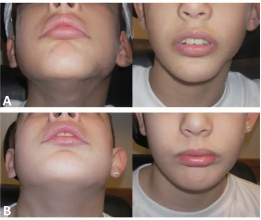
Three patients had undergone anterior or lateral skull base reconstruction (Figure 1) and three additional cas-es involved the use of acellular fetal bovine dermal matrix to provide improved contouring of facial defects or scars. In these cases the dermal matrix was placed in the subdermal or subperiosteal plane to address depressed scar or atrophic dermis and soft tissue (Figure 2).
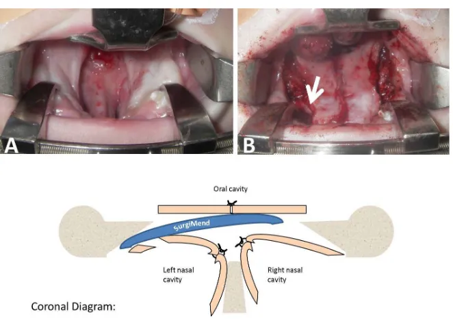
There was one case in which the matrix was used in an underlay fashion to prevent oronasal fistula formation in the context of a primary palatoplasty for closure of a wide cleft palate. The width of the cleft was wider than the width of the oral mucoperiosteal flaps in this case, preventing a proper multilayer closure along the lateral edges of the medially-advanced oral flaps. Additionally a small tear had occurred laterally during mobilization of the nasal mucoperiosteal flap on one side (Figure 3).
In the final case, the dermal matrix was used to repair a lateral pharyngeal wall defect, which could not be closed primarily due to excessive tension on the wound. The purpose of using a dermal matrix for repair in this case was to provide a scaffold for epithelial in growth while protecting the parapharygeal fat and carotid sheath from desiccation and gross contamination with oral flora.
Table 1. A summary of pediatric patients included in this study.
Subject Age Sex Indication Successful repair?
1 11 y F Transnasal endoscopic repair of traumatic sphenoid encephalocele
with cerebrospinal fluid leak. Yes, no leak at 1 year.
2 13 y M Open repair of lateral skull base defect following resection and
subsequent radiation of temporal bone rhabdomyosarcoma. Yes, 1 year.
3 17 y M Transnasal endoscopic repair of frontoethmoid encephalocele. Yes, no leak at 1 year.
4 1 y M Closure of wide cleft palate. Yes, no fistula at 2 years.
5 2 y M Transoral repair of iatrogenic lateral pharyngeal wall defect. Yes, complete healing by 3 months.
6 8 y M Recontouring of depressed, paramandibular facial scar. Yes, maintained symmetry at 3 years.
7 10 y M Recontouring of parotidectomy defect following excision of first
branchial cleft vestige. Yes, maintained symmetry at 2 years.
8 10 y F Recontouring of forehead and brow for progressive hemifacial atrophy (Parry Romberg syndrome). No, perceived resorption vs disease progression at 6 months.
Figure 1. Repair of a parasellar skull base encephalocele via an endo-nasal endoscopic approach. Dermal matrix placement into a right ante-rosuperior sphenoid region defect (A) (subject 1). Note pliability of gra-
fting material (B).
[image:3.595.169.430.458.677.2]Figure 3. Reinforcement of a tenuous cleft palate repair in subject 4, using a sheet of acel-lular fetal bovine dermal matrix as an interposition graft. (A) Wide cleft of the secondary palate. (B) Intraoperative appearance immediately following repair. White arrow points to allograft. A coronal diagram depicts graft placement.
handling as compared to the surgeon’s prior experience with human cadaveric acellular dermis. Additionally, every parent vocalized a greater comfort level with the use of a bovine product over the alternative of irradiated, human cadaveric tissue. Acellular fetal bovine dermal matrices are available in a wider variety of graft thick-nesses when compared to available human cadaveric products that are currently available, and the cost of fetal bovine derived acellular dermal matrix is lower than human cadaveric products (Table 2).
4. Discussion
Human acellular dermal matrix has been accepted as a useful product in pediatric soft tissue reconstruction and the endoscopic repair of skull base defects [6]-[9]. There are several reasons why acellular fetal bovine dermal matrix may be superior to human cadaveric tissue for soft tissue and skull base reconstruction. First, acellular fetal bovine dermal matrix is more pliable than human cadaveric tissue and easier to work with in tight spaces (e.g. a tight tissue pocket or endoscopically). In addition to inherent tissue characteristics that make this allograft more pliable than other options, it is available in variable 1, 2, 3, or 4 mm thicknesses. These details can be im-portant when performing surgery on children at various stages of development.
Second, acellular fetal bovine dermal matrix is available at a lower cost when compared to human tissue products. A recent review of dermal allografts found acellular fetal bovine dermal matrix to be one of the least costly products available, with an estimated cost of 23 dollars/cm2 as compared to 28 - 35 dollars/cm2 for human cadaveric dermal products [14][15]. Finally, acellular fetal bovine dermal matrix is classified as a medical de-vice rather than a tissue product. While cadaveric allografts are sterilized and irradiated to eradicate communic-able disease, some patients (especially parents of infants and children) are uncomfortcommunic-able with the use of a hu-man tissue product from an anonymous donor. In our initial experience, parents are more comfortable with the concept of sterilized xenografts than implanted human cadaveric allografts.
Table 2. Approximate price per square centimeter of currently available acellular dermal matrices, adapted from Cheng et al. [14] “SurgiMend” is acellular fetal bovine dermal matrix whereas all other products listed are human cadaveric tissue.
Product name Approximate price per cm2
AFBDM (SurgiMend) $23
AlloDerm $28
DermaMatrix $28.51 - $31.94
FlexHD $27.31 - $34.76
AlloMax $32.38
DermACELL $34
AFBDM: acellular fetal bovine dermal matrix.
5. Conclusion
Acellular fetal bovine dermal matrix (SurgiMend) may be a comparable material to human cadaveric acellular dermal matrices for pediatric head and neck and skull base reconstruction. Increased pliability, lower cost, and classification as a medical device rather than a tissue are potential advantages of this product, as compared with human cadaveric tissue. Further comparison studies are warranted.
Acknowledgements
The authors wish to acknowledge Dr. Jonathan Sillman and Dr. Mark Vecchiotti who performed the surgery for subject 2 in this series.
Financial Disclosures
None of the authors have an affiliation or financial interest in the makers of AlloDerm or SurgiMend. Andrew Scott, MD is a paid consultant for Advance Medical-expert medical second opinions.
Conflict of Interest
None.Source of Funding
Interdepartmental funds.References
[1] Banks, C.A., Palmer, J.N., Chiu, A.G., O’Malley Jr., B.W., Woodworth, B.A. and Kennedy, D.W. (2009) Endoscopic Closure of CSF Rhinorrhea: 193 Cases over 21 Years. Otolaryngology-Head and Neck Surgery, 140, 826-833.
http://dx.doi.org/10.1016/j.otohns.2008.12.060
[2] Shridharani, S.M. and Tufaro, A.P. (2012) Systematic Review of Acellular Dermal Matrices in Head and Neck Recon-struction. Plastic and Reconstructive Surgery, 130, 35S-43S. http://dx.doi.org/10.1097/PRS.0b013e31825eff7a
[3] Kridel, R.W., Foda, H. and Lunde, K.C. (1998) Septal Perforation Repair with Acellular Human Dermal Allograft.
Archives of Otolaryngology-Head and Neck Surgery, 124, 73-78. http://dx.doi.org/10.1001/archotol.124.1.73
[4] Sachsman, S.M. and Rice, D.H. (2007) Use of AlloDerm Implant to Improve Cosmesis after Parotidectomy. Ear, Nose Throat Journal, 86, 512-513.
[5] Girod, D.A., Sykes, K., Jorgensen, J., Tawfik, O. and Tsue, T. (2009) Acellular Dermis Compared to Skin Grafts in Oral Cavity Reconstruction. Laryngoscope, 119, 2141-2149. http://dx.doi.org/10.1002/lary.20548
[6] Munson, P.D. and Moore, E.J. (2010) Pediatric Endoscopic Skull Base Surgery. Current Opinion in Otolaryngology Head and Neck Surgery, 18, 571-576. http://dx.doi.org/10.1097/MOO.0b013e3283401fdc
[7] Achauer, B.M., VanderKam, V.M., Celikoz, B. and Jacobson, D.G. (1998) Augmentation of Facial Soft-Tissue Defects with AlloDerm Dermal Graft. Annals of Plastic Surgery, 41, 503-507.
[8] Aldekhayel, S.A., Sinno, H. and Gilardino, M.S. (2012) Acellular Dermal Matrix in Cleft Palate Repair: An Evidence- Based Review. Plastic and Reconstructive Surgery, 130, 177-182. http://dx.doi.org/10.1097/PRS.0b013e318254b2dc
[9] Shah, R.N., Surowitz, J.B., Patel, M.R., et al. (2009) Endoscopic Pedicled Nasoseptal Flap Reconstruction for Pediatric Skull Base Defects. Laryngoscope, 119, 1067-1075. http://dx.doi.org/10.1002/lary.20216
[10] Lorenz, R.R., Dean, R.L., Hurley, D.B., Chuang, J. and Citardi, M.J. (2003) Endoscopic Reconstruction of Anterior and Middle Cranial Fossa Defects Using Acellular Dermal Allograft. Laryngoscope, 113, 496-501.
http://dx.doi.org/10.1097/00005537-200303000-00019
[11] Clark, D.W., Citardi, M.J. and Fakhri, S. (2010) Endoscopic Management of Skull Base Defects Associated with Per-sistent Pneumocephalus Following Previous Open Repair: A Preliminary Report. Otolaryngology-Head and Neck Sur-gery, 142, 820-826. http://dx.doi.org/10.1016/j.otohns.2009.12.041
[12] Hadad, G., Bassagasteguy, L., Carrau, R.L., et al. (2006) A Novel Reconstructive Technique after Endoscopic Ex-panded Endonasal Approaches: Vascular Pedicle Nasoseptal Flap. Laryngoscope, 116, 1882-1886.
http://dx.doi.org/10.1097/01.mlg.0000234933.37779.e4
[13] Ismail, A.S., Costantino, P.D. and Sen, C. (2007) Transnasal Transsphenoidal Endoscopic Repair of CSF Leakage Us-ing Multilayer Acellular Dermis. Skull Base, 17, 125-131. http://dx.doi.org/10.1055/s-2007-970556
[14] Cheng, A. and Saint-Cyr, M. (2012) Comparison of Different ADM Materials in Breast Surgery. Clinics in Plastic Surgery, 39, 167-175. http://dx.doi.org/10.1016/j.cps.2012.02.004
[15] Butterfield, J.L. (2013) 440 Consecutive Immediate, Implant-Based, Single-Surgeon Breast Reconstructions in 281 Pa-tients: A Comparison of Early Outcomes and Costs between SurgiMend Fetal Bovine and AlloDerm Human Cadaveric Acellular Dermal Matrices. Plastic and Reconstructive Surgery, 131, 940-951.
http://dx.doi.org/10.1097/PRS.0b013e3182865ab3
![Table 2. Approximate price per square centimeter of currently available acellular dermal matrices, adapted from Cheng et al[14] “SurgiMend” is acellular fetal bovine dermal matrix whereas all other products listed are human cadaveric tissue](https://thumb-us.123doks.com/thumbv2/123dok_us/8115983.792516/5.595.92.538.110.222/approximate-centimeter-currently-available-acellular-surgimend-acellular-cadaveric.webp)
