Journal of Chemical and Pharmaceutical Research, 2012, 4(5):2712-2719
Research Article
CODEN(USA) : JCPRC5 ISSN : 0975-73842712
The Synthesis and Characterization of Core-Shell Quantum Dots via
Three-Phase System
Baoan Du
*, Lu Gao, Xueru Lian, Yongjing Jiao
College of Chemistry and Environmental Science, Hebei University, Baoding 071002, China
_____________________________________________________________________________________________
ABSTR ACT
The CdS/ZnS quantum dots (QDs) were synthesized via three-phase system. Transmission electron microscopy (TEM) and X-ray powder diffraction (XRD) techniques were used to characterize the as-synthesized QDs. The growing mechanism of the quantum dots in the phase-transfer process was discussed. As result, the QDs are narrow sized distribution, uniform morphology and cubic crystal structure. The result of PL spectroscopy measurements reveals that the exciton emission of CdS was greatly improved by covering with a layer of ZnS. That demonstrates the shell of ZnS modified the surface of CdS core and reduced the surface defect.
Keywords: CdS/ZnS, Quantum dots, Three-phase system.
_____________________________________________________________________________________________
INTRODUCTION
Semiconductor nanocrystals, also called quantum dots (QDs), show quantum size effects which distinct from these
of the corresponding bulk materials and have been widely used in light-emitting diodes (LEDs)[1-3], non-linear
optical devices[4], solar cell[5] and biological labeling[6,7], laser source[8] and so on. Among these semiconductor
materials, groupⅡ-Ⅵ semiconductor nanoparticles, especially CdS and CdSe nanoparticles, have been extensively
studied due to the ability to tune their emission in the visible range simply by changing their size, and due to the advances in their preparation[9].
Various kinds of high quality QDs such as CdSe and CdTe were successfully synthesized by organometallic approach[10-13]. The organometallic precursor route was firstly developed by Murry et. al. in 1993[10], which
involved the reaction of cadmium organometallic precursor Cd(CH3)2, with a selenium precursor, but it is generally
acknowledged that the reactant of Cd(CH3)2 is an expensive, pyrophoric, and hazardous materials. In 2000, Peng
et.al. developed a relatively simpler and greener nonorganometallic precursor route using inexpensive and little toxic
CdO instead of Cd (CH3)2 to produce CdSe nanocrystals[11,12].Recently, using noncoordinating solvents such as
octadecene (ODE) instead of coordinating solvent such as trioctylphosphine oxide (TOPO) has been extensively studied[14,15]. In general, Peng’s route and its alternatives involve the injection of a trioctylphosphine selenium (TOPSe) solution to a cadmium solution. Thus the reaction media involve at least three components, namely, the TOPO (or ODE) used as solvent, the cadmium solution, and the TOP-Se solution. These routes were called the TOP-based route[16] and high reaction temperature and Schlenk technique are always needed. It is possible for the cadmium precursor to decompose at high temperature. Therefore, developing a method for synthesizing nanocrystals under mild conditions and using simple manipulation techniques is still one of the important goals for the materials scientists.
2713
CdMA (myristic acid) and n-trioctylphosphine oxide (TOPO) or oleic oil (OA) in toluene and a solution of thiourea in water mixed and heated with stirring. The CdS nanocrystals grew at the liquid-liquid interface as the thiourea slowly decomposed, and the surface of the nanocrystals was capped by a monolayer of TOPO or OA as they formed. A general strategy for nanocrystals synthesis was recently proposed by Li et. al.[17], three phases was formed in their system: mixture of ethanol and linoleic acid as liquid phase, sodium linoleate as solid phase, and water ethanol solution containing noble metal ions as solution phase. This liquid-solid-solution (LSS) process can generate nanocrystals with a variety of properties such as, semiconductors, fluorescent, magnetic and dielectric, and nearly many the bandgap semiconductors can be effectively prepared though this simple method, but to our best knowledge, no core-shell nanocrystals were prepared by this strategy.
The high surface defects of the nanocrystals may lead to the reduced luminescence efficiency and photochemical degradation. Bandgap engineering concepts borrowed from materials science and electronics have lead to the development of core-shell nanocrystals sample with high room temperature quantum yields and much improved photochemical stability. By enclosing a core nanocrystals of one materials with a shell of another having a large bandgap, one can efficiently confine the excitation to the core, eliminating nonradiative relaxation pathways and preventing photochemical degradation[6] .Herein, we take the advantages of the autoclave to obtain the high temperature and high pressure for the nucleation and growth for the nanocrystals, we prepared the CdS core nanocrystals and the CdS/ZnS core-shell nanocrystals by the LSS phase transfer and separation process. Compared with the organometallic scheme, the method was environmental friendly, low-cost and security. The TEM and XRD show that the core-shell nanocrystals are monodispersed, narrow sizes distribution, cubic zinc—blend crystalline, and highly enhanced quantum yield. And no sophisticated equipments were needed. The mechanism of the core-shell nanocrystals formation was also discussed.
EXPERIMENTAL SECTION
Instruments and Reagents
F-4500 spectrofluorimeter (Hitachi, Japan) equipped with a 150W high-pressure Xenon lamp and a 1.0×1.0 cm quartz cell was used to measure the spectrum of the flourenence. A magnetic force stirring instrument and WH-861 vortex mixer (Huangjin Instrumental Corp., Jiangsu, China) was used to blend the solutions. The TEM images of the nanocrystals were acquired on a JEM-1200EX II transmission electron microscope (JEOL, Japan). High-speed freezing centrifugator was used to separate the samples. Constant temperature air-blast drying oven which can
control the temperature as high as 250℃ (Shanghai Anting scientific instrument factory, Shanghai China) was used
to keep the reaction temperature. KQ-250B ultrasonic cleaner was used to disperse the nanocrystals.
Cadmium acetate, zinc acetate, selenium powder, sulfur powder, sodium sulfur, sodium oleate, oleic acid, ethanol, cyclohexane are all purchased from Sinopharm Group Chemical Reagent Co. (China), the water used was doubly distilled. All reagents were analytical grade without further purification.
Preparation of CdS core nanocrystals
Cadmium acetate (0.400 g, 1.5mmol) dissolved in 17mL redistilled water, and sodium oleate (1.600 g),10 mL
ethanol, 2 mL oleic acid were added to a 100 mL beaker under stirring, then NaS ( 0.360 g ,1.5 mmol) dissolved in 3 mL water was dropped into the solution under stirring, when all the reagents were added, continued to stir for 15 min, and the solution turned to colloid. The colloid was loaded into the Teflon—lined stainless steel autoclave with a
volume of 50 mL. The autoclave was sealed and maintained at 180℃ for 4 hours. When the reaction was completed,
the autoclave was cooled to room temperature naturally, the supernatant was decanted, the CdS nanocrystals precipitated at the bottom of the autoclave was dissolved in 20 mL cyclohexane, the solution was ultrasonically dispersed for 20 min, then added the equal volume of ethanol to the solution, the CdS nanocrystals was separated by centrifugation under 5000 rmp for 5 min, decanted the supertant and the precipitate was washed for another twice
times with ethanol, dried at 50℃ for 5 hours. Little of the solid dispersed in cyclohexane was used for recording
UV-Vis and photoluminescence (PL) spectra, the solid was collected for X-ray powder diffraction (XRD), and transmission electron microscopy (TEM) measurement without any size sorting.
Preparation of CdS/ZnS core-shell nanocrystals
The crude solution of CdS nanocrystals prepared above was cooled to room temperature and then transferred to a 100 mL beaker, the solution of zinc acetate (0.330 g, 1.5 mmol) dissolved in 3 mL water was dropped into the beaker under stirring, then, sulfur powder (0.048 g, 1.5 mmol) was slowly added into the beaker under stirring, the solution was stirred for another 10 min, then transferred to the Teflon—lined stainless steel autoclave with the
volume of 50 mL, The autoclave was sealed and maintained at 180℃ for 8 hours. When the reaction was completed,
transmission electron microscopy (TEM) measurement were also obtained.
Mechanism
(a) (b)
Fig.1 Scheme of liquid-solid-solution transfer synthetic strategy for (a) the forming process of the CdS nanocrystals and (b) the forming process of the shell ZnS on the surface of the core CdS nanocrystals
According to Li’s process[17], the formation of the nanocrystals occurred at the interface between two of the three phases formed in this system: ethanol and oleic acid (liquid), sodium oleate (solid), and the water/ethanol containing the metal ions (solution). We proposed the different mechanism on the forming processes of the CdS nanocrystals and the CdS/ZnS core-shell nanocrystals. As shown in Fig.1 (a), when the cadmium acetate was added to the solution, it would first be coordinated by the oleic acid to form Cd-oleate complex and dispersed in the solid phase.
At the given temperature, the S2- dissolved in the ethanol-water solution react with Cd-complex to form the nuclei
which can then enter to the liquid phase as they formed, because the surface of the nuclei were caped by a layer of oleic acid, it was more easy for the caped nuclei to disperse in the liquid phase then in the solution phase, and the nanocrystals stop growing Their growth resumes when the nanocrystals return to the interface of the liquid and the solid. The separation between nucleation and growth stages is achieved spatially in this three phases approach. Thus a slow nucleation does not always lead to the polydispersed colloids. At the same time, the slow growth of nanocrystals is favorable for achieving monodispersed colloid, which is called “focusing” of the nanocrystals’ size distribution[18]. The other advantage of the approach is that the Ostwald ripening can be avoided, because in the Ostwald ripening, monomers can only be transferred though the homogeneous solution among the particles. Aggregating at the interface of the liquid and the solid, it is more difficult for the little particles to dissolve its monomer and to transfer them to the bigger particles, thus monodispersed nanocrystals were produced. So the size of the nanocrystals can be simply controlled by adjusting the reaction temperature and the reaction time.
In the forming process of the core-shell nanocrystals, the monomers such as sulfur powder and zinc acetate solution were added to the crude solution which was used to prepare the core quantum dots, the core quantum dots was used as “seed” to induced the shell crystals growing at the surface of the core. As shown in Fig.1(b), at the reaction
temperature, the sulfur powder was first reduced to S2- by ethanol in the solution, and then enters to the liquid. Zine
acetate reacts first with oleic acid to form the Zn-complex and dispersed in the solid phase[18]. In the synthesis of core-shell nanocrystals, the new ZnS nanocrystals may also be formed at the interface between the solid and liquid phases, but it is necessary for the formation of isolate new nanocrystals to firstly produce nuclei by the monomers reaction, then the growth of the nanocrystals. According to the surface free energy theory, the smaller the particles, the higher the surface free energy and the more difficult the formation of the new particles. At the interface of the two phases, there should exist competing pathways between the new nanocrystals growing at the core quantum dots surface and formation of complete isolate new nanocrystals. Since the formation of new nanocrystals needed two separate steps: the first nucleation and then the growth of the crystals, it needs to overcome very high free energy barrier for the formation of the much smaller nuclei, and high temperature is always needed to release high concentration precursor to quickly produce large amount of nucleus at short time. This is a higher energy pathway. At the same time, the new nanocrystals may be formed at the surface of the quantum dots already existed in the interface between the two phases. The diameter of the existing quantum dots is much bigger than the nuclei newly formed at the other pathway, so with much lower surface free energy, the formation of new crystals at the quantum dots surface needs overcoming much lower energy barrier, so the core-shell nanocrystals were always prior to be formed. As mentioned above, the core-shell nanocrystals were also monodispersed. The thickness of the shell can also be readily controlled by the reaction time and reaction temperature.
2715
Photoluminescence (PL) and absorption spectra are not only utilized to character semiconductor nanocrystals assisted with the TEM and other characterization means, but they are a powerful tool to confirm quantum-confined property of the nanocrystals. Fig.2 shows the PL spectra and the absorption spectra of the CdS nanocrystals. The same PL spectra was obtained with the nanocrystals being exited by 350 nm or 400 nm wavelengths, the emission wavelength of the nanocrystals is 475 nm, but there also exists an obvious emission peak with the wavelength at 570 nm (Fig. 2 a), which indicates that there are a large portion of the surface defects in the nanocrystals’ surface[19],and these defects constitutes the photon traps, the quantum-yield are therefore decreased. The narrow FWHM (full width at half maximum) in the photoluminescence spectra suggests that the sizes distribution of CdS nanocrystals is relatively narrow.
Fig.2 (a) The PL spectrum of the CdS nanocrystals. (b) The UV-vis spectrum of the CdS nanocrystals. The nanocrystals is dispersed in cyclohexane, the exited wavelength is 400 nm at room temperature..
Fig. 3 shows the typical TEM images of the as-prepared oleic acid capped CdS nanocrystals with the absorption peak at the 470 nm, the TEM image demonstrates that the sizes distribution of the nanocrystals are nearly monodispersed, the average size of the nanocrystals is about 4 nm.
Fig.3 The TEM image of the CdS core nanocrystals
[image:4.595.206.409.207.382.2] [image:4.595.210.402.473.666.2]Fig.4 The powder XRD pattern of the CdS core nanocrystals
The diffraction feature of the CdS nanocrystals shows that the sample has the characteristic features appearing at about 26.6°,44.2°, 52.1°which are respectively matching the (111), (220), and (311) plane of the cubic zinc-blend phase of the CdS compared to the standard spectrum of the bulk CdS (JCPDS card No: 89-0440, 26.5°, 43.9°, 52.0°respectively). According to the previous study, the zinc-blend phase is more readily to be formed at the lower temperature than the wurtzite phase, because in thermodynamics, the zinc—blend is the most stable form at lower temperature, while the wurtzite is more stable form at higher temperature[16]. In the present study, the zinc-blend
phase is preferred to be formed at the comparably lower temperature (180℃), while the wurtzite phase is preferred
for growth at higher temperature (about 300℃)[20].
Fig.5 (a) PL spectrum of the CdS nanocrystals. (b) PL spectrum of the core-shell CdS/ZnS nanocrystals. The nanocrystals is dispersed in cyclohexane, the exiting wavelength is 400 nm.
2717
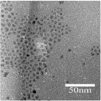
[image:6.595.215.397.106.291.2]As the TEM image shows in Fig. 6, the monodispersed core-shell nanocrystals was obtained by this method. The thickness of the nanocrystals is about 1.0 nm. These experiments were done without further sizes sorting.
Fig. 6 The TEM image of the CdS/ZnS core-shell nanocrystals
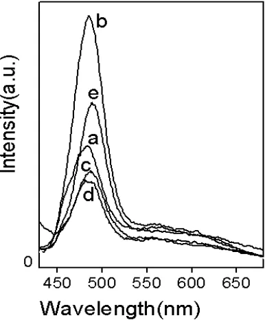
Fig.7 The effect of the monomer concentration on the shell thickness of the core /shell nanocrystals. The molar ratio of Cd2+ used to synthesize CdS core to Zn2+ used to synthesize the shell are (a) 1: 0.25, (b) 1: 0.5, (c) 1: 1.25, (d) 1: 1.5, (e) 1: 1.75 respectively. The molar ratio of sulfur powder to the Zn2+ is 1. The corresponding
emission peaks of the PL are (a) 483 nm, (b) 485 nm, (c) 486 nm, (d) 488 nm, (e) 489 nm. The reaction temperature is 180 and the reaction time is 8 h.
The effect of the monomer concentration on the thickness of the shell is shown in Fig.7. As the thickness increases,
the PL peaks will correspondingly red shift[22]. The result indicates that as the Zn2+ monomer increases, the
red-shift of the emission peaks of the PL increase with the thickness of the shell and that the thickness of the shell can be reflected by the optical properties of the nanocrystals. Since there already exists “the seed” for the new crystals growth on it, high monomer concentration is favorable for the reaction of the crystals growth and this stage is the so-called sizes focusing, which is favorable for the surface ordering and reconstructions during the growth of the nanocrystals, thus favorable for achieving the higher quantum yield before the Ostwald ripening. But too high monomer concentration would induce new isolate nucleus of ZnS nanocrystals, so the suitable monomer concentration is optimized.
[image:6.595.193.386.322.555.2]ones stay at the interface of the phases to continue the growth. To be coordinated by the oleic acid would obstacle the dissolving of the monomer from the smaller nanocrystals, and the higher monomer concentration in solid phase would keep the equilibrium to move to the nanocrystals growth, the spatial separation between the larger particles and the smaller particles is also unfavorable for the Ostwald ripening. Thus the separation between the sizes focusing with higher monomer concentration and the Ostwald ripening while the most monomer consumed can be readily controlled by adjusting the reaction time at certain temperature. The similar mechanism can also be used to explain the formation of the monodispersed core nanocrystals.
Fig. 8 The reaction time effect on the shell thickness of the core /shell nanocrystals. The molar ratio of Cd2+ used to synthesize CdS core to Zn2+ used to synthesize the shell is 1: 1.25. The molar ratio of sulfur
powder to the Zn2+ is 1: 1. The reaction temperature is 180 .
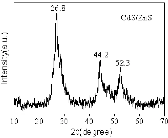
Fig.9 The powder XRD pattern of the CdS/ZnS core-shell nanocrystals
The reaction time effect on the shell thickness of the core /shell nanocrystals is shown in Fig. 8, as the time increase, the red-shift of the FL peaks will also increase, so do the thickness of the shells, but too thick shell will decrease the quantum yield of the core-shell nanocrystals, and too long reaction time will also deteriorate the optical properties of the nanocrystals, because of the Ostwald ripening mentioned above.
[image:7.595.166.434.409.629.2]2719
(JCPDS card No: 89-0440, 26.5°, 43.9°, 52.0°respectively). If there are no layer of the ZnS covering the core CdS nanocrystals, there should be the diffraction features of the isolate ZnS nanocrystals (JCPDS card No: 89-2201, 28.6°, 47.5°, 56.4°respectively) or the mixture of the CdS and ZnS, since the thickness of the ZnS shell is much smaller then the diameter of the core CdS, the diffraction features of ZnS is hardly shown in the powder XRD pattern of the core-shell nanocrystals, there should also shows the features of CdS diffraction, so the facts verify that the growth of ZnS nanocrystals certainly happens on the surface of the core CdS nanocrystals without newly isolate produced ZnS crystals[23].
CONCLUSION
In summary, the reagents used for synthesize the core-shell semiconductor nanocrystals are low-cost, safety, and environmental friendly, the reaction conditions are mild in comparison with the organometallic methods, and need no Schlenk techniques. The growth of the semiconductor nanocrystals can be readily controlled by the reaction temperature and time, CdS, CdS/ZnS core-shell monodispersed nanocrystals with high quantum yield and narrow PL spectra have be obtained by this method. The mechanism of the formation process of the nanocrystals has been discussed in detail, the formation of the nanocrystals in this three phases system can be reasonable explained by the mechanism. This method can also be applicable to synthesize the other core/shell semiconductor nanocrystals.
Acknowledgement
This work was supported by the Nature Science Foundation of Hebei Province (B2008000572) and the Foundation for the Introduction of Talent of Hebei University (2008-129)
REFERENCES
[1] MC Schlamp; XG Peng; AP Alivisatos. J. Appl. Phys., 1997, 82(11), 5837-5842 [2] WC Chan; SM Nie. Science, 1998, 281(5385), 2016-2018
[3] S Coe; WK Woo; JS Steckle; et al. Org. Electron., 2003, 4, 123-130. [4] VC Sundar; HJ Eisler; MG Bawendi. Adv. Mater., 2002, 14(10): 739-743. [5] NC Greenham; XG Peng; AP Alivistos. Phys. Rev. B, 1996, 54: 17628-17637. [6] MY Han ;XH Gao ; JZ Su ; et al. Nature Biotechnology, 2001, 19 (7) : 631-635.
[7] H Mattoussi; JM Mauro; ER Goldman; et al. J. Am. Chem. Soc., 2000, 122(49): 12142-12150. [8] VI Klimov; AA Mikhailovsky; S Xu; et al. Science, 2000, 290(5490),314-317.
[9] YG Yang; O Chen ; A Alexander et al. J. Am. Chem. Soc., 2008, 130 (46): 15649-15661 [10] ZA Peng; XG Peng. J. Am. Chem. Soc., 2001, 123(1), 183-184.
[11] ZA Peng; XG Peng. J. Am. Chem. Soc., 2002, 124 (13): 3343–3353. [12] L Qu; ZA Peng; XG Peng. Nano. Let., 2001, 1(6), 333-337.
[13] DV Talapin; AL Rogach; I Mekis; et al. Colloids Suef.A ,2002, 202, 145-154. [14] ZA Peng; XG Peng. J. Am. Chem. Soc., 2002, 124 (13): 3343–3353.
[15] CR Bullen; P Muvaney. Nano Lett., 2004, 4(12), 2303-2307.
[16] ZT Deng; LF Cao; QB Tang; et al. J. Phys. Chem. B, 2005, 109(35), 16671-16675. [17] X Wang; J Zhuang; Q Peng;et al. Nature, 2005, 437, 121-124.
[18] LH Qu; ZA Peng; XG Peng. Nano Letters, 2001, 1 (6): 333–337. [19] DC Pan; QS Wang; CX Jiang; et al. Adv. Mater., 2005, 17: 176 [20] Q Li; A Peng; XG Peng. Nano. Lett., 2001, 1(6), 333-337.