ISSN Online: 2333-9721 ISSN Print: 2333-9705
Anti-LGI1 Limbic Encephalitis—Case Report
Susana Sá, Miquelina Redondo
General Practice Departement, USF Sem Fronteiras, Santa Maria da Feira, Portugal
Abstract
Limbic encephalitis is characterized by acute or subacute changes in mood and be-haviour, by recent memory changes, seizures and cognitive dysfunction. In this arti-cle, the authors present a case report of a 53-year-old woman that resorted to con-sultation with clinical presentation suggestive of major depressive episode. However, the worsening of symptoms despite treatment and the testimony of seizures put on the trail of another diagnosis. She was admitted in Neurology Service of home area hospital, where she was diagnosed with Autoimmune Limbic Encephalitis (Anti- LGI1 positive). Currently, the patient keeps mensal immunotherapy and daily treat-ment with levetiracetam and valproic acid, without new seizures. However, she maintains memory and behavioural changes which interfere substantially with her daily functioning. The Family Physician had an essential role not only in the diagno-sis of Limbic Encephalitis, but also in helping the patient, through holistic and lon-gitudinal care, to accept and adapt to her illness and related limitations, relieving the suffering.
Subject Areas
Neurology, Psychiatry & Psychology
Keywords
Limbic Encephalitis, Encephalitis, Autoimmune Diseases
1. Introduction
The limbic area of the brain controls many types of activities including memory, learn-ing, and emotions such as sexual desire, love, anger, sadness and jealousy. The main re-gions of the limbic area include the hippocampus, thalamus, hypothalamus and amyg-dala [1].
There are several causes of Limbic Encephalitis (LE), and often the underlying cause is not found. Most forms of encephalitis fall into 2 main categories: infectious encepha-How to cite this paper: Sá, S. and Redondo,
M. (2016) Anti-LGI1 Limbic Encephalitis— Case Report. Open Access Library Journal, 3: e3219.
http://dx.doi.org/10.4236/oalib.1103219
Received: November 10, 2016 Accepted: December 5, 2016 Published: December 8, 2016
Copyright © 2016 by authors and Open Access Library Inc.
This work is licensed under the Creative Commons Attribution International License (CC BY 4.0).
litis; autoimmune encephalitis. Any infection of the brain can potentially cause an in-flammation of the limbic area. But a number of viruses seem to target this area particu-larly, such as the herpes simplex virus (HSV) [1]. There are broadly two forms of Auto- immune LE: Paraneoplastic Limbic Encephalitis (PLE) and Non-Paraneoplastic Limbic Encephalitis (NPLE). Most individuals with PLE will turn out to have a cancer of the lung, thymus gland, the breast or the testis. NPLE has only been clearly recognised in the last few years. The specific antibody targets may explain the variation of the clinical presentation. Voltage-gated potassium channel complex antibody targets proteins that are tightly linked in with the potassium channels in brain. The majority of these anti-bodies target LGI1 (leucine-rich glioma inactivated 1). LGI1 is a protein that appears to be important in controlling brain’s electrical activity. Antibodies against other protein in the voltage-gated potassium channel/LGI1 complex, such as Caspr2 (contactin- associated protein 2) and contactin-2, have been shown to also cause Autoimmune LE. AMPAR (α-amino-3-hydroxy-5-methyl-4-isoxazolepropionic-acid receptor) and GABABR (Gamma-aminobutyric-acid receptor) antibodies are now known to be less common causes Autoimmune LE. NMDAR (N-Methyl-D-Aspartate Receptor) anti-bodies usually cause encephalitis involving several brain regions, but it can rarely cause LE [2] [3].
LE is characterized by acute or subacute changes in mood and behaviour, by recent memory changes, seizures and cognitive dysfunction. In addition, electroencephalo-graphy (EEG) often shows focal or generalized slow wave or epileptiform activity, and magnetic resonance imaging (MRI) findings reveal hyperintense signals of the medial temporal lobes in T2-weighted or Fluid attenuation inversion recovery (FLAIR) images. However, the definitive diagnosis of encephalitis is pathological, but in practice it is es-tablished based on clinical presentation and by changing in analytical and imaging ex-ams [4] [5].
NPLE seems to have a better prognosis than PLE since a substantial proportion of NPLE may have a good response to immunotherapy, particularly if it is promptly initi-ated. The importance of early correct diagnosis and prompt treatment is emphasized by evidence that patients with a delay in the initiation of immunotherapy are less likely to respond to treatment [6].
The aim of this article was to facilitate recognition of the clinical features of LE, in order to sensitize clinicians to this entity, for rapid diagnosis and prompt treatment ini-tiation.
2. Case Report
Woman, 53-year-old, 9th grade, auxiliary of pharmacy. Pathological Background: Anxiety and Sleep Disorders and Obesity. Chronic medication: alprazolam 0.5 mg bid and trazodone 50 mg id.
altera-tions. It was prescribed hemogram and thyroid function, both normal, and venlafaxine 75 mg id, alprazolam 0.5 mg bid and ethyl loflazepate 2 mg in SOS.
In February 2014, the patient referred worsening of symptoms, particularly in mem-ory and “mind blocked”. During the appointment, the patient had stereotyped move-ments of the hands and mouth, without perception of these movemove-ments, no response to verbal stimulation or memory during this period (10 seconds). Brain computed tomo-graphy (CT) was requested, which was normal, and EEG recorded a crisis with parox-ysmal activity with left frontotemporal predominance. It was requested urgent evalua-tion by Neurology in the home area Hospital, where she was admitted in that same day.
The further study (Table 1) revealed alterations suggestive of Autoimmune LE (Anti- LGI1), having performed inpatient treatment with metilprednisolone and intravenous immunoglobulin.
During hospitalization, the patient presented uninhibited behaviour, with suggestive and seductive speech partially improved with the introduction of valproic acid and quetiapine.
She also presented confabulatory speech and visual hallucinations: “drops of water falling from the ceiling” and “figures behind her back.” The assessment by Neuropsy-chology revealed mnesic dysfunction and frontal deficits.
Currently, the patient keeps mensal immunotherapy and daily treatment with leveti-racetam and valproic acid, without new seizures. However, she maintains memory and behavioural changes, which interfere substantially with her daily functioning, being not capable to return to her habitual job.
3. Discussion
Anti-LGI1 LE is a rare neurological disorder, with few cases published in scientific lit-erature.
Table 1. Exams performed during the internment in Neurology Service of home area Hospital.
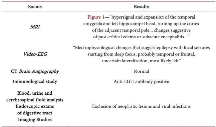
Exams Results
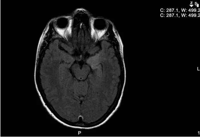
[image:3.595.192.554.493.705.2]MRI
Figure 1—“hypersignal and expansion of the temporal amygdala and left hippocampal head, turning up the cortex
of the adjacent temporal pole... changes suggestive of post-critical edema or subacute encephalitis...”
Video-EEG “Electrophysiological changes that suggest epilepsy with focal seizures starting from deep focus, probably temporal or frontal, uncertain lateralization, most likely left”
CT Brain Angiography Normal
Immunological study Anti-LGI1 antibody positive
Blood, urine and cerebrospinal fluid analysis
Endoscopic exams of digestive tract Imaging Studies
Figure 1. Axial fluid attenuation inversion recovery (FLAIR) MRI of the brain showing a bright signal temporal amygdala and left hippocampal head, turning up the cortex of the adjacent tem-poral pole, consistent with limbic encephalitis (arrow).
In this case report, the prompt diagnosis of LE was possible due to the testimony of stereotyped movements made by the patient, who never revealed changes in objective examination, raising the suspicion of a possible organic etiology. In fact, the clinical presentation manifested by the patient could easily be confused with an anxiety or de-pressive disorder. In addition, the patient presented personal psychiatric background and psychosocial stressors that would justify several of the presented symptoms.
Despite the rapid diagnosis of LE and the prompt initiation of treatment with metil-prednisolone and intravenous immunoglobulin, clinical improvement was incomplete. The patient had a highly responsible job in a local pharmacy for more than 20 years. However, the mnesic dysfunction and the frontal deficits maintained by the patient, barred the return to her work activity, which was a difficult situation to accept by the patient. The Family Physician had an essential role not only in the diagnosis of LE, but also in helping the patient, through holistic and longitudinal care, to accept and adapt to her illness and related limitations, relieving the suffering.
4. Conclusions
The initial stages of the LE may be similar to a depressive or anxiety disorder. A high level of suspicion is required in order to achieve the correct diagnosis.
in memory, cognition, and behaviour. Faciobrachial dystonic seizures can be observed, which are highly characteristic for LGI1 encephalitis. MRI shows medial temporal ab-normalities in more than half of the cases. Cerebrospinal fluid evaluation is usually normal. Hyponatremia is frequently associated and may confuse the initial diagnosis. Early recognition and prompt initiation of immunotherapies are of great importance. The clinical improvements often correlate with the antibody levels [7].
References
[1] Irani, S.R., Vicent, A. and Radcliffe, J. (2015) Limbic Encephalitis. The Encephalitis Society. https://www.encephalitis.info/support/information/practical-resources-on-encephalitis/typ es-of-encephalitis/limbic-encephalitis/
[2] Carvalho, F., et al. (2014) Neuropsychiatric Symptoms in Autoimmune Encephalopathies: A Clinician’s Guide. IJCNMH, 1, 11. https://doi.org/10.21035/ijcnmh.2014.1.11
[3] Leypoldt, F., et al. (2013) Autoimmune Encephalitis. European Neurological Review, 8, 31- 37. https://doi.org/10.17925/ENR.2013.08.01.31
[4] Lancaster, E. (2016) The Diagnosis and Treatment of Autoimmune Encephalitis. Journal of Clinical Neurology, 12, 1-13. https://doi.org/10.3988/jcn.2016.12.1.1
[5] Asztely, F. and Kumlien, E. (2012) The Diagnosis and Treatment of Limbic Encephalitis. Acta Neurologica Scandinavica, 126, 365-375.
https://doi.org/10.1111/j.1600-0404.2012.01691.x
[6] Flanagan, E.P., et al. (2010) Autoimmune Dementia: Clinical Course and Predictors of Immunotherapy Response. Mayo Clinic Proceedings, 85, 881-897.
https://doi.org/10.4065/mcp.2010.0326
[7] Szőts, M., et al. (2015) LGI1 Encephalitis: The First Hungarian Patient. Ideggyogy Sz, 68, 279-285. https://doi.org/10.18071/isz.68.0279
Submit or recommend next manuscript to OALib Journal and we will provide best service for you:
Publication frequency: Monthly
9 subject areas of science, technology and medicine
Fair and rigorous peer-review system Fast publication process
Article promotion in various social networking sites (LinkedIn, Facebook, Twitter, etc.)
Maximum dissemination of your research work