ISSN Online: 2162-2167 ISSN Print: 2162-2159
The Integrative Five-Fluid Circulation System in
the Human Body
Peter Chin Wan Fung
1, Regina Kit Chee Kong
21Division of Medical Physics, Department of Medicine, University of Hong Kong, Hong Kong, China
2School of Traditional Chinese Medicine, Southern Medical University, Guangzhou, China
Abstract
Water is the key medium to transport numerous constituents and to provide a plat-form for physiological processes to take place in the living organisms in general; it also participates actively in many of these processes. In humans, there are different vehicles to contain water and its constituents. Our objective is to find out whether there is an overall water-base circulation system in the human body by analyzing the updated findings of different research groups on the physiological functions of vari-ous seemingly isolated fluid systems. By 1963, there were five separate fluid systems discovered in mammalians: (i) The Primo Vasculature Fluid (PVF) with protein precursors and micro cells held in the Primo Vasculature System (PVS). (ii) Blood with its constituents held in the cardio vasculature. (iii) Extracranial interstitial fluid (ISF) whose vehicle had a very irregular structure—the interstitium all over the body. (iv) The cerebrospinal fluid had been considered to be within the brain ventricles and spinal canal. (v) The extra-cranial lymphatic system which drained ISF, and had been known to join the subclavian vein. Fluid (i) was first reported in 1963 and fluids (ii) to (v) have been known for many decades, but the failure to detect a lymphatic system inside the skull has also been a mystery for many decades. The intra-cranial ISF (which we name as BISF) has drawn little attention, apart from discussing the mechanism of the blood-brain-barrier. During the past decade, there has been direct evidence indicating that CSF and BISF are actually mixed. After that, the intracranial lymphatic system was discovered and confirmed in animal models only slightly over one year back, and we called such fluid as glymphatic-fluid. After reviewing the stated “classical” five fluid systems together with the new findings in Sections 2 - 7, we propose, for the first time, that the PVF, the blood, ISF, a mixture of CSF-BISF, and a mixture of glymphatic-fluid and lymph form an integrative circulation system in water base in the human and other mammalian bodies, as schematically represented in the last section. In this paper, we point out the positive correlation of chronic neuro degenerative diseases such as Alzheimer’s disease, Parkinson’s diseases How to cite this paper: Fung, P.C.W. and
Kong, R.K.C. (2016) The Integrative Five- Fluid Circulation System in the Human Body. Open Journal of Molecular and Inte-grative Physiology, 6, 45-97.
http://dx.doi.org/10.4236/ojmip.2016.64005
Received: October 18, 2016 Accepted: November 15, 2016 Published: November 18, 2016
Copyright © 2016 by authors and Scientific Research Publishing Inc. This work is licensed under the Creative Commons Attribution International License (CC BY 4.0).
and the insufficient brain wastes clearance by the glymphatic system. We also discuss the role played by the venous vessels as part of such clearance in upright posture. Moreover, simple non-invasive maneuver techniques are introduced here, as one example of enhancement of glymphatic fluid flow out of the skull to join the lym-phatic system. A series of questions are raised in Section 8, the answers to which would help us to understand the transition from physio- to pathological states in the development of many diseases. Detailed analysis of this paper leads us to consider that research in understanding this integrative circulation system is only at the in-fancy stage, and fluid dynamics investigation seems to be the plausible modality of approach in the near future.
Keywords
Glymphatic System, Primo Vasculature System, Cerebrospinal Fluid, Intra-Cranial and Extra-Cranial Interstitial Fluids, Lymph, Cardiovascular System, Integrative Five-Fluid Circulation System, Neuro Degenerative Diseases
1. Introduction
Water molecules have polar structure, and form protons/hydronium ions, hydroxyl ions among the water molecules themselves, making water a special fluid to function as the most convenient medium to transport both hydrophilic and hydrophobic bio-mo- lecules, and as a medium for numerous biochemical and biophysical interactions in the body. There are many different anatomical structures in different organs/organelles because they carry out different functions. Thus inevitably the fluid contents (water plus the solutes and insoluble peptides/proteins) in different parts of the body are dif-ferent. Moreover, the ratios of the amounts of bound and free water in organs such as skin, Achilles tendon, tracheal cartilage, muscle and others vary from site to site [1]. Thus knowledge of the state of water, its movement (carrying with it solutes and inso-luble biochemical constituents, proteins and cells) as well as the features/conditions of the physical vehicles that bring water and its constituents around is of prime impor-tance in medicine.
Note that the overall complicated integrative system contains several fluid systems. It will be confusing to the reader if we review the development and special features of each fluid system in details in this section. Such discussion will be presented in Sections 2-7. Rather, to begin with, we will present a possible origin of the first fluid system (PVS) here. After that, we will give a brief picture of each fluid system and outline the content of the paper, so that the reader might have an overall view of this investigation after reading this section.
nodes) were found. The progress was slow at first, due to the difficulty in finding the appropriate tracers, and the unawareness of the existence of such fluid-carrying vehicle system. During the past decade, many experimental revelations of such systems across species, including human, have been successful by application of different staining techniques. Starting from 2009, such a “new” system was named the Primo Vasculature System (PVS), the vessels are called Primo Vessels (PVs) and the joints/nodal structures are termed Primo Nodes (PNs), whereas the fluid inside such vessels is called Primo Vasculature Fluid (PVF).
The PVF has been found to contain stem cell niches, microcells, basophilic granules, hormones, peptides, precursors of proteins and proteins; a detailed review is presented in Section 2. This system has then been hypothesized to be a circulation system. We further hypothesize that PVS is the first circulation system before the blood circulation system develops in the embryo. The flow speed of the PVF has been measured to be within the range of 100 - 800 𝜇𝜇m/second in a number of animal models. These values are significantly higher than the lymph flow speed measured in lymphatic vessels asso-ciated with PVS; the flow speeds of PVF are much higher than the flow speed range of the interstitial fluids (~10 μm/second, Section 4) in different parts of the human body.
We hypothesize that the PVS helps to develop the endoderm, mesoderm, and later the ectoderm in the embryo. There is so far no concrete experimental report regarding to the origin of this system at this present time, and we can only speculate according to the simple model in relation to the embryonic growth [3], starting from the very early stage.
First, let us consider a mystery in embryonic development. When an immature egg cell (primary oocyte) undergoes its first division (meiosis), the daughter cells are diplo-id (each cell contains two chromosomes from each parent). Whereas the DNA of each daughter cell is the same, one daughter cell contains much more cellular contents (cy-toplasm) than the other. The much smaller cell is called a primary polar body and the cell with the much larger amount of cytoplasm is called a secondary oocyte. Then both cells undergo mitosis, a process leading to haploid daughter cells (each cell has only one chromosome from each parent). The secondary oocyte undergoes uneven cell division. This produces a mature ovum (mature egg cell) and another polar body. The primary polar body also undergoes mitosis and makes two secondary polar bodies. Usually, all polar bodies will die via apoptosis. The function of the polar bodies is poorly understood.
Based on the revelation of a complicated network on the vitelline and white thick al-bumin and some dots (which were speculated to be DNA containing bodies), it has been speculated in [3] that polar bodies are transformed into the PVS mentioned above as an extraembryonic tissue. The vessels in PVS with ample “ingredients” have been speculated to control the oocyte maturation process, providing a possible picture on the role and destination of the polar bodies. Further verification is needed to justify this hypothesis, but we have a tentative start of our five-fluid circulation model.
diffe-rentiate into the elements of the blood. The arteries and venous vessels develop at the same time. The heart, brain, and spine emerge as extensions of the blood vessels, and blood with the associated cells and proteins becomes the second member of the five- fluid circulation system of our model. We now jump to consider an adult human body. There has been ample investigation on the structure of the arterial vessels and the flow characteristics. In Section 3, we will give a very brief sketch and highlight a number of usually unfamiliar characteristics of the blood system, which are related to discussions in Sections 6, 7 & 8. Arterial blood supplies water, nutrients, oxygen, together with immunity cells to the third fluid of the five-fluid system, namely the interstitial fluid (ISF) which is kept in a very irregular anatomical structure of the whole body—the in-terstitium. The ISF participates in very important physiological functions by passing on the above mentioned constituents/elements to nourish the organs/organelles such as muscle cells, cartilage, bone cells plus other cell groups to perform their normal duties. Some proteins and carbon dioxide return from the ISF back to the blood. We use the term ISF to represent extra-cranial interstitial fluid. In Section 4, we briefly sketch some special features of the interstitium and highlight some crucial properties of ISF. We then analyze the generation of the classically known cerebrospinal fluid (CSF, which has passed through the blood-CSF barrier), including the contribution from arterial supply from extra-cranial source based on recent investigation, in Section 5. Whereas CSF is generated in the brain ventricles, arterial vessels in the brain parenchyma also supply interstitial fluid, via the blood-brain barrier to the brain cortex. We term this in-tracranial ISF as BISF. In Section 5, we also analyze the mixing process of CSF and BISF; we call the resulting mixture CSF-BISF, the fourth fluid in our system. This process has only been discovered within the past decade. The ISF must carry the meta-bolic wastes and debris of antigens (being broken down by immunity cells) in the in-terstitium to another fluid for identification and clearance—the lymphatic system, which is the fifth fluid system in this analysis. We review the function of the extracrani-al major lymphatic organs and lymphatic vessels in Section 6.
in analyzing physio-pathological states. We also raise some crucial questions to be ans-wered in future research.
2. The Primo Vasculature System
2.1. The PVSs Were Found in Rat, Mouse, Rabbit, Cow, Pig, Dog, and
Human
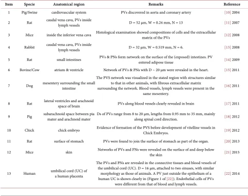
[image:5.595.41.553.317.722.2]Threadlike structures, different from all the circulation and nerve systems known, were first reported by Kim in 1963 [2][5]. Since then, quite a number of studies have re-ported the revelation of these transparent vessels [6][7] [8][9]. Table 1 lists a sum-mary of representative papers (not fully listed) in recent years, stating the locations, species of living organisms and some particulars where PVSs were found by experi-ments. The average of the largest diameters in a number of PVs revealed is indicated by D, if such data are available, and W is the average diameter of the lymph vessels en-closing the PVs, in cases where PVs are found to be inside lymph vessels. N is the
Table 1. Revelation of the Primo Vasculature System in Different Organisms.
Item Specie Anatomical region Remarks Reference
1 Pig/Swine cardiovascular system PVs discovered in aorta and coronary artery [10] 2004
2 Rat caudal vena cava, PVs inside lymph vessels D = 52 µm, W = 0.24 mm, N = 13 [11] 2007
3 Mice inside the inferior vena cava Histological examination showed compositions of cells and the extracellular matrix of the PVs [12] 2008
4 Rabbit caudal vena cava, PVs inside lymph vessels D = 32 µm, W = 0.519 mm, N = 6. [13] 2008
5 Rat small intestines PVs & PNs form network on the surface of the (exposed) intestines. PV entered adipose tissue [14] 2009
6 Bovine/Cow atrium & ventricle Network of PVs & PNs with D ~ 20 µm were revealed in the heart. [15] 2011
7 Dog mesentery surrounding the small intestine
The PVS network was visualized in the stated region with structures similar to that in other animals, with fibrous extracellular matrix surrounding the network. Blood vessels, lymph vessels were present in the
same mesentery.
[16] 2011
8 Rat lateral ventricles and arachnoid space of brain PVs along blood vessels clearly revealed in brain [17] 2011
9 Pig subarachnoid space between pia mater and arachnoid mater Ds of PVs range from 8 to 20 µm, lengths from 0.95 mm to 35 mm, mainly along spinal cord direction. [18] 2012
10 Chick chick embryo Evidence of formation of the PVS before development of vitelline vessels in Chick Embryos. [19] 2012
11 Rat surface of stomach PVs were found to join the surface of stomach as part of the organ. [20] 2013
12 Mice skin Networks of PVs and PNs were revealed on the surface of and deep below the skin [21] 2015
13 Human umbilical cord (UC) of a human placenta
The PVs and PNs are revealed in the connective tissues and blood vessels of the umbilical cord (UC). D = 16 μm, attached to two sinuses, with similar
morphology as those of animals. A PV just outside the epithelium of a human UC is shown clearly in (Figure 1 of [22]). Endothelial cells of PVs
were different from that of blood and lymph vessels.
number of specimens if available in the publications; special remark lists certain special features in brief. The last number in the last column is the year of publication.
2.2. Classification and General Characteristics of the Primo Vasculature
Systems among the Species Listed in the Last Section
(1) Definition of PVS
A unit of a PV, called a ductile (similar to a collagen molecule in a collagen fibril) in the PVS has a thin layer, composed of endothelial cells with rod-shaped nuclei. It is surrounded by an external membrane built of cells similar to smooth muscle cells or epithelial cells yet to be confirmed [23]. The endothelial cells have rod-shaped nuclei, and fine argentaffin fibers. A membrane wraps around several ductules to form a PV. Fibrous and amorphous materials are found in the interluminal space of a typical PV. PVs are joined to nodes called PNs to form a network.
(2) Intravascular PVS
There are reticular fibers, extracellular matrices (ECM) between ductules. One class of PVs (called intravascular class) are systematically distributed inside and along the blood and lymphatic vessels, often joined to one or a small number of corpuscles/ nodes. Some of these PVs are found to leave the lymphatic vessels and join the adipose tissues [5].
(3) Extravascular PVS
There are PVs and PNs running along, but outside the blood and lymphatic vessels, and nerves. This class of network may be called the extravascular class. There are thick connective tissues surrounding them. In the lumens of these PVs and inside the PNs, many chromaffin granules are revealed [5].
(4) Organ Surface PVS
Another class (called organ surface class) of PVs and PNs are joined together, form-ing a freely floatform-ing network on the surfaces of internal organs, such as that found in the adipose tissues on the intestine [14]. These PVs are not associated with blood or lymphatic vessels. In the lumens of the PVs and inside PVs in this class of network, there are cells possessing, bright cytoplasm and basophilic granules [15].
(5) Sub-system Nerve of PVS
Class N may be designated to those PVs and PNs floating in the cerebrospinal fluid (CSF) in the brain, with branches distributed in the parenchyma of the central nervous system. Sub-systems N (nerve) are also found in the peripheral nervous system [9].
(6) Intra-organ subsystem of PVS
Inside the parenchyma of internal organs, PVs and PNs are also found in animal models [9]. This class may be called class I, meaning intra-organ subsystem.
(7) Some features of PNs
colla-gen fibrils/fibers) supporting the blood vessels [9].
(8) The fluids inside the PVs found according to early studies are [2][5]: i) DNA & RNA
ii) Amino acids: 19 amino acids found, including some essential ones. iii) Nitrogen content: 3.12 to 3.40%.
iv) Lipid: 0.57% - 1.00%.
v) Reduced sugar: 0.10% - 0.12%. vi) Hyaluronic acid.
vii) Free mono nucleotides: 16 types so far found. (9) Motion of PVs
There are transverse and longitudinal oscillatory types of motion of a typical PV, presumably propelling the fluid (PVF) inside.
(10) PVS and Tumor
PVS has been found to be more densely populated in the proximity of a number of tumors [24].
We show in Figure 1 below some photos revealing the PVS in rabbit as reported in [25] with permission.
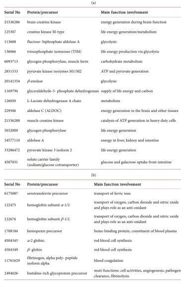
2.3. Contents of the Primo Fluid according to Recent Investigation, and
Its Implication
[image:7.595.246.500.489.613.2]The Primo Fluid inside a PVS system on the tissues above the intestine of rabbit model was extracted. Using a solution digestion technique, proteomic analysis carried out in [26] identified 81 proteins in such fluid and 207 proteins in the cells composing the ducts. Following, employing the gene ontology clustering analysis to study these pro-teins indicate that these propro-teins pertain to processes involved in various physiological processes and body development. We shall briefly analyze and classify 49, as examples, out of the 81 items in Table 2, according to their main functions in humans, though
Table 2. (a) Life energy generation and related metabolism; (b) Blood cells synthesis/regulation, iron ion transport, & angiogenesis; (c) Proteins interacting with key positive ions Ca2+, Na+, K+, or H+, and proteins involved in other signaling pathways; (d) Proteins involved in body growth and development; (e) Cholesterol/hormone transport or synthesis, Vitamins transport, mechano-transduction, tumor suppression and DNA repair.
(a)
Serial No Protein/precursor Main function involvement
21536286 brain creatine kinase energy generation during brain function
125307 creatine kinase M-type life energy generation/metabolism
113608 fluctose- biphosphate aldolase A glycolysis
136066 triosephosphate isomerase (TIM) life energy production via glycolysis
6093713 glycogen phosphorylase, muscle form carbohydrate metabolism
2851533 pyruvate kinase isozymes M1/M2 ATP and pyruvate generation
20141354 β-enolase glycolysis
1169794 glyceraldehyde-3- phosphate dehydrogenase supply of life energy and carbon
126050 L-Lactate dehydrogenase A chain metabolism
229506 aldolase C (ALDOC) energy generation in the brain and other tissues
21536288 muscle creatine kinase catalysis of ATP generation in heavy duty cells
5032009 glycogen phosphorylase life energy generation
34577110 aldolase A energy in liver, kidney and intestine
33286472 pyruvate kinase 3 isoform 2 life energy generation
4507031 solute carrier family (sodium/glucose cotransporter) glucose and galactose uptake from intestine
(b)
Serial No Protein/precursor Main function involvement 6175087 serotransferrin precursor transport of ferric ions
122475 hemoglobin subunit α-1/2 transport of oxygen, carbon dioxide and nitric oxide and plays role as an anti-oxidant
122676 hemoglobin subunit β-1/2 transport of oxygen, carbon dioxide and nitric oxide and plays role as an anti-oxidant
1708184 hemopexin precursor heme-binding protein, constituent of blood plasma
4504345 α-2 globin. red blood cell synthesis 4504349 β- globin red blood cell synthesis
11761629 fibrinogen, alpha poly- peptide isoform alpha blood coagulation
(c)
Serial No Protein/precursor Main function involvement
44889024 serum albumin precursor binding to the three most important positive ions, water, some hormones, it is the main protein in blood
118600944 parvalbuminα calcium ion is a multi-functioned second messenger. Parvalbumin α is a calcium-binding protein
62632750 ATPase, class VI, type II B transporter of positively charged ions
4557729 precursor of voltage-gated potassium channels sub-family, member 2 isoform regulate electro-cardiac signals, is transporter of positively charged ions
4506813 voltage-gated sodium channel provides means of electrical signals through nerves, skeletal muscle and heart
8923844 soluble adenylyl cyclase effector molecule of important signal transduction pathway, with special function of sensing bicarbonate
40254842 centaurin β2 multi-signaling pathways and hence involved in multi-cell functions
(d)
Serial No Protein/precursor Main function involvement
130488651 serpin peptidase inhibitor, grade F regulation of array of biological processes, including growth and immunity
21542114 lumican as a constituent in the interstitium, and cornea of the eye
4557871 transferrin transport of iron to proliferating cells
156523970 α-HS-glycoprotein (AHSG) brain, bone development with endocytosis capability 4502101 annexin 1 cell group formation and cell death signaling
22547186 serine hydroxymethy-transferase 1 isoform 1 cytoplasm synthesis
4503063 crystalline beta-B2 formation of eye lens 130498871 inter-α trypsin inhibitor heavy chain 2 connective tissue structure 112876 α1- antiproteinase F precursor connective tissues regulation
(e)
Serial No Protein/precursor Main function involvement
113996 apolipoprotein A-1 precursor transport of unused cholesterol back to liver
4557321 apolipoprotein A-1 preproprotein synthesis of high density lipoprotein and tissue reverse cholesterol transport as the last item
1722804 vitamin D-binding protein precursor vitamin D-transport 32483410 vitamin D-binding protein vitamin D-transport
136466 transthyretin (prealbumin) transportation of hormone and Vitamin A 139654 vitronectin precursor mechanotransduction (connective tissue)
15149463 caldesman 1 isoform 4 mechanotransduction of muscle and non-muscle cells 5453710 LIM and SH3 protein 1 intra-cellular and extra-cellular mechanotransduction
1485539842 deleted in malignant brain tumors 1 isoform b (DMBT1) may play role in suppressing tumor
these precursors of proteins or proteins were identified in the PVF of animal species. As many proteins have diversified functions, we put these 49 items in 5 groups (a-e). The number of each item follows that published in [26], for convenience of cross-examina- tion. The function of the related proteins can be found in general from NCBI Gene re-port. For example, information can be obtain from the data of “APOA1 apolipoprotein
A1 [Homosapiens (human)]. Gene ID: 335, updated on 9-Jul-2016” for the
Apolipo-protein A1. We will not put down such a similar and long list of references here. If we follow our table, we find that the above-mentioned proteins plays general roles in (a) Life energy generation and related metabolisms; (b) Blood cells synthesis/regu- lation, iron ion transport, angiogenesis; (c) Interaction with key positive ions Ca2+, Na+,
K+, H+, play roles involved in other signaling pathways; (d) Involved in body growth
and development; (e) Cholesterol/hormone transport or synthesis, Vitamins transport, mechanotransduction, tumor suppression and DNA repair.
Obviously, these proteins, if so developed from the precursors, participate in majori-ty of the major physiological functions. It is rather tempting to hypothesize that the PVs contain fluids with precursors of proteins for body development, life energy supply, and other key functions in physiological processes of the human body; note that the PVSs have been found in adult animal models and in the human placenta.
2.4. Proposed Functions of the PVS at the Embryo
reticular fibers to support the blood vessels. We do not have enough information to sketch the complete PV circulation system at the embryonic stage at present. However, we are rather certain that once the blood vessels are formed, the head and spine emerge from the blood vessels, an established procedure in developmental biology. Suppose the basic structure of the embryonic fetus has been developed, and we can now jump to an adult body to discuss the second circulation system in the next section. Note that stem cells-like cells and catecholamine producing endocrine organ have been identified in PVSs of adult animals [27].
3. The Second Fluid of the Five-Fluid Circulation System
3.1. Introduction
The cardio-vasculature system is proposed to be the Second Fluid circulation system, which has already been extensively studied. We will only sketch briefly some relevant properties and highlight some features to be followed up in later sections, or the fol-lowing papers of this series. In human, blood cells, glucose, proteins (majority is albu-min), mineral ions, hormones, oxygen, carbon dioxide are suspended or dissolved in the blood plasma (of which around 92% by volume is water). Albumin is the main pro-tein in plasma, and it functions to regulate the colloidal osmotic pressure of blood. Note that at any time about 84% of the whole blood volume of the body is circulating in the vessels, and 16% of blood is inside the heart and the lungs. Among the blood in the vessels, 64% is venous blood, 13% is inside arteries, whereas 7% is inside systemic arte-rioles and capillaries [28]. A blood vessel is composed of three layers—endothelial cells, smooth muscle cells and perivascular cells in larger blood vessels. These layers are se-parated by connective tissue layers and the outermost layer is the adventitia through which mechanical signals can be transmitted to the vessel. A capillary is much smaller and does not have smooth muscle cells (see details in Section 7). The wall of a venous vessel is thinner than that of an artery, and can hold more blood than an artery of the same type. Many veins have one-way valves for facilitating blood flow back to the heart. A common blood vessel disease, particular for women is the occurrence of varicose veins, commonly found at the back of legs. Such veins are enlarged veins caused by valve disorder/failure. The veins appear gnarled and engorged. Maintaining good blood circulation is of prime importance in health. When the small arteries in the hands and toes become narrow, poor blood circulation would result in these regions, and the Raynaud disease develops, usually in cold weather or under high stress.
5 - 10 microns) are some muscle fibers, the precapillary sphincters, which can contract and thus regulate the amount of blood going through the capillary bed. In the return journey, the blood flows through the other “half” of the capillary bed (deep blue in dia-grams of this paper and elsewhere, signifying the content of carbon dioxide) to join the smaller type of the venous vessels, i.e. the venules, which join the bigger venous vessels, with decreasing flow speed because the diameter of the transporting vessel is increasing in size. As the venous vessels have a total volume (when they are full of blood in the usual condition) much bigger than that of the artery vessels, 65% of the blood is re-tained in the venous vessels at any time. One sees bluish vessels near the skin surface, particular on the dorsal side of the hands. There are about 4.7 liters of blood in a hu-man weighing about 68.2 kilograms/150 pounds, at one time.
There is a significant difference in the flow speed in arteries during systole and dias-tole. One usually uses the parameter called pulsatility index (PI), to calculate such a difference. PI = (systole speed – diastole speed)/(mean speed at a site), which is de-creasing with inde-creasing distance from the heart [29]. From the fluid dynamics point of view, perhaps an interesting comparison is to relate the total cross-section area diame-ter of one or more blood vessels at a particular region (where the vessels, ardiame-tery or ven-ous, are located) with the flow speed. The flow speed at the (i) aorta (with cross- sectional area of 3 - 5 cm2) is about 40 cm/s, that at (ii) vena cavae inferior and superior
(with cross-sectional area of 14 cm2) is 15 cm/s, whereas that at (iii) a capillary bed with
a typical total cross-section area of 4500 - 6000 cm2, is ~0.03 cm/s [30][31]. Note that
the volume of blood passing through per second (found by simply multiplying the total cross-sectional area with the average flow speed), based on the above numbers, fall in the following ranges: (i) 120 - 200 cm3; (ii) 210 cm3; (iii) 135 - 180 cm3. These figures
are of the same order of magnitude, but should not be equal, because there is water ex-creting outside the vessels to the “Third Fluid system—the interstitial fluid”, as will be explained in Section 4, and represented schematically in Figure 2 there.
Normal blood plasma behaves like a Newtonian fluid, namely, whose viscous stress (due to the fluid flow) is linearly proportional to the local strain rate at every space point. The viscosity of normal human plasma at 37˚C is about 1.1 - 1.3 milli-Pascal- second [32]. An increase of 5˚C in temperature in the physiological range reduces plasma viscosity by about 10%. In other words, the blood becomes less viscous at mild hyperthermia. This property is relevant to the flow fluency of blood to be discussed in a flowing paper.
3.2. Blood Flow Rate Affects Remodeling of the Vessel Tissue through
Change in Elastin/Collagen Ratio
Due to difference in physiological demand in blood flow transportation, the ratio of elastin and collagen tend to decrease with increasing distance from the heart [33]. Note also that collagen has a faster turnover rate than elastin [34]. Collagen, and other com-ponents, of the ECM building the vessel cells and their surrounding ECM, are degraded by metalloproteinases (MMPs) and such degradation is regulated by inhibitors of me-talloproteinases (TIMPs) [35] [36]. Studies on the elastin/collagen ratio in different types of blood vessels, with respect to blood flow rates would be useful to understand diseases related to abnormality in blood vessel remodeling. In [33] the relationship be-tween blood flow rate and MMP/TIMP ratio in blood vessels is investigated. It has been remarked there that vessels with high blood flow rate showed a higher MMP-2 expres-sion as compared to that with low blood flow rate and viceversa. The elastin/collagen ratio has been found to decrease from aorta, to carotid artery, femoral artery, femoral vein, but the blood flow rate has been found to increase from femoral vein, femoral ar-tery, to carotid arar-tery, then aorta (see Figure 3(a) & Figure 3(b) of [33]). Passive tension vs stretch relationship for different vessel beds (as illustrated by aorta, carotid artery, vein) is shown in Figure 6 of [33]. For a fixed tension, the vena cava showed the mini-mum stretch, because this blood vessel has the minimini-mum elastin and maximini-mum amount of collagen, whereas the reverse is true for aorta, which has the maximum amount of elastin and minimum amount of collagen. In other words, the higher the elastin content in the tissue, the higher the compliance in that vessel. On the other hand, the result of Figure 5C in [33] suggests that increase in blood flow rate is associated with higher elastin-collagen ratio. Based on the above analysis, we propose that maneuver modality along the direction of blood flow on the veins (which appear superficially), would in-duce a higher blood flow rate. It is tempting to speculate that long term massage treat-ment might in turn induce a higher elastin-collagen ratio to be developed in the veins.
3.3. Cardiomyocytes Beat Best on a Matrix with Elasticity Similar to the
Heart
Under the healthy state, myocardium, cardiomyocytes are attached to extracellular ma-trix (ECM) built of collagen fibers which have just the right degree of elasticity/ compliancy for actomyosin’s action to pump the heart [37] [38] [39]. With proper blood flow rate, which is affected by the compliant property of the blood vessels, the pumping action is rhythmic with a frequency demanded by the physiological need. Clinically, myocardial infarct often leads to cardiac fibrotic rigidification, resulting in impairment of cardiac output [40].
In vitro investigation also shows that cardiomyocytes on rigid culture substrates
demonstrate progressive loss of rhythmic beating [41]. The elasticity E of ECM pro-vides cue for action of the heart cells during the stated mechanotransduction process.
maxi-mum contraction and E were obtained.
Since the cells are causing the contraction, cell strain εcell always exceeds, or at most
equal to the collagen matrix strain εout, i.e. εcell ≥ εout. The strain sustained within the cell
εin during contraction is: εin = εcell – εout. On soft matrices (E < 8 kPa in the experiment
with quail heart cells), cell strains have been transmitted almost completely to the ECM so that εin is significantly smaller than εout. (see Figure 1C left of [42]) and there is very
little contraction work done because there is no “rebounce”. On very stiffest matrices, εout is practically zero. Under this condition, the myosin-based contraction is fully
sus-tained by the cell (see Figure 1C right of [42]), hyperactivating the stretch-sensitive proteins, but the cells cannot “move” the ECM. Again rhythmic contraction cannot occur. Under physiological condition, the heart cells and the ECM must play the “push and pull” game in synchrony to save the heart cells from overwork. Such a synchro-nized condition happens only when at an optimized elasticity E* (in the quail experi-ment, E* = 10 kilo Pascal), at which εin = εout. Though embryonic cardiomyocytes were
used in the experiment [42], we anticipate that the same result holds true for heart muscles of subjects. In practice, thus we must let the connective tissues around the heart to be maintained at the optimum condition. Such a result means that we should not allow the occurrence of inflammation or fibrosis on the collagen fibers around the chest region; even though abnormality of the elasticity E of the collagen fibers occurs somewhere away from the heart region, such abnormality could indirectly influence the E value of the cardiac ECM because mechanical force can be transmitted throughout the connective tissues of the fascia to far distances [43]. Thus allowing the heart to work with the best efficiency possible condition by relaxing the associated connective tissues would protect the heart and ensure the best possible blood flow.
3.4. Circulation Function of the Emissary Veins
reduce the risk of infection in the “dangerous region” in modern medicine. To clear the fluid from internal jugular vein, we therefore consider that, with the subject in supine position, massage along directions of several nerve routes (which are adjacent to the veins emerging on the superficial layer of the face before joining the internal jugular vein) might be helpful to the process of brain fluid clearance, an issue to be discussed in details in Sections 7 and 8.
The second and third foramen of interest to us is the parietal foramen, which is lo-cated 2 - 5 cm anterior to the external occipital protuberance [48] and the parietal emissary vein(s) is located laterally to the sagittal suture, at the boundary between the posterior third and middle third of this suture. This foramen can occur bilaterally, un-ilaterally or completely absent, depending on the ossification process of the anterior fonticulus in individuals. The parietal foramen allows passage of the emissary vein (at that site) to connect the superficial veins on the scalp and the superior sagittal sinus in-side the skull. This foramen is also conin-sidered as “dangerous foramen” for spreading infection to the sinuses of the dura matter inside the brain. We also propose that mas-saging the superficial veins connected to the parietal emissary veins against direction flow into the skull with the subject in upright position could well be a non-invasive measure to avoid the stated risk of infection.
The sphenoidal emissary foramen (with the sphenoidal emissary vein) allows the passage of an emissary vein that connects the pterygoid venous plexus to the cavernous sinus.
As the superficial occipital veins join the occipital emissary vein, on route to the deep cervical vein, then the external jugular vein, we propose to massage, starting from the occipital foramen site, in a downwards direction when the subject is in upright posi-tion. Some people are short of some emissary veins, and we think that is a good sign because of the dangerous scenario of the two-way blood flow along emissary veins [49].
4. Interstitial Fluid as the Third Fluid of the Five-Fluid Circulation
System
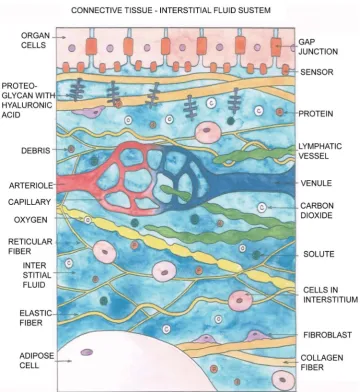
4.1. A Brief Sketch of the Interstitium
al-lowing diffusion of small molecules in and out of the tissue through the interstitial fluid (ISF) [43][50]. These protein complexes have two main classes (i) Proteoglycans (PG) with its subclasses Aggrecan, Glycosaminoglycan (GAG) and others. Note that hyalu-ronan is the most abundant and ubiquitous member of the GAGs [51]. The proteogly-can proteins, to which hyaluronan is attached, form negatively charged long chains. Hydronium ions together with layer of water are then bound to the hyaluronan, form-ing a thick interstitial fluid in the interstitium, called the ground substance. (ii) Glyco-proteins having subclasses such as Fibronectin , and Tenuscin. These two classes (i) and (ii) of proteins provide a potential bridge between the connective tissue matrix and the cells adjacent/within the interstitium. There are “residence cells” in the interstitium with almost constant populations: fibroblasts, mast cells, adipocytes, melanocytes. There are also “cells in transit” which migrate from the blood when in need: mono-cytes, macrophages, lymphomono-cytes, eosinophils, and plasma cells. They are mainly en-gaged in immunity duties.
4.2. Reasons for the Emergence of the Interstitial Fluid System as the
Third Fluid in the Circulation System
We know that arterial blood vessels are abundant in the body. The organs of the body are composed of specific cells (such as muscle cells, cardiomyocytes, hepatic cells, neu-rons,…) which must obtain, in addition to oxygen molecules, water, nutrients, signal-ing molecules (such as amino acids, sugar, solutes , specific proteins, hormones) to carry out physiological functions. The cells in the body are degraded and replaced by new cells (apart from the neurons in the brain) continuously. During infections and repair processes, the organs/organelles have to get help from immunity cells, Matrix metalloproteinases (MMPs), which are calcium-dependent zinc-containing endopepti-dases to degrade the damaged collagen (fibrils/fibers) of various classes. Other enzymes and proteins may also be needed during such infections/disorders and repairs. Stem cells, fibroblasts of various pheno-types, and other cells (such as white blood cells) also participate in the remodeling/repair processes. Thus, during physiological or patholog-ical conditions, part or all of the mentioned “necessary components” must be trans-ported to the target sites—tissues, or organs/organelles. As the artery vessels, including capillaries, are not directly “joined” to the cells of organs, other means must transport all the stated components to the target(s). Therefore, the interstitial fluid forms natu-rally the Third Fluid to carry out such transportation duties; this transportation in-cludes transportation of information—the interstitium with interstitial fluid (ISF) pro-vides a platform for efficient carriage of signaling peptides/proteins. Moreover, flow of the ISF provides a shear force to collagen fibers which in turn transmit the associated mechanical force to the integrins or other receptors in the cell membranes to trigger off mechanotransduction processes.
wa-ter must serve also as a medium of transportation of these molecules. In the blood cir-culation system, the lipid drops (which are typical examples of hydrophobic bio- mole-cules) are enclosed by a spherical structure (due to the hydrophobic nature) with a sin-gle lipid layer, called chylomicron. The chylomicrons transport fat from the blood stream to the liver, (via the ISF for a small part of the journey) where the lipid is broken down to form cholesterol, to be used to generate energy. Nutrients/bio-molecules hav-ing ionic property form solutes in water. The positive ions of water are bound to the hyaluronic acid structure, which is negatively charged and attached to the proteogly-cans; these are non-collagen proteins in the intersitium. A very viscous fluid is formed to impede the flow speed, so that the tissues/organs can receive their parcels (O2 and
nutrients etc) from the mailman. In summary, the ISF is doing very important work all the while, though the average flow speed is very slow, being only ~10 micron per second or even less, dependent on sites. The total volume of ISF passing through the body each day is not small—a human body treats about 2 - 4 liters per day. At 37 de-grees C, whereas the specific gravities of plasma, lymph, whole blood, and synovial fluid are very close to that of water-unity, the viscosities of these fluids are respectively 1.2, 1.5 - 2.2, 3.0 - 3.84, 10 - 104 milli-Pascal, covering a wide range [52]. In Figure 2, the
basic constituents of the interstitium with the ISF are schematically presented.
4.3. Basic Functions, and Some Physical Properties of the Interstitial
Fluid
In addition to the well-known function of providing transport of nourishment, oxygen to organs, interstitial flow also induces blood and lymphatic capillary morphogenesis (see evidence in in vitro experiments in [53][54][55][56] and lymphatic regeneration
in vivo [57]), upkeeps the physiological activity of chondrocytes and osteocytes [58]
[59][60], initiates fibroblast to differentiate [61][62], and triggers smooth muscle cells to produce cytokines [63]. The existence of suitable interstitial fluid pressure is critical for ocular health [64]. Even during embryonic development, cilia in motor protein drives convective ISF flow to pattern the early stage of life [65].
Up to 20% of the body’s mass is made up of interstitial fluid [66]. During inflamma-tion and acute edema, the flow speeds are greatly enhanced [67][68]. The ISF flow is drained by the lymphatic system which processes several liters (up to 8 liters) of lymph per day [69], with around 2/3 of the amount coming from the liver and intestines [66]. The ISF transport is not a one-way process. Note that oxygen, and plasma have leaked out of the blood capillaries arising from the hydrostatic and osmotic pressure differ-ences i.e. the Starling forces [70][71]. The plasma and carbon dioxide are returned to the blood circulation via the ISF.
and “absorb” (internalize) proteins, release proteases which can free the proteins that are bound to the ECM etc.
The composition and structure of the interstitium systems vary in different parts of the body for very good physiological reasons. The connective tissues, together with the help of the interstitial fluid, give mechanical support to cell groups/organs, based on which mechanical properties are specified. Normal leakage of plasma from blood ves-sels (due to Starling forces stated before) is the main source of fluid in the interstitium of most soft tissues. Therefore the interstitial fluid contains around 40% of the protein concentration of plasma, but with slightly different ions compositions for various rea-sons [66].
The magnitude of the ISF pressure (ISFP) at a location is influenced by many factors, such as exercise, blood pressure, tissue metabolism, hydration, ECM composition, and cell density at that location [75]. With regards to relation between ISFP and exercise, we would remark also that the interstitial flow in cartilage of joints is driven by time-de- pendent forces (compressing the tissues) resulting from motion such as walking [59] [76]. The ISFP, as enhanced during exercise, helps to transport nutrients, wastes, and proteins between chondrocytes (which are embedded in the cartilage). Remark that blood capillaries are at a distance because there is basically no blood circulation through the cartilage. In addition to passive causes, cells in the interstitium also play crucial active roles, such as maintaining tension in the ECM (in a β1 integrin-depen- dent manner) to regulate ISFP [77]. The magnitudes of ISFPs in some examples of tis-sues are listed in [78]. Pressure gradients between the interstitium and lymphatic fluid cause ISF to flow into the lymphatic system in general. In passing, we note that using radioactive tracers, there is experimental evidence that the interstitial fluid flow tracks in animal models and human are correlated with Traditional Chinese Medicine Meri-dians [43].
4.4. Interstial Fluid Flow Guides Lymphangiogenesis as an Introduction
to Another Fluid in the Circulation System
de-veloped in [81], a 2-mm-wide circumferential annulus of skin was removed (around half way) from the mouse tail, leaving the underlying bone, muscle, major blood ves-sels, and tendons intact (see Figure 1 of [81]). Examination of normal tail skin of mouse model, Atomic Force Microscopy (AFM) showed a regular hexagonal network of lym-phatic vessels in the skin (see particularly Figure 1a of [81]). There are many subcuta-neous blood vessels, but the interstitial fluid and lymphatic vessels are within the region of the skin being removed. Therefore blood flow to the tail remains uninterrupted, but the lymphatic network and the flow of interstitial fluid out of the tail tip are completely interrupted by the above surgery procedure. Such a condition, if untreated, would lead to significant edema of the tail [82]. However, in the experiment of [81], when a colla-gen dermal equivalent (CDE) was inserted into the site of the removed skin to provide path for fluid flow, no edema occurred (see Figure 1b through 1d of [81]). These au-thors used AFM to track lymph flow pathways (either as fluid channels or lymphatic vessels) within the CDE for a time interval of 60 days after the procedure. After at least 10 days, fluid channels were observed to be formed, bridging the distal and proximal portions of the lymphatic system; finally, those channels were replaced by an organized hexagonal network of lymphatic capillaries. This is concrete evidence supporting the proposal that interstitial fluid flow guides lymphangiogenesis.
5. The Fourth Fluid—A Mixture of the Cerebrospinal Fluid
and the Brain Interstitial Fluid
5.1. An Updated Concept of Cerebrospinal Fluid Generation
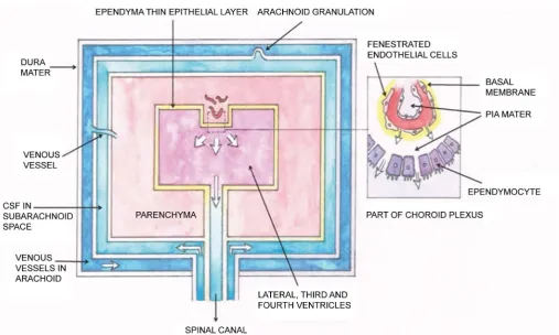
Figure 3. Schematic representation of the blood-CSF barrier and the classical model of brain drainage via vein vessels. The ependymal thin epithelial layer (yellowish color) encloses the lateral, third and fourth ventricles of the brain. The cells of this layer are called epen-dymocytes, joined together by tight junctions, but have some very small opening (<50 microns). The capillaries of the choroid plexus (part of it is enclosed by the dotted line) excrete water, glucose, oxygen, vitamins and ions into the mentioned ventricles and contributes the main part of the CSF in the brain. The capillary is enclosed by fenestrated endothelial cells, which allow small particles to go through. For immunity purples, macrophages and lymphocytes migrate from the blood and pass through the vessel wall and then the small open-ings among the ependymal layer, to the ventricles “outside”. The stated passage of this CSF is indicated by the white arrows. The CSF passes down to the end of the canal of the spinal cord and return to the sub-arachnoid space as shown by the white arrows. Following the pumping action of the heart, together with contribution of the arteries along the spinal region as described above, there is a resonance of the ventricles and the spinal column, and the characteristics of the fluid pulsation have been detected by MRI study [85]. Figure 3 is mod-ified from Figure 1 of [86]. This diagram was painted by author PCWF.
There are granulations from the subarachnoid space into the superior sagittal sinus (venous vessel), so that the CSF flows back to the blood circulation. The blood circula-tion in and out of the skull is well known and will not be discussed here. The CSF was considered to be a fluid isolated from the brain intersititial fluid. So this is the general picture of the CSF up to a few years ago. Let us turn first to a new addition to the gen-eration of the CSF discovered only a few years ago.
characteris-tics in large systemic arteries of young subjects were measured with MRI, whereas ce-rebral flow features were obtained from ultrasound Doppler analysis, and fluid pres-sures at various locations were measured with tonometry. Employing a similar metho-dology, a schematic representation of the arterial tree consists of 120 vessel segments contributing to the generation of CSF in the spinal cord and the brain region is de-picted in Figure 2A-C of [85]. Connection between this cardio-vascular system and the CSF system was constructed by a transfer function which is specified by a number of parameters: spinal volumetric (mechanical) compliance (as measured by the elastance coefficient Kein units of ml/[mmHg height]), fluid pressure, flow speed and flow waveform in the connected system [85]. These parameters were compared with the in vivo measured data of the stated combined system, giving results which support the suggestion that certain amount of CSF is supplied by the fluid of the blood circulation from systemic arteries plus cranial arteries. In particular, the CSF flow & CSF pressure were calculated, with input parameters from measurements, as functions of the spinal compliance (Ke, from flexible to stiff), taking the distance from the first cervical disc C1 as a parameter. It was noted that the CSF pressure wave reflection was greater at the caudal end of the spinal subarachnoid space (SSS) when the spine was more flexible (i.e. Ke = 0.04).
Here we refer the reader to Figure 3 which summarizes schematically the circulation of CSF in the brain and central canal of the spinal canal.
5.2. CSF Follows Special Pathways to Mix with BISF in the Brain
Parenchyma
We have briefly analyzed certain basic characteristics of the CSF. On the other hand, the neurons of the brain, as other organs of the body, are nourished by the interstitial fluid, which is different from the CSF. In fact, the interstitial fluid (ISF) exists in all connective tissues - including that surrounding the parenchymal cells of the brain and spinal canal. The water, nutrients in the form of solutes, plus proteins and some cells leave the blood vasculature and enter the interstitium as interstitial fluid, described in Section 4. The structure of the blood brain barrier (BBB) is well known. In brief, the ar-terial capillary in the parenchyma (neurons and astrocytes mainly) is surround by a layer of endothelial cells with tight junctions, which are further enclosed by a structure of basal lamina. There are also pericytes in this lamina layer. The external layer is em-bedded by endfeet of astrocytes; there are however small cleft (around 20-nm wide) or in layman term, “leak spaces” in between the endfeet. Thus the fluid from a capillary in the brain parenchyma could exit first to the lamina layer and then pass through the “leak spaces” to reach the brain cells. We call this brain interstitial fluid as BISF. The BBB, which regulates the water, nutrients, proteins and the amount of BISF that can reach the brain cells, and thus controlling the brain function [87].
move-ment of small ions, with the result of regulating blood-brain ionic traffic. (B) Restric-tion of small molecule permeaRestric-tion. (C) Specific transport of desirable small molecules needed by the brain. (D) Restriction and regulation of large molecular traffic. (E) Sepa-ration of peripheral and central neurotransmitter pools to avoid cross-talk between [88].
For at least a decade, the (classical) CSF was considered to be reabsorbed into the blood stream either through (i) the arachnoid granulations of the dura sinuses, or (ii) by emerging out of the cranial cavity along cranial nerve sheathes to the cervical lym-phatics [89][90]. However, the brain is a heavy-duty organ and large amount of wastes is generated continuously by the brain. Moreover, it is hard to believe that the relatively small amount of fluid carried out of the skull by processes (i) and (ii) could balance the water generated at the choroid plexus and SAS of the spinal canal. Therefore biomedi-cal scientists tried hard to find pathways that had not been discovered in the past dec-ade to carry fluid with soluble plus insoluble wastes out of the brain.
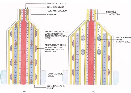
Tracers of different sizes were injected into the ventricular region or cisterna magna of mice models, and movement of the tracers were assessed using imaging technique in the parenchyma in [91]. Their findings were: (i) Small amount of the two small sized tracers was observed to cross ependyma of the lateral and third ventricles after 30 min of continuous infusion. (ii) After injection intracisternally of 40 kD tracers (FITC-d40), the fluid with tracers flew along the sheaths of arterial vessels in the sub-arachnoid space into the parenchyma. (iii) The amount of tracers found in the brain is inversely proportional their sizes. (iv) The authors inferred that these tracers followed two path-ways along one vessel: (a) the basement membrane (BM) between the vascular endo-thelial and the smooth muscle cell layers; (b) the sheath between the adventitia and the astrocyte endfeet (see Figure 4). The movement was inferred in [90] to be caused by arterial pulsation. We, however, consider that in general fluid flows from a high pres-sure point to a lower one, and the injection at the cisterna magna would certainly create a pressure gradient to drive the fluid (together with tracers) initially. This phenomenon was called the arterial influx (of CSF) to mix with BISF. The mixture with tracers was observed to follow the sheaths of venous vessels and drain into the SAS. At the mean-time, we modify the diagram in [91] to illustrate schematically the mixing of the CSF and BISF.
Although the tracers mentioned in Figure 4 were injected with fluid at the cisterna magna, yet under physiological condition, since there is a driving force on the CSF ori-ginated from pulsation of blood flow analyzed in Section (5.1), we anticipate that the same mixing process goes on continuously. The mixing rate may be different from that found in in vivo animal study [91]. The mixing process was confirmed with MRI using
the in vivo rat model in 2013 [92]. As the brain is a heavy-duty organ, a lot of wastes,
Figure 4. Experiment demonstrating the mixing of CSF and BISF. In [91], tracers were injected at the cistern magna so that they would pass through the subarachnoid space. It was found that the tracers were carried by the fluid up via two pathways; (i) along the sheath between the external arteriole wall (i.e. adventitia) and the astrocyte endfeet; (ii) along the basement membrane next to the endothelial cells. This influx occurs for arterial vessels, but not for veins. The tracers then were carried by the ISF bulb flow though the parenchyma, as indicated by the two wavy arrows. The yellowish pink parts represent parts of the astrocyte endfeet and the pink region represents the brain cortex. The fluid mixture was then carried through the sheath between the astrocyte endfeet and the adventitia (light yellow) of the venule (on the right side of the diagram), flowing downward with the tracers through a pia mater layer, the subarachnoid space, the arachnoid space with dura mater and then outside the skull. This diagram, which was painted by author PCWF, is modified from Figure 2 of [91]. In this diagram, the arterial vessel has penetrated into the subarachnoid space from somewhere not shown in the diagram and turn up to pass across a pia layer into the brain, whereas the venous vessel pass through the dura space, and other boundary layers to exit the skull.
lymphatic system is found practically to be associated with blood vessels, the absence of the vital clearance system inside the brain had been a mystery. We will therefore pro-vide a comprehensive analysis of the extra-cranial lymphatic system in Section 6 first, and then review the important discovery related to the stated mystery published only around one year ago in Section 7.
6. Lymph as the Fifth Fluid
6.1. How and When Is the Lymphatic System Developed
[95]. One might therefore consider that the main lymphatic vessels develop from the grand veins, whereas all the other lymphatics develop independently from the venous system by a process of canalization of the connective tissues clefts [96]. There is evi-dence that the lymphatic muscle cells are originated from mesenchymal progenitors [97]. There is also evidence that electrical action potentials trigger lymphatic smooth muscle contractions and vice versa [98]. In fact, the spontaneous and intrinsic na-ture/origin of these contractions suggests that each action potential/(rhythmic contrac-tion) is preceded by a pacemaker event, like cardiac contraction (see Figure 2 of [96]). Here we speculate that the Primo Vasculature System participates in supporting the function of the lymphatic system, based on the fact that the PVs have been found to be inside and in the vicinity lymph vessels.
6.2. Structure of the Lymphatic System
The ISF contains both some useful proteins and some debris, plus “unwanted cells” like cancer cells and bacteria. It is precisely the function of the lymphatic system to treat about 3 liters of ISF and return it back to the blood system, along directions towards the heart. Hence the lymphatic system is not a closed circulating system, but the constitu-ents in this fluid, the lymph, are different from that of the blood, ISF, and BISF and are different at different parts of the body. The lymphatic system plays important roles in immunity, and is not as simple as many would have imagined; in fact there are several organs, called lymphoids attached to the lymph vessels, to participate in carrying out the physiological duty just mentioned. Some relevant features of these lymphoids are briefly sketched below.
Major Lymphoid organs include (i) Bone marrow where production of T lympho-cytes and maturation of B lympholympho-cytes take place. (ii) Thymus where thymolympho-cytes are matured into T lymphocytes. Peripheral lymphoid organs can be classified into (a) Lymph nodes which are spaced along lymphatic vessels, form clusters in the chest, neck, pelvis, axilla, inguinal region, and near the blood vessels of the intestines. There are around five to six hundred lymph nodes in the human body. (b) Spleen contains over half of the body’s monocytes, and is a center of activity of the mononuclear pha-gocyte system. It produces lymphocytes and is considered to be a very large lymph node [99][100].
6.3. Entry of the Interstitial Fluid with Its Components into the
Lymphatic System through Initial Lymphatics and then the
Collecting Vessels
Introduction to entry of the interstitial fluid into the lymphatic system
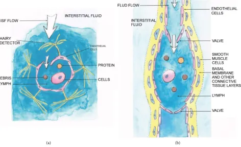
through the lymphatic capillaries, also known as initial lymphatics, or terminal lym-phatics [102]. This “entrance small tube” is built of a single layer of flattened endotheli-al cells usuendotheli-ally without a smooth muscle component. However, the over-lapping endo-thelial cells with connective tissue structures have hairy detectors (see Figure 5). These detectors are pushed by the flowing interstitial fluid, and the ISF can flow inside the duct as indicated by the white arrows, bringing with it the debris (circle in brown), protein(orange) from the interstitium, and cells (lymphocytes and other cells, represented by ellipsoid with nucleus). The interstitial fluid flows through overlapping edges of neighboring endothelial cells (microvalves). In contrast to the structure of a blood capillary, a lymphatic capillary has a thin wall with a wide lumen, irregular struc-ture; the wall does not contain pericytes and a basement membrane. Following the flow, the lymph is then drained into a collecting lymphatic vessel [103], which is surrounded
[image:26.595.80.553.267.555.2](a) (b)
by a basement membrane, a layer of smooth muscle cells (SMC) or pericytes (see Fig-ure 5, showing a basic unit of a collecting vessel, a lymphangion). These collecting lymph vessels contain luminal valves in order to prevent the back-flow. The one-way transport lymphatic system plays a key role in the maintenance of normal interstitial fluid volume and protein concentration. The immune competent cells also enter the lymph vessels and do their duties in the lymphatic nodes.
6.4. Intrinsic Propulsion of Fluid by the Lymph Pump
Even the ISF can enter the lymphatic initial duct, a propulsion mechanism is needed to drive the fluid flow. Historically, to look for the origin of such propulsion force driving lymph to flow through the peripheral to the major lymph duct (such as the thoracic duct), and then to the venous system, a series of experiments with unanaesthetized sheep as model were carried out half a century ago (see e.g. [104]).
Using a fistula system in the sheep model to study the lymph flow characteristics, it was found that lymph flow was intermittent with a well-defined rhythm that was unre-lated to muscle movements, unreunre-lated to respiration (except in the case of the thoracic duct for obvious reason based on anatomical structure).
The pulsatile pressures recorded from the various lymphatics ranged from 1 to 25 mm Hg, with pulse frequencies from 1 to 30/min. The magnitude & pulse rate of the pressure pulses increased as the lymph flow rate increased. When the various lymphatic cannulas (in the fistula system) were clamped to prevent lymph flow, the pressures in the lymphatics increased (reaching a high pressure up to 60 mm Hg) and the frequency of contractions also increased.
From experiment such as that just mentioned, we learn that one important driving force to cause lymph flow is the intrinsic contractile activity of the active lymph pump (composed of a series of lymphangions) whose function has been considered to be sim-ilar to the cardiac cycling action [105][106]. Note also that there are branching patterns of lymphatic system, making the flow profile in lymphatics even more complicated. In general there are no valves at the junctures of lymphatic branch points. Thus the con-tractile activity around the junctures is asymmetric and significant retrograde flow could well result. Hence it is difficult to predict the general direction of lymph flow in a local region unless the detailed structure of the vessel system, together with the nature and locations of valves are known. However, the early experimental result in [107] al-ready showed rather convincingly that intrinsic rhythmic contractions of the valved lymphatic vessels are mainly responsible for the propulsion of lymph from the peri-phery to the thoracic duct, with the function to control the removal of tissue fluid at a rate proportional to its rate of formation. Thus the intrinsic contraction mechanism has been considered to be established.
6.5. Extrinsic Forces Can Affect the Flow Characteristics
6.5.1. Definition of Extrinsic Propulsion Forces on Lymph Flowbeen caused by forces external to the lymph vessel, in a complicated manner: (i) The pulsation of the nearby blood vessels would aid lymph flow. (ii) Motion of skeletal muscles or tissues around the lymphatic vessels during exercise would squeeze the lymphatic vessels and aid fluid flow. However, it was reported long ago that sustained skeletal muscle activity would decrease lymph flow [107][108] because outflow resis-tance of peripheral lymphatics would be increased. We consider such a result is ex-pected because there is evidence that pulsatile pressure on the lymphatic vessels would enhance lymph flow; but compression with too large a frequency or too large a force would impede the lymphangion pulsations. (iii) The suction effects of respiration. Most of the time these forces help to move lymph centripetally. (iv) Note also that there are branching patterns of lymphatic system affecting the flow profile driven by both the in-trinsic and exin-trinsic forces. Now the term “exin-trinsic” lymph pump is meant to be a combination all extra-lymphatic forces which can influence (in general meaning en-hancement) lymph flow. The origin of these forces (which can be applied artificially) is not connected with active contractions of muscle cells in the lymphatic vessel wall.
6.5.2. Evidence of the Effect of Blood Vessel Pulsations on Lymph Pressure of Large Lymph Vessels
Since many branches of the artery and lymphatic vessels run roughly parallel to each other for long distances, intuitively one expects that movements from the great arterial trunks are imparted to the larger collecting lymphatics. In a number of studies, it has been inferred that the continual pulsatile motion of the lymphatic vessels are at least in part originated from the pulsation of the blood circulation. On the other hand, in the neck, where the thoracic duct is in close relation to large pulsating veins, venous pulsa-tions are considered to be transmitted to the lymph also (in particular, in the subclavian triangle area). Using dog’s model, the rhythmic intralymphatic pressure change in the supradiaphragmatic and cervical thoracic duct was investigated [109]. In both areas lymphatic pulses were found to be transmitted from the nearby large arteries. In the neck region, venous pulsations were also shown to be transmitted to the lymph in a similar manner. However, these pulsations were shown to propagate for only a few mil-limeters. In conclusion to this aspect, the blood circulation pulsation may trigger the pulsation of the lymph to certain extent. On the other hand, for smaller vessels in the peripheral regions, further study is needed to confirm such features relating to “trans-mitting of pulses between nearby small vessels”.
6.6. Physiological Functions of the Lymphatic System and the Causes of
Lymphedema [96]
important to health, implying lymphatic malfunction can be very series. Lymphatic dysfunction can be triggered by (i) gene mutations, or (ii) damage to the lymph vessels or valves. Tissue inflammation arising from external insult, or blood vessel leakage may lead to excessive storage of interstitial fluid and hence excessive increase in ISF pres-sure, resulting (ii) above. The overall consequence of lymphatic dysfunction is impaired immunity, chronic edema in the interstitium and accumulation of subcutaneous fat.
Colloid proteins and water are constantly filtrated from the arterial side of the capil-lary bed into the interstitium (red arrows in Figure 1 of [102]). Under physiological condition, majority of the filtrate is collected by the lymphatic capillaries (green arrows in Figure 1 of [102]) whereas a small amount of the fluid is reabsorbed into the venous capillaries (blue arrows in Figure 1 of [102]). However, during inflammation, a much larger amount of filtrate enters into the interstititum. If there is obstruction of the veins due to venous thrombosis or venous insufficiency, the reabsorption process will be im-peded. Further, if certain parts of the lymphatic drainage system are dysfunctional, edema would result. Therefore, lymphedema is now defined as tissue swelling due to a low output failure of the lymphatics, leading to accumulation of (macromolecule-rich) interstitial fluid in the interstitium. If the primary blockage of lymph flow occurs in the lymph conducting pathways inside the lymph vessels or lymph nodes, the phenomenon is called primary lymphedema. If the stated obstructing of lymph flow occurs elsewhere, the disease is called secondary lymphedema [111]. In both of these two situations, lymphatic smooth muscle dysfunction is the main cause of the pathology. Lymphatic filariasis is the most common cause of lymphedema in general, affecting over 100 mil-lion people world-wide. The most common symptoms of such a disease are lymphangi-tis, dilated lymphatics, and decreased lymphatic contractile function, leading to wea-kening of lymphatic contractile activity [111].
6.7. Proper Exercise or Massage Enhances Lymph Flow
Radioactively labelled serum albumin was injected bilaterally into the vastus lateralis muscles of eight subjects (n = 16). The scintographic method was employed in [112] to measure the total clearance of the radioactive tracer, but this method cannot distinguish between convective removal via the lymphatics and dissipative transport via blood ca-pillaries. It was noted, however, in the clearance of interstitially injected albumin, the convective transport of lymphatics removes at least 75% of the interstitial albumin [113]. The subjects performed 100 submaximal contractions exercise in 10 min as (i) dynamic knee extensions (CONS), (ii) isometric contractions with the knees at full ex-tension (IMExt), or (iii) isometric contractions with knees fixed at 90 deg angle flexion (IMFlex). The exercises were separated by 65 min periods in supine rest.
Reference [112] reported that there was a consistent three- to sixfold increase in the clearance rates due to muscle contractions by subjects performing three types of knee exercise stated above. Subjects: Eight healthy men (25 - 55 years) volunteered for the study.