Magis-Weinberg, L. and Custers, R. and Dumontheil, Iroise (2019) Rewards
enhance proactive and reactive control in adolescence and adulthood. Social
Cognitive and Affective Neuroscience , ISSN 1749-5016. (In Press)
Downloaded from:
Usage Guidelines:
Please refer to usage guidelines at or alternatively
For Peer Review
Rewards enhance proactive and reactive control in adolescence and adulthood
Journal: Social Cognitive and Affective Neuroscience Manuscript ID SCAN-19-008.R2
Manuscript Type: Original Manuscript Date Submitted by the
Author: n/a
Complete List of Authors: Magis-Weinberg, Lucía; University of California, Berkeley, Institute of Human Development; University College London, Experimental Psychology
Custers, Ruud; Universiteit Utrecht, Department of Psychology; University College London, Experimental Psychology
Dumontheil, Iroise; Birkbeck University of London, Department of Psychological Sciences
For Peer Review
Rewards enhance proactive and reactive control in adolescence and adulthood
(Revised manuscript, second round)
Lucía Magis-Weinberg1,2
Ruud Custers1,3
Iroise Dumontheil4
1. Department of Experimental Psychology, University College London
2. Institute of Human Development, University of California, Berkeley
3. Department of Psychology, Utrecht University
4. Department of Psychological Sciences, Birkbeck College, University of London
Corresponding author: Lucía Magis-Weinberg
Running title: Reward and proactive/reactive control
For Peer Review
A
BSTRACTCognitive control allows the coordination of cognitive processes to achieve goals. Control may be
sustained in anticipation of goal-relevant cues (proactive control) or transient in response to the cues
themselves (reactive control). Adolescents typically exhibit a more reactive pattern than adults in the
absence of incentives. We investigated how reward modulates cognitive control engagement in a
letter-array working memory (WM) task in 30 adolescents (12-17 years) and 20 adults (23-30 years) using a
mixed block- and event-related functional magnetic resonance imaging design. After a Baseline run
without rewards, participants performed a Reward run where 50% trials were monetarily rewarded.
Accuracy and reaction time (RT) differences between Reward and Baseline runs indicated engagement
of proactive control, which was associated with increased sustained activity in the bilateral anterior
insula (AI), right dorsolateral prefrontal cortex (PFC) and right posterior parietal cortex (PPC). RT
differences between Reward and No reward trials of the Reward run suggested additional reactive
engagement of cognitive control, accompanied with transient activation in bilateral AI, lateral PFC, PPC,
supplementary motor area, anterior cingulate cortex, putamen and caudate. Despite behavioural and
neural differences during Baseline WM task performance, adolescents and adults showed similar
modulations of proactive and reactive control by reward.
Keywords: cognitive control, development, reward, adolescence, fMRI
For Peer Review
I
NTRODUCTIONAdolescents’ ability to exert cognitive control is particularly susceptible to potential rewards and
affectively charged contexts (Cohen et al., 2016; Crone & Dahl, 2012; van Duijvenvoorde, Peters,
Braams, & Crone, 2016). Prevailing frameworks suggesting a maturational imbalance in adolescence
have focused on instances when cognitive control fails to constrain reward-sensitive systems, leading to
potentially negative outcomes, typically during risky decision-making (Casey, 2015). Less is known about
situations in which cognitive control might be enhanced by reward sensitivity (Strang & Pollak, 2014). In
this study, we explored whether adolescents and adults can adaptively engage cognitive control
processes as a function of the temporal dynamics of reward to maximise their performance in a working
memory (WM) task.
The dual mechanisms of control (DMC) framework distinguishes between two temporally distinct
cognitive control strategies (Braver, 2012). Proactive control refers to the sustained maintenance of
goal-relevant information in anticipation of a cue. Reactive control refers to the transient reactivation of
goals in response to a cue. Reactive control is less demanding than proactive control, but more
susceptible to interference (Braver, 2012; Chiew & Braver, 2017). While adults vary in the recruitment of
proactive and reactive control as a function of trait factors (Chiew & Braver, 2017; Locke & Braver,
2008), they can flexibly engage the most efficient mode of cognitive control to adapt to contextual
demands, as evidenced by changes in response to experimental manipulations (Braver, Paxton, Locke, &
Barch, 2009; Chiew & Braver, 2013). Mixed event-related/blocked functional magnetic resonance
imaging (fMRI) designs (Visscher et al., 2003) are specifically optimised to dissociate sustained versus
transient changes in neural activation within a single experimental paradigm. These designs have been
employed to study cognitive control (Brahmbhatt, White, & Barch, 2010; Marklund et al., 2007;
McDaniel et al., 2013), and the impact of reward manipulations on cognitive control strategies (Jimura,
For Peer Review
Locke, & Braver, 2010). fMRI studies have predominantly implicated the frontoparietal network in
implementing proactive and reactive control (Braver et al., 2009; Jimura et al., 2010). In addition, the
dorsal anterior cingulate cortex (ACC) is also involved in sustained control, and the rostral ACC is
involved in reactive compensations (Jiang, Beck, Heller, & Egner, 2015). Further, the anterior insula (AI)
participates in estimating the volatility of control demands and the caudate in predicting forthcoming
demands (Jiang et al., 2015).
Reliance on reactive control in early childhood shifts towards a mix of proactive and reactive control
depending on individual differences and task demands in mid- to late childhood (Chevalier, Martis,
Curran, & Munakata, 2015). By age 8, children seem to have the capacity to flexibly adapt strategies to
be more efficient (Blackwell & Munakata, 2014; Chatham, Frank, & Munakata, 2009; Chevalier et al.,
2015). In a handful of fMRI studies, more protracted proactive control development compared to
reactive control has been described in adolescence (Alahyane, Brien, Coe, Stroman, & Munoz, 2014;
Andrews-Hanna et al., 2011; Velanova, Wheeler, & Luna, 2009), and was associated with reduced
prefrontal activity in adolescents compared to adults in posterior dorsolateral prefrontal cortex (PFC)
(Andrews-Hanna et al., 2011) and with reduced sustained activity in children and adolescents compared
to adults in a region near the inferior frontal junction (Velanova et al., 2009). In contrast, Alahyane et al.
(2014) found that adolescents and adults had comparable fronto-parietal activity associated with
prosaccade and antisaccade preparation, which was higher than in children (8 -12 years old).
The balance between reactive and proactive cognitive control is sensitive to the motivational context
(Braver et al., 2014; Chiew & Braver, 2017), and interacts with reward circuitry in the presence of
incentives (Luna, Marek, Larsen, Tervo-Clemmens, & Chahal, 2015). Reward-driven enhancement of
performance may be driven by top-down control mechanisms that modulate the processing of
subsequent stimuli in preparatory fashion through increased sustained proactive control (Jimura et al.,
2010; Locke & Braver, 2008), or transient increases in reactive control on a trial-by-trial basis (Jimura et
For Peer Review
al., 2010). There is also evidence for some contribution by more automatic bottom-up processes,
suggesting increased saliency of reward-related features (Krebs, Hopf, & Boehler, 2015).
In the absence of reward, cognitive control continues to develop and become more stable during
adolescence (for working memory see review in Zanolie & Crone, 2018). Over the course of
development, cognitive control-related prefrontal activation becomes more attuned to varying
contextual demands (Chevalier, Jackson, Revueltas Roux, Moriguchi, & Auyeung, 2019). Adolescents can
improve their inhibitory control performance to match adults’ performance in the presence of reward
(Geier, Terwilliger, Teslovich, Velanova, & Luna, 2010; Luna et al., 2015; Padmanabhan, Geier, Ordaz,
Teslovich, & Luna, 2011; Zhai et al., 2015). Along these changes in performance, in the reward context,
adolescents show increased transient recruitment of cognitive control regions (frontal cortex along the
precentral sulcus) and reward regions (ventral striatum) during response preparation, compared to
adults (Geier et al., 2010; Padmanabhan et al., 2011).Corticostriatal coupling under high and low
rewards continues to develop in adolescence, underlying the increased capacity of adults to modulate
cognitive control selectively in the context of high rewards (Insel, Kastman, Glenn, & Somerville, 2017).
Overall, this suggests greater integration of executive control and motivation during development
(Smith, Steinberg, & Chein, 2014).
In contrast, Strang and Pollack (2014) found that in a task of proactive and reactive control
(AX-Continuous Performance Test (AX-CPT)) children, adolescents and adults between 9 and 30 years old
showed a similar ability to shift into a proactive control strategy in the context of reward, associated
with increased sustained activity in the right lateral PFC, right posterior parietal cortex (PPC) and right
AI, among other regions. An outstanding question is whether modulation of transient brain activity can
also be observed across age groups. Greater modulation of prefrontal activation in response to
For Peer Review
contextual demands has been proposed as one of the developmental mechanisms underlying cognitive
control development (Chevalier et al., 2019).
Here, we investigated the age-related increases in proactive and reactive cognitive control and their
modulation by a motivational (reward) context that varied trial-by-trial and across blocks. We employed
a mixed block and event-related fMRI design while adolescents and adults completed a WM task in
neutral and reward conditions adapted from Jimura et al. (2010). We used a mixed experimental design
which allowed us to detect sustained brain activity across blocks (proactive control) and transient
activity in response to trials (reactive control). We expected adolescents to be more reliant on reactive
control and to show greater sensitivity to a rewarding context, in terms of behaviour and transient
neural activity, while we expected adults to exhibit a more proactive control strategy, with associated
sustained frontoparietal activity across blocks.
M
ETHODSParticipants
Thirty adolescents (15 females, 12-17 years old, M= 14.6 ± 1.4 (SD))and 20 adults (10 females, 22-30
years old, M= 27.1 ± 1.9) participants took part in this study. Participants were reimbursed £20 (plus up
to £8 depending on their performance on the task) and their travel expenses. This study was approved
by the University College London Research Ethics Committee. Consent was obtained according to the
Declaration of Helsinki, adults and the parents of adolescents provided written consent while adolescent
themselves gave verbal consent. Adolescent and adult groups did not differ in their age-normed scores
on the Vocabulary subtest of the Wechsler Abbreviated Scale of Intelligence (WASI; Wechsler, 2011)
(adolescents: M = 66.9± 0.9 (SE); adults: M = 64.7± 1.7; t(28.8) = 1.15, p = .25).
For Peer Review
Experimental Design and Stimulus Material
Design
The fMRI task had one between-subjects factor (Age group: adults and adolescents) and two types of
within-subject factors: either sustained, run effects (Baseline run vs Reward run) or transient, trial
effects (Baseline trials vs Reward trials vs No reward trials). In the Reward run, half of the trials had
potential rewards (Reward trials) and half did not (No reward trials). Preceding the Reward run,
participants were unaware of the potential rewards, and hence all the Baseline trials in the Baseline run
were unrewarded (Figure 1A).
Insert Figure 1 about here
Letter array working memory task
We employed a fixed set-size Sternberg-item recognition task adapted from Jimura, Locke and Braver
(2010; Figure 1B). At the beginning of each WM trial, a cue indicated whether a potential reward could
be obtained on this trial (Reward trial) or not (Baseline trial or No reward trial). Five uppercase
consonants were then presented and after a retention interval a single lowercase probe letter.
Participants indicated by pressing one of two buttons on a handheld response box whether the probe
matched one of the letters from the memory set (right index finger) or not (right middle finger).
Participants were encouraged to respond both accurately and quickly. Visual feedback indicated
whether the response was incorrect, too slow, correct and not rewarded, or correct and rewarded
(Figure 1B). Cut-off times were individually set for each participant based on his/her own median correct
reaction time (RT) on trials performed in the practice.
For Peer Review
Other behavioural measures
After scanning, participants completed computerised versions of the Behavioural Activation and
Inhibition Scale (BIS/BAS; Carver & White, 1994), Sensitivity to Punishment and Sensitivity to Reward
Questionnaire (SPSRQ; Torrubia, Ávila, Moltó, & Caseras, 2001), WEBEXEC, a web-based short
self-report of executive functions (Buchanan et al., 2010) and a simple Go/No Go task (Humphrey &
Dumontheil, 2016; Simmonds, Pekar, & Mostofsky, 2008). Lastly, participants completed forward and
backward digit span tasks, and the Vocabulary subtest of the WASI. In addition, after scanning,
participants rated how rewarding they found both stars and money on a scale from 1 (not at all) to 5
(very rewarding).
Procedure
Participants were trained on the letter-array WM task outside the scanner. After receiving task
instructions, participants performed one block of ten trials with a cut-off time of 2.5 s and one further
block of 15 trials with their individual cut-off time limit (median RT in the first ten trials). This was done
to adapt task difficulty to each individual and quickly achieve a consistent level of performance.
In the scanner, participants first performed the Baseline run (30 Baseline trials) of the task. At this point
participants were naïve regarding the chance to earn further money based on their performance on the
task. Participants were then given further instructions regarding the reward component of the second
run, they were told: “In the second run, in some of the trials you can earn stars. Stars will turn into
money in the end. You can win up to £8.00”. Participants were also introduced to the reward cues and
reward feedback. Participants then performed the Reward run (15 Reward trials, 15 No reward trials).
The order of the trial types was fixed in one of two possible sequences, which were counterbalanced
across participants. Sequences started with the presentation of a Reward trial and did not present the
same trial type more than twice in a row (i.e. RNRRN RNRN RNRNN NRNRRN). Task blocks lasted
For Peer Review
95.0 s and alternated with fixation periods lasting 21.9-29.7 s. Block starts and ends were indicated by a
1.5 s instruction screen. The task was programmed in Cogent
(www.vislab.ucl.ac.uk/cogent_graphics.php) running in MATLAB (The MathWorks, Inc., Natick, MA).
Image acquisition
Functional data were acquired using the CMRR multiband echo-planar imaging sequence (Xu et al.,
2013) 2x acceleration, leak block on (Cauley, Polimeni, Bhat, Wald, & Setsompop, 2014) with
blood-oxygen-level dependent (BOLD) contrast (44 axial slices with a voxel resolution of 3 × 3 × 3 mm covering
most of the cerebrum; TR = 2 s; TE = 45 ms; TA = 2 s) in a 1.5 T MRI scanner with a 30-channel head coil
(Siemens TIM Avanto, Erlangen, Germany). Participants completed two scanning runs in which 321
functional volumes were obtained. A T1-weighted MPRAGE with 2x GRAPPA acceleration anatomical
image lasting 5 min 30 s was acquired after the functional runs.
Data Analysis
Behavioural data analysis
2 (Age group) x 3 (Trial type: Baseline, No reward, Reward) mixed-model repeated measures ANOVAs
were performed on correct trials mean RT and accuracy of the letter-array WM task. Models were fitted
in R 3.5.2 (R Development Core Team, 2018) using afex (Singmann, Bolker, Westfall, & Aust, 2018).
Greenhouse-Geisser correction was employed for violation of sphericity and Tukey correction for
multiple comparisons.
MRI data preprocessing
MRI data were preprocessed and analysed using SPM12 (Statistical Parametric Mapping, Wellcome
Trust Centre for Neuroimaging, http://www.fil.ion.ucl.ac.uk/spm/). Images were realigned to the first
analysed volume with a second-degree B-spline interpolation. The bias-field corrected structural image
For Peer Review
was coregistered to the mean, realigned functional image and segmented using Montreal Neurological
Institute (MNI)-registered International Consortium for Brain Mapping (ICBM) tissue probability maps.
Resulting spatial normalisation parameters were applied to obtain normalised functional imageswith a
voxel size of 3 x 3 x 3 mm, which were smoothed with an 8-mm full width at half maximum Gaussian
kernel. Realignment estimates were used to calculate framewise displacement (FD) for each volume
(Siegel et al., 2014). Volumes with an FD > 0.9 mm were censored and excluded from general linear
model (GLM) estimation by including a regressor of no interest for each censored volume. Adolescents
and adults did not differ in estimated movements (all p’s > .28) except adolescents had a lower root
mean square translational movement (M = 0.18, SD = .07) than adults (M = 0.24, SD = .12, p = .03).
Block and event-related fMRI data analysis
Sustained activity was modelled in Reward and No reward runs separately using extended boxcar
regressors representing task and fixation blocks. Transient activity was modelled using two boxcar
regressors of 10.5 s, representing correctly answered Reward trials and No reward trials (in the Baseline
run, this distinction was arbitrary but matched the order of Reward and No reward trials in the Reward
run). Other regressors were: start of blocks (1.5 s), end of blocks (1.5 s), incorrect trials (10.5 s),
censored volumes and session means. Regressors were convolved with a canonical haemodynamic
response function. The data and model were high-pass filtered at 1/128 Hz.
Two second-level whole-brain random-effect flexible factorial analyses were performed to look at
sustained and transient patterns of activation. The first included the factors Subject, Age group and
Block type ([Baseline blocks – fixation blocks], [Reward blocks – fixation blocks]), modelling Subject as a
main effect and the Age group x Block type interaction. The second analysis similarly included the
factors Subject, Age group and Trial type (Baseline trials, No reward trials, Reward trials event-related
activation).
For Peer Review
Statistical contrasts were thresholded at p < .001 at the voxel level with cluster size family wise error
(FWE) correction (p < .05) corresponding to a minimum cluster size of 82 voxels. In addition, activations
that survived whole-brain FWE correction at p < .05 are indicated. Automatic anatomical labelling was
done using AAL2 (Rolls, Joliot, & Tzourio-Mazoyer, 2015; Tzourio-Mazoyer et al., 2002), and manual
Brodmann area labelling with mricron (Rorden, Karnath, & Bonilha, 2007). Regions that exhibited mixed
sustained and transient effects were identified by running the transient contrasts inclusively masked by
the sustained contrasts. Reversely, to identify regions that were exclusively sustained or transient, the
relevant contrast was exclusively masked (puncorr < .05). Statistical maps for all whole-brain, voxel-wise
analyses are available at: https://neurovault.org/collections/WCYBQLBQ/.
Regions of interest analyses
Region of interest (ROI) analyses were performed on extracted mean signal within regions that exhibited
a mixed pattern of transient and sustained sensitivity to reward to explore possible interaction effects
between Task, Condition and Age group using the mixed block/events analysis parameter estimates.
ROIs were defined using MarsBar (Brett, Anton, Valabregue, & Poline, 2002) as 10 mm radius spheres
centred on the peak coordinates of clusters identified in the relevant contrasts.
R
ESULTSBehavioural results
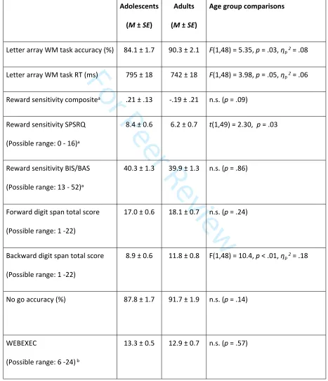
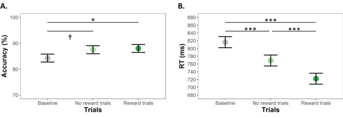
Accuracy and speed in the letter array WM task increased with age (Table 1), and differed between trial
types (accuracy: F(1.7,84.3) = 4.20, p = .02, ηp2 = .02, RT: (F(1.6,77.9) = 40.5, p < .0001, ηp2 = .13).
Participants were more accurate in Reward trials than in Baseline trials, with similar, but not significant,
increased accuracy in No reward trials (Figure 2A). Participants were faster in No reward trials than in
Baseline trials and even faster for Reward trials (Figure 2B).
For Peer Review
Insert Table 1 about hereInsert Figure 2 about here
There was a trend for a decrease in Reward Sensitivity (z-score normalised composite index of the two
self-report indices, SPSRQ and BIS/BAS) with age. A post-hoc analysis revealed adolescents were more
sensitive to rewards than adults when assessed with the SPSRQ, but not the BIS/BAS (Table 1).
Adolescents and adults earned comparable amounts of money although adolescents reported finding
money incentives more rewarding than adults (Table 1). Adults had greater backwards digit span scores
than adolescents, but the two age groups did no differ on the forward digit span task, WEBEXEC or
Go/No go task (Table 1). When including backward digit span score as a covariate in the mixed design
ANOVAs of the letter-array WM task, the difference between age groups in accuracy became non-
significant (F(1,46) = 1.21, p = .28), however, the RT difference remained (F(1,46) = 5.82, p = .020).
Neuroimaging results
Baseline activation during the WM task
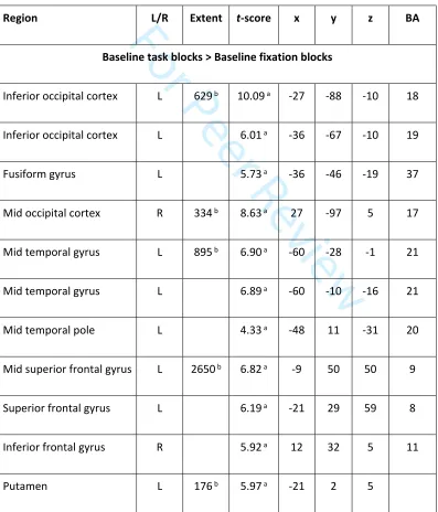
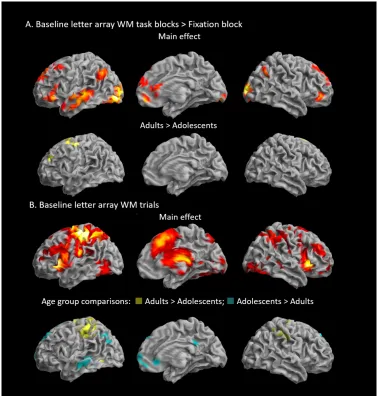
A broad network of regions showed sustained increased BOLD signal during letter array WM task blocks
compared to fixation blocks in the Baseline run (Table2 and Figure3A). Activation was overall more extensive in the left hemisphere, which may reflect the verbal nature of the task, but also overlapped
For Peer Review
with the “default mode network”. In the frontal lobes, bilateral activation was observed in the superior
frontal gyri (SFG) and anterior part of the inferior frontal gyri (IFG), extending along the medial wall into
the anterior aspect of the ACC. There was increased bilateral parietal activity in the left and right angular
gyri, as well as in the left middle temporal gyri and medial and left inferior occipital gyri. Activation in
the occipital cortex and angular gyrus bilaterally, left fusiform gyrus, middle temporal gyrus and
temporal poles, inferior and superior frontal gyri and putamen survived voxel-level whole brain
correction (Table 2). Compared to adolescents, adults showed increased activation, which survived
whole-brain correction at the cluster-level(but not voxel-level) in the left superior frontal and superior
medial gyri, extending into the ACC, precentral gyrus and supplementary motor area (SMA), and activity
in the lingual gyri (Table2 and Figure 3A).
Insert Figure 3 about here
Widespread transient increased BOLD activation was observed in frontal, parietal and temporal regions,
during WM task trials in the Baseline run, surviving both cluster-level and voxel-level whole-brain
correction (Table2 and Figure3B). In the frontal lobes, bilateral activation was observed in the SFG and
anterior part of the IFG, as well as orbitofrontal cortex, extending along the medial wall predominantly
into the middle cingulate cortex. There was increased bilateral activity in the insulae, in the angular gyri
in the parietal cortex, as well as in the middle temporal gyri and inferior occipital gyri. Increases in
subcortical activation were observed in the caudate and putamen, as well as in the thalamus and
hippocampus bilaterally. There was widespread bilateral activation in the cerebellum. Adults showed
increased activity in the precentral gyrus bilaterally, extending predominantly into the left postcentral
gyrus compared to adolescents (left peaks survived whole-brain correction at the voxel-level, except
clusters in left hippocampus and postcentral gyrus), while adolescents exhibited less deactivation in
medial prefrontal cortex and precuneus, than adults (Table2 and Figure3B).
For Peer Review
Insert Table 2 about hereReward effects
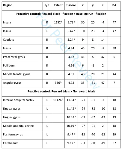
Sustained effects of reward were assessed by contrasting task block activation of the Reward run and
the Baseline run. This resulted in a large cluster peaking in the right insula and extending into bilateral
ventral PFC, right dorsolateral PFC and the left insula, as well as into the caudate and putamen
subcortically. There was an additional cluster in the right angular and supramarginal gyri. Bilateral
ventroprefrontal, insula, caudate and occipital peaks survived whole-brain correction at the voxel-level
(Table 3 and Figure 4A). The pattern of activation largely did not overlap with the sustained WM task
effects (Figure 3A vs. Figure 4A). No increased activation was observed in the reverse contrast [Baseline
blocks > Reward blocks].
Insert figure 4 about here
Transient effects of reward were assessed by contrasting activation in Reward trials with No reward
trials within the Reward run. This resulted in a large cluster peaking in inferior middle occipital gyrus and
extending into the middle occipital gyrus, the superior and inferior parietal cortex bilaterally, right
ventral and dorsolateral PFC and along the medial wall the medial frontal cortex and anterior and
middle cingulate cortex, as well as the right insula. There were additional clusters in the left insula and
right precentral and middle frontal gyri. Subcortical activity was observed bilaterally in caudate nucleus
extending slightly into accumbens, pallidum, thalamus, and bilateral hippocampi. There was widespread
activation of cerebellar regions. Peaks located in posterior occipital brain regions, inferior parietal
cortex, subcortical regions, and the cerebellum survived whole-brain correction at the voxel-level.
Among anterior brain regions the ACC and insulae were the only peaks surviving voxel-level correction
.
3For Peer Review
No increased activation was observed in the reverse contrast [No reward trials > Reward trials] (Table 3
and Figure 4B).
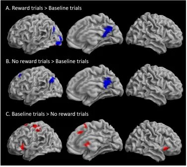
To further explore the pattern of changes in transient changes in BOLD signal according to reward,
Reward and No reward trials were contrasted to Baseline trials (Table 3). Reward trials were associated
with less deactivation of the precuneus and left lingual gyrus and middle occipital cortex (the latter was
not significant with voxel-level whole brain correction) than Baseline trials, and greater activation than
Baseline trials within a subset of the left lingual gyrus cluster (Figure 5A). No difference in activation was
observed in the reverse contrast [Baseline trials > Reward trials]. No reward trials showed, similarly to
Reward trials, less deactivation in the precuneus than Baseline trials, as well as less deactivation than
Baseline trials in the left superior frontal gyrus (the latter was not significant with voxel-level whole
brain correction) (Figure 5B). Finally, Baseline trials showed higher activation than No reward trials in
bilateral insulae, left precentral gyrus, medial frontal gyrus extending into middle cingulate gyrus, and
left inferior frontal cortex, there was also activation in the caudate, and inferior and middle occipital gyri
(Figure 5C). Only activations in the bilateral insulae survived voxel-level whole-brain correction. The
predominant pattern across regions showing transient increases in activation during the working
memory trials was therefore No reward trials < Baseline trials < Reward trials.
Insert Table 3 about here
Insert Figure 5 about here
Inclusive and exclusive masking contrasts indicated that bilateral insulae (surviving voxel-wise
whole-brain correction), right angular gyrus, and a subcortical cluster including right caudate nucleus, thalamus
For Peer Review
and left pallidum exhibited reward context-related changes in both transient and sustained activity.No
regions exhibited an exclusively sustained pattern of activation, but the more anterior aspect of the ACC
as well as some cerebellar and occipital areas exhibited transient changes only in response to reward.
Activations in the inferior occipital lobe and anterior cingulate cortex survived voxel-level whole-brain
correction (Table 3).
ROI analyses
To explore age effects in regions identified to have a mixed pattern of response to rewards, we
extracted mean parameter estimates in 10 mm radius spheres centred on the peaks of the four clusters
exhibiting modulation by reward of both transient and sustained activation (left and right insulae,
angular gyrus and caudate, see Table 3). The left AI showed a significant interaction between Run and
Age group: adolescents exhibited a greater increase in reward-dependent sustained activation than
adults (F(1,48) = 6.35, ηp2 = .05, p = .02, Figure 6A). The right AI showed a similar pattern (F(1,48) = 3.91,
p = .05) (Figure 6B). Analyses of transient activations showed that adults exhibited increased overall
activity in the right AI (F(1,48) = 4.79, ηp2 = .08, p = .03), but not the left AI (F(1,48) = 2.22, ηp2 = .04, p =
.14), across Reward and No reward trials compared to adolescents (Figure 6C-D). No other age effects
were identified.
Insert figure 6 about here
For Peer Review
D
ISCUSSIONWe examined the impact of reward on sustained and transient engagement of cognitive control, and
whether differences exist between adolescence and adulthood. In the letter-array WM task, high
accuracy rates can be achieved with a reactive control strategy. However, to produce accurate
responses that are fast enough, the optimal strategy is to proactively sustain the task set and rule use
across trials, in anticipation of the stimuli (Jimura et al., 2010). Results showed similar behavioural and
neural evidence for engagement of proactive and reactive strategies in the context of reward for
adolescents and adults.
Proactive control
Reaction times were faster in No reward than Baseline trials, suggesting sustained performance
improvement in the Reward run associated with a proactive cognitive control strategy (Jimura et al.,
2010). Reward blocks were associated with increased frontoparietal activity outside of the network
recruited in the main WM block contrast. Our results align with findings of reward-related increased
sustained activity in the right lateral PFC (middle frontal gyrus) and right PPC (angular gyrus), regions
associated with proactive control in adults (Jimura et al., 2010; Locke & Braver, 2008) and children,
adolescents and adults (Strang & Pollak, 2014). An increased cognitive control system engagement could
also be reflective of higher load due to the introduction of two different reward conditions, Reward and
No reward trials. However, this is unlikely to reflect a task switching load, as the working memory task
stays constant (there is no perceptual, response or set shifting needed [Kim, Cilles, Johnson, & Gold,
2012]), and we see a pattern of improved performance rather than the performance cost typically
associated with a switching context (Monsell, 2003). In addition, there was evidence for sustained
activation in regions typically associated with reward across age groups: the caudate nucleus, putamen
and orbitofrontal cortex (Silverman, Jedd, & Luciana, 2015).
For Peer Review
Reactive control
Adolescents and adults were fastest for Reward trials, with a similar trend for accuracy, which points to
a trial-by-trial reward enhancement reflecting reactive control. Reward trials were associated with
increased transient activity in cortical regions recruited overall during the letter-array WM task (AI, ACC
and parietal cortex), in contrast to the block effects of reward, which did not overlap with WM regions.
The overall pattern shows intermediate activations for baseline trials compared to No reward and
Reward trials.
A possible interpretation of these results is that, as transient activation is not as resource consuming as
sustained activation, there is still scope for increased trial-by-trial recruitment of working memory
regions to increase the chance of obtaining a reward, on top of working memory transient activation. In
addition to the sustained activation, Reward trials also recruited the right orbitofrontal cortex, the
caudate nucleus and putamen (Haber & Knutson, 2010).
In the present study, transient activity in the ACC might be related to increased monitoring in response
to reward. The ACC has been proposed to be involved in performance monitoring and, when conflict
arises, is thought to recruit higher control order structures in the lateral PFC (Botvinick & Braver, 2015).
In this case, conflict might signal a cost-benefit analysis where cost of task performance is weighed
against expect values of its outcome and necessary effort required (Shenhav, Botvinick, & Cohen, 2013).
Further, exclusively transient activations were observed in anterior aspects of the ACC, which fit with
previous evidence that this region is selectively involved in compensatory reactive control processes
(Jiang et al., 2015).
Mixed and exclusively transient regions
Bilateral insulae, the right PPC and the caudate exhibited a mixed pattern: with both higher sustained
activity for the Reward run than the Baseline run, and higher transient increases in activity for Reward
For Peer Review
trials compared to No reward trials, with Baseline trials showing intermediary levels of transient activity
(i.e. No reward < Baseline < Reward in most cases). The AI emerged as the key mixed region involved in
proactive and reactive control in response to reward, while the DLPFC had a more sustained pattern of
activation in the baseline working memory run. Sustained activation in the DLPFC in the reward context
may reflect increased top-down “boosting” excitatory connectivity to more posterior regions, which may
facilitate maintenance of representations over a delay (Edin et al., 2009). The AI has been implicated in
top-down control processes including task-set maintenance (Dosenbach, Fair, Cohen, Schlaggar, &
Petersen, 2008; Nelson et al., 2010), and tracking cognitive control demand stability (Jiang et al., 2015).
It has also been implicated in bottom-up salience detection of relevant cues (Menon & Uddin, 2010) as
part of the salience network and key cognitive-emotional hub (Menon & Uddin, 2010; Smith, Steinberg,
& Chein, 2014). The AI has been implicated in transient detection of salient stimuli and initiating
attentional control signals which are then sustained by the ACC and the ventrolateral and DLPFC (Menon
& Uddin, 2010). As one of the most commonly activated areas in fMRI studies, the AI has been
implicated in controlling attention as a function of task demands (see Nelson et al., 2010 for a review).
The caudate nucleus has been implicated in processing extrinsic reward related to monetary gains and
losses (Haber & Knutson, 2010; Richards, Plate, & Ernst, 2013). We found that activity in the caudate
nucleus was greater in Reward blocks than Baseline blocks, and that it tracked trial reward status as
baseline activity was maintained for Reward trials only, while activation was lower for No reward trials.
A speculative interpretation of these results is that once the explicit reward trials were introduced, the
value of the No reward trials dropped compared to their starting level. However, the caudate nucleus
has also been found to be activated in WM tasks in the absence of rewards (e.g. Ziermans et al., 2012),
and in the present study there were transient increases in activation in the caudate in Baseline WM task
trials, which suggests a non-exclusive reward role of the caudate.
For Peer Review
Our results speak to the debate surrounding two underlying configurations which have been proposed
for the DMC (Jiang et al., 2015). One of the accounts proposes that proactive and reactive control are
implemented by different dynamics within the same region of the right dorsolateral PFC (BA 46/9)
(Braver et al., 2009; Burgess & Braver, 2010; Jimura et al., 2010). Other accounts propose that different
strategies are implemented by distinct brain regions (De Pisapia & Braver, 2006; Jiang et al., 2015). Here,
we found evidence that both mechanisms might be at play. The bilateral AI, right PPC and caudate
nucleus showed a mixed pattern of response while the anterior aspects of the ACC showed transient
effects of reward only.
Developmental effects
In line with developmental studies of cognitive control (Humphrey & Dumontheil, 2016; Luna et al.,
2015), adults had greater overall accuracy and faster RT than adolescents, as well as greater backwards
digit span scores (Karakas, Yalin, Irak, & Erzengin, 2002). However, since speed thresholds were
determined individually, adolescents and adults earned comparable monetary rewards. Adolescents
reported finding monetary incentives more rewarding than adults. Comparable performance between
adolescents and adults might be driven by increased motivation to perform by the adolescents, perhaps
associated with finding money more rewarding.
Adolescents showed a trend for more reward sensitivity than adults (Galván, 2013; van Duijvenvoorde
et al., 2016). Post-hoc analyses revealed adolescents had significantly greater reward sensitivity than
adults on the SPSRQ (Torrubia et al. 2001, which assesses reward sensitivity per se, but not on approach
motivation subscale of the the BIS/BAS (Carver & White, 1994). However, we did not find age
differences reward-related brain activation. Although age differences between adolescents and adults
are often described in the neuroimaging reward literature, they are not consistent across stages of
reward processing or type of task (Galván, 2010, 2013).
For Peer Review
In the absence of rewards, previous developmental work has suggested that adolescents employ a more
reactive than proactive cognitive control strategy (Alahyane et al., 2014; Andrews-Hanna et al., 2011;
Velanova et al., 2009). In the present study we show that, like Strang and Pollack (2014), in the context
of potential rewards, adolescents, like adults, can sustain cognitive control proactively. By also
examining reactive control, in contrast to Strang and Pollack (2014), we provide evidence that
adolescents, like adults, show additional improvements in a trial-by-trial fashion.
In follow-up ROI analyses of the main results, we found that transient activation was overall greater in
the right AI in adults than in adolescents, but that both age groups showed similar increases in transient
activation in Reward trials compared to No reward trials. A different pattern was observed for sustained
activation, whereby adolescents showed a greater increase in activation in task blocks of the Reward run
compared to the Baseline run in the left AI, “catching up” with adult levels of activation. Adolescents
may be relying on an adaptive mechanism of sustained, but not transient, increase in AI activation. This
speaks to an immature proactive capacity in adolescents that is only engaged in the context of reward.
The role of the AI in adolescent decision-making processes is increasingly recognised, suggesting that the
relative immaturity of this cognitive-emotional hub, which is connected to both the lateral PFC and
striatum, may bias adolescents in affectively driven contexts (for a review see Smith et al., 2014). Here
we suggest that a sensitivity to reward context in the AI may support increased sustained engagement
of cognitive control in some instances.
Limitations and future directions
Varying between Reward and No reward trials could be an additional component of the task which may
have led to an increase in sustained activation of the cognitive control system by making the task more
engaging. However, a greater overall engagement could not account for transient differences between
reward and no-reward trials. Order effects are a limitation of the current study (as in Jimura et al.,
For Peer Review
2010). Counterbalancing the order of blocks was not possible to ensure participants were at first naïve
regarding potential rewards to determine their baseline performance. To minimise order effects related
to practice, we introduced a long practice period to ensure that participant’s performance stabilised
before the scanning runs. It is possible that the better performance observed in the second run could
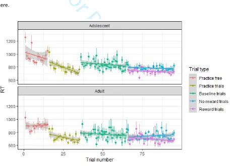
still be driven in part by practice effects. Plots of RT and accuracy as a function of trial number and block
number suggest indeed that in adolescents the RT difference between baseline and no-reward trials
may have been driven by practice effects (Supplementary Figure S1). However, this is not apparent in
adults, nor in the accuracy data (Supplementary Figure S2). Practice effects however could not explain
the difference in RT and brain activation between Reward and No reward trials. Although demanding,
the task was not very difficult, as reflected by high accuracy rates. It might be that the balance between
proactive and reactive strategies begins to emerge in more challenging cognitive control tasks, and
future studies could investigate this.
C
ONCLUSIONThis study shows behavioural and neuroimaging evidence of modulation of both proactive and reactive
control by reward in adults and in adolescents. Proactive and reactive control were found to be
supported both by partly separable frontoparietal neural circuitries, and by regions that exhibit both
sustained and transient modulation by reward. In the face of incentives, adolescents and adults can
sustain cognitive control in a proactive fashion, with additional transient readjustments in response to
the reward. There is some evidence of adaptive higher sustained activation in the anterior insula by
adolescents in the context of reward.
For Peer Review
A
CKNOWLEDGEMENTSThis work was supported by the Mexican National Council on Science and Technology (CONACYT)
[465015 to L.M.W].
R
EFERENCESAlahyane, N., Brien, D. C., Coe, B. C., Stroman, P. W., & Munoz, D. P. (2014). Developmental
improvements in voluntary control of behavior: Effect of preparation in the fronto-parietal
network? NeuroImage, 98, 103–17. https://doi.org/10.1016/j.neuroimage.2014.03.008
Andrews-Hanna, J. R., Seghete, K. L., Claus, E. D., Burgess, G. C., Ruzic, L., & Banich, M. T. (2011).
Cognitive control in adolescence: Neural underpinnings and relation to self-report behaviors.
PLoS One, 6(6), 27–30. https://doi.org/10.1371/journal.pone.0021598
Blackwell, K. A., & Munakata, Y. (2014). Costs and benefits linked to developments in cognitive control.
Developmental Science, 17(2), 203–11. https://doi.org/10.1111/desc.12113
Botvinick, M. M., & Braver, T. S. (2015). Motivation and Cognitive Control: From Behavior to Neural
Mechanism. Annual Review of Psychology, 66(1), 83–113.
https://doi.org/10.1146/annurev-psych-010814-015044
Brahmbhatt, S. B., White, D. A., & Barch, D. M. (2010). Developmental differences in sustained and
transient activity underlying working memory. Brain Research, 1354, 140–151.
https://doi.org/10.1016/j.brainres.2010.07.055
For Peer Review
Braver, T. S. (2012). The variable nature of cognitive control: A dual mechanisms framework. Trends in
Cognitive Sciences, 16(2), 106–113. https://doi.org/10.1016/j.tics.2011.12.010
Braver, T. S., Krug, M. K., Chiew, K. S., Kool, W., Westbrook, J. A., Clement, N. J., … Somerville, L. H.
(2014). Mechanisms of motivation–cognition interaction: Challenges and opportunities.
Cognitive, Affective, & Behavioral Neuroscience, 14(2), 443–472.
https://doi.org/10.3758/s13415-014-0300-0
Braver, T. S., Paxton, J. L., Locke, H. S., & Barch, D. M. (2009). Flexible neural mechanisms of cognitive
control within human prefrontal cortex. Proceedings of the National Academy of Sciences of the
United States of America, 106(18), 7351–7356. https://doi.org/10.1073/pnas.0808187106
Brett, M., Anton, J.-L., Valabregue, R., & Poline, J.-B. (2002). Region of interest analysis using an SPM
toolbox. Presented at the 8th International Conference on Functional Mapping of the Human
Brain, June 2-6, 2002, Sendai, Japan.
Buchanan, T., Heffernan, T. M., Parrott, A. C., Ling, J., Rodgers, J., & Scholey, A. B. (2010). A short
self-report measure of problems with executive function suitable for administration via the Internet.
Behavior Research Methods, 42(3), 709–714. https://doi.org/10.3758/BRM.42.3.709
Burgess, G. C., & Braver, T. S. (2010). Neural Mechanisms of Interference Control in Working Memory:
Effects of Interference Expectancy and Fluid Intelligence. PLoS ONE, 5(9), e12861.
https://doi.org/10.1371/journal.pone.0012861
Carver, C. S., & White, T. L. (1994). Behavioural inhibition, behavioural activation, and affective
responses to impending reward and punishment: The BIS/BAS scales. Journal of Personality and
Social Psychology, 67, 319 – 333.
Casey, B. J. (2015). Beyond Simple Models of Self-Control to Circuit-Based Accounts of Adolescent
Behavior. Annual Review of Psychology, 66(1), 295–319.
https://doi.org/10.1146/annurev-psych-010814-015156
For Peer Review
Cauley, S. F., Polimeni, J. R., Bhat, H., Wald, L. L., & Setsompop, K. (2014). Interslice leakage artifact
reduction technique for simultaneous multislice acquisitions. Magnetic Resonance in Medicine,
72(1), 93–102. https://doi.org/10.1002/mrm.24898
Chatham, C. H., Frank, M. J., & Munakata, Y. (2009). Pupillometric and behavioral markers of a
developmental shift in the temporal dynamics of cognitive control. Proceedings of the National
Academy of Sciences, 106(14), 5529–5533. https://doi.org/10.1073/pnas.0810002106
Chevalier, N., Jackson, J., Revueltas Roux, A., Moriguchi, Y., & Auyeung, B. (2019). Differentiation in
prefrontal cortex recruitment during childhood: Evidence from cognitive control demands and
social contexts. Developmental Cognitive Neuroscience, 36, 100629.
https://doi.org/10.1016/j.dcn.2019.100629
Chevalier, N., Martis, S. B., Curran, T., & Munakata, Y. (2015). Metacognitive processes in executive
control development: The case of reactive and proactive control. Journal of Cognitive
Neuroscience, 27(6), 1125–1136. https://doi.org/10.1162/jocn_a_00782
Chiew, K. S., & Braver, T. S. (2013). Temporal dynamics of motivation-cognitive control interactions
revealed by high-resolution pupillometry. Frontiers in Psychology, 4(1), 15.
https://doi.org/10.3389/fpsyg.2013.00015
Chiew, K. S., & Braver, T. S. (2017). Context processing and control in the human brain: From gating
models to dual mechanisms. The Wiley Handbook of Cognitive Control, 143–166.
Cohen, A. O., Breiner, K., Steinberg, L., Bonnie, R. J., Scott, E. S., Taylor-Thompson, K. A., … Casey, B. J.
(2016). When Is an Adolescent an Adult? Assessing Cognitive Control in Emotional and
Nonemotional Contexts. Psychological Science, 27(4), 549–562.
https://doi.org/10.1177/0956797615627625
For Peer Review
Crone, E. A., & Dahl, R. E. (2012). Understanding adolescence as a period of social–affective engagement
and goal flexibility. Nature Reviews Neuroscience, 13(9), 636–650.
https://doi.org/10.1038/nrn3313
De Pisapia, N., & Braver, T. S. (2006). A model of dual control mechanisms through anterior cingulate
and prefrontal cortex interations. Neurocomputing, 69, 1322–1326.
Dosenbach, N. U. F., Fair, D. A., Cohen, A. L., Schlaggar, B. L., & Petersen, S. E. (2008). A dual-networks
architecture of top-down control. Trends in Cognitive Sciences, 12(3), 99–105.
https://doi.org/10.1016/j.tics.2008.01.001
Edin, F., Klingberg, T., Johansson, P., McNab, F., Tegnér, J., & Compte, A. (2009). Mechanism for
top-down control of working memory capacity. Proceedings of the National Academy of Sciences,
106(16), 6802–6807. https://doi.org/10.1073/pnas.0901894106
Galván, A. (2010). Adolescent development of the reward system. Frontiers in Human Neuroscience.
https://doi.org/10.3389/neuro.09.006.2010
Galván, A. (2013). The Teenage Brain: Sensitivity to Rewards. Current Directions in Psychological Science,
22(2), 88–93. https://doi.org/10.1177/0963721413480859
Geier, C., Terwilliger, R., Teslovich, T., Velanova, K., & Luna, B. (2010). Immaturities in reward processing
and its influence on inhibitory control in adolescence. Cerebral Cortex, 20(7), 1613–1629.
https://doi.org/10.1093/cercor/bhp225
Haber, S. N., & Knutson, B. (2010). The Reward Circuit: Linking Primate Anatomy and Human Imaging.
Neuropsychopharmacology, 35(1), 4–26. https://doi.org/10.1038/npp.2009.129
Humphrey, G., & Dumontheil, I. (2016). Development of Risk-Taking, Perspective-Taking, and Inhibitory
Control During Adolescence. Developmental Neuropsychology, 41(1–2), 59–76.
https://doi.org/10.1080/87565641.2016.1161764
For Peer Review
Insel, C., Kastman, E. K., Glenn, C. R., & Somerville, L. H. (2017). Development of corticostriatal
connectivity constrains goal-directed behavior during adolescence. Nature Communications,
8(1), 1605. https://doi.org/10.1038/s41467-017-01369-8
Jiang, J., Beck, J., Heller, K., & Egner, T. (2015). An insula-frontostriatal network mediates flexible
cognitive control by adaptively predicting changing control demands. Nature Communications,
6(5), 8165. https://doi.org/10.1038/ncomms9165
Jimura, K., Locke, H. S., & Braver, T. S. (2010). Prefrontal cortex mediation of cognitive enhancement in
rewarding motivational contexts. Proceedings of the National Academy of Sciences of the United
States of America, 107(19), 8871–8876. https://doi.org/10.1073/pnas.1002007107
Karakas, S., Yalin, A., Irak, M., & Erzengin, Ö. U. (2002). Digit Span Changes From Puberty to Old Age
Under Different Levels of Education. Developmental Neuropsychology, 22(2), 423–453.
https://doi.org/10.1207/S15326942DN2202_1
Kim, C., Cilles, S. E., Johnson, N. F., & Gold, B. T. (2012). Domain general and domain preferential brain
regions associated with different types of task switching: A meta-analysis. Human Brain
Mapping, 33(1), 130–142. https://doi.org/10.1002/hbm.21199
Krebs, R. M., Hopf, J.-M., & Boehler, C. N. (2015). Within-Trial Effects of Stimulus-Reward Associations.
In T. S. Braver (Ed.), Motivation and Cognitive Control (1st ed.). Taylor & Francis Group,
Routledge.
Locke, H. S., & Braver, T. S. (2008). Motivational influences on cognitive control: Behavior, brain
activation, and individual differences. Cognitive, Affective, & Behavioral Neuroscience, 8(1), 99–
112. https://doi.org/10.3758/CABN.8.1.99
Luna, B., Marek, S., Larsen, B., Tervo-Clemmens, B., & Chahal, R. (2015). An Integrative Model of the
Maturation of Cognitive Control. Annual Review of Neuroscience, 38(1), 151–170.
https://doi.org/10.1146/annurev-neuro-071714-034054
For Peer Review
Marklund, P., Fransson, P., Cabeza, R., Petersson, K. M. K., Ingvar, M., & Nyberg, L. (2007). Sustained and
transient neural modulations in prefrontal cortex related to declarative long-term memory,
working memory, and attention. Cortex, 43(1), 22–37.
https://doi.org/10.1016/S0010-9452(08)70443-X
McDaniel, M. A., LaMontagne, P., Beck, S. M., Scullin, M. K., & Braver, T. S. (2013). Dissociable Neural
Routes to Successful Prospective Memory. Psychological Science, 24(9), 1791–1800.
https://doi.org/10.1177/0956797613481233
Menon, V., & Uddin, L. Q. (2010). Saliency, switching, attention and control: A network model of insula
function. Brain Structure & Function, 214(5–6), 655–667.
https://doi.org/10.1007/s00429-010-0262-0
Monsell, S. (2003). Task switching. Trends in Cognitive Sciences, 7(3), 134–140.
https://doi.org/10.1016/S1364-6613(03)00028-7
Nelson, S. M., Dosenbach, N. U. F., Cohen, A. L., Wheeler, M. E., Schlaggar, B. L., & Petersen, S. E. (2010).
Role of the anterior insula in task-level control and focal attention. Brain Structure and Function,
214(5–6), 669–680. https://doi.org/10.1007/s00429-010-0260-2
Padmanabhan, A., Geier, C., Ordaz, S. J., Teslovich, T., & Luna, B. (2011). Developmental changes in brain
function underlying the influence of reward processing on inhibitory control. Developmental
Cognitive Neuroscience, 1(4), 517–529. https://doi.org/10.1016/j.dcn.2011.06.004
Richards, J. M., Plate, R. C., & Ernst, M. (2013). A systematic review of fMRI reward paradigms used in
studies of adolescents vs. Adults: The impact of task design and implications for understanding
neurodevelopment. Neuroscience and Biobehavioral Reviews, 37(5), 976–91.
https://doi.org/10.1016/j.neubiorev.2013.03.004
For Peer Review
Rolls, E. T., Joliot, M., & Tzourio-Mazoyer, N. (2015). Implementation of a new parcellation of the
orbitofrontal cortex in the automated anatomical labeling atlas. NeuroImage, 122, 1–5.
https://doi.org/10.1016/J.NEUROIMAGE.2015.07.075
Rorden, C., Karnath, H.-O., & Bonilha, L. (2007). Improving lesion-symptom mapping. Journal of
Cognitive Neuroscience, 19, 1081–1088.
Shenhav, A., Botvinick, M. M., & Cohen, J. D. (2013). The expected value of control: An integrative
theory of anterior cingulate cortex function. Neuron, 79(2), 217–240.
https://doi.org/10.1016/j.neuron.2013.07.007
Silverman, M. H., Jedd, K., & Luciana, M. (2015). Neural networks involved in adolescent reward
processing: An activation likelihood estimation meta-analysis of functional neuroimaging
studies. NeuroImage, 122, 427–439. https://doi.org/10.1016/j.neuroimage.2015.07.083
Simmonds, D. J., Pekar, J. J., & Mostofsky, S. H. (2008). Meta-analysis of Go/No-go tasks demonstrating
that fMRI activation associated with response inhibition is task-dependent. Neuropsychologia,
46(1), 224–232. https://doi.org/10.1016/j.neuropsychologia.2007.07.015
Singmann, H., Bolker, B., Westfall, J., & Aust, F. (2018). afex: Analysis of Factorial Experiments. [R
package version 0.22-1]. Retrieved from https://CRAN.R-project.org/package=afex
Smith, A. R., Steinberg, L., & Chein, J. M. (2014). The role of the anterior insula in adolescent decision
making. Developmental Neuroscience, 36(3–4), 196–209. https://doi.org/10.1159/000358918
Strang, N. M., & Pollak, S. D. (2014). Developmental continuity in reward-related enhancement of
cognitive control. Developmental Cognitive Neuroscience, 10, 34–43.
https://doi.org/10.1016/j.dcn.2014.07.005
Torrubia, R., Ávila, C., Moltó, J., & Caseras, X. (2001). The Sensitivity to Punishment and Sensitivity to
Reward Questionnaire (SPSRQ) as a measure of Gray’s anxiety and impulsivity dimensions.
For Peer Review
Personality and Individual Differences, 31(6), 837–862.
https://doi.org/10.1016/S0191-8869(00)00183-5
Tzourio-Mazoyer, N., Landeau, B., Papathanassiou, D., Crivello, F., Etard, O., Delcroix, N., … Joliot, M.
(2002). Automated anatomical labeling of activations in SPM using a macroscopic anatomical
parcellation of the MNI MRI single-subject brain. NeuroImage, 15(1), 273–289.
https://doi.org/10.1006/nimg.2001.0978
van Duijvenvoorde, A. C. K., Peters, S., Braams, B. R., & Crone, E. A. (2016). What motivates adolescents?
Neural responses to rewards and their influence on adolescents’ risk taking, learning, and
cognitive control. Neuroscience & Biobehavioral Reviews, 70, 135–147.
https://doi.org/10.1016/j.neubiorev.2016.06.037
Velanova, K., Wheeler, M. E., & Luna, B. (2009). The maturation of task set-related activation supports
late developmental improvements in inhibitory control. Journal of Neuroscience, 29(40), 12558–
12567. https://doi.org/10.1523/JNEUROSCI.1579-09.2009
Visscher, K. M., Miezin, F. M., Kelly, J. E., Buckner, R. L., Donaldson, D. I., McAvoy, M. P., … Petersen, S. E.
(2003). Mixed blocked/event-related designs separate transient and sustained activity in fMRI.
NeuroImage, 19(4), 1694–1708.
Wechsler, D. (2011). Weschler Abbreviated Scale of Intelligence (WASI-II).
https://doi.org/10.1177/0734282912467756
Xu, J., Moeller, S., Auerbach, E. J., Strupp, J., Smith, S. M., Feinberg, D. A., … Uğurbil, K. (2013).
Evaluation of slice accelerations using multiband echo planar imaging at 3 T. NeuroImage, 83,
991–1001. https://doi.org/10.1016/j.neuroimage.2013.07.055
Zanolie, K., & Crone, E. A. (2018). Development of Cognitive Control across Childhood and Adolescence.
In Ghetti, Simona (Ed.), The Stevens’ Handbook of Experimental Psychology and Cognitive
Neuroscience (4th ed., Vol. 4). New York: Wiley.
For Peer Review
Zhai, Z. W., Pajtek, S., Luna, B., Geier, C., Ridenour, T. A., & Clark, D. B. (2015). Reward-Modulated
Response Inhibition, Cognitive Shifting, and the Orbital Frontal Cortex in Early Adolescence.
Journal of Research on Adolescence, 25(4), 753–764. https://doi.org/10.1111/jora.12168
Ziermans, T., Dumontheil, I., Roggeman, C., Peyrard-Janvid, M., Matsson, H., Kere, J., & Klingberg, T.
(2012). Working memory brain activity and capacity link MAOA polymorphism to aggressive
behavior during development. Translational Psychiatry, 2(2), e85.
https://doi.org/10.1038/tp.2012.7
For Peer Review
Table 1: Summary statistics of measures collected in adolescent and adult participants. BIS/BAS: Behavioural
Inhibition/Activation scales; M: Mean; SE: standard error; RT: reaction time; SPSRQ: Sensitivity to Punishment and
Sensitivity to Reward Questionnaire; WM: working memory.
Adolescents
(M ± SE)
Adults
(M ± SE)
Age group comparisons
Letter array WM task accuracy (%) 84.1 ± 1.7 90.3 ± 2.1 F(1,48) = 5.35, p = .03, ηp2 = .08
Letter array WM task RT (ms) 795 ± 18 742 ± 18 F(1,48) = 3.98, p = .05, ηp2 = .06
Reward sensitivity compositea .21 ± .13 -.19 ± .21 n.s. (p = .09)
Reward sensitivity SPSRQ
(Possible range: 0 - 16)a
8.4 ± 0.6 6.2 ± 0.7 t(1,49) = 2.30, p = .03
Reward sensitivity BIS/BAS
(Possible range: 13 - 52)a
40.3 ± 1.3 39.9 ± 1.3 n.s. (p = .86)
Forward digit span total score
(Possible range: 1 -22)
17.0 ± 0.6 18.1 ± 0.7 n.s. (p = .24)
Backward digit span total score
(Possible range: 1 -22)
8.9 ± 0.6 11.8 ± 0.8 F(1,48) = 10.4, p < .01, ηp2 = .18
No go accuracy (%) 87.8 ± 1.7 91.7 ± 1.9 n.s. (p = .14)
WEBEXEC
(Possible range: 6 -24) b
13.3 ± 0.5 12.9 ± 0.7 n.s. (p = .57)
For Peer Review
Total money earned (£)(Possible range: 0 – 8)
6.50 ± 0.24 6.67 ± 0.23 n.s. (p = .63)
Rating of monetary incentive
(Possible range: 1 – 5)c
4.17 ± 0.14 3.45 ± 0.30 F(1,48) = 6.02, p = .02,ηp 2 = .11
a Higher scores indicate more sensitivity to reward
b Higher scores indicate more executive function failures
c Higher scores indicate finding money more rewarding
For Peer Review
Table 2: Letter array working memory task neuroimaging results in the Baseline run. Coordinates and t-values are
listed for regions showing a significant difference in BOLD signal in the whole brain analysis of block effects of
working memory ([Baseline blocks – Fixation blocks]) and trial effects of working memory ([Baseline trials]). Both
main effects and age group differences are reported. x, y, and z = Montreal Neurological Institute (MNI)
coordinates. a indicates voxels where p
FWE < .05 at the voxel-level, b indicates clusters where pFWE < .05 at the
cluster-level. BA = Brodmann area, L/R = left/right hemisphere.
Region L/R Extent t-score x y z BA
Baseline task blocks > Baseline fixation blocks
Inferior occipital cortex L 629 b 10.09 a -27 -88 -10 18
Inferior occipital cortex L 6.01 a -36 -67 -10 19
Fusiform gyrus L 5.73 a -36 -46 -19 37
Mid occipital cortex R 334 b 8.63 a 27 -97 5 17
Mid temporal gyrus L 895 b 6.90 a -60 -28 -1 21
Mid temporal gyrus L 6.89 a -60 -10 -16 21
Mid temporal pole L 4.33 a -48 11 -31 20
Mid superior frontal gyrus L 2650 b 6.82 a -9 50 50 9
Superior frontal gyrus L 6.19 a -21 29 59 8
Inferior frontal gyrus R 5.92 a 12 32 5 11
Putamen L 176 b 5.97 a -21 2 5
For Peer Review
Angular gyrus L 323 b 5.64 a -42 -58 29 39
Angular gyrus L 5.57 a -45 -67 47 39
Angular gyrus R 125 b 5.50 a 51 -61 32 39
Adults [Baseline blocks – Baseline fixation blocks] > Adolescents [Baseline blocks –
Baseline fixation blocks]
Superior frontal gyrus L 168 b 4.64 -18 2 56 6
Precentral gyrus L 4.58 -39 -4 56 6
Supplementary motor
area
L
3.49 -6 17 65 6
Medial superior frontal
gyrus L 102 b 4.43 -9 17 41 32
Anterior cingulate cortex L 3.76 -6 32 26 32
Lingual gyrus L 160 b 4.23 -18 -46 -7 30
Lingual gyrus L 3.96 -21 -67 -13 18
Superior medial frontal
gyrus L 113 b 4.21 -30 47 26 46
Superior frontal gyrus L 4.05 -30 38 47 9
Baseline trials
For Peer Review
Precentral gyrusL 18304
b 26.63 a -36 -7 68 6
Insula L 24.92 a -30 20 5 48
Postcentral gyrus L 23.83 a -45 -37 53 2
Middle frontal gyrus R 530 b 12.82 a 42 38 32 46
Middle frontal gyrus R 9.14 a 39 56 14 46
Orbitofrontal cortex R 97 b 7.07 a 21 44 -16 11
Adults Baseline trials > Adolescents Baseline trials
Postcentral gyrus L 695 b 8.18 a -36 -43 62 2
Inferior parietal lobule L 5.71 a -51 -28 50 2
Precuneus L 339 b 7.26 a -21 -46 8 37
Inferior temporal gyrus L 5.80 a -39 -55 -7 37
Hippocampus L 4.08 -36 -31 -10 37/20
Hippocampus R 167 b 6.23 a 24 -40 8 37
Hippocampus L 3.80 39 -37 -7 37
Superior frontal gyrus R 364 b 6.21 a 33 -4 68 6
Paracentral lobule R 6.09 a 6 -19 80 6
Superior frontal gyrus L 5.79 a -33 -4 68 6
For Peer Review
Postcentral gyrus R 148 b 4.56 42 -31 50 3
Adolescents Baseline trials > Adults Baseline trials
Medial orbitofrontal
cortex 521 b 7.41 a 0 56 -4
10
Medial orbitofrontal
cortex 6.70 a 0 32 -10
11
Caudate 3.76 0 11 -13 25
Superior frontal gyrus L 431 b 7.05 a -9 47 47 9
Medial superior frontal
gyrus 5.59 a 0 44 20 32
Medial superior frontal
gyrus
R
4.19 15 53 41 9
Mid temporal gyrus L 250 b 6.23 a -60 -25 -16 20
Posterior cingulate cortex L 136 b 4.60 -3 -55 32 23