N A N O E X P R E S S
Open Access
Gelatin-layered and multi-sized porous
b
-tricalcium
phosphate for tissue engineering scaffold
Sung-Min Kim
1, Soon-Aei Yi
1, Seong-Ho Choi
2, Kwang-Mahn Kim
1and Yong-Keun Lee
1*Abstract
The multi-sized porousb-tricalcium phosphate scaffolds were fabricated by freeze drying followed by slurry coating using a multi-sized porous sponge as a template. Then, gelatin was dip coated on the multi-sized porousb -tricalcium phosphate scaffolds under vacuum. The mechanical and biological properties of the fabricated scaffolds were evaluated and compared to the uniformly sized porous scaffolds and scaffolds that were not coated by gelatin. The compressive strength was tested by a universal testing machine, and the cell viability and differentiation behavior were measured using a cell counting kit and alkaline phosphatase activity using the MC3T3-E1 cells. In comparison, the gelatin-coated multi-sized porousb-tricalcium phosphate scaffold showed enhanced compressive strength. After 14 days, the multi-sized pores were shown to affect cell differentiation, and gelatin coatings were shown to affect the cell viability and differentiation. The results of this study demonstrated that the multi-sized porousb-tricalcium phosphate scaffold coated by gelatin enhanced the mechanical and biological strengths.
Keywords:β-tricalcium phosphate scaffold, multi-sized pores, gelatin coating, mechanical property, biological property
Introduction
Tissue engineering is one of the important methods of constructing biological tissues or devices for reconstruc-tion and repair of the organ structures in order to main-tain and improve their function [1]. The production of scaffolds, which are used for framework and initial support for the cells to attach, proliferate and differentiate, and form an extracellular matrix, is one area of tissue neering [2]. The goal of scaffold production in tissue engi-neering is to fabricate reproducible, bioactive, and bioresorbable 3D scaffolds with appropriated properties that are able to maintain their structure for predictable times, even under load-bearing conditions [3]. The bio-ceramic scaffold is commonly used as a replacement of hard tissue through the 3D scaffold. The hydroxyapatite [HA], Ca10(PO4)6(OH)2, andb-tricalcium phosphate [b
-TCP], Ca3(PO4)2, are well-known bioceramics which are
biocompatible and bioactive. These materials exhibit a close resemblance in chemical composition to the human
bone, a high biocompatibility with the surrounding living tissue, and high osteoconduction characteristics [4].
With the current tissue engineering, the scaffolds have suffered from limited cell depth viability when cultured in vitro, with viable cells existing within the outer 250 to 500μm from the fluid-scaffold interface because of the lack of nutrient delivery into and waste removal from the inner regions of the scaffold construct [5,6]. To achieve better bioactive 3D scaffolds, bioceramic scaffolds with multi-pores were created to enhance bio-logical properties as they have improved oxygen diffu-sion and fluid permeability.
The bioceramics have disadvantages of being brittle, and the composites of calcium phosphate ceramic with a protein-based polymer were of interest as the bone tis-sues repair materials due to their better mechanical prop-erties as well as having adequate biological propprop-erties. The natural-based materials such as polysaccharides (starch, alginate, chitin/chitosan, hyaluronic acid deriva-tives) or proteins (soy, collagen, gelatin) in combination with a reinforcement of a variety of biofibers are one of the protein-based polymers, and the others are synthetic biodegradable polymers such as saturated poly(a-hydroxy * Correspondence: leeyk@yuhs.ac
1Department and Research Institute of Dental Biomaterials and
Bioengineering, Yonsei University College of Dentistry, Seoul 120-752, South Korea
Full list of author information is available at the end of the article
esters), including poly(lactic acid), poly(-glycolic acid), and poly(lactic-coglycolide) copolymers [7]. Gelatin is obtained by thermal denaturation or physical and chemi-cal degradation of collagen through the breaking of the triple-helix structure into random coils [8]. When com-pared with collagen, gelatin does not express antigenicity under physiological conditions; it is completely resorb-ablein vivo, and its physicochemical properties can be suitably modulated; furthermore, it is much cheaper and easier to obtain in concentrated solutions [9]. Gelatin is also clinically proven as a temporary defect filler and wound dressing because of its biodegradability and cyto-compatibility [10,11]. However, the mechanical proper-ties of gelatin itself are not satisfactory for hard tissue applications. Hence, the purpose of the present study was to create a 3D scaffold with enhanced mechanical and biological properties through multi-pore formation and gelatin coating.
Materials and methods
Preparation of multi-sized porousb-TCP scaffold and gelatin coating
Theb-TCP scaffold was fabricated using template coating
and freeze drying methods. Theb-TCP slurry was made
by dispersing the nanob-TCP powders (OssGen Co.,
Daegu, South Korea) into distilled water. The organic additives (5% polyvinyl alcohol, 1% methyl cellulose, 5% ammonium polyacrylate dispersant, and 5%N,N -dimethyl-formamide drying agent) were added to the slurry to improve the sintering force and to stabilize the scaffold structure. The polyurethane sponges used as template were coated with slurry and dried at room temperature or using the freeze drying method for 12 h, and theb-TCP scaffold was sintered at 1,250°C for 3 h. After the first coating, the micro-sized pore on the scaffold surface was fabricated by needle. Theb-TCP scaffold was coated again with slurry and resintered. The finalb-TCP scaffold size was 5 × 5 × 5 mm.
The 3% gelatin powder from the bovine skin was melted in distilled water at 45°C. After cross-linking with 0.2% glutaraldehyde, the gelatin was coated on theb-TCP scaf-fold through the dip-coating method at vacuum environ-ment. Compressed air was blown into theb-TCP scaffold to remove the residual gelatin slurry. The gelatin-coated
b-TCP scaffold was dried at 40°C in a vacuum drying oven for removal of the glutaraldehyde. The four types of sam-ple were prepared by the above processes and designated with a code for the purpose of this paper [see Additional file 1].
Characterization of theb-TCP scaffold
The surface morphologies of the sintered and
gelatin-coatedb-TCP scaffold were showed by a field emission
scanning electron microscope [FE-SEM] (S-800; Hitachi,
Tokyo, Japan) at an accelerating voltage of 20 kV. The detailed porosity and thickness of the structure were observed with micro-CT (Skyscan 1076; Skyscan Co.,
Antwerp, Belgium). The resolution was set at 9 μm,
rotation step was 0.6° and rotation angle was 180°. The compressive strength was measured by a universal
testing machine (3366, Instron®Co. Ltd. Norwood, MA,
USA) at a crosshead speed of 1.0 mm/min. The com-pressive strength was calculated from the maximum load by the following equation:
S=F/A
where Sis the compressive strength (in megapascals),
Fis the maximum compressive load (in newton), andA
is the surface area of theb-TCP scaffold perpendicular to the load axis (in square millimeters).
Biological evaluation
The biological properties were measured by cell prolifera-tion and differentiaprolifera-tion. The mouse osteoblast cell, MC3T3-E1 cell, (ATCC, Rockville, MD, U.S.A.) was used for in vitrotests. The cells (1 × 10⁵ cells/100μl) were seeded on each scaffold for 1, 3, 7, and 14 days in a 37°C, 5% CO2incubator. The cell viability was measured by the
Cell Counting Kit-8 [CCK-8] (Dojindo Laboratories, Kumamoto, Japan). The tetrazolium salt, 2-(2-methoxy-4- nitrophenyl)-3-(4-nitrophenyl)-5-(2,4-disulfophenyl)-2H-tetrazolium, monosodium salt (WST-8), was reduced by the dehydrogenases in the cells to show an orange-colored product (formazan). The absorbance was read at 450 nm with an ELISA reader (Benchmark Plus, Hercules, CA, USA).
The cell differentiation was measured by measuring the level of alkaline phosphatase [ALP] activity using the Sensolyte®pNPP ALP Assay Kit (Anaspec, Inc., Fremont, CA, USA). The cells were lysed by Triton X-100 (Ana-spec, Inc., Fremont, CA, USA) into the kit and reacted with the working solution. The final solution shows a yel-low-colored product. The absorbance was measured at 405 nm.
Results and discussion
Characterization of theb-TCP scaffold
Figure 1 shows the surface morphologies of theb-TCP
scaffolds that were not coated by gelatin. It was noticed that while the SP (Figures 1a,b) had a dense surface, the MP (Figures 1c,d) fabricated by freeze drying methods had micro-size pores on the surface. The micro-CT results have shown that the TCP had a similar pore size at all of the cross-section area (Figure 2a), whereas MP had a macro-size pore in the middle of theb-TCP scaf-folds (Figure 2b). Table 1 in Additional file 1 shows the porosity and mean structure thickness of all samples.
Kimet al.Nanoscale Research Letters2012,7:78 http://www.nanoscalereslett.com/content/7/1/78
The porosities of SP, MP, SPGC, and MPGC were 78.04 ± 1.58, 82.65 ± 4.17, 77.29 ± 0.68, and 85.83 ± 1.02%, respec-tively. The mean structure thicknesses were 116.83 ± 6.18, 122.40 ± 12.39, 124.93 ± 4.29, and 112.90 ± 4.14μm in SP, MP, SPGC, and MPGC, respectively. The porosity and mean structure thickness were similar between gelatin-coated and ungelatin-coated samples.
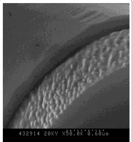
Figure 3 shows theb-TCP scaffolds coated by gelatin. As shown by Figure 3, the gelatin was uniformly coated on the surface of theb-TCP scaffold with thickness around 180 nm. The compressive strength was measured by a uni-versal testing machine and shown in Figure 4. The maxi-mum compressive strengths were 0.15 ± 0.03, 0.11 ± 0.01, 0.78 ± 0.03, and 0.53 ± 0.05 MPa in SP, MP, SPGC, and MPGC, respectively. The compressive strength of the
gelatin-coated scaffolds was about five times higher than that of the non-coated scaffolds. Most of the other studies using the mixed form of bioceramics and gelatin showed that the compressive strength was increased about two to four times [9,12,13]. The gelatin coating maintained the porosity and structure thickness of the scaffold which is similar to the uncoated scaffold. However, the high elasti-city of gelatin as a polymer enhanced the compressive strength of the scaffold [14,15].
Biological properties of theb-TCP scaffold
Figure 5a shows the cell viability results of the scaffolds following 1, 3, 7, and 14 days of culturing that was mea-sured using the CCK-8 assay. The cells on the scaffolds continued to proliferate. The optical density value was
(a)
(b)
[image:3.595.59.538.86.492.2]
(c) (d)
similar between SP and MP and between SPGC and MPGC. This result shows that the multi-sized pores did not affect cell viability. However, the cell viability results on the gelatin-coated scaffolds were higher than those on the uncoated scaffolds. Hence, it was evident that gelatin coating enhanced the cell viability.
Figure 5b shows the ALP activity of the seeded cells on each scaffold. The MP and MPGC having multi-sized pores have shown a higher ALP activity compared to the scaffolds having uniformly sized pores. In addi-tion, the gelatin coating on the scaffold enhanced the
ALP activity compared to the uncoated samples. After 14 days, the MPGC showed the highest ALP activity than the others. This result, wherein the ALP activity was enhanced by increasing gelatin content, is in agree-ment with the previous research by Takahashi et al. [16].
Conclusion
The scaffold having multi-sized pores were successfully fabricated using template coating and freeze drying methods. The gelatin-coated scaffold was fabricated uni-formly by dip coating. The compressive strength of the
b-TCP scaffold was enhanced about five times by gelatin coating. The scaffold having multi-sized pores resulted in improved cell differentiation, and gelatin coating enhanced the cell proliferation and differentiation. This study provides significant data regarding the mechanical and biological properties of the b-TCP scaffold accord-ing to the multi-sized pores and gelatin coataccord-ing.
[image:4.595.58.542.88.266.2](a) (b)
[image:4.595.58.292.450.700.2]Figure 2Images of the cross-section ofb-TCP scaffold with (a) SP and (b) MP.
Figure 3SEM morphologies of theb-TCP scaffold surface coated by gelatin.
SP MP SPGC MPGC
0.0 0.2 0.4 0.6 0.8 1.0
Compr
es
si
ve S
tr
engt
h (
M
P
[image:4.595.306.539.552.714.2]a)
Figure 4The compressive strength ofb-TCP scaffolds.
Kimet al.Nanoscale Research Letters2012,7:78 http://www.nanoscalereslett.com/content/7/1/78
Additional material
[image:5.595.60.540.87.282.2]Additional file 1: Designation code, porosity, and mean thickness. A table showing the designation code, porosity, and mean thickness of the structure of the samples.
Acknowledgements
This study was supported by a grant of the Korea Healthcare Technology R&D Project, Ministry of Health, Welfare & Family Affairs, Republic of Korea (A101578).
Author details 1
Department and Research Institute of Dental Biomaterials and
Bioengineering, Yonsei University College of Dentistry, Seoul 120-752, South Korea2Department of Periodontology, Yonsei University College of Dentistry, Seoul 120-752, South Korea
Authors’contributions
SMK carried out the overall experiments including characterization of the scaffold as well as biological evaluation as the first author. SAY was in charge of cell culture. SHC participated in the biological evaluation and performed the statistical analysis. KMK participated in the biological evaluation. YKL conducted the design and analysis of all experiments as a corresponding author. All authors read and approved the final manuscript.
Competing interests
The authors declare that they have no competing interests.
Received: 2 September 2011 Accepted: 17 January 2012 Published: 17 January 2012
References
1. Wu X, Liu Y, Li X, Wen P, Zhang Y, Long Y, Wang X, Guo Y, Xing F, Gao J:
Preparation of aligned porous gelatin scaffolds by unidirectional freeze-drying method.Acta Biomater2010,6:1167-1177.
2. Liu C, Xia Z, Czernuszka JT:Design and development of three-dimensional scaffolds for tissue engineering.Chem Eng Res Des2007,
85:1051-1064.
3. Guarino V, Cause F, Ambrosio L:Bioactive scaffolds for bone and ligament tissue.Expert Rev Med Dev2007,4:405-418.
4. Descamps M, Richart O, Hardouin P, Hornez JC, Leriche A:Synthesis of macroporousβ-tricalcium phosphate with controlled porous architectural.Ceramics Int2008,34:1131-1137.
5. Buckley CT, O’Kelly KU:Fabrication and characterization of a porous multidomain hydroxyapatite scaffold for bone tissue engineering investigations.J Biomed Mater Res B Appl Biomater2010,93B:459-467. 6. Susan L, Genevieve M, Michael J, Antonios G:Three-dimensional culture of
rat calvarial osteoblasts in porous biodegradable polymers.Biomaterials
1998,19:1405-1412.
7. Bakhtiari L, Reza Rezaie H, Mohamad Hosseinalipour S, Ali Shokrgozar M:
Investigation of biphasic calcium phosphate/gelatin nanocomposite scaffolds as a bone tissue engineering.Ceram Int2010,36:2421-2426. 8. Veis A:The Macromolecular Chemistry of GelatinNew York and London:
Academic Press; 1964.
9. Bigi A, Panzavolta S, Rubini K:Relationship between triple-helix content and mechanical properties of gelatin films.Biomaterials2004,
25:5675-5680.
10. Sundaram J, Durance TD, Wang R:Porous scaffold of gelatin-starch with nanohydroxyapatite composite processed via novel microwave vacuum drying.Acta Biomater2008,4:932-942.
11. Yaylaoglu MB, Korkusuz P, Ors U, Korkusuz F, Hasirci V:Development of a calcium phosphate-gelatin composite as a bone substitute and its use in drug release.Biomaterials1999,20:711-719.
12. Panzavolta S, Fini M, Nicoletti A, Bracci B, Rubini K, Giardino R, Bigi A:
Porous composite scaffolds based on gelatin and partially hydrolyzedα -tricalcium phosphate.Acta Biomater2009,5:636-643.
13. Landi E, Valentini F, Tampieri A:Porous hydroxyapatite/gelatine scaffolds with ice-designed channel-like porosity for biomedical applications.Acta Biomater2008,4:1620-1626.
14. Roohani-Esfahani S, Nouri-Khorasani S, Lu Z, Appleyard R, Zreiqat H:The influence hydroxyapatite nanoparticle shape and size on the properties of biphasic calcium phosphate scaffolds coated with hydroxyapatite-PCL composites.Biomaterials2010,31:5498-5509.
15. Kobayashi S, Yamadi S:Strain rate dependency of mechanical properties of TCP/PLLA composites after immersion in simulated body
environments.Compos Sci Technol2010,70:1820-1825.
16. Takahashi Y, Yamamoto M, Tabata Y:Enhanced osteoinduction by controlled release of bone morphogenetic protein-2 from biodegradable sponge composed of gelatin andβ-tricalcium phosphate.Biomaterials
2005,26:4856-4865.
doi:10.1186/1556-276X-7-78
Cite this article as:Kimet al.:Gelatin-layered and multi-sized porousb -tricalcium phosphate for tissue engineering scaffold.Nanoscale Research Letters20127:78.
1day 3day 7day 14day 0.0 0.8 1.6 2.4 Ce ll pr oli fe ra ti on (OD at 450nm )
SP MP
SPGC MPGC
1day 3day 7day 14day
0.0 0.1 0.2 0.3 0.4 0.5
SP MP SPGC MPGC
Cel l di ff er ent ia ti on ( OD at 405nm )
[image:5.595.305.539.324.687.2]

