PRIMER
Wnt signalling: conquering complexity
Katrin E. Wiese1, Roel Nusse2,* and Renée van Amerongen1,*ABSTRACT
The history of the Wnt pathway is an adventure that takes us from mice and flies to frogs, zebrafish and beyond, sketching the outlines of a molecular signalling cascade along the way. Here, we specifically highlight the instrumental role that developmental biology has played throughout. We take the reader on a journey, starting with developmental genetics studies that identified some of the main molecular players, through developmental model organisms that helped unravel their biochemical function and cell biological activities. Culminating in complex analyses of stem cell fate and dynamic tissue growth, these efforts beautifully illustrate how different disciplines provided missing pieces of a puzzle. Together, they have shaped our mechanistic understanding of the Wnt pathway as a conserved signalling process in development and disease. Today, researchers are still uncovering additional roles for Wnts and other members of this multifaceted signal transduction pathway, opening up promising new avenues for clinical applications.
KEY WORDS: Wnt signalling, Cancer, Model organisms, Stem cells
Introduction
Most researchers today will not stop to think about the marvel of accessing an entire genome sequence with a few easy clicks. Nowadays, almost anyone can run gene ontology or gene set enrichment analyses to get an idea of the signalling pathways contributing to a phenotype of interest. It is important to remember, however, that all of this information has only become structured in hindsight. Until the 1990s, for example, deciphering the sequence of a gene, one base at the time, continued to be a huge challenge, let alone figuring out the actual biological function of the encoded protein.
The history of the Wnt signalling field serves as a prime example: starting with mouse andDrosophilagenetics (Nusse and Varmus, 2012; Nusse and Varmus, 1982; Nüsslein-Volhard and Wieschaus, 1980), almost 40 years of basic research in different model systems have painted a picture of a signalling cascade that is absolutely essential for the development of all multicellular animals, and for the growth and maintenance of various adult tissues. Although our understanding of this complex pathway is still somewhat lacking, we would not be where we are today without these earlier efforts. From geneticists to cell biologists and from embryologists to hard-core biochemists, researchers from various disciplines have contributed essential bits and pieces to resolve the molecular mysteries of Wnt signalling. And the more insight we gained into the diverse physiological roles of Wnt proteins, the better we began to recognize and grasp aberrant signalling and its involvement in the
pathogenesis of many human diseases, ranging from bone and metabolic disorders to multiple forms of cancer (Nusse and Clevers, 2017).
Here, we will primarily review the history of the Wnt pathway from a developmental biology perspective. We will highlight how instrumental this particular discipline has been in unravelling specific aspects of Wnt signalling and discuss its impact on current clinical research and the progress of novel therapeutic avenues. By necessity, we have had to be selective. Our choice to focus mainly on developmental genetics and so-called‘Wnt/β-catenin signalling’ means that we had to omit many beautiful and insightful studies. We hope that after reading this piece, the reader will be primed to explore this broad and exciting research field further according to their own interests.
Wnt signalling in a nutshell
Wnts are secreted proteins that mediate cell-cell communication, either contact dependent or across a short distance. In Wnt-producing cells, the O-acyltransferase porcupine (Porcn) is required for lipid modification of Wnts with palmitoleic acids in the endoplasmic reticulum (ER) (Takada et al., 2006; Willert et al., 2003). Wnt trafficking to the plasma membrane further relies on the multipass transmembrane and putative sorting receptor Evi/ Wntless/Sprinter (Bartscherer et al., 2006; Bänziger et al., 2006; Goodman et al., 2006) (Fig. 1, top).
Once secreted, the hydrophobic Wnt protein shows limited diffusion in the more aqueous extracellular environment (Willert et al., 2003). Therefore, Wnt proteins usually act on neighbouring or nearby cells where they bind Frizzled/Lrp heterodimeric receptor complexes (Bhanot et al., 1996; Wehrli et al., 2000). The first Wnt/ Frizzled crystal structure revealed that the lipid moiety actively engages the receptor and is thus a crucial part of the receptor binding domain (Janda et al., 2012).
A main downstream effector of Wnt signalling in a target cell is the transcriptional co-activator β-catenin (Fig. 1, bottom). In the absence of Wnts, cytoplasmicβ-catenin is bound by Axin and APC,
Advocating developmental biology
This article is part of Development’s advocacy collection–a series of
review articles which make compelling arguments for the field’s
importance. The series is split into two: one set of articles, including
this one, addresses the question‘What has developmental biology ever
done for us?’We want to illustrate how discoveries in developmental
biology have had a wider scientific and societal impact, and thus both
celebrate our field’s history and argue for its continuing place as a core
biological discipline. In a complementary set of articles, we asked
authors to explore ‘What are the big open questions in the field?’
Together, the articles will provide a collection of case studies looking
backwards to the field’s achievements and forwards to its potential, and a
resource for students, educators, advocates and researchers alike. To see the full collection as it grows, go to http://dev.biologists.org/content/ advocating-developmental-biology.
1Section of Molecular Cytology and Van Leeuwenhoek Centre for Advanced Microscopy, Swammerdam Institute for Life Sciences, University of Amsterdam, Science Park 904, 1098 XH Amsterdam, The Netherlands.2Department of Developmental Biology, Howard Hughes Medical Institute, Stanford University, School of Medicine, 265 Campus Drive, Stanford, CA 94305-5458, USA.
*Authors for correspondence ([email protected]; [email protected])
R.N., 0000-0001-7082-3748; R.v.A., 0000-0002-8808-2092
DEVEL
O
phosphorylated by Gsk3 and Ck1, and ubiquitylated by the E3 ligase β-TrCP (reviewed by Stamos and Weis, 2013). Hence, without an incoming Wnt signal this‘destruction complex’ensures that newly synthesizedβ-catenin is continuously eliminated by the proteasome. In the presence of Wnt ligands, the cytoplasmic tail of Lrp6 is phosphorylated by Gsk3 and Ck1, resulting in binding of Axin (Tamai et al., 2004; Zeng et al., 2005). In a process that involves Dishevelled (Dvl), which binds to the Frizzled cytoplasmic tail, these membrane-proximal signalling events result in the formation of large ‘signalosomes’(Bilic et al., 2007; Gammons et al., 2016; Gerlach et al., 2018). This ultimately leads to either translocation or disruption of the destruction complex, causing the stabilization and accumulation ofβ-catenin. Upon translocation to the nucleus, direct transcriptional activation of target genes is mediated by association of β-catenin with Tcf/Lef transcription factors (Behrens et al., 1996; Brunner et al., 1997; Molenaar et al., 1996). Although most of these are cell and tissue specific, several targets, such as Axin2, a common target gene in mammals, are
components of the pathway itself, implicating feedback regulation as an important feature for ensuring a robust signalling response (Jho et al., 2002).
Stabilization of β-catenin and subsequent Tcf/Lef-dependent gene expression changes are arguably the most studied response to Wnt ligand binding in a target cell. However, tissue morphogenesis studies in flies, fish and frogs have each also revealed distinct physiological Wnt pathway responses that proceed independently from, or sometimes appear to counteract, signalling through β -catenin (reviewed by van Amerongen, 2012). We will come back to these responses in more detail below, but not before we take a closer look at how the first individual players were originally discovered.
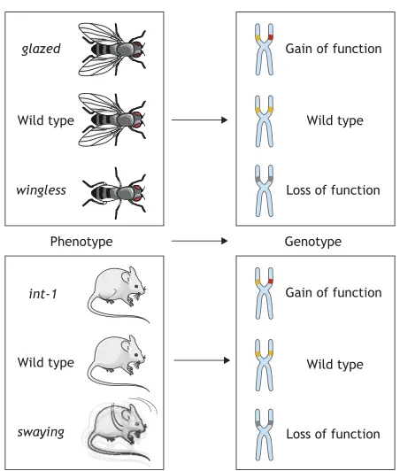
Mapping out a pathway: genetic screens inDrosophila Owing to elegant genetic tools, studies in flies were the first to provide many of the Wnt pathway components we know today and arrange them in a functional order. In fact, from a fly geneticist’s perspective, the first‘discovery’of a Wnt-related phenotype can be traced back to 1936, when Thomas Hunt Morgan and colleagues at Caltech described aDrosophilamutant with glazed eyes (Morgan et al., 1936). Many years later, scientists in India discovered wingless flies and named the hypomorphic allele accordingly (Sharma, 1973; Sharma and Chopra, 1976). It was only revealed later thatglazedis a gain-of-function allele ofwingless, caused by a retrotransposon insertion (Brunner et al., 1999) (Fig. 2, top).
The possibility of (straight-)forward genetic screens in Drosophila allowed systematic searches for interesting mutations causing developmental phenotypes. In 1980, awingless(wg) null allele was (re-)discovered among a group of Nobel Prize-winning genes that caused lethal segmentation defects in developing fly embryos. More specifically,wgbelongs to a subclass of genes that affect polarity within individual body segments (Nüsslein-Volhard and Wieschaus, 1980).
Over the course of the next decade, more refined screens led to the identification of additional so-called ‘segment polarity mutants’, among themarrow(Lrp),armadillo(β-catenin),dishevelled(Dvl), porcupine(Porcn) andshaggy/zeste-white 3(Gsk3) (Perrimon and Mahowald, 1987; Wieschaus and Riggleman, 1987; Wieschaus et al., 1984). Similar to wg, each of these mutations affected embryonic patterning, but their connection was initially unclear, as was their link to an oncogene driving mammary tumour formation in mice, for that matter, which led a parallel existence asint1(Nusse and Varmus, 1982). In the 1930s, when Morgan and colleagues first described theglazedmutant, other biologists sought to understand the contribution of a hereditary ‘milk factor’to mammary gland carcinoma formation in mice (Bittner et al., 1945; Korteweg, 1936). Little did they know at the time that these tumours arose due to integration of the mouse mammary tumour virus (MMTV) in the vicinity ofint1, in a manner ultimately not that different from the transposon-based activation ofwgin theglazedmutant. It was not until 1987 thatint1was recognized as the mouse homologue of the Drosophila wggene (Cabrera et al., 1987; Rijsewijk et al., 1987) (Fig. 2, bottom part). After a whole panel of related genes was discovered in mice (Gavin et al., 1990), a new nomenclature was established to show their common ancestry: theWnt(forwingless -typeMMTV integration site) gene family was born (Nusse et al., 1991).
One gene does not make a pathway, however. Devising a whole signalling cascade requires analysis of functional order and dependencies between genes. Although much of our knowledge of the underlying biochemistry was acquired in other model systems, some of the core relationships between Wnt pathway
Porcupine
Wnt
ER
Golgi
Wnt
Wnt
APC
β-Catenin Tcf/Lef Gsk3 Axin
Dvl
Lgr5 Axin2
Ck1
Wnt
Wnt
Frizzled Lrp
Secreting cell
Target cell
β-Catenin
[image:2.612.104.245.56.449.2]β-Catenin Wntless
Fig. 1. Wnt signalling 101.Simplified model of Wnt secreting (top) and Wnt/β -catenin-responsive cells (bottom) featuring main pathway components. See
text for details ofβ-catenin-independent responses. APC, adenomatous
polyposis coli; Ck1, Casein kinase 1; Dvl, Dishevelled; Gsk3, Glycogen synthase kinase 3; Lgr5, Leucine-rich repeat-containing G-protein-coupled receptor 5; Tcf/Lef, T-cell factor/lymphoid enhancer factor.
DEVEL
O
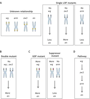
components were first established by epistasis analyses. These types of experiments are perhaps one of the most powerful tools in the field of developmental biology, essentially comparing the phenotype of a double mutant with that of single mutants (Fig. 3). For example, to determine the role of the Gsk3 homolog Zw3/Sgg in the Wg pathway, expression of Engrailed (En), one of the first Wg target genes described, was examined in eitherwg single-,zw3single- orwg/zw3double-mutantDrosophilaembryos. Whereas En was destabilized in wg mutants (implicating Wg as positive regulator),zw3mutants showed enhanced En expression (implicating Zw3 as negative regulator). Double mutants determined the order of action: the En protein expression pattern after loss of bothwgandzw3mimicked the phenotype ofzw3single mutants, demonstrating that Wg acts upstream of Zw3 to antagonize Zw3-mediatedenrepression (Siegfried et al., 1992). Likewise, in search of genes that mediated the Wg signal, other segment polarity mutants were systematically investigated in transgenic wg Drosophila strains. This led to the discovery of Dsh and Arm (vertebrate Dvl andβ-catenin, respectively) as essential components downstream of Wg ultimately placing Arm downstream of Wg and Zw3, with Wg positively and Zw3 negatively regulating the abundance of Arm (Noordermeer et al., 1994; Peifer et al., 1994).
Featuring an elaborate set of ligands, (co-)receptors, effectors and feedback regulators, the complexity of the Wnt pathway as we know it today has increased immensely since these early studies. Nowadays, novel genetic and functional interactions can be elegantly revealed by high-throughput genome-wide screening in cell lines (DasGupta et al., 2005; Lebensohn et al., 2016). However, it was this first batch of invertebrate genetic analyses that clearly helped to compose a blueprint for one of the most crucial developmental signalling cascades in the animal kingdom.
Adding layers: (alternative) Wnt effects during development Although a role for Wnt signalling in invertebrate pattern regulation was evident from the beginning, a similar function during vertebrate development became clear when the pathway was shown to impact on formation of the primary body axis inXenopus laevisembryos. Upon ectopic expression of a murineWnt1mRNA in frog oocytes, embryos developed a secondary body axis (McMahon and Moon, 1989). This axis duplication was also induced by excessβ-catenin and Dishevelled, confirming their role in the pathway (McCrea et al., 1993; Sokol et al., 1995). Perhaps, more importantly, these experiments ultimately led to the realization that Wnt/β-catenin signalling is crucial for establishing the primary body axis during the development of virtually all multicellular animals, ranging from sea urchins (Logan et al., 1999) to mice (Liu et al., 1999).
Of note, one long-missing piece of the puzzle, the identity of a transcription factor that could translate the Wnt signal into a specific target gene response, was first postulated based on the same Xenopusaxis duplication phenotype. A dominant-negative allele of XTcf-3, a frog homolog of the HMG box transcription factors from the Tcf/Lef family, suppressed the induction of axis duplication and was therefore a likely candidate to mediate the transcriptional effects ofβ-catenin in the nucleus (Molenaar et al., 1996). Soon this was confirmed from several angles and in different species, including Drosophila. Here, the Tcf/Lef1 homologue pangolin (pan) was isolated in a suppressor screen as a crucial component of the wg pathway. Pan was shown to interact with Arm, both physically and genetically, thus completing the picture (Brunner et al., 1997).
Studies in gastrulatingXenopusembryos continue to advance our mechanistic understanding of Wnt-dependent gene regulation today. A recent analysis of β-catenin chromatin association suggested that only a subset of genes that are bound by the protein are also immediately transcriptionally regulated in response to Wnt signalling (Nakamura et al., 2016). Such a‘priming’role to poise genes for subsequent rapid activation had already been proposed during the early cleavage stages of frog embryos (Blythe et al., 2010). Depending on the context, additional inputs from other signalling pathways are integrated after β-catenin recruitment to determine the final transcriptional output (Nakamura et al., 2016). As alluded to above, alternative,β-catenin-independent responses also exist. For example, multiple developmental processes require that large collectives of cells‘coordinate’their polarized behaviour and align across the plane of a given tissue. This phenomenon is called planar cell polarity (PCP) and is responsible for, among other things, the precise and uniform orientation of trichomes (hairs) in the fly wing and of the stereocilia on sensory hair cells in the mammalian inner ear. A key feature of PCP is the asymmetric subcellular localization of signalling molecules. Among them are the core Wnt pathway components Frizzled and Dishevelled (Krasnow et al., 1995; Vinson and Adler, 1987), which in this case signal through small RhoGTPases and JNK rather than through β-catenin to alter cytoskeletal dynamics (Axelrod et al., 1998; Shulman et al., 1998; Wallingford and Habas, 2005). In fact, PCP defects were described infzmutant flies (Gubb and García-Bellido, 1982) long before its homologue fz2 was identified as the wg receptor (Bhanot et al., 1996). At the same time, this β-catenin independent‘flavour’of the Wnt pathway is still poorly understood mechanistically and a direct requirement for Wnt ligands in PCP has not been shown definitively under all circumstances.
Studies in fish and frogs, however, did firmly establish a role for selected Wnt ligands and a PCP-like‘alternative’Wnt pathway in regulation of vertebrate gastrulation movements. During so-called
Phenotype Genotype
glazed
wingless
swaying
Wild type Wild type Gain of function
Loss of function
Wild type
int-1
Wild type Gain of function
[image:3.612.61.287.52.318.2]Loss of function
Fig. 2. From phenotype to genotype in flies and mice.Gain- and
loss-of-function alleles ofWntwere independently discovered based on different
phenotypes in flies (top) and mice (bottom). Interesting parallels can be observed in these vastly different model organisms. The wild-type allele is depicted in orange. Gain of-function mutations are shown in red, whereas loss-of-function mutations are shown in grey.
DEVEL
O
convergent extension movements, cells align and intercalate, leading to simultaneous narrowing and lengthening of the embryo along the body axis (Wallingford et al., 2002). Polarized cell behaviour and convergent extension are disrupted in frog embryos lacking Dishevelled (Wallingford et al., 2000). In addition, in both Xenopus and zebrafish, this process is crucially dependent on Wnt11 and Wnt5a (Heisenberg et al., 2000; Kilian et al., 2003; Moon et al., 1993; Tada and Smith, 2000). The same two ligands also regulate convergent extension movements and epithelial-mesenchymal transition (EMT) during murine gastrulation, indicating a conserved β-catenin-independent Wnt signalling mechanism for these specialized forms of directed cell migration (Andre et al., 2015).
In conclusion, research in Xenopus and other experimentally amenable vertebrate embryos provided convenient functional assays for Wnt signalling components. Those, in turn, revealed unexpected layers of complexity by affecting cell polarity and directional migration as important aspects of Wnt signalling during development (Jussila and Ciruna, 2017). For many years, Wnt ligands were also stereotypically divided into two classes primarily based on the embryological assays described above: induction of axis duplication, on the one hand (e.g. Wnt1, Wnt3a, Wnt8A/B), and regulation of convergent extension, on the other (e.g. Wnt4, Wnt5a, Wnt11) (Du et al., 1995). It now appears that this division is not so black and white: A complex of Wnt5a and Wnt11 can induce bothβ-catenin-dependent and -independent signalling in the early Xenopus embryo (Cha et al., 2008), and Wnt5a can elicit both responses by engaging different receptors bothin vitroandin vivo (Mikels and Nusse, 2006; van Amerongen et al., 2012b). Although it may not be news to insiders that Wnts have context-dependent activities, this complexity is bound to confuse newcomers to the
field and before we will be able to see the complete picture there is still much to learn and understand at the molecular level. As evidence is starting to accumulate that these developmental processes are hijacked by cancer cells, either to drive abnormal cellular migration during metastasis or to escape from drug treatment (Anastas et al., 2014; Grossmann et al., 2013; Weeraratna et al., 2002), we will likely continue to call on these model systems for help in resolving the complexity of these cellular signalling events (Fig. 4).
From embryology to adult tissue maintenance: probing Wnt pathway function in mouse models
Wnt signalling is conserved across evolution and is a basic mechanism of intercellular communication used by all multicellular animals. But if years of research have taught us one thing, it is that there is nothing basic and simple about this pathway. Apart from an intricate network of intracellular components and molecular responses, the 19 different Wnt, 10 Frizzled and two Lrp co-receptor genes in mammalian genomes offer numerous possibilities for promiscuous interactions on the cell surface. In addition, individual Wnt ligands and receptors show staggeringly dynamic expression patterns during development (Kemp et al., 2005; Lickert et al., 2001; Summerhurst et al., 2008).
Mice have proved a useful model organism with which to start resolving this complexity. The possibility of manipulating the mouse genome first appeared on the technological horizon in the 1980s. Where forward genetics inDrosophilahad discovered Wnt pathway genes responsible for a particular phenotype (‘from phenotype to genotype’), reverse genetics in mice allowed researchers to interrogate and dissect the physiological functions of many Wnt signalling components by creating knockout mice A
Unknown relationship
wg arm zw3 en
B
No
wg ; zw3
More
en Double mutant
No
wg
Less
en
No
zw3
More
en Single LOF mutants
C
No arm
Less
en
More
wg
More
en GOF mutant
D
More
wg
No
arm
Less
en Suppressor
mutant Pathway
wg
arm
[image:4.612.48.350.54.385.2]en zw3
Fig. 3. Epistasis experiments reveal the relationship between genes.(A) Loss-of-function (LOF) mutants for individual genes that result in similar phenotypes (in
this case affecting the expression ofen) can hint at a functional
relationship between the encoded gene products. Mutation of a negative regulator results in the opposite phenotype (in this
case: nowg, lessen; nozw3, moreen). (B) For positive and
negative regulators, the phenotype of a double mutant can help determine the functional order of genes in a pathway. The activity of the most downstream gene determines the phenotype of the double mutant. (C) If a loss-of-function mutant
(in this case noarm) can rescue the phenotype induced by a
gain-of-function (GOF) mutant (in this case morewg), this
also allows the order of signalling events to be delineated. (D) Together, these analyses allow investigators to build a pathway from previously unknown relationships. The wild-type allele is depicted in orange. Gain of-function mutations are shown in red, whereas loss-of-function mutations are shown in grey.arm,armadillo;en,engrailed;wg,wingless;zw3, zeste-white 3.
DEVEL
O
(‘from genotype to phenotype’). In accordance with the diverse expression patterns of Wnt ligands, the phenotypes of individual knockouts differed considerably, ranging from abnormal placental development to urogenital defects and brain anomalies.
For example, knockout ofWnt1, one of the first genes ever to be targeted by homologous recombination in mouse embryonic stem cells, results in severe defects in brain development, ranging from loss of most of the midbrain and cerebellum (McMahon and Bradley, 1990) to severe cerebellar ataxia phenotypes in surviving homozygous mutants (Thomas and Capecchi, 1990). Interestingly, it is here that the Wnt-target gene Engrailed makes another appearance: proper expression of this gene is crucial for formation of the midbrain-hindbrain boundary, an area also known as the isthmus organizer, thus explaining the phenotype and once again showing similar genetic interactions in flies and mice. Around the same time, mouse geneticists also came to the realization that a long-known spontaneous mouse mutant with ataxia, known as swaying(Lane, 1967), was the result of a mutatedWnt1allele that resulted in premature truncation of the protein (Thomas et al., 1991). This situation too is reminiscent of that encountered by their Drosophilacounterparts upon discovering the connection between winglessandglazed(Fig. 2).
Another clue to the diverse (and at the same time highly specific) functions of pathway components came from knockout studies for Lef1 and Tcf1, which were identified as DNA-binding proteins in hematopoietic cells well before their involvement in Wnt/β-catenin signalling became apparent (Travis et al., 1991; van de Wetering et al., 1991). WhereasTcf1knockouts only presented with defects in thymus development (Verbeek et al., 1995),Lef1-null mice showed multiple abnormalities, including the loss of skin appendages such as hair follicles, mammary glands and teeth (van Genderen et al., 1994). Compound knockout mice also revealed considerable
redundancy, with double knockouts of Lef1and Tcf1 mimicking the early developmental defects ofWnt3a-null mice (Galceran et al., 1999). Although genetic redundancy ensures developmental robustness, it definitely makes the life of a developmental biologist much harder, and the complex mouse crosses that are required to reveal the plethora of phenotypes associated with a given gene family typically require multiple PhD or postdoc appointments. However, technological improvements, including various ‘flavours’ of inducible and conditional mouse strains coupled with ongoing progress in genetic engineering strategies continue to offer attractive new opportunities, and have brought Wnt research to the next level. As a result of such advances, in the past two decades we have begun to fully appreciate that Wnt signalling is not only important during early development but continues to have considerable influence on the maintenance of multiple different tissues.
The firstin vivostudies with so-called TOPGAL reporter mice, in which cells with active Wnt/β-catenin signalling can be identified because they express aβ-galactosidase reporter gene (lacZ) under the control of three consensus Tcf/Lef1 binding sites, suggested a regulatory role for Wnt/β-catenin signalling in hair follicle morphogenesis and differentiation (DasGupta and Fuchs, 1999). Further insight that the Wnt pathway is involved in cell fate decisions in multiple tissues came from more sophisticated approaches using inducible mouse models for lineage-tracing purposes. In lineage tracing, a single cell is labelled with a permanent mark (e.g. expression of a fluorescent protein) at a given time point so that all daughter cells, which inherit this mark, can be recognized and followed over multiple generations. This technique was not invented by mouse geneticists, of course: it was pioneered, in a much simpler form, by developmental biologists such as Edwin Conklin in the early 20th century (Conklin, 1905). In mice, lineage
Regeneration Repair Tumour
growth Metastasis Proliferation
Differentiation Migration Cell fate
specification
3D organization
Wnt
Fzd/Dvl
Gsk3
β-Catenin
Target genes
RhoA/Rac1
Rock/Jnk
Actin cytoskeleton
[image:5.612.49.356.54.366.2]Development Maintenance Aging
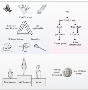
Fig. 4. Towards resolving the complexity of Wnt signalling.Studies performed in different developmental model organisms have contributed vital clues to our understanding of Wnt signalling, revealing its importance for balancing cell proliferation, cell fate specification,
differentiation and migration, ultimately allowing multicellular animals to properly organize their cells into functional 3D tissues (top left). At the same time, these model organisms have been instrumental in identifying and organizing the core players into the pathway we know today, and we are now beginning to appreciate the elaborate downstream signalling network that influences a vast array of cellular processes (top right). A more applied challenge is to improve human health via context-specific Wnt activation or inhibition: harnessing its regenerative power to promote tissue maintenance and repair, thereby counteracting degeneration and aging, while at the same time inhibiting its oncogenic effects by blocking Wnt-mediated tumour growth and progression (bottom). Fzd.Dvl, frizzled/dishevelled; Gsk3, Glycogen synthase kinase 3; RhoA/Rac1, Ras homolog family member A/Rac family small GTPase 1; Rock/Jnk, Rho associated coiled-coil containing protein kinase/c-Jun N-terminal kinase.
DEVEL
O
tracing is usually achieved through genetic recombination with the Cre-loxPsystem, with a ubiquitous promoter controlling marker or reporter gene expression. Expression of the marker or reporter gene is prevented by a ‘floxed’ STOP cassette, however, until Cre recombinase excises the stop sequence and allows the reporter to be induced (Kretzschmar and Watt, 2012). Cre activation can be controlled in several ways. First, spatial control can be achieved by expressing it under a promoter of choice (ideally making it cell-type specific). Second, fusing Cre to the modified hormone-binding domain of the human oestrogen receptor (CreERT2) for example,
allows the timing of recombination to be experimentally controlled by the addition of tamoxifen (Feil et al., 1997).
The phenotype ofTcf4knockout mice had already provided a first hint towards an important role for Wnt signalling in maintaining stem cells in the small intestine (Korinek et al., 1998). Almost a decade later, a mouse model expressing an inducible Cre recombinase under the control of the Lgr5 promoter led to the realization that this Wnt target gene specifically marks fast-dividing adult stem cells in both the small intestine and colon (Barker et al., 2007). The Lgr5-CreERT2mouse, together with others such as the
Axin2-CreERT2 lineage-tracing model have since provided
invaluable information about the existence of Wnt-responsive stem cells in multiple tissues, ranging from the mammary gland and the skin, to the brain, liver and ovaries (Bowman et al., 2013; Lim et al., 2013; Ng et al., 2014; van Amerongen et al., 2012a; Wang et al., 2015).
Translational relevance: fighting cancer
By now it may be obvious that our travels through Wnt history are characterized by (forgotten) discoveries, re-discoveries and conceptual frameworks built on observations and homologies in different species. As mentioned earlier, the first mammalian Wnt gene (Wnt1) was originally identified as an oncogene driving mammary tumour formation in mice. Setting out to find putative host cell genes that would be activated by insertional mutagenesis of the mouse mammary tumour virus (MMTV), theint1gene (for first common integration site) was identified after 2 years of careful mapping efforts (Nusse and Varmus, 1982). However, in contrast to other prominent proto-oncogenes that were discovered around that time, such as Myc and Ras, neither dominant activating mutations in int1nor alterations of the int1 locus were found in human cancers. Although its initial discovery thus implicatedint1 as a bona fide proto-oncogene (Tsukamoto et al., 1988), primary research efforts slowly drifted away from tumorigenesis to elucidating the function of int1(by then known asWnt1) during development. It was not until more than 10 years later that the important connection between Wnt pathway alterations and cancer was re-discovered.
A region on human chromosome 5q21 had been suspected to have an association with a hereditary cancer syndrome called familial adenomatous polyposis (FAP) and other types of colon cancer. The linkedAPCgene (for adenomatous polyposis coli) was cloned in 1991 (Groden et al., 1991; Kinzler et al., 1991; Nishisho et al., 1991) and subsequently found to interact with β-catenin (Rubinfeld et al., 1993; Su et al., 1993). The ultimate realization of how important this interaction might be in human cancer, however, only came with the observation that APC is a crucial part of a destruction complex that degradesβ-catenin in the absence of Wnt ligands (Korinek et al., 1997; Morin et al., 1997; Munemitsu et al., 1995).APCis now well known to be a‘gatekeeper’gene that is crucial for cell expansion in the early stages of colorectal carcinogenesis (Kinzler and Vogelstein, 1996).
As it turns out, both dominant mutations, not restricted to APC, as well as more subtle deregulation of Wnt signalling, are a recurrent theme in many types of human tumours (Fig. 5). As a result, both β-catenin-dependent and -independent Wnt signalling are now considered to be promising therapeutic targets (Daulat and Borg, 2017; Zhang and Hao, 2015) and after 40 years of basic developmental research, the first clinical trials with Wnt inhibitors are now being conducted (www.clinicalTrials.gov). But why has it taken so long to translate all of our scientific knowledge into clinical action? Given the frequent occurrence of mutations in more downstream components, such as APC andβ-catenin, many drug development efforts were initially focused on blocking the activity ofβ-catenin/Tcf complexes. Success in this area has been limited and the results have been disappointing–a common finding for drug development efforts directed against transcription factors. Efforts are still ongoing, however, and new drugs continue to be developed. Although tumours in which the Wnt pathway is activated by dominant mutations in more downstream signalling components, such as the APC and β-catenin mutations observed in colorectal cancer, have dominated the literature, promising therapeutic results have been obtained by focusing attention on upstream signalling events rather than on events within the receiving cell. For example, multiple different tumour types have shown promising responses to Wnt pathway inhibition with decoy receptors or receptor blocking antibodies in pre-clinical studies (Gurney et al., 2012; Takebe et al., 2015). As mentioned earlier, Wnt secretion and activity is crucially dependent on palmitoyl groups attached by the Porcn enzyme (Janda et al., 2012; Takada et al., 2006). In 2009, Lum and colleagues were the first to isolate a Porcn small molecule inhibitor (IWP2) and in 2013 Harris and colleagues identified a second, LGK974 (Chen et al., 2009; Liu et al., 2013), which is currently in phase I/II clinical trials.
Multiple Multiple
Amplification/deletion Amplification/deletion
Mutation Mutation Ligands
and receptors e.g. WNTs, FZDs Intracellular
components e.g. APC,
-Catenin Colorectal Lung SCC
Liver Ovarian
Head and neck
MelanomaProstate Lung AC Breast
Pancreas GBM
Mesothelioma AML
90 80 70 60 50 40 30 20 10 0
Alteration frequency (%)
[image:6.612.313.565.426.627.2]Key
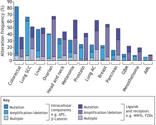
Fig. 5. Frequent alteration of Wnt pathway members in human cancers. In silicoanalysis of selected datasets from The Cancer Genome Atlas (TCGA) for mutations, amplifications and deletions of either intracellular (e.g. APC,
β-catenin; in blue) or extracellular ligand and receptor (e.g. WNTs, FZDs; in
purple) pathway components. DNA amplifications and deletions of different Wnt pathway components are a common feature across different human tumour types, highlighting the potential for therapeutic targeting of the Wnt-pathway. AC, adenocarcinoma; AML, acute myeloid leukaemia; APC, adenomatous polyposis coli; FZD, Frizzled; GBM, glioblastoma multiforme; SCC, squamous cell carcinoma. This figure was generated using data
available at www.cbioportal.org.
DEVEL
O
Although clinical efficacy remains to be shown, in our opinion this hopeful development may highlight the fact that many different cancers show deregulation of the Wnt pathway, thereby essentially hijacking a developmental growth control pathway to boost tumour expansion (Fig. 5). Given the molecular complexity of Wnt signal transduction, however, additional drugs with more specific modes of action, such as the Wnt5a hexapeptide Foxy5 (Säfholm et al., 2008), might be necessary to specifically block (or enhance) independent arms of the pathway. This is particularly important as evidence is accumulating that the different Wnt signalling branches may be intimately linked to balance cell proliferation and differentiation during development, tissue maintenance, ageing and regeneration (Stoick-Cooper et al., 2007). Given that cancer may be viewed as normal development ‘gone wrong’, the chances are that the fate of a cancer cell is likewise determined by the balance of β-catenin-dependent and -independent signals. Considering the history of Wnt research, it is likely that new insights for therapeutic avenues will continue to come from the fundamental questions curious developmental biologists ask.
Why we need developmental biology: looking back and gazing ahead
Experiments in the model organisms described above have been instrumental in revealing the molecular players of Wnt signalling and their functional relationships to each other. Many of these studies were conducted with the sole purpose of understanding how a particular tissue or organism develops, with the realisation of the societal relevance of the discoveries only coming much later. Instead of building our story around one or two‘Eureka’moments, we deliberately chose to present a small selection of efforts from the history of Wnt pathway research. In doing so, we have undoubtedly omitted other deserving discoveries, such as those related to cell fate specification and migration in the development of the roundworm C. elegans or the exciting studies in regenerating planarian flatworms. Still, our selection reflects what happens in science: pieces ultimately fall into place from different angles (Fig. 4). It has really been this unique mix of questions, concepts and discoveries that brought us where we are today in our understanding of Wnt signalling. As such, one experimental system or approach cannot easily be disregarded as being inferior to another.
For example, a screen for novel Wnt/β-catenin-modulating genes executed in HEK293T cells, arguably the cell biologist’s work horse, identified R-spondins as natural Wnt signalling enhancers (Kazanskaya et al., 2004). In hindsight, this was a major breakthrough: we now know from different experimental systems that Lgr4/5/6, which mark fast dividing stem cells in multiple tissues, act as receptors for R-spondins (Carmon et al., 2011; de Lau et al., 2011; Glinka et al., 2011). Thanks to transgenic mouse models, we discovered the ability of human RSPO1 to promote massive intestinal crypt proliferation (Kim et al., 2005). Soon after, R-spondin-based media were used to establish the first intestinal 3D organoid cultures (Sato and Clevers, 2013; Sato et al., 2009). In fact, we are now able to keep stem cells from a variety of tissues and sources successfully in 3D culture. In almost every case, they are crucially dependent on some form of Wnt pathway activation, either purified Wnt proteins or agonists–admittedly among other factors and 3D matrix (Nusse and Clevers, 2017). Those stem cell-derived organoid models were elected as‘Method of the Year’by Nature Methods in 2017 (de Souza, 2018), illustrating their great potential to study human diseases and promising to advance regenerative medicine.
Developmental biologists often have to hear that their favourite organism does not fully recapitulate all the complexities of human physiology and disease. This objection is correct: after all, they are called model systems for a reason. Still, the record shows that in many cases what we have learned about the molecular mechanism of Wnt signalling in diverse organisms has turned out to be directly translatable and relevant for human health. Importantly, these similarities and connections all too often only reveal themselves later, once the same concepts are re-discovered in a human (disease) context. Therefore, it seems fair to say that developmental biologists should continue to investigate biology for its own sake, without direct and immediate translation as a prerequisite, and, crucially, to be offered the funding to do so. History has proven time and time again that societal impact will follow. If developmental biology has done one thing for us, it was to show us that new ideas and concepts originating from basic curiosity-driven research have the potential to contribute to biomedical applications and eventually fuel new treatment innovations for human diseases.
Acknowledgements
We celebrate the multidisciplinary efforts that have led to our current understanding and accumulated knowledge of the Wnt pathway and apologize to all colleagues whose important work could not be cited due to space constraints.
Competing interests
The authors declare no competing or financial interests.
Funding
R.v.A.’s research is funded by KWF Kankerbestrijding (ANW 2013-6057, UVA 2015-8014, UVA 11082-2017) and an NWO VIDI grant from the Nederlandse Organisatie voor Wetenschappelijk Onderzoek (864.13.002). K.E.W. is a Marie Skłodowska-Curie Actions Individual Fellowship fellow (European Union’s Horizon 2020 research and innovation programme, 706443). R.N. is an investigator of the Howard Hughes Medical Institute and receives research funding from the California Institute of Regenerative Medicine.
References
Anastas, J. N., Kulikauskas, R. M., Tamir, T., Rizos, H., Long, G. V., von Euw,
E. M., Yang, P.-T., Chen, H.-W., Haydu, L. et al. (2014). WNT5A enhances
resistance of melanoma cells to targeted BRAF inhibitors.J. Clin. Invest.124, 2877-2890.
Andre, P., Song, H., Kim, W., Kispert, A. and Yang, Y.(2015). Wnt5a and Wnt11
regulate mammalian anterior-posterior axis elongation. Development 142, 1516-1527.
Axelrod, J. D., Miller, J. R., Shulman, J. M., Moon, R. T. and Perrimon, N.(1998). Differential recruitment of Dishevelled provides signaling specificity in the planar cell polarity and Wingless signaling pathways.Genes Dev.12, 2610-2622.
Barker, N., van Es, J. H., Kuipers, J., Kujala, P., van den Born, M., Cozijnsen, M.,
Haegebarth, A., Korving, J., Begthel, H., Peters, P. J. et al. (2007).
Identification of stem cells in small intestine and colon by marker gene Lgr5.
Nature449, 1003-10U1.
Bänziger, C., Soldini, D., Schütt, C., Zipperlen, P., Hausmann, G. and Basler, K.
(2006). Wntless, a conserved membrane protein dedicated to the secretion of Wnt proteins from signaling cells.Cell125, 509-522.
Bartscherer, K., Pelte, N., Ingelfinger, D. and Boutros, M.(2006). Secretion of
Wnt ligands requires Evi, a conserved transmembrane protein.Cell125, 523-533.
Behrens, J., von Kries, J. P., Kühl, M., Bruhn, L., Wedlich, D., Grosschedl, R.
and Birchmeier, W. (1996). Functional interaction of beta-catenin with the
transcription factor LEF-1.Nature382, 638-642.
Bhanot, P., Brink, M., Samos, C. H., Hsieh, J.-C., Wang, Y., Macke, J. P., Andrew,
D., Nathans, J. and Nusse, R.(1996). A new member of the frizzled family from
Drosophila functions as a Wingless receptor.Nature382, 225-230.
Bilic, J., Huang, Y.-L., Davidson, G., Zimmermann, T., Cruciat, C.-M., Bienz, M.
and Niehrs, C. (2007). Wnt induces LRP6 signalosomes and promotes
dishevelled-dependent LRP6 phosphorylation.Science316, 1619-1622.
Bittner, J. J., Evans, C. A. and Green, R. G.(1945). Survival of the mammary tumor milk agents of mice.Science101, 95-97.
Blythe, S. A., Cha, S.-W., Tadjuidje, E., Heasman, J. and Klein, P. S.(2010). beta-Catenin primes organizer gene expression by recruiting a histone H3 arginine 8 methyltransferase, Prmt2.Dev. Cell19, 220-231.
Bowman, A. N., van Amerongen, R., Palmer, T. D. and Nusse, R.(2013). Lineage
tracing with Axin2 reveals distinct developmental and adult populations of Wnt/β -catenin-responsive neural stem cells.Proc. Natl. Acad. Sci. USA110, 7324-7329.
DEVEL
O
Brunner, E., Peter, O., Schweizer, L. and Basler, K.(1997). pangolin encodes a Lef-1 homologue that acts downstream of Armadillo to transduce the Wingless signal in Drosophila.Nature385, 829-833.
Brunner, E., Brunner, D., Fu, W., Hafen, E. and Basler, K.(1999). The dominant
mutation Glazed is a gain-of-function allele of wingless that, similar to loss of APC, interferes with normal eye development.Dev. Biol.206, 178-188.
Cabrera, C. V., Alonso, M. C., Johnston, P., Phillips, R. G. and Lawrence, P. A.
(1987). Phenocopies induced with antisense RNA identify the wingless gene.Cell 50, 659-663.
Carmon, K. S., Gong, X., Lin, Q., Thomas, A. and Liu, Q.(2011). R-spondins
function as ligands of the orphan receptors LGR4 and LGR5 to regulate Wnt/beta-catenin signaling.Proc. Natl. Acad. Sci. USA108, 11452-11457.
Cha, S.-W., Tadjuidje, E., Tao, Q., Wylie, C. and Heasman, J.(2008). Wnt5a and
Wnt11 interact in a maternal Dkk1-regulated fashion to activate both canonical and non-canonical signaling in Xenopus axis formation. Development 135, 3719-3729.
Chen, B., Dodge, M. E., Tang, W., Lu, J., Ma, Z., Fan, C.-W., Wei, S., Hao, W., Kilgore, J., Williams, N. S. et al.(2009). Small molecule-mediated disruption of Wnt-dependent signaling in tissue regeneration and cancer.Nat. Chem. Biol.5, 100-107.
Conklin, E. G.(1905).The Organization and Cell-lineage of the Ascidian Egg.
Philadelphia, PA: Academy of Natural Sciences.
DasGupta, R. and Fuchs, E. (1999). Multiple roles for activated LEF/TCF
transcription complexes during hair follicle development and differentiation.
Development126, 4557-4568.
DasGupta, R., Kaykas, A., Moon, R. T. and Perrimon, N.(2005). Functional
genomic analysis of the Wnt-wingless signaling pathway.Science308, 826-833.
Daulat, A. M. and Borg, J.-P. (2017). Wnt/planar cell polarity signaling: new
opportunities for cancer treatment.Trends Cancer3, 113-125.
de Lau, W., Barker, N., Low, T. Y., Koo, B.-K., Li, V. S. W., Teunissen, H., Kujala,
P., Haegebarth, A., Peters, P. J., van de Wetering, M. et al.(2011). Lgr5
homologues associate with Wnt receptors and mediate R-spondin signalling.
Nature476, 293-297.
de Souza, N.(2018). Organoids.Nat. Meth.15, 23-23.
Du, S. J., Purcell, S. M., Christian, J. L., McGrew, L. L. and Moon, R. T.(1995). Identification of distinct classes and functional domains of Wnts through expression of wild-type and chimeric proteins in Xenopus embryos.Mol. Cell. Biol.15, 2625-2634.
Feil, R., Wagner, J., Metzger, D. and Chambon, P.(1997). Regulation of Cre
recombinase activity by mutated estrogen receptor ligand-binding domains.
Biochem. Biophys. Res. Commun.237, 752-757.
Galceran, J., Fariñas, I., Depew, M. J., Clevers, H. and Grosschedl, R.(1999). Wnt3a-/–like phenotype and limb deficiency in Lef1(-/-)Tcf1(-/-) mice.Genes Dev. 13, 709-717.
Gammons, M. V., Renko, M., Johnson, C. M., Rutherford, T. J. and Bienz, M.
(2016). Wnt signalosome assembly by DEP domain swapping of dishevelled.Mol. Cell64, 92-104.
Gavin, B. J., McMahon, J. A. and McMahon, A. P.(1990). Expression of multiple
novel Wnt-1/int-1-related genes during fetal and adult mouse development.
Genes Dev.4, 2319-2332.
Gerlach, J. P., Jordens, I., Tauriello, D. V. F., van‘t Land-Kuper, I., Bugter, J. M., Noordstra, I., van der Kooij, J., Low, T. Y., Pimentel-Muiños, F. X., Xanthakis,
D. et al.(2018). TMEM59 potentiates Wnt signaling by promoting signalosome
formation.Proc. Natl. Acad. Sci. USA115, E3996-E4005.
Glinka, A., Dolde, C., Kirsch, N., Huang, Y.-L., Kazanskaya, O., Ingelfinger, D.,
Boutros, M., Cruciat, C.-M. and Niehrs, C.(2011). LGR4 and LGR5 are
R-spondin receptors mediating Wnt/β-catenin and Wnt/PCP signalling.EMBO Rep. 12, 1055-1061.
Goodman, R. M., Thombre, S., Firtina, Z., Gray, D., Betts, D., Roebuck, J.,
Spana, E. P. and Selva, E. M.(2006). Sprinter: a novel transmembrane protein
required for Wg secretion and signaling.Development133, 4901-4911.
Groden, J., Thliveris, A., Samowitz, W., Carlson, M., Gelbert, L., Albertsen, H., Joslyn, G., Stevens, J., Spirio, L. and Robertson, M.(1991). Identification and characterization of the familial adenomatous polyposis coli gene. Cell 66, 589-600.
Grossmann, A. H., Yoo, J. H., Clancy, J., Sorensen, L. K., Sedgwick, A., Tong,
Z., Ostanin, K., Rogers, A., Grossmann, K. F., Tripp, S. R. et al.(2013). The
small GTPase ARF6 stimulatesβ-catenin transcriptional activity during WNT5A-mediated melanoma invasion and metastasis.Sci. Signal.6, ra14.
Gubb, D. and Garcı́a-Bellido, A.(1982). A genetic analysis of the determination of cuticular polarity during development in Drosophila melanogaster.J. Embryol. Exp. Morphol.68, 37-57.
Gurney, A., Axelrod, F., Bond, C. J., Cain, J., Chartier, C., Donigan, L., Fischer, M., Chaudhari, A., Ji, M., Kapoun, A. M. et al.(2012). Wnt pathway inhibition via the targeting of Frizzled receptors results in decreased growth and tumorigenicity of human tumors.Proc. Natl. Acad. Sci. USA109, 11717-11722.
Heisenberg, C.-P., Tada, M., Rauch, G.-J., Saúde, L., Concha, M. L., Geisler, R.,
Stemple, D. L., Smith, J. C. and Wilson, S. W. (2000). Silberblick/Wnt11
mediates convergent extension movements during zebrafish gastrulation.Nature 405, 76-81.
Janda, C. Y., Waghray, D., Levin, A. M., Thomas, C. and Garcia, K. C.(2012).
Structural basis of Wnt recognition by Frizzled.Science337, 59-64.
Jho, E.-H., Zhang, T., Domon, C., Joo, C.-K., Freund, J.-N. and Costantini, F.
(2002). Wnt/beta-catenin/Tcf signaling induces the transcription of Axin2, a negative regulator of the signaling pathway.Mol. Cell. Biol.22, 1172-1183.
Jussila, M. and Ciruna, B.(2017). Zebrafish models of non-canonical Wnt/planar
cell polarity signalling: fishing for valuable insight into vertebrate polarized cell behavior.Wiley Interdiscip. Rev. Dev. Biol.6, e267.
Kazanskaya, O., Glinka, A., del Barco Barrantes, I., Stannek, P., Niehrs, C. and
Wu, W.(2004). R-Spondin2 is a secreted activator of Wnt/beta-catenin signaling
and is required for Xenopus myogenesis.Dev. Cell7, 525-534.
Kemp, C., Willems, E., Abdo, S., Lambiv, L. and Leyns, L.(2005). Expression of
all Wnt genes and their secreted antagonists during mouse blastocyst and postimplantation development.Dev. Dyn.233, 1064-1075.
Kilian, B., Mansukoski, H., Barbosa, F. C., Ulrich, F., Tada, M. and Heisenberg,
C. P.(2003). The role of Ppt/Wnt5 in regulating cell shape and movement during
zebrafish gastrulation.Mech. Dev.120, 467-476.
Kim, K.-A., Kakitani, M., Zhao, J., Oshima, T., Tang, T., Binnerts, M., Liu, Y.,
Boyle, B., Park, E., Emtage, P. et al.(2005). Mitogenic influence of human
R-spondin1 on the intestinal epithelium.Science309, 1256-1259.
Kinzler, K. W. and Vogelstein, B.(1996). Lessons from hereditary colorectal
cancer.Cell87, 159-170.
Kinzler, K. W., Nilbert, M. C., Su, L. K., Vogelstein, B., Bryan, T. M., Levy, D. B.,
Smith, K. J., Preisinger, A. C., Hedge, P. and McKechnie, D. (1991).
Identification of FAP locus genes from chromosome 5q21.Science253, 661-665.
Korinek, V., Barker, N., Morin, P. J., van Wichen, D., de Weger, R., Kinzler, K. W., Vogelstein, B. and Clevers, H.(1997). Constitutive transcriptional activation by a beta-catenin-Tcf complex in APC-/- colon carcinoma.Science275, 1784-1787.
Korinek, V., Barker, N., Moerer, P., van Donselaar, E., Huls, G., Peters, P. J. and
Clevers, H.(1998). Depletion of epithelial stem-cell compartments in the small
intestine of mice lacking Tcf-4.Nat. Genet.19, 379-383.
Korteweg, R.(1936). On the manner in which the disposition to carcinoma of the
mammary gland is inherited in mice.Genetica18, 350-371.
Krasnow, R. E., Wong, L. L. and Adler, P. N.(1995). Dishevelled is a component of the frizzled signaling pathway in Drosophila.Development121, 4095-4102.
Kretzschmar, K. and Watt, F. M.(2012). Lineage tracing.Cell148, 33-45.
Lane, P. W.(1967). Swaying.Mouse News Lett.36, 40.
Lebensohn, A. M., Dubey, R., Neitzel, L. R., Tacchelly-Benites, O., Yang, E., Marceau, C. D., Davis, E. M., Patel, B. B., Bahrami-Nejad, Z., Travaglini, K. J.
et al.(2016). Comparative genetic screens in human cells reveal new regulatory
mechanisms in WNT signaling.Elife5, 410.
Lickert, H., Kispert, A., Kutsch, S. and Kemler, R.(2001). Expression patterns of Wnt genes in mouse gut development.Mech. Dev.105, 181-184.
Lim, X., Tan, S. H., Koh, W. L. C., Chau, R. M. W., Yan, K. S., Kuo, C. J., van Amerongen, R., Klein, A. M. and Nusse, R.(2013). Interfollicular epidermal stem cells self-renew via autocrine Wnt signaling.Science342, 1226-1230.
Liu, P., Wakamiya, M., Shea, M. J., Albrecht, U., Behringer, R. R. and Bradley, A.
(1999). Requirement for Wnt3 in vertebrate axis formation.Nat. Genet. 22, 361-365.
Liu, J., Pan, S., Hsieh, M. H., Ng, N., Sun, F., Wang, T., Kasibhatla, S., Schuller, A. G., Li, A. G., Cheng, D. et al.(2013). Targeting Wnt-driven cancer through the inhibition of Porcupine by LGK974.Proc. Natl. Acad. Sci. USA110, 20224-20229.
Logan, C. Y., Miller, J. R., Ferkowicz, M. J. and McClay, D. R.(1999). Nuclear
beta-catenin is required to specify vegetal cell fates in the sea urchin embryo.
Development126, 345-357.
McCrea, P. D., Brieher, W. M. and Gumbiner, B. M.(1993). Induction of a
secondary body axis in Xenopus by antibodies to beta-catenin.J. Cell Biol.123, 477-484.
McMahon, A. P. and Bradley, A.(1990). The Wnt-1 (int-1) proto-oncogene is
required for development of a large region of the mouse brain.Cell62, 1073-1085.
McMahon, A. P. and Moon, R. T.(1989). Ectopic expression of the proto-oncogene
int-1 in Xenopus embryos leads to duplication of the embryonic axis.Cell58, 1075-1084.
Mikels, A. J. and Nusse, R.(2006). Purified Wnt5a protein activates or inhibits beta-catenin-TCF signaling depending on receptor context.PLoS Biol.4, e115.
Molenaar, M., van de Wetering, M., Oosterwegel, M., Peterson-Maduro, J., Godsave, S., Korinek, V., Roose, J., Destrée, O. and Clevers, H.(1996). XTcf-3 transcription factor mediates beta-catenin-induced axis formation in Xenopus embryos.Cell86, 391-399.
Moon, R. T., Campbell, R. M., Christian, J. L., McGrew, L. L., Shih, J. and Fraser, S.(1993). Xwnt-5A: a maternal Wnt that affects morphogenetic movements after overexpression in embryos of Xenopus laevis.Development119, 97-111.
Morgan, T. H., Bridges, C. B. and Schultz, J.(1936). Constitution of the germinal material in relation to heredity.Yb. Carnegie Instn. Wash.35, 289-297.
Morin, P. J., Sparks, A. B., Korinek, V., Barker, N., Clevers, H., Vogelstein, B. and Kinzler, K. W.(1997). Activation of beta-catenin-Tcf signaling in colon cancer by mutations in beta-catenin or APC.Science275, 1787-1790.
Munemitsu, S., Albert, I., Souza, B., Rubinfeld, B. and Polakis, P.(1995).
Regulation of intracellular beta-catenin levels by the adenomatous polyposis coli (APC) tumor-suppressor protein.Proc. Natl. Acad. Sci. USA92, 3046-3050.
DEVEL
O
Nakamura, Y., de Paiva Alves, E., Veenstra, G. J. C. and Hoppler, S.(2016). Tissue- and stage-specific Wnt target gene expression is controlled subsequent toβ-catenin recruitment to cis-regulatory modules.Development143, 1914-1925.
Ng, A., Tan, S., Singh, G., Rizk, P., Swathi, Y., Tan, T. Z., Huang, R. Y.-J.,
Leushacke, M. and Barker, N.(2014). Lgr5 marks stem/progenitor cells in ovary
and tubal epithelia.Nat. Cell Biol.16, 745-757.
Nishisho, I., Nakamura, Y., Miyoshi, Y., Miki, Y., Ando, H., Horii, A., Koyama, K.,
Utsunomiya, J., Baba, S. and Hedge, P.(1991). Mutations of chromosome 5q21
genes in FAP and colorectal cancer patients.Science253, 665-669.
Noordermeer, J., Klingensmith, J., Perrimon, N. and Nusse, R. (1994).
dishevelled and armadillo act in the Wingless signalling pathway in Drosophila.
Nature367, 80-83.
Nusse, R. and Clevers, H.(2017). Wnt/β-catenin signaling, disease, and emerging therapeutic modalities.Cell169, 985-999.
Nusse, R. and Varmus, H. E. (1982). Many tumors induced by the mouse
mammary tumor virus contain a provirus integrated in the same region of the host genome.Cell31, 99-109.
Nusse, R. and Varmus, H.(2012). Three decades of Wnts: a personal perspective
on how a scientific field developed.EMBO J.31, 2670-2684.
Nusse, R., Brown, A., Papkoff, J., Scambler, P., Shackleford, G., McMahon, A.,
Moon, R. and Varmus, H.(1991). A new nomenclature for int-1 and related
genes: the Wnt gene family.Cell64, 231.
Nüsslein-Volhard, C. and Wieschaus, E.(1980). Mutations affecting segment
number and polarity in Drosophila.Nature287, 795-801.
Peifer, M., Sweeton, D., Casey, M. and Wieschaus, E.(1994).winglesssignal and
Zeste-white 3 kinase trigger opposing changes in the intracellular distribution of Armadillo.Development120, 369-380.
Perrimon, N. and Mahowald, A. P.(1987). Multiple functions of segment polarity
genes in Drosophila.Dev. Biol.119, 587-600.
Rijsewijk, F., Schuermann, M., Wagenaar, E., Parren, P., Weigel, D. and Nusse, R.(1987). The Drosophila homolog of the mouse mammary oncogene int-1 is identical to the segment polarity gene wingless.Cell50, 649-657.
Rubinfeld, B., Souza, B., Albert, I., Müller, O., Chamberlain, S. H., Masiarz, F. R.,
Munemitsu, S. and Polakis, P.(1993). Association of the APC gene product with
beta-catenin.Science262, 1731-1734.
Sato, T. and Clevers, H.(2013). Growing self-organizing mini-guts from a single
intestinal stem cell: mechanism and applications.Science340, 1190-1194.
Sato, T., Vries, R. G., Snippert, H. J., van de Wetering, M., Barker, N., Stange, D. E., van Es, J. H., Abo, A., Kujala, P., Peters, P. J. et al.(2009). Single Lgr5 stem cells build crypt-villus structures in vitro without a mesenchymal niche.
Nature459, 262-265.
Säfholm, A., Tuomela, J., Rosenkvist, J., Dejmek, J., Härkönen, P. and
Andersson, T.(2008). The Wnt-5a-derived hexapeptide Foxy-5 inhibits breast
cancer metastasis in vivo by targeting cell motility. Clin. Cancer Res. 14, 6556-6563.
Sharma, R. P. (1973). Wingless a new mutant in Drosophila melanogaster.
Drosophila information service134-134.
Sharma, R. P. and Chopra, V. L.(1976). Effect of the Wingless (wg1) mutation on
wing and haltere development in Drosophila melanogaster. Dev. Biol. 48, 461-465.
Shulman, J. M., Perrimon, N. and Axelrod, J. D.(1998). Frizzled signaling and the developmental control of cell polarity.Trends Genet.14, 452-458.
Siegfried, E., Chou, T.-B. and Perrimon, N.(1992). wingless signaling acts
through zeste-white 3, the Drosophila homolog of glycogen synthase kinase-3, to regulate engrailed and establish cell fate.Cell71, 1167-1179.
Sokol, S. Y., Klingensmith, J., Perrimon, N. and Itoh, K.(1995). Dorsalizing and
neuralizing properties of Xdsh, a maternally expressed Xenopus homolog of dishevelled.Development121, 3487.
Stamos, J. L. and Weis, W. I.(2013). Theβ-catenin destruction complex.Cold
Spring Harb. Perspect. Biol.5, a007898-a007898.
Stoick-Cooper, C. L., Weidinger, G., Riehle, K. J., Hubbert, C., Major, M. B.,
Fausto, N. and Moon, R. T. (2007). Distinct Wnt signaling pathways have
opposing roles in appendage regeneration.Development134, 479-489.
Su, L. K., Vogelstein, B. and Kinzler, K. W.(1993). Association of the APC tumor
suppressor protein with catenins.Science262, 1734-1737.
Summerhurst, K., Stark, M., Sharpe, J., Davidson, D. and Murphy, P.(2008). 3D
representation of Wnt and Frizzled gene expression patterns in the mouse embryo at embryonic day 11.5 (Ts19).Gene Expr. Patterns8, 331-348.
Tada, M. and Smith, J. C.(2000). Xwnt11 is a target of Xenopus Brachyury:
regulation of gastrulation movements via Dishevelled, but not through the canonical Wnt pathway.Development127, 2227-2238.
Takada, R., Satomi, Y., Kurata, T., Ueno, N., Norioka, S., Kondoh, H., Takao, T. and Takada, S.(2006). Monounsaturated fatty acid modification of Wnt protein: its role in Wnt secretion.Dev. Cell11, 791-801.
Takebe, N., Miele, L., Harris, P. J., Jeong, W., Bando, H., Kahn, M., Yang, S. X.
and Ivy, S. P.(2015). Targeting Notch, Hedgehog, and Wnt pathways in cancer
stem cells: clinical update.Nat. Rev. Clin. Oncol.12, 445-464.
Tamai, K., Zeng, X., Liu, C., Zhang, X., Harada, Y., Chang, Z. and He, X.(2004). A mechanism for Wnt coreceptor activation.Mol. Cell13, 149-156.
Thomas, K. R. and Capecchi, M. R.(1990). Targeted disruption of the murine int-1
proto-oncogene resulting in severe abnormalities in midbrain and cerebellar development.Nature346, 847-850.
Thomas, K. R., Musci, T. S., Neumann, P. E. and Capecchi, M. R.(1991). Swaying
is a mutant allele of the proto-oncogene Wnt-1.Cell67, 969-976.
Travis, A., Amsterdam, A., Belanger, C. and Grosschedl, R.(1991). LEF-1, a
gene encoding a lymphoid-specific protein with an HMG domain, regulates T-cell receptor alpha enhancer function [corrected].Genes Dev.5, 880-894.
Tsukamoto, A. S., Grosschedl, R., Guzman, R. C., Parslow, T. and Varmus, H. E.
(1988). Expression of the int-1 gene in transgenic mice is associated with mammary gland hyperplasia and adenocarcinomas in male and female mice.Cell 55, 619-625.
van Amerongen, R.(2012). Alternative Wnt pathways and receptors.Cold Spring
Harb. Perspect. Biol.4, a007914-a007914.
van Amerongen, R., Bowman, A. N. and Nusse, R.(2012a). Developmental stage
and time dictate the fate of Wnt/β-catenin-responsive stem cells in the mammary gland.Cell Stem Cell11, 387-400.
van Amerongen, R., Fuerer, C., Mizutani, M. and Nusse, R.(2012b). Wnt5a can
both activate and repress Wnt/β-catenin signaling during mouse embryonic development.Dev. Biol.369, 101-114.
van de Wetering, M., Oosterwegel, M., Dooijes, D. and Clevers, H.(1991).
Identification and cloning of TCF-1, a T lymphocyte-specific transcription factor containing a sequence-specific HMG box.EMBO J.10, 123-132.
van Genderen, C., Okamura, R. M., Fariñas, I., Quo, R. G., Parslow, T. G., Bruhn,
L. and Grosschedl, R.(1994). Development of several organs that require
inductive epithelial-mesenchymal interactions is impaired in LEF-1-deficient mice.
Genes Dev.8, 2691-2703.
Verbeek, S., Izon, D., Hofhuis, F., Robanus-Maandag, E., te Riele, H., van de Wetering, M., Oosterwegel, M., Wilson, A., MacDonald, H. R. and Clevers, H.
(1995). An HMG-box-containing T-cell factor required for thymocyte differentiation.Nature374, 70-74.
Vinson, C. R. and Adler, P. N.(1987). Directional non-cell autonomy and the
transmission of polarity information by the frizzled gene of Drosophila.Nature329, 549-551.
Wallingford, J. B. and Habas, R. (2005). The developmental biology of
Dishevelled: an enigmatic protein governing cell fate and cell polarity.
Development132, 4421-4436.
Wallingford, J. B., Rowning, B. A., Vogeli, K. M., Rothbächer, U., Fraser, S. E.
and Harland, R. M.(2000). Dishevelled controls cell polarity during Xenopus
gastrulation.Nature405, 81-85.
Wallingford, J. B., Fraser, S. E. and Harland, R. M.(2002). Convergent extension: the molecular control of polarized cell movement during embryonic development.
Dev. Cell2, 695-706.
Wang, B., Zhao, L., Fish, M., Logan, C. Y. and Nusse, R.(2015). Self-renewing
diploid Axin2(+) cells fuel homeostatic renewal of the liver.Nature524, 180-185.
Weeraratna, A. T., Jiang, Y., Hostetter, G., Rosenblatt, K., Duray, P., Bittner, M. and Trent, J. M.(2002). Wnt5a signaling directly affects cell motility and invasion of metastatic melanoma.Cancer Cell1, 279-288.
Wehrli, M., Dougan, S. T., Caldwell, K., O’Keefe, L., Schwartz, S.,
Vaizel-Ohayon, D., Schejter, E., Tomlinson, A. and DiNardo, S.(2000). arrow encodes
an LDL-receptor-related protein essential for Wingless signalling.Nature407, 527-530.
Wieschaus, E. and Riggleman, R.(1987). Autonomous requirements for the
segment polarity gene armadillo during Drosophila embryogenesis.Cell 49, 177-184.
Wieschaus, E., Nüsslein-Volhard, C. and Jürgens, G.(1984). Mutations affecting the pattern of the larval cuticle inDrosophila melanogaster : III. Zygotic loci on the X-chromosome and fourth chromosome.Wilehm Roux Arch. Dev. Biol.193, 296-307.
Willert, K., Brown, J. D., Danenberg, E., Duncan, A. W., Weissman, I. L., Reya, T., Yates, J. R. and Nusse, R.(2003). Wnt proteins are lipid-modified and can act as stem cell growth factors.Nature423, 448-452.
Zeng, X., Tamai, K., Doble, B., Li, S., Huang, H., Habas, R., Okamura, H.,
Woodgett, J. and He, X.(2005). A dual-kinase mechanism for Wnt co-receptor
phosphorylation and activation.Nature438, 873-877.
Zhang, X. and Hao, J.(2015). Development of anticancer agents targeting the Wnt/
β-catenin signaling.Am J. Cancer Res.5, 2344-2360.