The Bionic Eye - “Ray of Hope” – A Review
Prakash Chandra Bharti
PG StudentJUET Guna (MP), India
Ashutosh
PG Student JUET Guna (MP), IndiaRanu Gupta
LecturerJUET Guna (MP), India
ABSTRACT
Among all the five senses the eye has highest priority, it gives vision to us but there are millions of people whose vision is not perfect but their eyes can be corrected with the help of glasses. And millions of people who are unable to see the beauty of nature. They are perfectly blind. The glasses won’t help those who are completely blind. There is no alternative way by which they can see without eyes because the eye is the only mechanism which act in between the outer world and the brain. This means the eye can convert the visual signal to an electronic signal which is understandable by our brain. Now the technology has been given many pathways for the mankind. As technology has improved up to an extent where the integration of engineering with the medical sciences become possible. Now a day the whole human body can be controlled using electronic chip. We have seen that the prosthetics have played important role in restoring the damaged parts of the body like limbs, heart, kidney, pancreas etc. But in case of human eyes the visual signals can be captured by other means and transferred to the human brain this is the possible way by which the vision can be restored. That can be done by the help of bionic eye or artificial eye. The devices are designed to mimic the retina or the function of the human eye. There are several methods like chips can be developed to mimic the function of the retina and this chip can be embedded into the eye by the help of microsurgery. Now the scientist made it possible to integrate the electronics and engineering with the biotechnology that provide the artificial vision. In this process of bionic eye development all have played an important role in the development of the device, whether a biomedical, computer, electronics, mechanical or any other. This bionic eye has the ray of hope for the blind. Development of this device adds life to the blind people around the world.
General Terms
Bionic eye, electronic eye.
Keywords
Keywords are your own designated keywords which can be used for easy location of the manuscript using any search engines.
1.
INTRODUCTION
In this paper the various methods for the restoration of vision have been presented. As we all know that there is no replacement for the human sight because the artificial vision can be incomparable to that of the human eye. For the engineers and scientist belonging to the community of engineering there is no frontier where we cannot conquer. As the scientist and engineers are correlated scientist do research and give birth to the ideas and our engineers put the life into their ideas. There are many fields where the scientist and engineers have created a wave. If we talk about the robotics then the artificial intelligence played an important role.
Likewise there have been possibility of development of bionic Eye and that device replace the functionality of the human eye. The study and development in the field of bionic eye is very new but it is quite successful and it restores the vision to the people who have lost their vision for their lifetime. The bionic eye mimics the original functionality of the human eye by stimulating the visual nerve which is activated by the electrical impulse. For simulating the electronic eye a small retinal implant [4-5] can be implanted in the human eye and a device is attached to the human body that implant receive the radio signal and transfer it to the brain through the optical nerve. There are several research have been done and the research has been already in progress earlier the photo detectors have been made based on silicon and called as artificial silicon retina. But the exhibits harmful effects on the human body and reacts with the fluid filled with the eye. So the scientist has developed a new material based on tiny ceramic photocells and restore 2the human vision up to an extent.
2.
ANATOMY OF HUMAN EYE
Before the study of the artificial eye one should have the knowledge of natural eye there are several parts of the human eye [1-8].
[image:1.595.313.543.472.664.2]Like cornea, pupil, iris, lens, ciliary body, cilliary muscle, aqueous humour, vitreous humour, visual axis, zonules, fovea, scalera, retina, choroid, macula, optic nerve as shown in the diagram below:
Figure 1. Parts of Human Eye
Cornea: The white transparent part of the human eye in the front is called as cornea that covers the iris, pupil and anterior chamber. The cornea has nerve endings which are sensitive to the temperature, touch, chemical etc.
Iris: Iris is a thin ring shaped membrane behind the cornea of the eye. Responsible for controlling the diameter and size of the pupil and thus the amount of light reaching the retina.
Lens: Human eye has a crystalline lens that is transparent, biconvex shape attached along with the cornea that helps to refract the light to be focused on the retina by changing its shape and focal length. Its power is approximately 18 diopters.
Ciliary body: The ciliary body is the circumferential tissue inside the eye composed of the ciliary muscle.
Ciliary muscle: The ciliary muscle is a ring of straight smooth muscle in the eye’s middle layer.
Aqueous Humour: The transparent gelatinous fluid of low protein content. It is secreted from the ciliary epithelium.
Visual Axis: Visual axis is the straight line between the intended observer's eye and the object. It is also called line of sight.
Zonules: It is a ring of fiber strands connecting the ciliary body with the crystalline lens of the eye.
Fovea: It is a part of the eye located in the center of the macula region of the retina. It is responsible for the sharp central vision which is necessary for reading, driving etc.
Vitreous Humour: It is the clear gel that fills the space between the lens and the retina of the eyeball.
Sclera: The opaque fibers and white part of the eye it acts as the protective layer of the eye containing collagen and elastic fiber.
Choroid: The vascular layer of the eye containing connecting tissue and lying between the retina and the sclera.
Macula: It is oval shaped highly pigmented yellow spot near the center of the retina of the human eye.
Optic Nerve: It is a connecting nerve from the retina to the brain which transmits the optical signal to the brain.
The working of the eye is as follows: Human can be able to see when the light incident on the object which we want to see and reflected from that object and enters our eye through the cornea. Then the light is projected to the retina. And the received signal from the retina is converted and sends to the brain via optic nerve and brain decipher or decode the signal and perform accordingly.
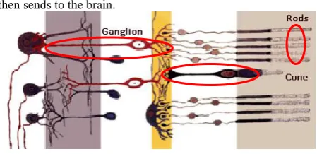
In human retina there are three types of cell [3] which is sensitive to the light are described below:
Rod Cell Cone Cell Ganglion Cell
There are millions of photosensitive cells in the human eye among which 125 million rods and cone cells in the human eye. The rods function in the low light condition and create a grayscale image. The cones are sensitive to color detail and it gives a color vision to human being. The total of approximately 1 million ganglion cells in the human eye. The signal received from the rods and cons are then transferred to the ganglion cell which interpret the received message and
[image:2.595.317.542.74.183.2]then sends to the brain.
Figure 2: Retinal Cells
2.1
Diseases of Eye
Vision impairment can be of various types like vision can be lost due to some accident but another type of vision impairment can be due to degeneration of malfunction of a part of the human eye. In this type of malfunctioning there is degradation of the optic nerve, degradation of brain portion, or any visual part like photosensitive cells etc.. And there are several diseases due to which the vision can be lost and which can lead to blindness. The two diseases which are notable are shown below [3-8]:
1) Retintis Pigmentosa (RP)
2) Age Related Macular Degradation (AMD)
Retintis Pigmentosa (RP): It is an inherited disease of the eye causes severe vision impairment and offer blindness. The symptoms of this disease are as follows:
Night Blindness, Tunnel Vision, peripheral vision, Aversion to glare, Slow adjustment from dark to light environment, Blurring of vision, Poor Color Separation etc.
Age Related Macular Degradation (AMD): AMD is a medical condition which usually affects older adults and results in the loss of vision in the center of visual field i.e. macula because the damage to the retina. It is of two types i.e. (Dry AMD and Wet AMD).
3.
VISION RESTORATION
The common and old vision restoration procedure is cornial transplant in which there is a surgical removal of the opaque and damaged cornea and replaced with the new one which is donated by someone. The tissue of cornea doesn't have blood vessels and it is purely vascular. Sometimes the surgical transplantation of the cornea doesn't fit compatible into the body of the defected person because the immune system doesn't accept that eye because it doesn't have blood vessel to carry the antibodies to the transplanted tissue and it takes very long time to get the success in the transplantation of the damaged cornea with the new one. The artificial vision is always fascinated the biomedical engineers. It has lots of technical issues as well as medical compatibilities apart from the biological considerations.
disease like RP and AMD in this the cells behind they are active only the retina has been damaged, so in this case the ophthalmologist thought that instead of sending signals to the brain if connect the signal directly to the ganglion behind the retina then it will be easy. Then in 1990s scientist developed the retinal implant. Now this process of development goes on and in the year 2000 the optobionics has been developed named artificial eye the involves the light sensitive chip under the retina.
4.
VARIOUS SYSTEM AND
TECHNICAL CONSIDERATIONS
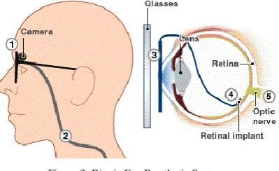
[image:3.595.316.542.102.226.2]Visual prosthetics have been developed as a potentially very valuable device for blind and it gives a hope to live life once more [6-7]. There are several different devices like Artificial Silicon Retina (ASR), Argus retinal Implant [9] etc. The bionic eye system is shown below:
Figure 3: Bionic Eye Prosthetic System
Artificial Silicon Retina (ASR) [2-4-9]: Alan Chow and Vincent Chow both are brothers; they have developed a solid state device which has a microchip that contains an array of 3500 photoreceptor diodes. Photoreceptors are the device similar to the retina that converts the light is a signal to the electrical signal and which mimic the functionality of the retina. The artificial silicon retinal chip is 2mm in diameter and 1/1000 inch in the thickness. Each diode has its own stimulating electrode.
Figure 4: (a) Artificial Silicon Retina
(b) ASR Device Implanted in Human Eye
The micro sized photodiodes that are designed in such a way that it converts the light energy from the images into the thousands of an electrical impulse to simulate the retinal cells of the retina in the defected person. This ASR doesn’t have externally powered it works solely by the incident light. ASR is surgically implanted under the retina i.e. sub retinal space
The visually impaired person can see the object or light by stimulating the ganglion cells behind the retina. The ganglion is the cells which are the connecting mechanism between the retinal cells and the optic nerve. So in the ASR scientist set out to create a device that could translate the image signals and restore the vision artificially. The size of ASR can be
[image:3.595.69.269.266.388.2]shown by the comparison of Black Square with the penny as shown below:
Figure 5: Visual Dimension of ASR compared with Penny
This ASR can be implanted into the sub retinal space with the precision and without damaging the other parts of the eye this implantation can be done by the ophthalmic surgeon.
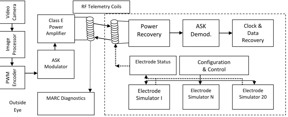
[image:3.595.316.541.464.565.2]MARC System [3]: MARC System stands for the multiple unit ASR. It consists of an external camera that captures the external environmental object and sends the image to the secondary receiving coil in the form of an electrical signal. The receiving coil is attached close to the proximity to the cornea. The other marc component like power and signal transceiver, a processing chip and a simulation current driver and a proposed electrode array fabricated in material such as silicon rubber, thin silicon or polyamide with ribbon cables containing the devices. The bio-compatibility of polyamide has been tested and a thin light weight consistency suggested its possible use as a non intrusive material for an electrode. Titanium tracks or cyanoacrylate glue may be used to hold the electrode array in place. The diagram below shows the image formation with the MARC system and a method to extend the MARC system with flexible electrode and polyamide thin flexible extension.
Figure 6: Image Formation with the MARC System
Figure 7: Method of Extendin
g MARC System with Flexible Electrode Array
[image:3.595.55.278.499.605.2] [image:3.595.318.537.605.705.2]higher frequency carrier signal. The signal will be rectified and filtered and MARC will be capable of extracting power, data and a clock signal. The subsequently derived signal will be simulated upon the retina of the impaired person. The MARC system consists of the two parts which separately resides into the interior and exterior parts of the eyeball. The
[image:4.595.58.544.161.359.2]primary coil can be driven by a 0.5 – 10 MHz carrier signal accompanied by the 10 KHz amplitude modulated (AM/ASK) signal which provides data for setting the configuration of the stimulating electrodes. The diagram below shows the functional block diagram of the bionic eye system.
Figure 8: Circuit of MARC System
The DC signal obtained by the rectification of the incoming RF signal. The receiver on the secondary side extracts four bits of data for each pixel from the incoming RF signal and provides filtering, demodulation, and amplification. The extracted data are interpreted by the electrode signal driver which finally generates appropriate current for the stimulating electrode in terms of magnitude pulse width and frequency. The optic nerve must be at least partly functional to provide sight.
The working of MARC system: The visual sensation to the brain is given by an artificial eye by simulating the optical nerve. Optical nerves are activated by the electrical impulse. In the case the patient has a small device implanted into the body that can receive the radio signal and transmit the radio signal to the nerves.
The Argus II Retinal Implant [5-10]: The Argus II retinal implant consists of an array of an electrode that are attached to the retina and used in conjunction with an external camera and a video processing system to provide rough sight to implant person. Argus II retinal prosthesis system can provide sight to the person who is suffering from the disease like RP and AMD. Argus II replaces the retina and connects the signal to the optic nerve via ganglion cell.
The second incarnation of second sight retinal prosthesis consists of five main parts:
Digital camera attached to the eyeglasses which capture the real time images and send it to the microchip.
Video processing microchip which is a handheld processing system that processes images into electrical pulses representing the patterns of light and dark. Send pulses to the radio transmitter in glasses.
Radio transmitter that transmits the pulse to the receiver implant under the eye.
Radio receiver receives the signal via thin hair implant wire. Retinal implant is an array of 60 electrodes on a chip
measuring 1mm x 1mm.
The whole system runs by the external battery power placed in the video processing unit. Whenever the picture has been taken then it forms patterns of light and dark pixels. This pattern of images is sent to the video processing unit which converts the series of pattern into the series of electrical impulses that represents the light and dark pixels. The video processor sends the pattern to the radio transmitter on the glasses, which then transmit the pulse into radio form to the receiver. The receiver is directly connected via wire to the electrode array implanted at the back of the eye and it sends the pulses to the wire. The electrical pulse executes the electrode array and the electrode array behaves like the retina. The electrical signal then transferred to the neural signal and goes to the brain visual center. All this need first training the device. At first they see mostly the light and dark spots and after a while they learn what the brain actually wants to show them. This bionic eye helps the blind to see the object and recognize them. The Argus II retinal implant was CE marked in February 2011 and the company has launched two UK centers in late 2011-early 2012. The cost of the device is estimated £66.000 excluding VAT. Additional cost estimated by the company includes £6,200 for surgery and £3,600 for clinical follow up. And Argus II has got FDA approval for commercialization [11].
Comparative analysis of ASR and MARC system:
ASR and MARC microchip is an electronic device so it requires the power supply for their proper functioning. As in the early visual prosthetic system there is a need for implanting the battery inside of the body or place the battery near the ear and connect the power supply by the micro wires. But the ASR system eliminates the need of external power
V
id
eo
Camer
a
Ima
ge
Pro
ce
ss
o
r
PW
M
En
co
d
er
RF Telemetry Coils
Class E Power Amplifier
ASK Modulator
MARC Diagnostics Outside
Eye
Power
Recovery
ASK
Demod.
Clock & Data Recovery
Electrode Simulator I
Electrode Simulator N
Electrode Simulator 20 Electrode Status Configuration
supply. It is designed in such a way that the power is taken from the incident light.
Advantage of ASR:
It replaces the function of damaged photoreceptor cells in the eye.
Problem associated with the ASR prosthetics:
In the dim light condition the current delivery is low, so insufficient to electrically activate the neurons in the retina with damaged photoreceptors.It has been observed that after experimentation [13] and testing that the sub retinal prosthetic approach is not practical without an additional source of energy to power the implant.
Advantage of MARC system:
There are many advantages of the MARC system as compared to ASR and earlier prosthetics. The problem of cell activation in the low light condition is eliminated in this system as the power is fed to the MARC system by the RF signal. There is a circuit in MARC chip for the power recovery which extracts the power from the received RF signal and used for the functioning of the chip. MARC system is compact better heat dissipation etc.
5.
CONCLUSIONS
The artificial eye is being developed to replace the defective or degraded vision and restore the vision. Argus II retinal implant has been developed successfully and been tested and get CE certification and approval for commercialization. The system gives life to millions of people who are hopeless due to the loss of vision. The device has been developed yet has helped people to recognize faces, read books, distinguish object etc. The visual prosthetics have changed the world of people who have lost their vision. Researchers and scientist have already been trying to make a device which has 1,000 electrodes as the Argus II has only 16 electrodes. Which allow the people to recognize the faces and see the colored object. The device has some limitation that it doesn’t restore the vision perfectly as there is no any replacement to the natural vision. And it will not helpful in restoring the loss of vision from birth.
6.
ACKNOWLEDGMENTS
The author would like to thank Prof. Rajiv Saxena for their guidance. The author would like to thank to their family members and close friends for many fruitful Discussions.
7.
REFERENCES
[1] Michael O. Hughes, “Anatomy of Anterior Eye for Ocularists”, Journal of Opthalmic Prosthesis
[2] Kareem A. Zaghloul, Kwabena Boahen, “A Silicon Retina that Reproduces Signal in The Optic Nerve”, Journal of Neural Engineering 3 (2006) pp 257-267
[3] Praveen Kumar Narayanan and Guhan Senthil, “Bionic Eye Powered By Nanogenerator Design of Electronic Eye for Visually Impaired”, 2011 International Conference on Life Science and Technology IPCBEE Vol3 (2011)
[4] Alice C. Parker and Adi N. Azar, “A Hierarchical Artificial Retina Architecture”, Viterbi School of Engineering, University of Southern California Los Angles CA 90089-2562
[5] News Brief, “Argus IITM Retinal Prosthesis System for Peripheral Retinal Degradation” National HorizonTM Scanning Center, June 2011
[6] Fact Sheet “Bionic Eye” Bionics Institute 384-388Albert Street East Melbourne Vic 3002 03 9667 7500
[7] “Bionic Eye Fact Sheet”, Center for Eye Research Australia 17 Dec 2009
[8] Figure Anatomy of human eye available at : http://seedoctoradams.com/the_amazing_human_eye
[9] ASR Device Available at:
http://optobionics.com/asrdevice.shtml.
[10]“Bionic Eye Comes To Market” Available at: http://www.technologyreview.com/news/423216/a-bionic-eye-comes-to-market/
[11]FDA Approves First Retinal Implant for Rare Use: Available at:
http://www.reuters.com/article/2013/02/14/us-
secondsight-fda-eyeimplant-idUSBRE91D1AK20130214.
[12] Advances in Biotechnology, Available at: http://biotech17.blogspot.in/2007/12/chronological-recap.html