Supraclavicular brachial plexus block with and without
Dexamethasone – A Comparative Study
Dr R. G. Pathak
1, Dr Anand P. Satkar
2, Dr Rajendra N. Khade
31
Professor and head, department of anaesthesiology, Dr. S. C. G. M. C. Nanded. 2
Senior resident, department of anaesthesiology, Dr S. C. G. M. C. Nanded 3
Senior resident, department of anaesthesiology, Dr S. C. G. M. C. Nanded
Abstract
-INTRODUCTION: Brachial plexus block was performed using supraclavicular approach and a mixture ofadrenalized lignocaine and bupivacaine either aloneor combined with dexamethasone was administered. Dexamethasone is selected as an adjuvant to localanaesthetics in brachial plexus block because it has been reported to prolong duration of action of local anaesthetics.
AIMSANDOBJECTIVES: To compare effect of addition of dexamethasone to local anaesthetic drugs on-
onset of analgesia. onset of motor blockade. duration of analgesia. duration of motor blockade. complications / side effects if any.
MATERIALSANDMETHODS:
50 patients with ASA grade 1 and 2, in age group 18 to 70 years, are grouped into two.
Group 1 (cases) – received 1.5% adrenalized xylocaine (20ml) and 0.5% bupivacaine (16ml) + dexamethasone 8mg (2ml) Group 2 (control) - received 1.5% adrenalized xylocaine (20ml) and 0.5% bupivacaine (16ml) + 0.9% normal saline (2ml)
RESULTS:
mean onset of sensory block in grp 1 in mins 5.92 +/- 2.8 mean onset of sensory block in grp 2 in mins 6.6 +/- 2.95 mean onset of motor block in grp 1 in mins 15.8 +/- 5.6 mean onset of motor block in grp 2 in mins 16.6 +/- 5.11 mean duration of analgesia in grp 1 in mins 834 +/- 78.1 mean duration of analgesia in grp 2 in mins 276 +/- 38.73 mean duration of motor block in grp 1 in mins 376.4 +/- 39.99 mean duration of motor block in grp 2 in mins 175.2 +/-26.94
DISCUSSION:
Mean age in grp 1 and grp 2 was 28.04 +/- 12.24 years and 27.60 +/- 9.73years respctively.
Mean wt in grp 1 and grp 2 was 57.39 +/- 9.725kg and 61.52 +/- 4.76kg respectively.
Mean ht in grp 1 and grp 2 was 169.3 +/- 4.66 cm and 168.6 +/- 4.620cm respectively.
Duration of surgery in grp 1 and grp 2 was 167.4 +/- 37.14min and 154.2 +/- 28.89min respectively.
Mean onset of sensory block in grp 1 and grp 2 was 6.02 +/- 2.8 min and 6.7 +/- 2.9 in mins.
Mean onset of motor block in grp 1 and grp 2 in mins 16.0 +/- 5.6 and 16.6 +/- 5.2 mins.
CONCLUSION:
Addition of dexamethasone to local anaesthetic drugs in brachial plexus block significantly prolongs the duration of analgesia and motor block in patients undergoing upper limb surgeries and is remarkably safe and cost effective method of providing post operative analgesia.
INDEXTERMS:
Supraclavicular brachial plexus block, Dexamethasone
I. INTRODUCTION
rachial plexus block is a popular and widely employed regional nerve block of the upper extremity. Various approaches to brachial plexus block have been described but supraclavicular approach is the easiest and most consistent method for anaesthesia and perioperative pain management in surgery below the shoulder joint. Supraclavicular brachial plexus block is an excellent technique in experienced hands. Pneumothorax (1-6%)1,2,3, Hemothorax, Horner’s syndrome and phrenic nerve block are the potential complications.
Nowadays different drugs have been used as adjuvant with local anesthetics in brachial plexus block to achieve quick, dense and prolonged block. Drugs like morphine, pethidine, clonidine, dexmedetomidine, butorphanol, buprenorphine are commonly used along with local anesthetics for this purpose. Since morphine, buprenorphine, pethidine are associated with side effects like heavy sedation, respiratory depression and psychomimmetic effects, drugs with minimal of these side effects are always looked for.
Recently, dexamethasone has been studied as an adjuvant to local anesthetic in peripheral nerve block4,5. Steroids have nerve block prolonging effects. They produce analgesia by blocking transmission of nociceptive myelinated c-fibers and suppressing ectopic neuronal discharge. They might bring about this effect by altering the function of potassium channels in the excitable cells. Thus, dexamethasone was selected as an adjuvant
to local anesthetics in brachial plexus block in our study because it has been reported to prolong duration of action of local anesthetics and respiratory depression is not a major problem. In a randomized, prospective, double blinded and controlled study, we included 50 patients of ASA grade 1-2 between age group 18-70 years undergoing upper limb surgery below shoulder joint. The brachial plexus block was performed using supraclavicular approach and a mixture of adrenalised lignocaine and bupivacaine either alone or combined with Dexamethasone was administered. The block was performed using a nerve locator in all cases. The onset and duration of analgesia and motor block and any complications were evaluated.
II. AIMSANDOBJECTIVES
To compare the effect of addition of dexamethasone and clonidine to local anesthetic drug in supraclavicular brachial plexus block on
Onset of analgesia Onset of motor blockade Duration of analgesia Duration of motor blockade Complications/ side effects if any
III. METHODS AND MATERIALS
After obtaining approval from hospital academic and ethics committee and written, informed valid consent, 50 patients were enrolled in the study. The study population included patients of either sex, ASA grade 1 and 2 in the age range of 18-70 years. All patients were posted for upper extremity surgeries below the shoulder joint and received brachial plexus block by supraclavicular approach.
Study design:
The study was a randomised, prospective, double blinded and controlled study.
Inclusion criteria:
- Age group 18-70 years
- ASA grade 1 and 2
- Upper limb surgery below shoulder joint (both elective and emergency surgery)
Exclusion criteria:
- Consent not given
- ASA Grade 3 and 4
- Any bleeding disorder and patient on anticoagulants - Severe respiratory disease
- Neuro deficit involving brachial plexus - Local infection at the injection site - History of allergy to local anaesthetic
- Patients with a history of peptic ulcer disease, diabetes mellitus, hepatic or renal failure (contraindication to steroids)
- pregnant women
Patients were randomly allocated to one of the two groups using a standard randomisation code.
Group Ι (cases): Patients in this group received 1.5% adrenalized xylocaine (20ml) and 0.5% bupivacaine(16ml) plus dexamethasone 8mg(2ml) making a total volume of 38ml. Group Џ (control): Patients in this group received 1.5% adrenalized xylocaine (20ml) and 0.5% bupivacaine(16ml) plus 0.9% normal saline (2ml) making a total volume of 38ml. In each patient, thorough history was elicited. Patient was clinically examined in details and investigated.
Investigations: Complete hemogram X-ray chest
Blood urea nitrogen, Blood Sugar Electrocardiogram, if >40 years
Drug solution used and dosage:
- Xylocaine 5% ampoule was used. 6ml of it was diluted to 20ml with 0.9% normal saline to make it 1.5%. Xylocaine was used in a dose not exceeding 7mg/kg. - Adrenaline 100µg i.e. 0.1ml of 1:1000 Adrenaline to
make a dilution of 1:200000 or 5µg/ml was taken with tuberculin syringe and added to xylocaine solution. - Bupivacaine 0.5% ampoule was used. 16ml of it was
taken in 20 ml syringe. It was used in the dose not exceeding 2mg/kg.
- Dexamethasone ampoule (4mg/ml; 2ml) was used. 2ml was added to xylocaine in group Ι patients.
Total volume of solution in both groups was 38ml.Drug solutions was prepared by an independent anaesthesiologist not involved in the study.
Monitoring:
Standard monitors were attached-
- Pulse oximetry for saturation(SpO2) - Cardio scope for rate and rhythm
- Manual blood pressure monitoring using
sphygmomanometer
An intravenous drip was started before undertaking the procedure which continued throughout the length of surgery. Vital parameters were observed throughout the procedure and oxygen at the rate of 2l/min administered through oxygen mask.
Instruments:
A set containing following was used: 1. Insulated Stimulator needle: 2. Peripheral nerve stimulator 3. ECG electrode.
4. Two 20 ml leurlock syringes. 5. Skin marker pencil
6. One tuberculin syringe of 1 ml.
7. Two stainless sterile bowls one each for iodine and spirit. 8. Sterile gauze pieces, one sterile swab holding forceps and one sterile drape.
Technique of supraclavicular brachial plexus block:
The brachial plexus block was carried out after thorough explanation of the procedure and emphasising the need for patient cooperation.
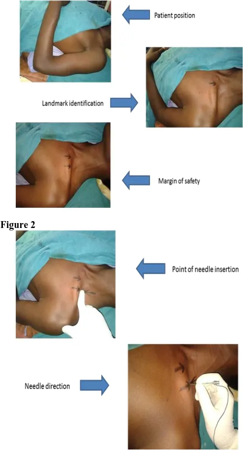
1) We used the classical approach to supraclavicular block using a single-injection, nerve-stimulator technique. The patient was asked to be in the dorsal recumbent position without a pillow, arms at his/her sides and head turned to side opposite to the one being blocked. Small pad was placed below bilateral shoulder. The patient was asked to lower the shoulder and flex the elbow, so that the forearm rests on his/her lap. The wrist was supinated so the palm of the hand faced the patient’s face. This manoeuvre allowed for detection of any subtle finger movement produced by nerve stimulation
2) Part of neck was aseptically cleaned and draped. 3) The operator stood on the side to be blocked so for a left side block the palpation was done with the left hand and the needle was manipulated with the right. For a right side block exactly the opposite was done so that operator manipulated the needle with the left hand and palpated with the right.
4) With the patient in the above described position and the shoulder down, the lateral (posterior) border of the sternocleidomastoid(SCM) muscle was identified and followed distally to the point where it met the clavicle. The point of needle entrance was about 1 in (2.5 cm) lateral to the insertion of the SCM in the clavicle or one “thumb breadth” lateral to the SCM. Palpation of the subclavian artery at this site confirms the landmark. The palpating index finger was placed at this site. 5) Local infiltration of 1ml of 2% lignocaine was given at the proposed puncture site.
6) We used an insulated needle to perform this technique. The needle was connected to nerve locator by the electrodes and was properly grounded with the help of ECG lead. We started the stimulation with an intensity of 2.0 mA and a pulse width of 100 µs. Once the desired response was obtained – that is a muscle twitch of the fingers that is clearly visible - we started to decrease the current gradually upto 0.6mA. If still, we got the desired response the drug solution was injected. If the response was obtained at 0.4mA also, then the needle was repositioned again so as to get response at 0.6mA but not at 0.4mA.
7) If we did not get adequate response or if reposition of the needle was necessary, the needle was withdrawn and the penetration angle was adjusted in the anteroposterior plane, either slightly more posterior or slightly more anterior, but always parallel to the midline. As a goal we aimed to elicit an isolated muscle twitch in all fingers either in flexion or extension.
8) Following injection, area was massaged so as to help the solution to track along the plexus.
9) The site of injection was sealed with a tincture benzoin seal.
[image:3.612.313.560.103.564.2]10) During the conduct of block and thereafter, the patient was observed vigilantly for any complications of the block and for the toxicity of the drugs injected.
Figure 1
Supraclvicular brachial plexus block
Figure 2
Prevention of deleterious effects:
Following precautions were taken during conduct of the block-
1) Repeated aspiration before injection to prevent intravascular spread.
2) Administration of a test dose of 1-2 ml of adrenalized lignocaine solution to observe for cardiovascular or central nervous toxicity.
The following parameters were studied: 1) Onset of sensory block:
i.e. the time from injection to onset of analgesia in each of the major peripheralnerve distributions( ulnar, radial, medial and musculocutaneous). Sensory block was assessed by pinprick using the blunt end of a 27-gauge needle at 0, 2, 5,10, 15, 20, and 30 min. Sensoryblock was graded according to the following scale: 0 = no block (normal sensation), 1 = partial block (decreased sensation),and 2 = complete block (no sensation). 2) Onset of motor block:
i.e. the time from injection to the inability of the patient to move his/her fingers or raise their hand. Motor block was measured at 0, 10, 20, and30 min by assessing the following motor functions: flexion atthe elbow (musculocutaneous nerve), extension of the elbow andthe wrist (radial nerve), opposition of the thumb and indexfinger (median nerve), and opposition of the thumb and smallfinger (ulnar nerve). Motor block was graded according to the following scale: 0 = no block (full muscle activity), 1 = partialblock (decreased muscle activity), and 2 = complete block (nomuscle activity).
3) Duration of analgesia:
During the procedure, anaesthesia was considered satisfactory if the patient did not complain of any pain or discomfort and if no sedation was necessary.
Post operative follow up was carried out in the recovery and post operative ward. The duration of analgesia was noted according to 0-10 visual analogue score (VAS) for pain at every half an hour for first 10 hours and then hourly till 24 hours. When the patients began to experience the worst pain (VAS =8-10), it was considered that analgesic action of the drugs was terminated and rescue analgesic (IM Diclofenac 1-1.5mg/kg) given.
4) Duration of motor block:
The duration of motor block postoperatively was assessed every hourly by asking the patients to move their fingers and to see whether they are able raise the hand or not. This time was recorded and taken as cessation of motor block effect.
5) Possible side effects of brachial plexus block:
Incidence of drowsiness, pruritus, nausea/vomiting, Horner’s syndrome, phrenic nerve palsy, pneumothorax, respiratory depression and sign and symptoms for local anaesthetic toxicity were looked for and noted, if any.
The above assessments were carried out by the principal investigator who was blinded to the drugs administered in the plexus block.
Management of unsuccessful block:
In the circumstance of inadequate or patchy action of the block, the block would be supplemented with general anaesthesia.
If in case surgery was unduly prolonged and the effect of the block wore off, rescue analgesia with IV propofol or IV ketamine would be given.
Statistical analysis :
The data obtained in this study was analysed using unpaired ‘t’ test which gives p value to be applied as follows :
- If p> 0.05, it means that there is no significant difference between the means of two groups studied.
- If p=0.05, it indicates that there is a significant difference at 5% level of significance. ( i.e. out of 100, in 95 cases there is a significant difference).
- If p< 0.01, it indicates that the data is significant at 1% level of significance ( i.e. out of 100, in 99 cases there is a significant difference). If p<0.001, it is highly significant
IV. RESULTS
TABLE 1
TABLE 2.
Groups Group1 Group2 P value
Onset of sensory block(min)
5.92 6.6 0.4101(>0.05)
Onset of motor block(min)
15.8 16.6 0.6005(>0.05)
Duration of
analgesia(min)
834 276 <0.0001
Duration of
motor block(min)
376 175 <0.0001
mean onset of sensory block in grp 1 in mins 5.92 +/- 2.8 mean onset of sensory block in grp 2 in mins 6.6 +/- 2.95 mean onset of motor block in grp 1 in mins 15.8 +/- 5.6 mean onset of motor block in grp 2 in mins 16.6 +/- 5.11 mean duration of analgesia in grp 1 in mins 834 +/- 78.1 mean duration of analgesia in grp 2 in mins 276 +/- 38.73 mean duration of motor block in grp 1 in mins 376.4 +/- 39.99 mean duration of motor block in grp 2 in mins 175.2 +/-26.94
V. DISCUSSION
Although general anesthesia continues to be used for most of the surgical procedures, regional anesthesia has been increasing in popularity in recent years. This is mainly because of the fact that the regional anesthesia techniques can be utilized for analgesia not only during the operative period, but during the postoperative period as well and avoids complications of general anaesthesia.
The brachial plexus block consists of injecting local analgesic drugs in the fascial spaces surrounding the nerve plexus, thereby blocking the autonomic, sensory and motor fibres supplying the upper extremity. It is a simple, safe and effective
Groups Group1 Group2 P value
Mean age(years)
28.04 12.24 >0.05
Duration of surgery(min)
167.4 154.2 > 0.05
Weight(kg) 57.39 61.52 >0.05
Height(cm) 169 168 >0.05
Sex distribution
Male Female Male Female >0.05
technique of anesthesia having distinct advantages over general and intravenous regional anesthesia. A regional technique should always be considered whenever general condition of the patient is poor, or the patient is not adequately prepared or in the presence of associated conditions like uncontrolled diabetes, cardiovascular or respiratory diseases. It is also useful when the patient prefers to retain his consciousness during surgery and when it is important for the patient to remain ambulatory.
Advantages of brachial plexus block compared with general anaesthesia are –
1) Early discharge for outpatients 2) Smooth transition to pain control 3) Increased blood flow to extremity 4) Less nausea and vomiting 5) Less drowsiness
6) Tracheal intubation not required
We had selected supraclavicular approach to brachial plexus block. Supraclavicular brachial plexus block is widely employed regional nerve block to provide anaesthesia and analgesia for the upper extremity surgery. Supraclavicular block provides a rapid, dense and predictable anesthesia of the entire upper extremity in the most consistent manner of any brachial plexus technique. It is the most effective block for all the portions of the upper extremity and is carried out at the “division” level of the brachial plexus; with high volume the “trunk” level of the plexus may also be blocked in this approach1,2,3. Perhaps that is why there is often little or no sparing of peripheral nerve if an “adequate” paresthesia or stimulation is obtained.
The alleviation of the suffering is of course a primary concern of the anaesthesiologist. Any method of postoperative pain relief must meet three basic criteria. It should be effective, safe, and feasible. Currently available local anesthetics can provide analgesia for limited period of time when used as single injection. To extend the analgesia period beyond the operating rooms, various methods have been tried with the aim of prolonging the local anaesthetic action, like continuous infusion of local anesthetics via indwelling catheters, use of different additives in local anaesthetics.
In our study, dexamethasone was used as adjuvant in local anesthetic. Our study was a randomised, prospective, double blinded and controlled study. Fifty patients posted for upper limb surgeries below shoulder joint were given brachial plexus block by supraclavicular approach. The patients were randomly allocated into two groups using standard randomisation code. The group Ι (cases or study group) received 1.5% adrenalized
xylocaine (20ml) and 0.5% bupivacaine (16ml) plus
dexamethasone 8mg(2ml) making a total volume of 38ml. The group II (control group) received 1.5% adrenalized xylocaine (20ml) and 0.5% bupivacaine (16ml) plus 0.9% normal saline (2ml) making a total volume of 38ml.
Five patients (three patients in control group and two patients in study group) failed to achieve satisfactory levels of anaesthesia and required induction of general anaesthesia. They were excluded from the study.
The assessment of onset and duration of block was carried out by the principal investigator who was blinded to the drugs administered in the block.
In our study, the mean age of patients in group Ι was 28.04 ± 12.24 years. Mean age of patients in group Џ was 27.60 ± 9.734 years. Using unpaired‘t’ test; there was no significant difference in the two groups statistically (p > 0.05). (Table 1) Mean weight of the patients in group Ι and group Џ were 57.39 ± 9.725 and 61.52 ± 4.76 kgs respectively. Mean height of the patients n group Ι and group Џ were 169.3 ± 4.668 and 168.6 ± 4.620. This was not statistically significant (p > 0.05). (Table 1)
There were more male patients than female in both the groups. There was no significant difference regarding the sex distribution between two groups. (Table 1)
In group Ι, mean duration of surgery was 167.4 ± 37.14 minutes. While in group Џ it was 154.2 ± 28.89 minutes. Thus the duration of surgery was similar in both the groups (p > 0.05). (Table 1)
So there was random allocation of the patients in the two groups and both the groups were statistically alike. Thus bias in patient selection was excluded.
The mean onset of sensory block in minutes was 5.92 ± 2.827 and 6.6 ± 2.958 in group Џ (p = 0.4101). (Table 2)
The mean onset of motor block in minutes in group Ι and group Џ was 15.8 ± 5.605 and 16.6 ± 5.115 respectively (p = 0.6005). (Table 2)
Both these data were not significant statistically as p > 0.05. So our study showed that there was no significant difference in the onset time of sensory and motor block between two groups. In one study by Shrestha BR, Maharjan SK, Tabedar S4 onset of action was 10-30 minutes in local anesthetic group (mean 18.15 ± 4.25) and 10-20 minutes (mean 14.5 ± 2.10) in the local anesthetic plus steroid group. They found statistically significant difference between two groups. However another study by Ali Movafegh, Mehran
Razazian, Fatemeh Hajimaohamadi et al5 found that the onset
time of sensory and motor blockade was similar in both the groups. Our study also showed that dexamethasone does not produce significant difference in the onset time of sensory and motor block.
In our study, the mean duration of analgesia in group Ι was 834 ± 78.1 minutes whereas in group Џ it was 276 ± 38.73 minutes (p < 0.0001).(Table 2) And the mean duration of motor block in group Ι and group Џ were 376.4 ± 39.99 and 175.2 ± 26.94 minutes respectively (p < 0.0001).(Table 2) Both these data’s were highly significant statistically which showed that both the duration of analgesia and motor block were significantly prolonged compared to the control group.
Several studies have shown that addition 4-8 mg of dexamethasone to local anesthetics effectively and significantly prolongs the duration of analgesia.
In a study by Castillo J, Curley J, Hotz J et al6 a prolonged percutaneous block of sciatic nerve in rat using bupivacaine – dexamethasone microspheres was demonstrated. Kathrine Holte, Mads U. Werner, Peter G. Lacouture et al7 found that addition of small amounts of dexamethasone to bupivacaine incorporated in microcapsules prolonged local analgesia compared with microcapsules with plain bupivacaine after subcutaneous administration in humans.
bupivacaine microspheres significantly prolonged intercostal nerve block in sheep. These authors believe that there is a causative relationship between the suppression of inflammation and the remarkably longer duration of effect.
Kopacz DJ, Lacouture PG, Wu D et al9 also reported that the intercostal injection of dexamethasone- containing bupivacaine microspheres produces a prolonged duration of anaesthesia and analgesia.
Estebe IP, Le corry P, Clement R, Duplessis L et al10 studied the effect of dexamethasone on motor brachial plexus
block with bupivacaine and with bupivacaine-loaded
microspheres in a sheep model and found that the incorporation
of dexamethasone in bupivacaine-loaded microspheres
dramatically increases the duration of action.
Other preliminary data suggest that methylprednisolone can increase the duration of sensory and motor block. In a study by
Stan T, Goodman E, Cardida B, Curtis RH11 the patients were
divided into 2 groups to receive solutions containing 20 mL mepivacaine, 20 mL bupivacaine, 0.2 mL epinephrine, and, in one group, 40 mg methylprednisolone was added to this solution. The authors found that the duration of sensory analgesia (23 hours versus 16 hours; P < 0.01) and motor block (19 hours versus 13 hours; p < 0.001) were significantly longer in the steroid group. The authors believed that the applicability of these findings to clinical practice should be verified in a randomized prospective clinical trial.
One such randomised prospective trial was done by
Shrestha BR, Maharjan SK, Tabedar S4. In their study forty
patients undergoing arm, forearm and hand surgeries were randomly selected. The forty patients were divided in two groups of 20 each. In-group one, a brachial plexus block was done with 40-50 ml of local anesthetic with 1:200,000 adrenaline and in the other group the block was performed with the same amount of local anesthetic with dexamethasone. The authors found that there was significant faster onset of action (14.5 ± 2.10 minutes verses 18.15 ± 4.25 minutes; p<0.05) and prolonged duration of analgesia (12.75 ± 5.33 hours verses 3.16 ± 0.48 hours; p = 0.00) in the dexamethasone group than in the other group.
Ali Movafegh, Mehran Razazian, Fatemeh
Hajimaohamadi, and Alipasha Meysamie5 did a prospective,
randomized, double-blind study to evaluate the effect of dexamethasone added to lidocaine on the onset and duration of axillary brachial plexus block. Sixty patients scheduled for elective hand and forearm surgery under axillary brachial plexus block were randomly allocated to receive either 34 mL lidocaine 1.5% with 2 mL of isotonic saline chloride (control group, n = 30) or 34 mL lidocaine 1.5% with 2 mL of dexamethasone (8 mg) (dexamethasone group, n = 30). Neither epinephrine nor bicarbonate was added to the treatment mixture. They used a nerve stimulator in all of the patients. They found that the duration of surgery and the onset times of sensory and motor block were similar in the two groups. The duration of sensory (242 ± 76 versus 98 ± 33 min) and motor (310 ± 81 versus 130 ± 31 min) blockade were significantly longer in the dexamethasone than in the control group (P < 0.01).
Although corticosteroids have been used successfully for postoperative pain relief in oral, general, and orthopaedic surgery12,13, other studies have not corroborated these reports14,15.
The mechanism of the analgesia induced by corticosteroids is not fully understood. This effect is suspected to be mediated by their anti-inflammatory or immune-suppressive effects16,17. The use of corticosteroids as an adjuvant to local anaesthetic for peripheral nerve blocks rarely has been described, and its mechanism of action is not clearly understood. Corticosteroids cause skin vasoconstriction on topical application. The vasoconstriction effects of topical steroids are mediated by occupancy of classical glucocorticoid receptors rather than by nonspecific pharmacological mechanisms16,17. According to the traditional theory of steroid action, steroids bind to intracellular receptors and modulate nuclear transcription. However, dexamethasone produced a relatively rapid effect which cannot be explained by the above mechanism18. Therefore, vasoconstriction, the presumed mechanism of action for epinephrine’s adjunctive effect on local anaesthetics, is probably not responsible for block prolongation by dexamethasone. Corticosteroids may have a local effect on the nerve; the dexamethasone effect may be related to this action19.
Many authors believe that the block prolonging effect of dexamethasone is due to its local action and not a systemic one8. They found that steroids produce analgesia by blocking transmission in nociceptive c-fibres and suppressing ectopic neuronal discharge. Local application of methylprednisolone has been found to block transmission in c-fibres but not in a and B fibres20. The effect was reversible, suggesting a direct membrane action of steroids. Steroids might bring about this effect by altering the function of potassium channels in the excitable cells.21,22,23,24
There are others who believe that analgesic properties of corticosteroids are the result of their systemic effect13,14. The safety of dexamethasone use in a nerve sheath may raise some concerns. In animal experiments, repeated intrathecal injections of small-dose betamethasone25 and triamecilonon acetate26 did not induce spinal neurotoxicity. In one study, after approximately 2000 intrathecal injections of dexamethasone (8 mg) in 200 patients for treatment of posttraumatic visual disturbance, no neurological disorders were found at 1-month follow up27. Nerve injury is a rare complication of dexamethasone injection, and it usually occurs in the context of needle trauma28,29.
Indeed in our study, one side effect which was observed was one case of Horner’s syndrome. It is a known complication of supraclavicular block and it subsided as the effect of block wore off.
The dose of dexamethasone as an adjuvant to local anaesthetics for peripheral nerve block has not been described; we used a dose of 8 mg because administration of this dose seems to be safe in adults. Adverse effects with a single dose of dexamethasone are probably extremely rare and minor in nature, and previous studies have demonstrated that short-term (< 24 hours) use of dexamethasone was safe15,30.
Offering the pain free period to the patient during postoperative time is essential on humanitarian grounds. Besides it eliminates the stress response to surgery and helps in smoother transition of the patient from surgery to the routine preoperative state.
using supraclavicular approach, produced prolonged motor blockade and effective postoperative analgesia which lasted longer than that produced by local anaesthetics alone without any significant side effects. Also it is a very cost effective way of providing analgesia as the cost of one ampoule of dexamethasone is Rs 3.32 only and the patient does not require other analgesic drug in immediate postoperative period.
VI. CONCLUSION
To conclude:
Addition of dexamethasone to local anesthetic drugs in brachial plexus block significantly prolongs the duration of analgesia and motor block in patients undergoing upper limb surgeries and is a remarkably safe and cost effective method of providing post operative analgesia.
REFERENCES
[1] Moore D: Supraclavicular approach for block of the brachial plexus, in Moore D(ed): Regional block. A handbook for use in the clinical practice of medicine and surgery, 4th ed. Springfield, Charles C Thomas Publisher, 1981; pp 221-42
[2] Lanz E, Theiss D, Jankovic D: The extent of blockade following various techniques of brachial plexus block. Anesth Analg 62:55-8, 1983
[3] Urmey W: Upper extremity blocks, in Brown D (ed): Regional anesthesia and analgesia. Philadelphia, W.B. Saunders Company, 1996; pp 254-78 [4] Shrestha BR, Maharjan SK, Tabedar S: Supraclavicular brachial plexus
block with and without dexamethasone - A comparative study. Kathmandu University Medical Journal (2003) Vol. 1, No. 3, 158-160
[5] Ali Movafegh, Mehran Razazian, Fatemeh Hajimaohamadi et al: Dexamethasone Added to Lidocaine Prolongs Axillary Brachial Plexus Blockade. Anesth Analg 2006; 102:263–7
[6] Castillo J, Curley J, Hotz J et al: Glucocorticoids prolong rat sciatic nerve blockade in vivo from bupivacaine microspheres. Anesthesiology1996; 85:1157–66.
[7] Kopacz DJ, Allen HW, Holte K, Swanton R, Lacouture P: Dexamethasone Prolongs Local Analgesia after Subcutaneous Infiltration of Bupivacaine Microcapsules in Human Volunteers. Anesthesiology 96(6):1331-1335, June 2002
[8] Droger C, Benziger D, Gao F, Berde CB: Prolonged intercostals nerve blockade in sheep using controlled-release of bupivacaine and dexamethasone from polymer microspheres. Anesthesiology1998; 89:969– 74.
[9] Kopacz DJ, Lacouture PG, Wu D, et al. The dose response and effects of dexamethasone on bupivacaine microcapsules for intercostals blockade (T9 to T11) in healthy volunteers. Anesth Analg 2003; 96:576–82.
[10] Estebe IP, Le Corry P, Clement R, Duplessis L, Chevanne F et al: Effect of dexamethasone on motor brachial plexus block with bupivacaine and with bupivacaine-loaded microspheres in a sheep model. European Journal of Anaesthesiology, Volume 20, Number 4, 1 April 2003, pp. 305-310(6)
[11] Stan T, Goodman E, Cardida B, Curtis RH. Adding methylprednisolone to local anesthetic increases the duration of axillary block. Reg Anesth Pain Med 2004; 29:380–1.
[12] Aasboe V, Raeder JC, Groegaard B: Betamethasone reduces postoperative pain and nausea after ambulatory surgery. Anesth Analg 1998; 87:913–7. [13] Baxendale BR, Vater M, Lavery KM: Dexamethasone reduces pain and
swelling following extraction of third molar teeth. Anaesthesia 1993; 48:961–4.
[14] Liu K, Hsu CC, Chia YY: Effect of dexamethasone on postoperative pain and emesis. Br J Anaesth 1998; 80:85–6.
[15] Tan P, Liu K, Peng CH, et al: The effect of dexamethasone on postoperative pain and emesis after intrathecal neostigmine. Anesth Analg 2001; 92:228– 32.
[16] 60. McCormack K: The spinal actions of nonsteroidal anti-inflammatory drugs and the dissociation between their anti-inflammatory and analgesic effects. Drugs 1994; 47:28–45.
[17] Ahlgren SC, Wang JF, Levine JD: C-fiber mechanical stimulus response functions are different in inflammatory versus neuropathic hyperalgesia in the rat. Neuroscience 1997; 76:285–90.
[18] Taguchi H, Shingu K, Okuda H, Matsumoto H: Analgesia for pelvic and perineal cancer pain by intrathecal steroid injection. Acta Anaesthesiol Scand 2002; 46:190–3.
[19] Devor MD, Gorvin-Lippmann R, Raber P: Corticosteroids suppresses ectopic neural discharge originating in experimental neuromas. Pain 1985; 22:127–37.
[20] Honorio T. Benzon, Epidural Steroids: In P. Prithvi Raj. Pain medicine, a comprehensive review. Mosby publications 1999 Page 259 – 263
[21] Attali B, Latter H, Rachamim N, Garty H: A corticosteroid-induced gene expressing an “IsKlike” K+ channel activity in Xenopus oocytes. Proc Natl Acad Sci USA 92:6092-6, 1995
[22] Takimoto K, Levitan ES: Glucocorticoid induction of Kv1.5 K+ channel gene expression in ventricle of rat heart. Circ Res 75:1006-13, 1994 [23] Pennington AJ, Kelly JS, Antoni FA: Selective enhancement of an A type
potassium current by dexamethasone in a corticotroph cell line. J Neuroendocrinol 6:305-15, 1994
[24] Attardi B, Takimoto K, Gealy R, Severns C, Levitan ES: Glucocorticoid induced upregulation of a pituitary K+ channel mRNA in vitro and in vivo. Receptors Channels 1:287-93, 1993
[25] Latham JM, Fraser RD, Moore RJ, et al: The pathologic effects of intrathecal betamethasone. Spine 1997; 22:1558–62.
[26] Abram SE, Marsala M, Yaksh TL. Analgesic and neurotoxic effects of intrathecal corticosteroids in rats. Anesthesiology 1994; 81:1198–205. [27] Sugita K, Kobayashi S, Yokoo A, et al. Intrathecal steroid therapy for
post-traumatic visual disturbance. Neurochirurgia (Stuttg) 1983; 26:112–7. [28] Frederick HA, Carter PR. Injection injuries to the median and ulnar nerves
at the wrist. J Hand Surg Am 1992; 17:645–7.
[29] Stahl S, Kaufman T. Ulnar nerve injury at the elbow after steroid injection for medial epicondylitis. J Hand Surg Br 1997;22:69–70.
[30] Splinter WM, Rhine EJ. Low-dose ondansetrone with dexamethasone more effectively decreases vomiting after strabismus surgery in children than does high-dose ondansetron. Anesthesiology 1998; 88:72–5.
AUTHORS
First Author – DR. R. G. PATHAK. Professor and head, department of anaesthesiology,
Dr. S. C. G. M. C. Nanded. Second Author – DR. ANAND P. SATKAR. Senior resident, department of anaesthesiology, Dr S. C. G. M. C. Nanded. Email id. [email protected]
Third Author – DR. RAJENDRA N. KHADE. Senior resident, department of Anaesthesiology. Dr S. C. G. M. C. Nanded. Email id. [email protected]