A Modified Extraction Method of Circulating Free DNA
for Epidermal Growth Factor Receptor Mutation Analysis
Haihua Yuan,
1Zhong-Zheng Zhu,
2Yachao Lu,
3Feng Liu,
1Wenying Zhang,
1Gang Huang,
4Guanshan Zhu,
3and Bin Jiang
11Department of Oncology, No. 3 People’s Hospital, School of Medicine, Shanghai Jiaotong University, Shanghai;
2Department of Oncology, No.113 Hospital of People’s Liberation Army, Ningbo;
3Tumor Genetics Capability, Innovation Center China, Astrazeneca Global R&D, Shanghai;
4Department of Nuclear Medicine, Renji Hospital, School of Medicine, Shanghai Jiaotong University, Shanghai, China.
Received: November 25, 2010 Revised: February 1, 2011 Accepted: February 11, 2011 Corresponding author: Dr. Bin Jiang, Department of Oncology, No. 3 People’s Hospital, School of Medicine, Shanghai Jiaotong University, No. 280 Mohe Road, Shanghai 201900, China.
Tel: 86-21-56691101-6172, Fax: 86-21-56691662 E-mail: [email protected]
∙ The authors have no financial conflicts of interest.
© Copyright:
Yonsei University College of Medicine 2012
This is an Open Access article distributed under the terms of the Creative Commons Attribution Non-Commercial License (http://creativecommons.org/ licenses/by-nc/3.0) which permits unrestricted non-commercial use, distribution, and reproduction in any medium, provided the original work is properly cited.
Purpose: Circulating free DNA (cfDNA) in plasma is promising to be a surrogate for tumor tissue DNA. However, not all epidermal growth factor receptor (EGFR) mutations in tumor tissue DNA has been detected in matched cfDNA, at least partly due to inefficient cfDNA extraction method. The purpose of this study was to
estab-lish an efficient plasma cfDNA extraction protocol. Materials and Methods: The
yield of plasma cfDNA extracted by our modified phenol-chloroform (MPC) meth-od from non-small-cell lung cancer (NSCLC) patients was compared with that by QIAamp MinElute Virus Spin kit (Qiagen kit) as control, using the Wilcoxon rank-sum test. TaqMan quantitative polymerase chain reaction (qPCR) assays were used to quantify the plasma cfDNA extracted. Both Mutant-enriched PCR (ME-PCR) coupled sequencing and DxS EGFR mutation test kit were used to evaluate the
im-pact of extraction method on EGFR mutation analysis. Results: MPC method
ex-tracted more plasma cfDNA than Qiagen kit method (p=0.011). The proportion of longer fragment (≥202 bp) in cfDNA extracted by MPC method was significantly higher than by Qiagen kit method (p=0.002). In the sequencing maps of ME-PCR products, a higher mutant peak was observed on plasma cfDNA extracted by MPC method than by Qiagen kit method. In DxS EGFR mutation test kit results, plasma cfDNA extracted by MPC method contained more tumor-origin DNA than by
Qia-gen kit method. Conclusion: An improved plasma cfDNA extraction method of
MPC is provided, which will be beneficial for EGFR mutation analysis for patients with NSCLC.
Key Words: Circulating free DNA, DNA extraction, methodology, EGFR muta-tion, non-small-cell lung cancer
INTRODUCTION
Non-small-cell lung cancer (NSCLC) accounts for about 80% of all patients with
lung cancer,1 and most of NSCLC patients are first diagnosed at advanced stage. In
Extraction of cfDNA
The stored plasma sample was thawed at room temperature and centrifuged at 15,700 g for 15 min at 4°C to remove re-sidual precipitated cellular components. Three methods were applied for the extraction of cfDNA, i.e. MPC meth-od, traditional phenol-chloroform (PC) methmeth-od, and com-mercial QIAamp MinElute Virus Spin kit (Qiagen kit).
For PC/MPC method, 1 mL of plasma was mixed with 50 µL of 25% sodium lauryl sulfate (SDS) and 30 µL of 20 mg/mL proteinase K (Qiagen, Hilden, Germany) and incu-bated at 55°C for 16 h. After digestion, equal volume of water-saturated phenol was added into the sample and mixed. Then, the mixture was centrifuged at 16,000 g at room temperature for 15 min (PC method), or transferred into a Phase Lock Gel tube (MaXtract Low Density tube, Qiagen, Hilden, Germany) followed by centrifugation at 16,000 g at room temperature for 5 min (MPC method). The supernatant was transferred into a new 2 mL eppendorf tube and mixed with equal volume of chloroform/isoamyl alcohol (24 : 1) mixture. The mixture was centrifuged at 16,000 g for 15 min (PC method), or transferred into a Phase Lock Gel tube (MaXtract High Density tube, Qiagen, Hilden, Germany) followed by centrifugation at 16,000 g at room temperature for 5 min (MPC method). Sodium ace-tate (3 mM) with 1 : 10 ratio of the supernatant and equal volume of isopropanol were added for DNA precipitation overnight at -20°C. DNA was dissolved in 50 µL of DNA hydration solution (Qiagen, Hilden, Germany) and stored at -20°C until use.
For Qiagen kit (Qiagen, Hilden, Germany) method, ex-periment was performed according to manufacturer’s pro-tocol, with the exception of increasing sample volume to 1 mL and accordingly adding the reagent amount to 5-fold. Briefly, 1 mL of plasma was mixed with 62.5 µL of prote-ase and 500 µL of buffer AL. After incubation at 56°C for 15 min, 250 µL ethanol was added. The mixture was fil-tered through the column and rinsed by AW1 and AW2 in order. DNA was dissolved in 50 µL of buffer AVE.
Quantification and purity measurement of plasma cfDNA
TaqMan quantitative polymerase chain reaction (qPCR) as-says targeting at DNA template of a single copy gene SER-PINA1, whose copy number is confirmed to be relatively stable in solid tumors and has been used as internal refer-ence gene indicating copy number of genomic DNA, were used to quantify the extracted cfDNA on ABI7900 real time
rate is only 17-22%.2 Tyrosine kinase inhibitors (TKI)
tar-geting epidermal growth factor receptor (EGFR) have been used for the treatment of advanced NSCLC, and EGFR ac-tivating mutation, which occurs in 30-50% of NSCLC pa-tients in Asian and in 10% in Caucasian, has been proved to
be the critical factor for TKI efficacy.3-5 The response rate is
around 75% for NSCLC patients with tumor EGFR activat-ing mutation versus less than 10% for those with wild-type
EGFR.6 Therefore, EGFR mutation analysis is important for
decision of TKI therapy for NSCLC patients.
Tumor tissue is the most optimal sample type for genomic DNA extraction for EGFR mutation analysis. However, tu-mor tissue samples are not always available from advanced NSCLC patients in clinical practice. Circulating free DNA (cfDNA) in plasma has thus been used as a surrogate for tu-mor tissue DNA for EGFR mutation analysis, and results
from preliminary studies seemed promising.7-9 However, not
all EGFR mutations in tumor tissue DNA has been detected
in matched cfDNA,7-9 at least partly due to inefficient cfDNA
extraction method. In the present study, we provided an im-proved plasma cfDNA extraction method, i.e. modified phe-nol-chloroform (MPC) method. Since QIAamp MinElute Vi-rus Spin kit, in comparison with several other commercially available kits, has been reported to yield the highest amount
of cfDNA,10 we compared our MPC method to Qiagen kit
method on cfDNA extraction yield and the impact of differ-ent extraction method on EGFR mutation analysis.
MATERIALS AND METHODS
Subjects and samples
A total of 25 patients with advanced lung adenocarcinoma were recruited from No.3 People’s Hospital, School of Medi-cine, Shanghai Jiaotong University, China between March and September 2009. Written informed consent was ob-tained from each subject. This study was approved by the ethics review committee of the Institutional Review Board of the hospital.
for 30 s, 56°C for 60 s, and 72°C for 60 s. A 5 µL aliquot of 1st PCR product was digested for 4 h at 37°C in a 20 µL re-action volume containing 5 units of Mse I enzyme (New England Biolabs, Beverly, MA, USA), to digest the TTAA sequence within the deletion target region of wild-type DNA. For the 2nd PCR, forward and reverse primers were 5’-ACT GTA AAA CGA CGG CCA GTA TCC CAG AAG GTG AGA AAG ATA AAA TTC-3’ and 5’-ACC AGG AAA CAG CTA TGA CCA CAC AGC AAA GCA GAA ACT CAC ATC GAG-3’, respectively. The 2nd PCR was performed with 25 µL reaction volume containing 5 µL of the digested product above and the same concentra-tions of primers and AmpliTaq Gold PCR Master Mix as the 1st PCR reaction, with the cycling condition of 95°C for 10 min followed by 40 cycles of 94°C for 30 s, 60°C for 30 s, 72°C for 40 s, ended with 72°C for 10 min. The 2nd PCR product was applied for Sanger sequencing according to the standard protocol in the manual of ABI prism 3730XL DNA analyzer (Applied Biosystems, Foster City, CA, USA) using common M13R primer (sequence: ACC AGG AAA CAG CTA TGA CC). Sequencing results were ana-lyzed using the SeqScape software v2.5 (Applied Biosys-tems, Foster City, CA, USA).
A DxS EGFR mutation test kit (DxS Ltd, Manchester, UK), which combines amplification refractory mutation system and Scorpion technologies, was also used to detect known EGFR mutations in real-time PCR as described
pre-viously.11,12 All reactions were performed in 25 uL volumes
including 5 uLof template DNA, 16 uL of reaction buffer mix, 0.6 uL of Taq polymerase and 3.4 uL water. Real-time PCR was carried out by using MX3005P real-time PCR machine (Stratagene, La Jolla, CA, USA) under the follow-ing conditions: initial denaturation at 95°C for 10 min, 45 cycles of 95°C 30 s, 61°C 60 s with fluorescence FAM reading at the end of each cycle. Data analysis was per-formed with MxPro v4.10 (Stratagene, La Jolla, CA, USA). The Cycle threshold (Ct) represents the threshold at which the signal is detected above background fluorescence. Sam-ple ΔCt values are calculated as the difference between the mutation Ct and control Ct. If the sample’s ΔCt is lower than the cut-off ΔCt value, it is judged as positive for a mu-tation detected by this assay. The cut-off ΔCt value is 12 for deletion in exon 19 of EGFR. The bigger the ΔCt is, the less mutation the sample contains.
Statistical analysis
The nonparametric comparison of median cfDNA yield be-PCR machine (Applied Biosystems, Foster City, CA,
USA). Forward primers for 77 bp, 123 bp and 202 bp PCR products were 5’-TAC TCA AGG GAA AAT TGT GGA TTT-3’, ACA CCG AAG AGG CCA AGA A-3’ and 5’-GGC CTG AAG CTA GTG GAT AAG TT-3’, respectively. Common reverse primer was 5’-AGA AGA TGT AAT TCA CCA GAG CAA A-3’. The probe sequence was 5’-FAM-TGT GTC TCT GTC AAG CTC CTT GAC-3’BHQ1. The 15 µL reaction mixture contained 7.5 µL platinum qPCR superMix-UDG (Invitrogen, Paisley, UK), 4.3 µL of pure water, 0.6 µL of 0.1 µM/L primer, 0.6 µL of 0.2 µM/L probe, and 2 µL of cfDNA. PCR was performed at 95°C for 10 min, followed by 40 cycles of 94°C for 30 s, 60°C for 60 s and 72°C for 15 s. The results were analyzed with soft-ware SDS 2.3. Each sample was tested in three replicates and their average was used in the statistical analysis. Quan-tity of cfDNA was expressed in copies/mL.
Purity of the plasma cfDNA extracted by PC, MPC and Qiagen kit method was spectrophotometrically evaluated using OD (260 nm/280 nm) value determined with the NanoDrop spectrometer ND-1000 (NanoDrop, Rockland, Delaware, USA).
EGFR mutation analysis
For all the 25 patients recruited in the present study, tumor tissue DNA has previously been examined for mutation in exon 19 of the EGFR gene by PCR-based sequencing, and only one small fragmental deletion, 2235-2249delGGAAT-TAAGAGAAGC, was identified in one patient (unpublished data). Thus, EGFR mutation analysis was performed on plasma cfDNA from this patient, by Mutant-enriched PCR (ME-PCR) coupled sequencing and DxS EGFR mutation test kit. Plasma cfDNA extracted by MPC and Qiagen kit method was used for EGFR mutation analysis.
The principle of the ME-PCR coupled sequencing meth-od was that a wild-type specific enzyme digestion step was inserted before the second PCR of the nest-PCR process, which enriched mutant-type PCR products for subsequent
sequencing analysis.8 For the 1st step PCR, forward and
In contrast to the moderate impurities of plasma cfDNA extracted by PC method, the MPC and Qiagen kit methods extracted similarly better purity of cfDNA, as determined by spectrophotometric measurements (data not shown).
Comparison of fragment distribution between cfDNA extracted by MPC and Qiagen kit method
The percentage of cfDNA size of 77-122 bp, 123-201 bp and ≥202 bp extracted by MPC method were 8.2%, 37.6% and 54.2%, respectively, whereas 15.4%, 52.4% and 32.2%, re-spectively, extracted by Qiagen kit (Fig. 2). The proportion of longer fragment (≥202 bp) in cfDNA extracted by MPC method (54.2%) was significantly higher than by Qiagen kit method (32.2%, p=0.002) (Fig. 2).
Impact of extraction method on EGFR mutation analysis
ME-PCR coupled sequencing method and the DxS EGFR mutation test kit were used for EGFR mutation analysis of tween groups were evaluated using the Wilcoxon rank-sum
test. Exact chi-square test was used to evaluate the differ-ence in fragment distribution of cfDNA between MPC meth-od and Qiagen kit methmeth-od. All tests were two-sided and a p value of less than 0.05 was considered significant. Statistical analyses were conducted using SPSS 13.0 software (SPSS, Inc., Chicago, IL, USA).
RESULTS
Comparison of yield and purity of the plasma cfDNA extracted by MPC, PC and Qiagen kit method
[image:4.595.314.512.342.477.2] [image:4.595.100.295.349.482.2]Plasma cfDNA was extracted from all 25 patients by MPC, PC and Qiagen kit methods. As expected, MPC method ex-tracted more cfDNA than PC method (p=0.005). When com-pared with the Qiagen kit method, significantly elevated cfDNA yield was also observed by MPC method (p=0.011) (Fig. 1).
Fig. 1. Comparison of cfDNA yield extracted by modified phenol-chloroform (MPC) method and QIAamp MinElute Virus Spin kit (Qiagen kit).
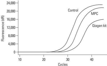
Fig. 3. Sequencing maps of known EGFR deletion in exon 19 of plasma cfDNA. cfDNA was extracted by modified phenol-chloroform (MPC) method (A) and QIAamp MinElute Virus Spin kit (Qiagen kit) (B). In (A), the height of mutant peak is similar to that of wild-type peak, while the height of mutant peak in (B) is much lower than that of wild-type peak. EGFR, epidermal growth factor receptor.
Fig. 2. Comparison of fragment distribution of plasma cfDNA ex-tracted by modified phenol-chloroform (MPC) method and QIAamp MinElute Virus Spin kit (Qiagen kit).
0 0%
25% 50% 75% 100%
2 4 6 8
cf
DN
A
co
pi
es
(1
0
3 co
pi
es
/m
L)
Fr
ag
m
en
t d
ist
rib
ut
io
n
MPC Qiagen kit MPC Qiagen kit
p=0.011 ≥202 bp 123-201 bp 77-122 bp
G G C T T T T C G G G A G A A G NT C G T C G T N C AN C C G T A C G T G T C CA A T G T
G G C N T T T C G N G A NN A T G T T N G T C G T T C AT C CT T A N G T G T C CA TAT G T
A
[image:4.595.103.503.525.703.2]tracted by PC method which is consistent with previous data
by Müller, et al.,19 the purity of cfDNA extracted by MPC
method was better, similar to that by Qiagen kit method. By comparing several commercially available cfDNA
extraction kits, Board, et al.10 reported that QIAampVirus
Spin kit was the one yielding the highest amount of plas-ma cfDNA from patients with splas-mall cell lung cancer (SCLC).Therefore, we chose QIAamp Virus Spin kit as a reference method to test the performance of the MPC meth-od for cfDNA extraction yield in plasma. Our data showed that MPC method extracted more plasma cfDNA than Qia-gen kit method, and that cfDNA extracted by MPC method contained higher percentage of longer fragment (≥202 bp). cfDNA extracted by MPC method was more intact than by Qiagen kit method. Furthermore, we compared the impact of different extraction method of MPC or Qiagen kit on EGFR mutation analysis. Results from both sequencing maps of ME-PCR products and DxS EGFR mutation test suggested that MPC method is superior to Qiagen kit on plasma cfD-NA extraction for analysis of mutant EGFR. It has been suggested that tumor-specific cfDNA is enriched in the DNA portion containing shorter fragments, while serum cfDNA isolated by the Qiagen kit method has been
demon-strated to be enriched in high-molecular-weight DNA.19,20
Thus, the enhanced sensitivity of detection of mutant EGFR by MPC method versus Qiagen kit method may partly be attributable to more tumor-specific cfDNA extracted by MPC method. Considering that small DNA molecules are
partially lost by the Qiagen kit extraction method,19,20 a new
kit from Qiagen for the extraction of cfDNA, QIAmp Cir-culating Nucleic Acid Kit, has most recently been devel-oped, which improves the extraction of small DNA frag-ments. Further studies are needed to compare our MPC method to the new Qiagen kit method in cfDNA extraction yield, as well as fragment distribution and the impact on EGFR mutation analysis.
In summary, we described an improved cfDNA extrac-tion method of MPC which will be beneficial for EGFR mutation analysis for patients with NSCLC. Additional studies with sufficient number of patients are needed to val-idate the efficiency and reliability of the MPC method for cfDNA extraction.
ACKNOWLEDGEMENTS
This work was supported by Shanghai Education Commit-plasma cfDNA from one patient with a known EGFR
frag-mental deletion in exon 19, to evaluate the impact of extrac-tion method on EGFR mutaextrac-tion analysis. In the sequencing maps of ME-PCR products, the mutant peak, based on plas-ma cfDNA extracted by MPC method (Fig. 3A), was high-er than that on plasma cfDNA extracted by Qiagen kit (Fig. 3B). In DxS EGFR mutation test kit results, ΔCt of amplifi-cation of cfDNA extracted by MPC method and Qiagen kit was 2.24 and 5.45, respectively (Fig. 4).
DISCUSSION
Increasing evidence has shown that gene mutation in tumor
tissue DNA can be detected in blood using cfDNA.7,8,12 But,
three key characteristics of cfDNA in blood from cancer patients hindered the establishment of a reliable and stan-dardized cfDNA extraction method, including that 1) cfD-NA concentration is very low with only 10-1,200 ng/mL in
plasma;13 2) cfDNA fragment is relatively short with peak
fragment of around 180 bp;14 and 3) the percentage of
tu-mor-origin DNA in cfDNA can be very low, especially in
serum cfDNA versus plasma cfDNA,10,15,16 since cfDNA is
derived from both tumor cells and non-tumor cells.17,18 In
the present study, we described an improved cfDNA extrac-tion method, i.e. MPC method, by incorporating Qiagen MaXtract Low and High Density tubes into the traditional PC method, which markedly elevated cfDNA yield in plas-ma. In addition, this modification offered a better separation between supernatant layer and organic solvents layer, thus minimizing the contamination of PCR inhibition in the ex-tracted cfDNA, as well as making manipulation process much easier and safer. Purity comparision showed that, in contrast to the moderate impurities of plasma cfDNA
ex-Fig. 4. Amplification plot of cfDNA extracted by modified phenol-chloro-form (MPC) method and QIAamp MinElute Virus Spin kit (Qiagen kit) in DxS EGFR mutation test kit. EGFR, epidermal growth factor receptor.
0 4,000 8,000 12,000 16,000 20,000 24,000
Flu
or
es
ce
nc
e
(d
R)
10 20 30 40
Cycles Control
MPC
[image:5.595.55.281.67.207.2]10. Board RE, Williams VS, Knight L, Shaw J, Greystoke A, Ranson M, et al. Isolation and extraction of circulating tumor DNA from patients with small cell lung cancer. Ann N Y Acad Sci 2008;1137: 98-107.
11. Kimura H, Kasahara K, Kawaishi M, Kunitoh H, Tamura T, Hol-loway B, et al. Detection of epidermal growth factor receptor mu-tations in serum as a predictor of the response to gefitinib in pa-tients with non-small-cell lung cancer. Clin Cancer Res 2006;12: 3915-21.
12. Kimura H, Suminoe M, Kasahara K, Sone T, Araya T, Tamori S, et al. Evaluation of epidermal growth factor receptor mutation sta-tus in serum DNA as a predictor of response to gefitinib (IRES-SA). Br J Cancer 2007;97:778-84.
13. Jahr S, Hentze H, Englisch S, Hardt D, Fackelmayer FO, Hesch RD, et al. DNA fragments in the blood plasma of cancer patients: quantitations and evidence for their origin from apoptotic and ne-crotic cells. Cancer Res 2001;61:1659-65.
14. Suzuki N, Kamataki A, Yamaki J, Homma Y. Characterization of circulating DNA in healthy human plasma. Clin Chim Acta 2008; 387:55-8.
15. Lee TH, Montalvo L, Chrebtow V, Busch MP. Quantitation of ge-nomic DNA in plasma and serum samples: higher concentrations of genomic DNA found in serum than in plasma. Transfusion 2001;41:276-82.
16. Taback B, O’Day SJ, Hoon DS. Quantification of circulating DNA in the plasma and serum of cancer patients. Ann N Y Acad Sci 2004;1022:17-24.
17. van der Drift MA, Hol BE, Klaassen CH, Prinsen CF, van Aarssen YA, Donders R, et al. Circulating DNA is a non-invasive prognos-tic factor for survival in non-small cell lung cancer. Lung Cancer 2010;68:283-7.
18. Yoon KA, Park S, Lee SH, Kim JH, Lee JS. Comparison of circu-lating plasma DNA levels between lung cancer patients and healthy controls. J Mol Diagn 2009;11:182-5.
19. Müller I, Beeger C, Alix-Panabières C, Rebillard X, Pantel K, Schwarzenbach H. Identification of loss of heterozygosity on cir-culating free DNA in peripheral blood of prostate cancer patients: potential and technical improvements. Clin Chem 2008;54:688-96. 20. Wang M, Block TM, Steel L, Brenner DE, Su YH. Preferential
isolation of fragmented DNA enhances the detection of circulating mutated k-ras DNA. Clin Chem 2004;50:211-3.
tee Foundation grant (No. 08YZ47) and Shanghai Science and Technology Committee Foundation grant (No. 10JC 1409200). We thank technician Yun Sun from Tumor Genet-ics Capability, Innovation Center China, Astrazeneca Global R&D, Shanghai, China, for her kindly technical assistance.
REFERENCES
1. Alberg AJ, Samet JM. Epidemiology of lung cancer. Chest 2003;123:21S-49S.
2. Schiller JH, Harrington D, Belani CP, Langer C, Sandler A, Krook J, et al. Comparison of four chemotherapy regimens for advanced non-small-cell lung cancer. N Engl J Med 2002;346:92-8. 3. Jänne PA, Engelman JA, Johnson BE. Epidermal growth factor
receptor mutations in non-small-cell lung cancer: implications for treatment and tumor biology. J Clin Oncol 2005;23:3227-34. 4. Mok TS, Wu YL, Thongprasert S, Yang CH, Chu DT, Saijo N, et
al. Gefitinib or carboplatin-paclitaxel in pulmonary adenocarcino-ma. N Engl J Med 2009;361:947-57.
5. Sharma SV, Bell DW, Settleman J, Haber DA. Epidermal growth factor receptor mutations in lung cancer. Nat Rev Cancer 2007;7: 169-81.
6. Riely GJ, Politi KA, Miller VA, Pao W. Update on epidermal growth factor receptor mutations in non-small cell lung cancer. Clin Cancer Res 2006;12:7232-41.
7. Bai H, Mao L, Wang HS, Zhao J, Yang L, An TT, et al. Epidermal growth factor receptor mutations in plasma DNA samples predict tumor response in Chinese patients with stages IIIB to IV non-small-cell lung cancer. J Clin Oncol 2009;27:2653-9.
8. He C, Liu M, Zhou C, Zhang J, Ouyang M, Zhong N, et al. Detec-tion of epidermal growth factor receptor mutaDetec-tions in plasma by mutant-enriched PCR assay for prediction of the response to gefi-tinib in patients with non-small-cell lung cancer. Int J Cancer 2009;125:2393-9.