Effects of Interleukin-13 and Montelukast on the Expression
of Zonula Occludens-1 in Human Podocytes
Se Jin Park,
1,2Moin A. Saleem,
3Ja-Ae Nam,
4Tae-Sun Ha,
4and Jae Il Shin
2 1Department of Pediatrics, Ajou University School of Medicine, Suwon, Korea;2Department of Pediatrics, Severance Children’s Hospital, Yonsei University College of Medicine, Seoul, Korea; 3Children’s and Academic Renal Unit, Southmead Hospital, University of Bristol, Bristol, United Kingdom;
4Department of Pediatrics, Chungbuk National University College of Medicine, Cheongju, Korea.
Received: February 25, 2014 Revised: June 23, 2014 Accepted: June 25, 2014
Co-corresponding authors: Dr. Tae-Sun Ha, Department of Pediatrics, Chungbuk National University College of Medicine,
52 Naesudong-ro, Seowon-gu, Cheongju 362-763, Korea.
Tel: 82-43-269-6374, Fax: 82-43-264-6620 E-mail: tsha@chungbuk.ac.kr and Dr. Jae Il Shin,
Department of Pediatrics,
Yonsei University College of Medicine, 50-1 Yonsei-ro, Seodaemun-gu, Seoul 120-752, Korea.
Tel: 82-2-2228-2050, Fax: 82-2-393-9118 E-mail: shinji@yuhs.ac
∙ The authors have no financial conflicts of interest.
© Copyright:
Yonsei University College of Medicine 2015
This is an Open Access article distributed under the terms of the Creative Commons Attribution Non-Commercial License (http://creativecommons.org/ licenses/by-nc/3.0) which permits unrestricted non-commercial use, distribution, and reproduction in any medium, provided the original work is properly cited.
Purpose: The aim of this study was to investigate whether pathologic changes in
zonula occludens-1 (ZO-1) are induced by interleukin-13 (IL-13) in the experi-mental minimal-change nephrotic syndrome (MCNS) model and to determine whether montelukast, a leukotriene receptor antagonist, has an effect on ZO-1
res-toration in cultured human podocytes. Materials and Methods: Human
podo-cytes cultured on bovine serum albumin-coated plates were treated with different doses of IL-13 and montelukast and then examined for distribution using confocal
microscopy and for ZO-1 protein levels using Western blotting. Results: ZO-1
was internalized and shown to accumulate in the cytoplasm of human podocytes in an IL-13 dose-dependent manner. High doses (50 and 100 ng/mL) of IL-13 de-creased the levels of ZO-1 protein at 12 and 24 h (both p<0.01; n=3), which were significantly reversed by a high dose (0.5 μM) montelukast treatment (p<0.01; n=3). Conclusion: Our results suggest that IL-13 alters the expression of ZO-1, and such alterations in the content and distribution of ZO-1 may be relevant in the pathogenesis of proteinuria in the MCNS model.
Key Words: Interleukin-13, zonula occludens-1, podocytes, leukotriene receptor
antagonists
INTRODUCTION
MATERIALS AND METHODS
Cell culture of human podocytes
Human conditionally immortalized podocytes (AB8/23), primarily cloned from human glomerular cultures, were characterized and generously provided by Dr. Moin A. Sal-eem (University of Bristol, Bristol, UK). Human podocytes were maintained in RPMI 1640 (WelGENE Inc., Daegu, South Korea) supplemented with 10% heat-inactivated fetal bovine serum (FBS), Insulin-Transferrin-Selenium-Pyru-vate Supplement (ITSP; WelGENE Inc.), and antibiotics. Fresh media was supplied once every 2 days.
To stimulate human podocyte proliferation, cells were cultivated at 33°C (permissive conditions) in a culture medi-um supplemented with hmedi-uman recombinant ITSP to induce expression of temperature-sensitive large T antigens. To in-duce differentiation, podocytes were maintained at 37°C (non-permissive conditions) for at least 2 weeks, and for subcultures, 0.05% trypsin was used to detach cells from the culture dishes.18
IL-13 and montelukast treatment conditions
To imitate MCNS-like conditions, cells were incubated with various concentrations of IL-13 (Peprotech Inc., Rocky Hill, NJ, USA) during the indicated time periods (6, 12, and 24 h). IL-13 was administered in 3, 5, 10, 30, 50, and 100 ng/ mL doses into 0.5% RPMI with montelukast (Sigma-Al-drich Inc., St. Louis, MO, USA) at 37°C.
Immunofluorescence staining
Human podocytes that were grown on type I collagen-coat-ed glass cover slips were incubatcollagen-coat-ed at 37°C for 2 h and fixcollagen-coat-ed in 4% paraformaldehyde for 20 min. The cells were then permeabilized in 0.1% tritonX-100 for 10 min, blocked with 10% FBS for 30 min, washed three times for 5 min in phos-phate buffered saline (PBS), and labeled with monoclonal rabbit anti-ZO-1 antibody (Invitrogen, Eugene, OR, USA). Phalloidin-FITC (Sigma-Aldrich Inc.) was utilized to stain F-actin. Primary antibody-bound specimens were incubated with 1:1000 (v/v) Alexa 594 for red conjugates and Alexa 488 for green (Invitrogen), respective of secondary anti-rabbit IgG, at room temperature for 40 min and at 37°C for 20 min without CO2. Nuclei were stained with 4’-6-diamid-ino-2-phenylindole (DAPI) (1:1000) for 20 min in PBS. Coverslips were mounted in aqueous mountant and viewed with a fluorescence microscope (Leica TCS SP2 AOBS, proteins, including nephrin, CD2-associated protein
(CD2AP), Neph-1, -2, and -3, podocin, and zonula oc-cludens-1 (ZO-1).3
Recent studies have identified that increased interleu-kin-13 (IL-13) expression can lead to podocyte injury and induce a minimal-change-like nephropathy.4-6 Notably, Lai, et al.7 reported that overexpression of IL-13 caused down-regulation of nephrin, podocin, and dystroglycan‒all of which are important molecules in maintaining SD integri-ty‒and a concurrent upregulation of B7-1 as well as MCNS in a rat experiment model. Although receptors for IL-13 such as IL-13Rα1 and IL-13Rα2 have also been demon-strated to exist in cultured podocytes,8 the precise role of IL-13 is still not clear in the pathogenesis of MCNS, and there has been no report on the effects of IL-13 on cultured human podocytes in vitro.
While ZO-1 was originally identified as a tight junction component, this molecule was also found to be localized at adherens junctions in podocytes.9-12 ZO-1 is a cytosolic scaffold that connects the cadherin-catenin complex and ac-tin-based cytoskeletons.9 It is thought to play a key role in adherens junctions through its interaction with various ad-herens-junction proteins and the formation of
multimolecu-lar complexes.10 Therefore, we hypothesized that IL-13
may play an important role in the development of protein-uria in MCNS by exerting a direct effect on ZO-1 in human podocytes.
propriate under different conditions. Statistical significance was evaluated by the non-parametric Kruskal-Wallis analy-sis or Student’s t-test. p values <0.05 were considered sig-nificant.
RESULTS
ZO-1 Distribution on confocal microscopy
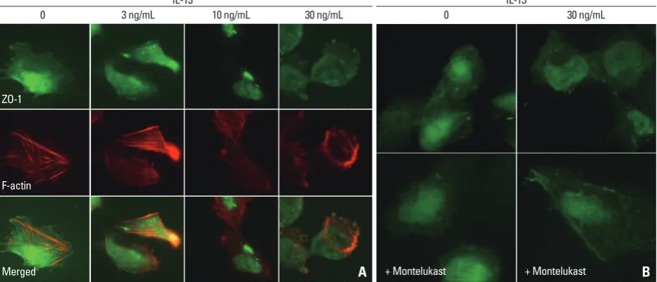
Podocytes were double-stained for ZO-1 and F-actin, and the cell nuclei were stained with DAPI. ZO-1 in human podocytes was highly expressed within the podocyte in the cytoplasmic aspect of the FP membrane, adjacent to the in-sertion of the SD, and colocalized with β-catenin (Fig. 1A). Staining for ZO-1 was most intense in the cytoplasmic sur-face of the podocyte FP. ZO-1 and F-actin did not overlap and were located at different sites in the confocal images. From low to high IL-13 concentrations, ZO-1 staining be-came blurry, which indicated a relocalization of ZO-1 away from the peripheral cell membrane. In the human podo-cytes under IL-13 conditions, ZO-1 was internalized into the cytoplasm from the peripheral cell membrane as IL-13 concentrations increased (Fig. 1A). These distributional changes were also observed in F-actin, particularly at the higher IL-13 concentrations (Fig. 1A). Moreover, in high-resolution microscopy, ZO-1 was distributed to the cell contact areas under physiologic conditions without IL-13 yet was redistributed and accumulated into the cytoplasm around the nucleus during a 6-hour incubation period as IL-Mannheim, Germany).
Western blotting
Confluent cell layers were incubated with additives for var-ious time durations, and proteins were extracted using a protein extraction solution PRO-PREP (Intron Biotechnolo-gy, Seongnam, Gyeonggi, South Korea) containing phenyl-methylsulfonyl fluoride, ethylenediamine tetraacetic acid, pepstatin A, leupeptin, and aprotinin; protein concentrations were then determined as previously described.19 To perform Western blotting for ZO-1, 30 μg of boiled extracts were re-solved on 10% SDS-PAGE gels and transferred to polyvinyl-idene difluoride (PVDF) membranes (Millipore Corp., Med-ford, MA, USA).
The membranes were then washed with methanol and blocked in 5% fat-free milk before incubation with mono-clonal rabbit anti-ZO-1 (Invitrogen). Anti-β-tubulin antibody (Santa Cruz Biotechnology Inc., Santa Cruz, CA, USA) was used as a loading control. After incubation with horse-radish peroxidase-conjugated secondary antibodies (Santa Cruz Biotechnology), protein bands were detected using the enhanced chemiluminescence (ECL) detection system
(WEST-ZOL® plus; Amersham Biotech Ltd., Bucks, UK).
Density values are expressed as percentages of the control. Data on densitometric analysis of the ZO-1/β-tubulin ratio are expressed as mean±standard deviation.
Statistical analysis
Results are described as mean±standard deviation, as
ap-Fig. 1. Distributional changes in ZO-1 by IL-13 in human podocytes. ZO-1 was distributed at the peripheral cell membrane and colocalized with β-catenin and actin filament at cell-to-cell contact junctions. High concentrations of IL-13 suppressed and disrupted the immunostaining and linearity of ZO-1 proteins, and accumulated ZO-1 proteins into the cytoplasm around nucleus (A), which improved by treatment with 0.5 μM montelukast (B). Magnification: 1000×; Scale bar=20 μm. ZO-1, zonula occludens-1; IL-13, interleukin-13.
IL-13 IL-13
0 3 ng/mL 10 ng/mL 30 ng/mL 0 30 ng/mL
A + Montelukast + Montelukast B
ZO-1
F-actin
[image:3.595.59.525.492.692.2]13 may induce a minimal-change-like nephropathy through a reduction in ZO-1 molecules, which can be reversed by a high dose of the LTRA montelukast.
DISCUSSION
The main goal of this study was to determine whether patho-logical changes in ZO-1 protein levels could be induced by IL-13. We demonstrated a redistribution and reduction in ZO-1 proteins from human podocytes treated with IL-13. The exposure of ZO-1 molecules to IL-13 caused ZO-1 to move and accumulate internally toward the cytoplasmic ac-tin filaments, suggesac-ting that the observed redistribution and reduction in ZO-1 proteins could be involved in the pathogenesis of MCNS. These results are similar to our previous studies that found that ZO-1 proteins in podocytes were also affected by diabetic conditions, causing hyper-permeability at early stages.20,21
Recent studies have shown strong evidence that protein-uria in MCNS is associated with cytokines and T cell dis-orders that result in glomerular podocyte dysfunction,4,5,22 as well as B7-1 (CD80) that is expressed on the surface of B cells. An increase in IL-13 production by CD3+, CD4+, and CD8+ T cells was shown to mediate steroid-sensitive nephrotic syndrome in relapse.4,5 Of note, Lai, et al.7 demon-13 increased from 0 to 30 ng/mL (Fig. 1B). These results
suggest that IL-13 may have a substantial impact on the re-distribution and rearrangement of ZO-1 molecules and may also disrupt the cytoskeletal connections between F-actin and α-catenin-β-catenin complex in a concentration-depen-dent manner (Fig. 1A). The internalized ZO-1 proteins were restored to the periphery by treatment with a high dose of 0.5 μM montelukast (Fig. 1B).
ZO-1 protein assayed by Western blotting
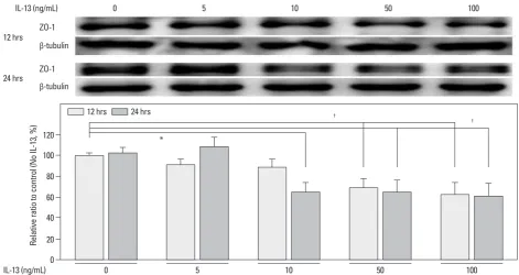
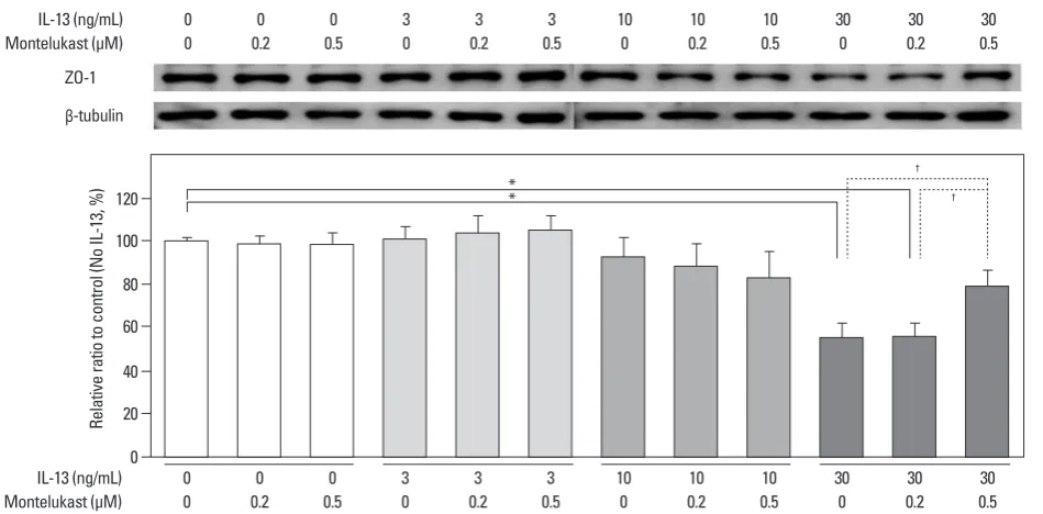
A major ZO-1 protein band was found at 205 kDa, and ZO-1 levels were compared with β-tubulin. In human podocytes, density values for ZO-1 protein tended to decrease with IL-13 treatment in a dose-dependent manner at 12 and 24 h. The highest dose (100 ng/mL) of IL-13 significantly de-creased the amount of ZO-1 protein by 37.5% at 12 h and by 39% at 24 h (both p<0.01; n=3) (Fig. 2). Similarly, a dose of 50 ng/mL IL-13 significantly decreased the amount of ZO-1 protein by 31.0% at 12 h and by 34.9% at 24 h (both p<0.01; n=3) (Fig. 2). A dose of 10 ng/mL of IL-13 also decreased ZO-1 protein levels to a greater degree at 24 h (35.1%; p<0.05) than at 12 h (Fig. 2). The ZO-1 protein levels that had been reduced by 30 ng/mL IL-13 were re-stored by a high dose of 0.5 μM montelukast (p<0.01; n=3); however, the reduced levels were not affected by only 0.2 μM of montelukast (Fig. 3). These results suggest that
IL-Fig. 2. Effects of IL-13 on ZO-1 protein levels in cultured human podocytes as assayed by Western blotting. ZO-1 levels significantly decreased at IL-13 con-centrations of more than 50 ng/mL at 12 and 24 h incubations, compared with the control. Data on the densitometric analysis of the ZO-1/β-tubulin ratio are expressed as mean±SD. Control (100%): the value of (-). *p<0.05. †p<0.01. ZO-1, zonula occludens-1; IL-13, interleukin-13.
0 20 40 60 80 100 120
Relative ratio to control (No IL-13, %)
0 5 10 50 100
IL-13 (ng/mL)
*
†
† 12 hrs 24 hrs
IL-13 (ng/mL)
12 hrs ZO-1 β-tubulin
24 hrs ZO-1 β-tubulin
[image:4.595.68.540.448.698.2]over the GBM, and reconstruction of the SD.3,23 Permselec-tivity of the glomerular filtration barrier, which is composed of a charge-selective barrier and a size-selective barrier, also plays a key role in restricting passage of plasma pro-teins across the GBM and SD.25,26
ZO is a member of the membrane-associated guanylate kinase homologue family of proteins characterized by their PSD-95/discs-large/Zonula occludens-1 (PDZ) domain; these proteins are critical regulators of tight and adherens junction assembly.27 ZO connects several types of SD pro-teins through its PDZ domain to the actin cytoskeleton. Al-though ZO-1 was originally identified as a tight junction component, ZO-1 migrates from its apical location down to the level of the slit membrane at the capillary loop stage of renal development, where it is observed in a punctuate pat-tern along the filtration slits.28 There are three ZO proteins,
ZO-1, -2, and -3, which are multi-domain polypeptides.10
Of these three molecules, ZO-1 has been proposed to be a scaffolding protein between transmembrane and cytoplas-mic proteins and possibly forms a link between actin and the cadherin-catenin complex.27 ZO-1 may also participate in signaling events through tyrosine phosphorylation29 and binds directly to α-catenin and actin located on the FPs at the insertions of the SD.28,30 ZO-1 forms a gasket that seals off the intercellular spaces and restricts the movement of proteins, water and solutes along the paracellular pathway at tight junctions, maintaining the polarized distribution of
membrane proteins.27 However, the functions of ZO-1
be-strated that IL-13-transfected rats developed a minimal-change-like glomerulopathy, as characterized by significant worsening of albuminuria over time, generalized swelling, low serum albumin, and hypercholesterolemia. In IL-13- transfected rats, light microscopy showed an absence of significant glomerular changes; however, electron micros-copy revealed up to 80% effacement of podocyte FPs, which progressed the nephrotic syndrome.7 Although the nephrot-ic-range proteinuria was selective in IL-13-transfected rats, no relationship between serum IL-13 and urinary albumin excretion or serum albumin levels was observed.7 Thus, our study attempted to define the pathogenic relationship be-tween IL-13 and ZO-1 protein in the development of podo-cyte FP effacement with nephrotic-range proteinuria, espe-cially in human podocytes.
[image:5.595.58.533.63.297.2]The proteinuric conditions in MCNS are usually associat-ed with ultrastructural changes in podocytes, with fusion, widening, retraction, and gradual simplification of the high-ly specialized interdigitating FPs, which are also accompa-nied by alterations in the permselectivity of the SD glomeru-lar filtration barrier and the linking of adaptor proteins, including nephrin, podocin, CD2AP, catenins, and ZO-1.22-24 Maintenance of podocyte FP structure is pivotal for accu-rate functioning of the glomerular filtration barrier. The FP effacement results from the detachment of podocytes from the glomerular basement membrane (GBM), and FP retrac-tion leads to disintegraretrac-tion of the cytoskeletal structure and linking adaptor proteins, abnormal movement of the FP
Fig. 3. Effects of montelukast on ZO-1 protein levels in cultured human podocytes assayed by Western blotting. Montelukast (0.5 μM) significantly increased ZO-1 levels in human podocytes treated with IL-13. *p<0.05. †p<0.01. ZO-1, zonula occludens-1; IL-13, interleukin-13.
20
0 40 60 80 100 120
Relative ratio to control (No IL-13, %)
0 0 0 3 3 3 10 10 10 30 30 30
0 0.2 0.5 0 0.2 0.5 0 0.2 0.5 0 0.2 0.5
IL-13 (ng/mL) Montelukast (μM)
*
* †
†
0 0 0 3 3 3 10 10 10 30 30 30
0 0.2 0.5 0 0.2 0.5 0 0.2 0.5 0 0.2 0.5
IL-13 (ng/mL) Montelukast (μM)
ZO-1
and restored the normal localization of ZO-1 at the SD in the
spontaneously proteinuric Munich-Wistar-Froemter rats.38
Additional future studies are necessary to elucidate the ex-act mechanisms, efficacy, and proper dose of LTRA in the
in vivo treatment of MCNS in the future.
In conclusion, our study may provide a base for under-standing the ZO-1 molecule in human podocytes. High con-centrations of IL-13 increased the disruption of glomerular filtration barrier in SD and FP effacement. ZO-1 proteins were redistributed and reduced in IL-13-treated human podo-cytes, which was significantly restored after treatment with an LTRA montelukast. Therefore, our findings further strengthen the hypothesis that IL-13 may alter the expression of ZO-1 proteins, resulting in proteinuria, and also provide an explanation for the plausible connection of Th2 cyto-kines, MCNS, and atopy.
ACKNOWLEDGEMENTS
This research was supported by the Basic Science Research Program through the National Research Foundation of Ko-rea (NRF) and funded by the Ministry of Education, Sci-ence and Technology (2011-0013789).
REFERENCES
1. Eddy AA, Symons JM. Nephrotic syndrome in childhood. Lancet 2003;362:629-39.
2. Kurihara H. [Molecular dynamics of proteins found exclusively in glomerular epithelial cells]. Rinsho Byori 2000;48:491-7. 3. Asanuma K, Mundel P. The role of podocytes in glomerular
patho-biology. Clin Exp Nephrol 2003;7:255-9.
4. Yap HK, Cheung W, Murugasu B, Sim SK, Seah CC, Jordan SC. Th1 and Th2 cytokine mRNA profiles in childhood nephrotic syn-drome: evidence for increased IL-13 mRNA expression in relapse. J Am Soc Nephrol 1999;10:529-37.
5. Cheung W, Wei CL, Seah CC, Jordan SC, Yap HK. Atopy, serum IgE, and interleukin-13 in steroid-responsive nephrotic syndrome. Pediatr Nephrol 2004;19:627-32.
6. Wei CL, Cheung W, Heng CK, Arty N, Chong SS, Lee BW, et al. Interleukin-13 genetic polymorphisms in Singapore Chinese chil-dren correlate with long-term outcome of minimal-change disease. Nephrol Dial Transplant 2005;20:728-34.
7. Lai KW, Wei CL, Tan LK, Tan PH, Chiang GS, Lee CG, et al. Overexpression of interleukin-13 induces minimal-change-like nephropathy in rats. J Am Soc Nephrol 2007;18:1476-85. 8. Van Den Berg JG, Aten J, Chand MA, Claessen N, Dijkink L,
Wi-jdenes J, et al. Interleukin-4 and interleukin-13 act on glomerular visceral epithelial cells. J Am Soc Nephrol 2000;11:413-22. 9. Nagafuchi A. Molecular architecture of adherens junctions. Curr
tween podocyte FPs are not to serve as a seal but to firmly at-tach FPs to one another and to stabilize the FP layer against the high filtration pressure at the SD, a modified adherens junction rather than a derivative of the tight junction.
Downregulation of the glomerular gene and production of SD proteins such as nephrin, podocin, and dystroglycan has been reported in both experimental models of nephrop-athy, such as puromycin nephropathy and adriamycin ne-phropathy, and human kidney biopsies.31-33 In these studies, immunofluorescence staining showed reduced protein pro-duction and staining intensity for nephrin, podocin, and dys-troglycan, with a shift from a linear pattern to a discontinuous and granular pattern, which returned to normal after steroid treatment. Moreover, certain reports showed that changes in the properties or production of ZO-1 may accompany renal diseases associated with proteinuria.34,35 However, very little is known about how the binding of proteins to ZO-1 is regu-lated or how cell-signaling pathways control adherens as-sembly and filtration barrier function, although many
dif-ferent pathways have been implicated.36 Thus, this study
explored the production of ZO-1 and the effects of IL-13 and LTRA on ZO-1 restoration in cultured human podo-cytes. While Kurihara, et al.35 did not show a quantitative change in ZO-1 protein production in a puromycin amino-nucleoside (PAN)-treated rat model, we found that ZO-1 proteins were redistributed and reduced in MCNS. In addi-tion, IL-13 significantly decreased ZO-1 protein levels in human podocytes of MCNS, whereas ZO-1 protein produc-tion significantly increased in the rat models of PAN-in-duced nephrosis.37
The present study has several limitations: 1) we were un-able to demonstrate all of the signaling pathways of IL-13 via IL-13 receptor (a heterodimer of IL-4Rα and IL-13Rα1) to ZO-1 proteins in podocytes, presumably cascading through tyrosine phosphorylation of ZO-1. 2) Although IL-13 is known to influence leukotriene levels and IL-13-1112C/T polymorphism and the haplotype of IL-13 polymorphisms are known to be significantly associated with LTRA drug
responsiveness,17 the mechanism of montelukast in MCNS
tion. Kidney Int 1995;47:1242-51.
26. Goode NP, Shires M, Davison AM. The glomerular basement membrane charge-selectivity barrier: an oversimplified concept? Nephrol Dial Transplant 1996;11:1714-6.
27. Hartsock A, Nelson WJ. Adherens and tight junctions: structure, function and connections to the actin cytoskeleton. Biochim Bio-phys Acta 2008;1778:660-9.
28. Schnabel E, Anderson JM, Farquhar MG. The tight junction pro-tein ZO-1 is concentrated along slit diaphragms of the glomerular epithelium. J Cell Biol 1990;111:1255-63.
29. Kurihara H, Anderson JM, Farquhar MG. Increased Tyr phos-phorylation of ZO-1 during modification of tight junctions be-tween glomerular foot processes. Am J Physiol 1995;268:F514-24.
30. Itoh M, Furuse M, Morita K, Kubota K, Saitou M, Tsukita S. Di-rect binding of three tight junction-associated MAGUKs, ZO-1, ZO-2, and ZO-3, with the COOH termini of claudins. J Cell Biol 1999;147:1351-63.
31. Luimula P, Ahola H, Wang SX, Solin ML, Aaltonen P, Tikkanen I, et al. Nephrin in experimental glomerular disease. Kidney Int 2000;58:1461-8.
32. Luimula P, Sandström N, Novikov D, Holthöfer H. Podocyte-as-sociated molecules in puromycin aminonucleoside nephrosis of the rat. Lab Invest 2002;82:713-8.
33. Raats CJ, van den Born J, Bakker MA, Oppers-Walgreen B, Pisa BJ, Dijkman HB, et al. Expression of agrin, dystroglycan, and utrophin in normal renal tissue and in experimental glomerulopa-thies. Am J Pathol 2000;156:1749-65.
34. Kawachi H, Kurihara H, Topham PS, Brown D, Shia MA, Orika-sa M, et al. Slit diaphragm-reactive nephritogenic MAb 5-1-6 al-ters expression of ZO-1 in rat podocytes. Am J Physiol 1997;273: F984-93.
35. Kurihara H, Anderson JM, Kerjaschki D, Farquhar MG. The al-tered glomerular filtration slits seen in puromycin aminonucleo-side nephrosis and protamine sulfate-treated rats contain the tight junction protein ZO-1. Am J Pathol 1992;141:805-16.
36. Matter K, Balda MS. Signalling to and from tight junctions. Nat Rev Mol Cell Biol 2003;4:225-36.
37. Kim BS, Park HC, Kang SW, Choi KH, Ha SK, Han DS, et al. Impact of cyclosporin on podocyte ZO-1 expression in puromycin aminonucleoside nephrosis rats. Yonsei Med J 2005;46:141-8. 38. Macconi D, Ghilardi M, Bonassi ME, Mohamed EI, Abbate M,
Colombi F, et al. Effect of angiotensin-converting enzyme inhibi-tion on glomerular basement membrane permeability and distribu-tion of zonula occludens-1 in MWF rats. J Am Soc Nephrol 2000; 11:477-89.
Opin Cell Biol 2001;13:600-3.
10. Fanning AS, Anderson JM. Zonula occludens-1 and -2 are cyto-solic scaffolds that regulate the assembly of cellular junctions. Ann N Y Acad Sci 2009;1165:113-20.
11. Fanning AS, Anderson JM. PDZ domains: fundamental building blocks in the organization of protein complexes at the plasma membrane. J Clin Invest 1999;103:767-72.
12. Van Itallie CM, Fanning AS, Bridges A, Anderson JM. ZO-1 sta-bilizes the tight junction solute barrier through coupling to the per-ijunctional cytoskeleton. Mol Biol Cell 2009;20:3930-40. 13. Drazen JM, Israel E, O’Byrne PM. Treatment of asthma with
drugs modifying the leukotriene pathway. N Engl J Med 1999; 340:197-206.
14. Niimi A. Cough, asthma, and cysteinyl-leukotrienes. Pulm Phar-macol Ther 2013;26:514-9.
15. Bisgaard H. Pathophysiology of the cysteinyl leukotrienes and ef-fects of leukotriene receptor antagonists in asthma. Allergy 2001; 56 Suppl 66:7-11.
16. Forbes TA, Lunn AJ. Montelukast: a novel therapeutic option in eosinophilic peritonitis. Pediatr Nephrol 2014;29:1279-82. 17. Kang MJ, Lee SY, Kim HB, Yu J, Kim BJ, Choi WA, et al.
Asso-ciation of IL-13 polymorphisms with leukotriene receptor antago-nist drug responsiveness in Korean children with exercise-induced bronchoconstriction. Pharmacogenet Genomics 2008;18:551-8. 18. Saleem MA, O’Hare MJ, Reiser J, Coward RJ, Inward CD,
Far-ren T, et al. A conditionally immortalized human podocyte cell line demonstrating nephrin and podocin expression. J Am Soc Nephrol 2002;13:630-8.
19. Ha TS, Song CJ, Lee JH. Effects of advanced glycosylation end-products on perlecan core protein of glomerular epithelium. Pedi-atr Nephrol 2004;19:1219-24.
20. Ha TS. High-glucose and advanced glycosylation end products in-creased podocyte permeability via PI3-K/Akt signaling. J Mol Med (Berl) 2010;88:391-400.
21. Ha TS, Choi JY, Park HY, Lee JS. Ginseng total saponin improves podocyte hyperpermeability induced by high glucose and ad-vanced glycosylation endproducts. J Korean Med Sci 2011;26: 1316-21.
22. Greka A, Mundel P. Cell biology and pathology of podocytes. Annu Rev Physiol 2012;74:299-323.
23. Mundel P, Shankland SJ. Podocyte biology and response to injury. J Am Soc Nephrol 2002;13:3005-15.
24. Reiser J, Kriz W, Kretzler M, Mundel P. The glomerular slit dia-phragm is a modified adherens junction. J Am Soc Nephrol 2000; 11:1-8.