INTRODUCTION
Multiple myeloma (MM) is a disease of cancerous plasma cells that expand in bone marrow, causing symptoms of ane-mia, hypercalceane-mia, and osteolytic bone lesions.1 The disease
is characterized by uncontrolled proliferation of a plasma cell clone with an accumulation of monoclonal immunoglobu-lins.2 Myeloma and stromal cells produce osteoclast activating
factors, such as IL-6, IL-3, receptor activator nuclear factor kappa B ligand (RANKL), TNF-α, and MIP1α, which are re-sponsible for bone resorption.3-7 Furthermore, myeloma cells
Cyclized Oligopeptide Targeting LRP5/6-DKK1
Interaction Reduces the Growth of Tumor Burden
in a Multiple Myeloma Mouse Model
Bo Mi Park
1, Eun Jin Kim
2, Hee Jin Nam
3, Dongdong Zhang
1, Chu Hyun Bae
1, Myeongmo Kang
2,
Heeyoun Kim
4, Weontae Lee
4, Bjarne Bogen
5, and Sung-Kil Lim
1,31Brain Korea 21 PLUS Project for Medical Science, Yonsei University, Seoul;
2Institute of Biomedical Sciences, Yonsei University, Seoul;
3Division of Endocrinology and Endocrine Research Institute, Department of Internal Medicine, Yonsei University College of Medicine, Seoul; 4Department of Biochemistry, Yonsei University, Seoul;
5Centre for Immune Regulation, Institute of Immunology, University of Oslo and Oslo University Hospital, Oslo, Norway.
Purpose: Dickkopf 1 (DKK1) has been extensively investigated in mouse models of multiple myeloma, which results in osteolytic bone lesions. Elevated DKK1 levels in bone marrow plasma and serum inhibit the differentiation of osteoblast precursors. Pres-ent pharmaceutical approaches to target bone lesions are limited to antiresorptive agPres-ents. In this study, we developed a cyclized oligopeptide against DKK1-low density lipoprotein receptor-related protein (LRP) 5/6 interaction and tested the effects of the oli-gopeptide on tumor burden.
Materials and Methods: A cyclized oligopeptide based on DKK1-LRP5/6 interactions was synthesized chemically, and its nucle-ar magnetic resonance structure was assessed. Luciferase reporter assay and mRNA expressions of osteoblast mnucle-arkers were eval-uated after oligopeptide treatment. MOPC315.BM.Luc cells were injected into the tail vein of mice, after which cyclized oligopep-tide was delivered subcutaneously 6 days a week for 4 weeks.
Results: The cyclized oligopeptide containing NXI motif bound to the E1 domain of LRP5/6 effectively on surface plasmon reso-nance analysis. It abrogated the Wnt-β-catenin signaling inhibited by DKK1, but not by sclerostin, dose dependently. RT-PCR and alkaline phosphatase staining showed increased expressions of osteoblast markers according to the treatment concentrations. Bioluminescence images showed that the treatment of cyclized oligopeptide reduced tumor burden more in oligopeptide treated group than in the vehicle group.
Conclusion: The cyclized oligopeptide reported here may be another option for the treatment of tumor burden in multiple myeloma.
Key Words: Multiple myeloma, DKK1, oligopeptide, tumor, burden, Wnt signaling
pISSN: 0513-5796 · eISSN: 1976-2437
Received: August 12, 2016 Revised: January 20, 2017 Accepted: January 26, 2017
Corresponding author: Dr. Sung-Kil Lim, Division of Endocrinology and Endocrine Research Institute, Department of Internal Medicine, Yonsei University College of Medicine, 50-1 Yonsei-ro, Seodaemun-gu, Seoul 03722, Korea.
Tel: 82-2-2228-0838, Fax: 82-2-2227-8129, E-mail: [email protected]
•The authors have no financial conflicts of interest. © Copyright: Yonsei University College of Medicine 2017
This is an Open Access article distributed under the terms of the Creative Com-mons Attribution Non-Commercial License (http://creativecomCom-mons.org/licenses/ by-nc/4.0) which permits unrestricted non-commercial use, distribution, and repro-duction in any medium, provided the original work is properly cited.
induce changes in the bone marrow microenvironment, thereby the production of osteoblastic inhibitor factors, such as TGF-β, TNF-α, and IL-3, is increased.8,9 Accordingly, an
im-balance in osteoclast and osteoblast activity with the preva-lence of bone resorption leads to marrow destruction.
The Wnt signaling pathway plays important role in the pro-cess of osteogenic differentiation and in the maintenance of mesenchymal stem cells, as well as in embryonic develop-ment.10 Several attempts have been made to target the Wnt
signaling pathway by inhibiting endogenous antagonists and by regulating intracellular mediators for the treatment of os-teogenic disorders.11 Antibodies against endogenous Wnt
an-tagonists, such as sclerostin, have shown bone-forming and fracture healing effects.12 Dickkopf 1 (DKK1) is another
im-portant endogenous inhibitor of the canonical Wnt signaling pathway, which binds to low density lipoprotein receptor-re-lated protein (LRP) 5/6, together with Kremen receptor, to in-hibit the canonical Wnt signaling pathway.13
DKK1 has been extensively investigated in murine models of MM where osteolytic lesions occur. Many studies have con-firmed that the interaction of MM cells with the bone marrow microenvironment causes the bone lesions of myeloma.14
MM cancer cells secrete DKK1, which disrupts the balance of osteoblastogenesis and osteoclastogenesis.15 DKK1 disrupts
the differentiation of mesenchymal stem cells to osteoblast lineage cells, and this results in a shift of the RANKL/osteo-protegerin (OPG) ratio, leading to excessive bone resorption and marrow destruction.15 Enhanced bone resorption is not
followed by neo-matrix deposition due to the inhibition of further differentiation of osteoblast precursors by DKK1.16
Therefore, DKK1 has been a potential therapeutic target for the treatment of MM.
Presently approved medications that target bone diseases are limited to anti-resorptive agents and bone-anabolic agents,17 and bone-protecting anti-resorptive agents have
failed to confer significant antitumor activities in clinical stud-ies.18 A small number of papers have reported that the
treat-ment of antibodies against DKK1 not only reduces bone le-sions but also reduces the tumor burden of the disease.19,20
However, exact mechanisms remain under investigation. We hypothesized that DKK1-inhibiting oligopeptide would bind in the place of DKK1 of the Wnt signaling pathway, reducing the binding of the overexpressed DKK1 protein by MM cells. The present study aimed to develop a cyclized oligopeptide containing the NXI motif already known in DKK1 and to test the effects of the oligopeptide on tumor burden.
MATERIALS AND METHODS
Synthesis and purification of peptides
The peptide consisted of 10 amino acids with one disulfide bond; it contains NXI motif, which is found in DKK1. Peptides
were synthesized via Fmoc solid phase peptide synthesis us-ing ASP48S (Peptron Inc., Daejeon, Korea) and purified via the reverse phase high-performance liquid chromatography using a Vydac Everest C18 column (250×22 mm, 10 μm). Elu-tion was carried out with a water-acetonitrile linear gradient [3–40% (v/v) of acetonitrile] containing 0.1% (v/v) trifluoro-acetic acid. Molecular weights of the purified peptide were confirmed using Liquid Chromatography/Mass Spectrometry (Agilent HP1100 series, Agilent Technologies, Waldbronn, Germany).
Cell culture
MOPC315.BM.Luc cells21 were obtained from Bogen Lab.
Briefly, cells were developed from a MOPC315 cell line through s.c. injection twice to obtain MOPC315.4 and i.v. in-jection to obtain final cell line MOPC315.BM. Then, cells were tagged with luciferase gene. Cells were cultured in RPMI 1640 medium supplemented with fetal bovine serum (FBS) and penicillin-streptomycin (Welgene, Gyeongsan, Korea). ST2 cells were cultured in dulbecco’s modified eagle’s medicum medium, and luciferase transfected MC3T3-E1 cells were cul-tured in alpha-minimum essential medium with FBS and penicillin-streptomycin.
Nuclear magnetic resonance (NMR) spectroscopy Nuclear magnetic resonance (NMR) spectra were acquired at 283 K on a Bruker DRX-500 spectrometer equipped with a tri-ple resonance probe with an x, y, and z-shielded pulsed-field gradient coil. Two-dimensional (2D) NMR spectra were re-corded in a phase-sensitive mode using time proportional phase increment for quadrature detection in the t1 domain.
Total correlation spectroscopy (TOCSY)22 using a dipsi-2
spin-lock pulse sequence with a mixing time of 70 ms and nuclear overhauser enhancement spectroscopy (NOESY)23 with
mix-ing times of 250–600 ms were performed. All NMR spectra were acquired with 2048 complex data points in t2 and 256
incre-ments in the t1 dimension, with 64 scans per each increment.
All NMR data were processed using nmrPipe/nmrDraw or XWIN-NMR software (Bruker Instruments, Karlsruhe, Ger-many) and analyzed using the Sparky 3.95 program.
Structure calculations
Distance restraints were derived from the NOESY spectra in 100% deuterated DMSO solution. The solution structures were calculated using the hybrid distance geometry and dy-namical simulated annealing (SA) protocol using the CNS 1.1 program on a Linux workstation. Final structures were ana-lyzed using the PROCHECK20 and MOLMOL programs.24
Surface plasmon resonance (SPR) assay
LRP5 E1 domains were tethered (immobilized) onto a biosen-sor chip surface. Then, five different concentrations (1, 3, 10, 30, and 100 µM) of the oligopeptide were passed over the LRP5 E1 domain in order for binding interactions to occur. The Langmuir model of the ProteOn ManagerTM program
(Bio-Rad Laboratory) was used for SPR data analysis.
Transfected MC3T3-E1 cell luciferase reporter assay In the Wnt signaling study, MC3T3-E1 cells were transfected as previously reported.25 Seeded cells were treated in different
concentrations (50 nM, 500 nM, 5 µM) of oligopeptide along with Wnt7a and DKK1 for 24 hours. Cells were collected after lysis in 1X passive lysis buffer (Promega, Madison, WI, USA) and measured for luciferase reporter activity following the Promega protocol.
ST2 cell alkaline phosphatase staining assay (ALP staining)
ST2 cells were seeded and treated with Wnt7a, DKK1, and dif-ferent concentrations of oligopeptide: 50 nM, 500 nM, 5 µM, and 50 µM. After 48 hours, alkaline phosphatase (ALP) stain-ing assay was performed usstain-ing an ALP kit (Sigma-Aldrich, St. Louis, MO, USA) according to the manufacturer’s protocol. Images were taken using an Olympus IX73 inverted micro-scope at magnification ×40.
Mice and injections
Five-week-old BALB/c female mice were purchased (Orient-bio, Seongnam, Korea) and housed with five mice per cage at the Yonsei Biomedical Research Institute. Mice were main-tained for 1 week to allow them to adapt to the new environ-ment. A total of 30 mice were divided into three groups (ten mice each): a negative control, a positive control, and an ex-perimental group. The negative control group received 150 µL of PBS via tail vein injection, while the other 20 mice received 2×10⁵ MOPC315.BM.Luc cells in 150 µL of PBS via tail vein
in-jection. Five days after the cell injection, oligopeptide (75 µg/150 µL/mouse) and vehicle (150 µL of phosphate-buffered saline/mouse) treatments were administered via subcutane-ous injection 6 days per week for 4 weeks.
Bioluminescence imaging (BLI)
All mice underwent bioluminescence imaging (BLI) using an IVIS Spectrum (Caliper-Xenogen, Hopkinton, MA, USA) 5 days after the tail vein cell injection and 4 weeks after oligopeptide treatment. D-luciferin (VivoGlo Luciferin, Promega, Madison, WI, USA) was injected (150 mg/kg) with an anesthesia mix-ture of tiletamine+zolazepam (30 mg/kg) and xylazine (10 mg/kg) 10 minutes prior to taking the image via intraperito-neal injection. Bioluminescence was measured from the dor-sal and ventral sides of mice. Images were evaluated with Liv-ing Image® 4.4 (Caliper-Xenogen, Hopkinton, MA, USA).
Ethics statement
All experiments were conducted according to the guidelines for the care and use of laboratory animals (National Research Coun-cil, Washington, DC, USA), the IACUC, and the 3R principles.
Statistics
Values are expressed as mean±standard deviation and medi-an, and statistical analysis was performed using and Graph-Pad Prism 5 software. Mann-Whitney U test was used to ana-lyze statistical significance, and differences were considered to be statistically significant when p<0.05.
RESULTS
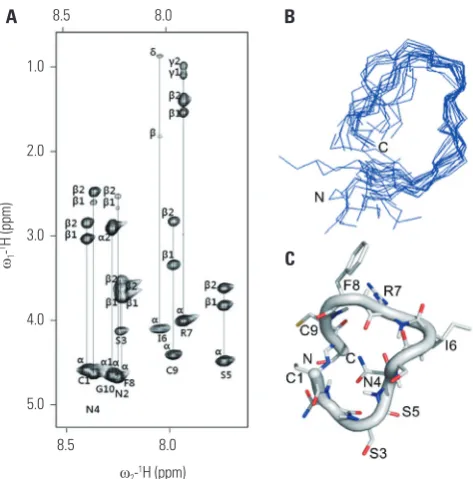
Solution structures of the cyclized oligopeptide Once the individual spin systems had been classified, back-bone sequential resonance assignment was easily completed via dαN (i, i+1) NOE connectivities in the 2D-NOESY spectra. The side chain assignment was performed via TOCSY con-nectivities (Fig. 1A). All NOEs were observed at mixing times of 600 ms. A total of 100 starting structures were calculated in the initial SA stage. The 20 lowest-energy structures (<SA>k)
were selected out of the 100 final simulated-annealing
struc-Fig. 1. NMR structure of cyclized oligopeptide. (A) 2D TOCSY spectrum of DKK1 (1 mM) in 100% DMSO at pH 7.0, 283 K, recorded with a spin-lock mixing time of 70 ms. The spectrum shows the identification of amino acid spin systems based on scalar correlation of the backbone amide protons of (ω2), with the respective side-chain protons (ω1) of each spin system. (B) A
backbone superposition of the energy-minimized average structure (<SA>kr)
over the family of 20 final SA structures (<SA>kr). (C) A ribbon diagram of
DKK1 with side-chain orientations is displayed as a stick model. NMR, nuclear magnetic resonance; 2D, two-dimensional; TOCSY, total correla-tion spectroscopy; DKK1, dickkopf 1; SA, simulated annealing.
1.0
2.0
3.0
4.0
5.0
8.5 8.0
8.5 8.0
ω2-1H (ppm) ω1
-1H (ppm)
A B
[image:3.595.316.553.389.629.2]tures for structural analysis. The average structure was gener-ated from the geometrical average of 20 structure coordinates and was subjected to restrained energy minimization to cor-rect bond length and angle distortions. The average NMR struc-ture exhibited 0.68-Å root-mean-square deviation for backbone atoms with respect to 20 (<SA>k) structures. A best-fit
super-position of all final structures and the backbone conformation for the average restrained-energy minimized structure (<SA>kr)
are displayed in Fig. 1B. The overall fold of the cyclized oligo-peptide is presented as a ribbon diagram in Fig. 1C. The cyclized oligopeptide shows a loop conformation with no regular sec-ondary structure.
Cyclized oligopeptide binds to the LRP5 E1 site and targets Wnt signaling
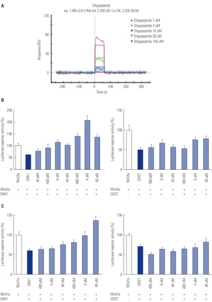
The binding activity increased as the flowing oligopeptide concentration increased from 1 µM to 100 µM (Fig. 2A). MC3T3-E1-S-Top cells were treated with Wnts, Wnt inhibitors, and oligopeptide. When the cells were treated with Wnt3a and DKK1, the activity increased as the concentration of oli-gopeptide increased; however, when treated with sclerostin, the activity could not overcome the inhibition effect (Fig. 2B). Similar results were observed with Wnt7a treatment (Fig. 2C). These results suggest that the oligopeptide is relatively DKK1 specific and that the oligopeptide targets the Wnt signaling pathway, abrogating the inhibition effects of DKK1.
Cyclized oligopeptide reverses the inhibitory effect of DKK1 on osteoblasts in vitro
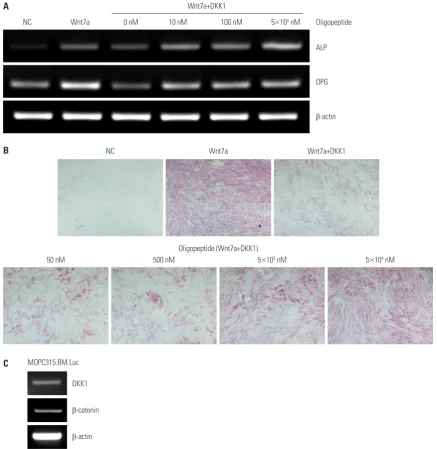
ST2 cells were treated with Wnt7a, DKK1, and oligopeptide. Wnt7a treatment increased ALP and OPG mRNA expression levels, and the addition of DKK1 decreased mRNA expression levels. However, when cells were treated with oligopeptide, the mRNA expressions of ALP and OPG increased again (Fig. 3A). This result indicated that the oligopeptide activates the canonical Wnt signaling that is inhibited by DKK1. To further confirm this result, ST2 cells were treated with Wnt7a, DKK1, and oligopeptide for ALP staining assay. The results of the ALP staining assay were similar to those seen in the RT-PCR. Cells treated with only Wnt7a expressed a stronger color, and the treatment of DKK1 decreased the staining. Cells treated with Wnt7a, DKK1, and oligopeptide could overcome the inhibi-tion effect of DKK1 dose dependently, shown by the increase in staining intensity (Fig. 3B). These results together suggested that this oligopeptide has bone-anabolic effects in vitro.
Cyclized oligopeptide inhibits tumor growth in a multiple myeloma mouse model
To determine the effect of oligopeptide on MM cell growth in vivo, DKK1 expression was confirmed. The mRNA expression of both DKK1 and β-catenin were detected (Fig. 3C), indicat-ing that the canonical Wnt signalindicat-ing pathway may have cer-tain effects on MM progression and bone lesions. After the
ex-pression of DKK1 was proven, we injected MOPC315.BM.Luc cells into the tail vein of 20 BALB/c mice (day -5), and five days later (day 0) mice were injected subcutaneously with ei-ther the oligopeptide or vehicle alone (Fig. 4A). Tumor burden was measured via BLI (Fig. 4B). On day 0, no significant radi-ance (p/sec/cm2/sr) was detected in any of the representative
mice of three groups. In contrast, on day 28, all mice injected with MM cells invoked a BLI signal. Importantly, larger radi-ances were detected in the vehicle group than in the oligo-peptide group. No significant radiances were detected in the control group, as no cells were injected. The signals of oligo-peptide-treated mice were predominantly detected in the spleen, while the signals of the vehicle-treated mice were more diffused all over the body, especially stronger in femur areas. The median radiance fold change of the vehicle group had the highest value, and the oligopeptide group was about three times lower (p=0.046) than the vehicle group (Fig. 4C). This data was divided into three ranges, 10 to less than 50, 50 to 500, and more than 500. In the lower range of fold change, there were more mice from the oligopeptide group (n=7) than the vehicle group (n=3). However, in the fold changes of 50 and higher, the number of the vehicle group mice was much larger (n=7) than the oligopeptide group mice (n=3) (Fig. 4D). All mice from the control group had fold changes of less than two. In accordance with the BLI results, the ELISA assessed M315 protein level of the peptide group showed lowering trend, compared to the vehicle group, with no significance (data not shown). This may be resulted because of the small number of mice in each group and their large variations of the M315 protein level. This observation showed that oligopep-tide affects the growth of MM cells in vivo, although the de-tailed mechanism remains unknown.
Cyclized oligopeptide has no direct effect on MOPC315.BM.Luc cell proliferation in vitro
One concern of using this oligopeptide is that it may affect the Wnt signaling pathway and stimulate cancer cell growth. In our in vivo experiment, however, we found that the tumor burdens were reduced with the treatment of the oligopeptide. To better define the effect of the oligopeptide on MM cell growth, we tested the oligopeptide in vitro, examining cell proliferation via WST assay. The MM cell population did not vary significantly in the differently treated groups (data not shown). This result demonstrated that oligopeptide does not affect MM cell growth directly, despite the MM cells exhibit-ing a Wnt/β-catenin signalexhibit-ing pathway.
Fig. 2. Luciferase reporter activity of cells treated with cyclized oligopeptide and ProteOn XPR36 analysis. MC3T3-E1-S-Top cells were treated with Wn-t3a, DKK1, sclerostin, and oligopeptide. (A) This graph shows the oligopeptide bound directly to the LRP5 E1 domain and the activity response increased dose dependently. (B) Graph on the left shows the dose increasing with luciferase reporter activity as the concentration of the oligopeptide increased. However, in the graph on the right, when treated with sclerostin, the same trend is not seen. (C) The same cells were treated with Wnt7a, DKK1, scleros-tin and oligopeptide. DKK1, dickkopf 1; LRP, lipoprotein receptor-related protein; SOST, sclerositn.
120
80
40
0
-200 -100 0 100 200 300 Time (s)
Oligopeptide
ka: 1.00E+0.8 1/Ms kd: 2.25E+03 1/s DK: 2.25E-05 M
Oligopeptide 1 uM Oligopeptide 3 uM Oligopeptide 10 uM Oligopeptide 30 uM Oligopeptide 100 uM
Response (RU)
250
200
150
100
50
0
150
100
50
0
150
100
50
0
150
100
50
0 Wnt3a + + + + + + + + +
DKK1 - + + + + + + + +
Wnt7a + + + + + + + + DKK1 - + + + + + + +
Wnt3a + + + + + + + + SOST - + + + + + + +
Wnt7a + + + + + + + + SOST - + + + + + + + Wnt3a DKK1 40 pM 400 pM 4 nM 40 nM 400 nM 4 uM 40 uM
Wnt7a DKK1 400 pM 4 nM 40 nM 400 nM 4 uM 40 uM
Wnt3a SOST 300 pM 3 nM 30 nM 300 nM 3 uM 30 uM
Wnt7a SOST 400 pM 4 nM 40 nM 400 nM 4 uM 40 uM
Luciferase reporter activity (%)
Luciferase reporter activity (%)
Luciferase reporter activity (%)
Luciferase reporter activity (%)
A
B
burden; however, further studies are needed.
DISCUSSION
In the present study, we showed that a cyclized oligopeptide treatment reduced tumor burden in the MOPC315.BM.Luc MM mouse model. To study the effects of this oligopeptide targeting the DKK1-LRP5/6 binding pocket in MM bone dis-ease, DKK1 NXI motif-containing cyclized oligopeptide was developed, and a series of in vitro and in vivo experiments
was performed.
Wnt signaling is well known for the osteoblast lineage speci-fication from mesenchymal cells, further differentiation of os-teoblast precursor cells, and skeletal development.24 DKK1,
an endogenous Wnt inhibitor, binds to LRP5/6 with Kremen to inhibit the canonical Wnt signaling pathway. Most MM cells express DKK1;14 furthermore, the secreted DKK1 disrupts
the RANKL/OPG expression ratio, which enhances osteoclast-mediated osteolysis and simultaneous osteoblast inhibition.15
[image:6.595.72.510.229.679.2]As DKK1 has been a potential therapeutic target for the treat-ment of MM, some effective therapies targeting DKK1 have
Fig. 3. Reverse transcription-PCR and Alkaline phosphatase staining assay. (A) ST2 cells were treated with Wnt7a, DKK1, and oligopeptide then the mRNA expression of both ALP and OPG assessed. (B) When Wnt7a alone was used, the staining was stronger than when Wnt7a and DKK1 were used
together. The staining intensity also increased dose dependently with increasing concentration of oligopeptide treatment. Original magnification ×40. (C)
The expressions of DKK1 and β-catenin were checked in the MOPC.315.BM.Luc cells prior to the experiment. DKK1, dickkopf 1; ALP, alkaline phosphatase; OPG, osteoprotegerin.
50 nM
NC
NC Wnt7a 0 nM 10 nM 100 nM 5×10⁴ nM Wnt7a+DKK1
Oligopeptide
ALP
OPG
β-actin
DKK1
β-catenin
β-actin
Wnt7a Wnt7a+DKK1
500 nM
Oligopeptide (Wnt7a+DKK1)
MOPC315.BM.Luc
5×10³ nM 5×10⁴ nM
Fig. 4. Tumor growth was reduced in the MOPC315.BM.Luc cell induced multiple myeloma mouse model. (A) A brief timeline of the animal study is shown. (B) One representative mouse from each group was chosen based on the closest radiance activity to the median value. Images taken on day 0 and day 28 are shown and compared. On day 0, BLI was taken before the oligopeptide treatment. No radiance was detected in the untreated mouse. (C) Biolumi-nescence radiance fold changes of each group from day 0 to day 28 are shown. Each group was comprised of 10 mice. (D) Mice numbers according to
the fold changes of bioluminescence radiance are shown. Fold changes were calculated simply by dividing the final value by the initial value. *p<0.05 and
***p<0.0001 by Mann-Whitney U test. MM, multiple myeloma.
Untreated
Vehicle
Oligopeptide
Untreated
Vehicle
Oligopeptide
Bioluminescence imaging (BLI)
s.c. injection of vehicle and oligopeptide 6 days/week for 4 weeks
Day 0 Day 28
MM cell injection (vehicle & oligopeptide group)
Day -5 Day 0 Day 28
2.0
1.5
1.0
0.5
6.0
5.0
4.0
3.0
2.0
1.0
×107
×106 Sacrifice
104
103
102
101
100
10-1
8
6
4
2
0
Control
10–<50 3
7
5
3
2
0
Vehicle
50–500
Biolunimescence fold change Vehicle Oligopeptide
Oligopeptide
>500 ***
*** *
Radiance fold change
Number of mice
A
B
been developed, including DKK1 neutralizing antibodies, proteasome inhibitors, DKK1 vaccines, and tumor-produced endothelin-1.26 A study using the SCID-rab mouse model of
myeloma showed that anti-DKK1 antibody treatment in-creased bone formation and reduced tumor burden in a rab-bit bone implanted with MM.19 Human anti-DKK1
monoclo-nal antibody (BHQ880) was also found to increase osteoblasts and blocked MM cell proliferation when MM cells were co-cultured with bone marrow stromal cells (BMSC).20
The cyclized oligopeptide against the DKK1-LRP5/6 bind-ing pocket abrogated the Wnt-β-catenin signalbind-ing inhibited by DKK1 (not by sclerostin) dose dependently (IC50≈5×10⁻⁸ M).
The NMR structure was obtained by complete proton reso-nance assignment. Isoleucine residue of the cyclized peptide should target the second pocket of LRP5/6. A ribbon diagram (Fig. 1C) showed that DKK1 is a linear peptide chain with a loop conformation.
The MM mice model we studied was developed in the Bo-gen lab.21 It was stated that the MM mice developed tumor
burdens mostly around long bone areas and also developed tumor cells in the bone marrow. How the MM cells affect the bone metabolism or bone marrow, however, was not deeply studied. The administration of the oligopeptide abrogated the suppression of canonical Wnt signaling by DKK1 and inhibit-ed tumor burden significantly. As matrix protein contains many growth factors and cytokines stimulating proliferation of tumor cells, inhibition of bone resorption by decreasing the RANKL/OPG ratio via the oligopeptide could reduce the re-lease of these growth factors and cytokines from matrix pro-teins and inhibit the overgrowth of MOPC315.BM.Luc cells. Similar to what we have achieved, the DKK1 neutralizing anti-body BHQ880 has been shown to not only improve bone for-mation but also to reduce tumor burden.20 Additionally,
stud-ies have reported that DKK1 suppressed β-catenin in myeloid-derived suppressor cells (a heterogeneous population of myeloid lineage immune cells in the stromal compartment) and thus inhibited tumor growth in mice.27 To explore the
mechanism of the tumor burden reduction, we studied the ef-fects of oligopeptide on proliferation of tumor cells. However, we could not find any direct effects of oligopeptide on the proliferation of tumor cells, despite the fact that DKK1 is ex-pressed in MOPC315.BM.Luc cells (data not shown). This re-sult was supported by a previous study showing that, while BHQ-880 did not have a direct anti-tumor effect on myeloma cells, it inhibited myeloma growth in the presence of BMSC.20
The MM niches contain bone marrow, fat cells, and immune cells, and they interact with each other. Several other cell types within the bone microenvironment produce significant amounts of DKK1, including megakaryocytes, endothelial cells, and os-teoblasts, and may contribute to the regulation of tumor gr-owth.28,29 TGF-β, VEGF, and FGF, as well as the increase of bone
resorption by osteoclasts, seem to support MM cell growth.30,31
In a previous study, DKK1 indirectly contributed to myeloma
growth by regulating IL-6 in the bone microenvironment. BHQ-880 significantly reduced the production of IL-6 levels in BMSC supernatants, and the addition of IL-6 reversed the ma-jority of growth inhibitory effects.20 Therefore, the overall results
we obtained may have derived from DKK1 acting on these cells differently.
In conclusion, the cyclized oligopeptide based on the DKK1-LRP5/6 interaction abrogates the suppression of canonical Wnt signaling by DKK1. Treatment of the oligopeptide re-duced tumor burden significantly in the MOPC315.BM.Luc MM mouse model. Cyclized oligopeptide may be an option for proper management of tumor burden in MM; however, further research is required to determine the details of the mechanisms involved.
ACKNOWLEDGEMENTS
MOPC315.BM.Luc cells and ab2.1-4 antibodies were obtained as generous gift from Bogen lab. This study was supported by Mid-career Research Program (NRF-2013R1A2A2A01068963) through NRF grant funded by the MEST; and Bo Mi Park, Dong-dong Zhang, Chu Hyun Bae and Heeyoun Kim are recipients of Brain Korea 21 PLUS grant.
REFERENCES
1. Raab MS, Podar K, Breitkreutz I, Richardson PG, Anderson KC. Multiple myeloma. Lancet 2009;374:324-39.
2. Balakumaran A, Robey PG, Fedarko N, Landgren O. Bone mar-row microenvironment in myelomagenesis: its potential role in early diagnosis. Expert Rev Mol Diagn 2010;10:465-80.
3. Ballester OF, Moscinski LC, Lyman GH, Chaney JV, Saba HI, Spi-ers AS, et al. High levels of interleukin-6 are associated with low tumor burden and low growth fraction in multiple myeloma. Blood 1994;83:1903-8.
4. Lee JW, Chung HY, Ehrlich LA, Jelinek DF, Callander NS, Rood-man GD, et al. IL-3 expression by myeloma cells increases both osteoclast formation and growth of myeloma cells. Blood 2004; 103:2308-15.
5. Heider U, Zavrski I, Jakob C, Bängeroth K, Fleissner C, Langelotz C, et al. Expression of receptor activator of NF-kappaB ligand (RANKL) mRNA in human multiple myeloma cells. J Cancer Res Clin Oncol 2004;130:469-74.
6. Sati HI, Greaves M, Apperley JF, Russell RG, Croucher PI. Expres-sion of interleukin-1beta and tumour necrosis factor-alpha in plasma cells from patients with multiple myeloma. Br J Haematol 1999;104:350-7.
7. Choi SJ, Cruz JC, Craig F, Chung H, Devlin RD, Roodman GD, et al. Macrophage inflammatory protein 1-alpha is a potential os-teoclast stimulatory factor in multiple myeloma. Blood 2000;96: 671-5.
8. Mukai T, Otsuka F, Otani H, Yamashita M, Takasugi K, Inagaki K, et al. TNF-alpha inhibits BMP-induced osteoblast differentiation through activating SAPK/JNK signaling. Biochem Biophys Res Commun 2007;356:1004-10.
10. Boland GM, Perkins G, Hall DJ, Tuan RS. Wnt 3a promotes prolif-eration and suppresses osteogenic differentiation of adult human mesenchymal stem cells. J Cell Biochem 2004;93:1210-30. 11. Kim JH, Liu X, Wang J, Chen X, Zhang H, Kim SH, et al. Wnt
sig-naling in bone formation and its therapeutic potential for bone diseases. Ther Adv Musculoskelet Dis 2013;5:13-31.
12. Li X, Ominsky MS, Warmington KS, Morony S, Gong J, Cao J, et al. Sclerostin antibody treatment increases bone formation, bone mass, and bone strength in a rat model of postmenopausal osteo-porosis. J Bone Miner Res 2009;24:578-88.
13. Fedi P, Bafico A, Nieto Soria A, Burgess WH, Miki T, Bottaro DP, et al. Isolation and biochemical characterization of the human Dkk-1 homologue, a novel inhibitor of mammalian Wnt signaling. J Biol Chem 1999;274:19465-72.
14. Mitsiades CS, McMillin DW, Klippel S, Hideshima T, Chauhan D, Richardson PG, et al. The role of the bone marrow microenviron-ment in the pathophysiology of myeloma and its significance in the development of more effective therapies. Hematol Oncol Clin North Am 2007;21:1007-34.
15. Qiang YW, Chen Y, Stephens O, Brown N, Chen B, Epstein J, et al. Myeloma-derived Dickkopf-1 disrupts Wnt-regulated osteoprote-gerin and RANKL production by osteoblasts: a potential mecha-nism underlying osteolytic bone lesions in multiple myeloma. Blood 2008;112:196-207.
16. Tian E, Zhan F, Walker R, Rasmussen E, Ma Y, Barlogie B, et al. The role of the Wnt-signaling antagonist DKK1 in the develop-ment of osteolytic lesions in multiple myeloma. N Engl J Med 2003;349:2483-94.
17. Rachner TD, Hadji P, Hofbauer LC. Novel therapies in benign and malignant bone diseases. Pharmacol Ther 2012;134:338-44. 18. Rachner TD, Göbel A, Benad-Mehner P, Hofbauer LC, Rauner M.
Dickkopf-1 as a mediator and novel target in malignant bone dis-ease. Cancer Lett 2014;346:172-7.
19. Yaccoby S, Ling W, Zhan F, Walker R, Barlogie B, Shaughnessy JD Jr. Antibody-based inhibition of DKK1 suppresses tumor-induced bone resorption and multiple myeloma growth in vivo. Blood 2007; 109:2106-11.
20. Fulciniti M, Tassone P, Hideshima T, Vallet S, Nanjappa P, Etten-berg SA, et al. Anti-DKK1 mAb (BHQ880) as a potential
therapeu-tic agent for multiple myeloma. Blood 2009;114:371-9.
21. Hofgaard PO, Jodal HC, Bommert K, Huard B, Caers J, Carlsen H, et al. A novel mouse model for multiple myeloma (MOPC315. BM) that allows noninvasive spatiotemporal detection of osteo-lytic disease. PLoS One 2012;7:e51892.
22. Davis DG, Bax A. Assignment of complex proton NMR spectra via two-dimensional homonuclear Hartnabb-Hahn spectroscopy. J Am Chem Soc 1985;107:2820-1.
23. Jeener J, Meier BH, Bachman P, Ernst RR. Investigation of exchange processes by two-dimensional NMR spectroscopy. J Chem Phys 1979;71:4546-53.
24. Koradi R, Billeter M, Wüthrich K. MOLMOL: a program for dis-play and analysis of macromolecular structures. J Mol Graph 1996; 14:51-5.
25. Jami A, Gadi J, Lee MJ, Kim EJ, Lee MJ, Jung HS, et al. Pax6 ex-pressed in osteocytes inhibits canonical Wnt signaling. Mol Cells 2013;35:305-12.
26. Zhou F, Meng S, Song H, Claret FX. Dickkopf-1 is a key regulator of myeloma bone disease: opportunities and challenges for thera-peutic intervention. Blood Rev 2013;27:261-7.
27. D’Amico L, Capietto AH, Zamani A, Faccio R, Bumpass D. Dick-kopf-realtaed protein 1 (Dkk1) exerts immune suppressive effects in cancer by regulating expansion and function of myeloid de-rived suppressor cells. Seattle: Paper presented at Annual Meet-ing of the American Society for Bone and Mineral Research; 2015. 28. Wong D, Winter O, Hartig C, Siebels S, Szyska M, Tiburzy B, et al.
Eosinophils and megakaryocytes support the early growth of mu-rine MOPC315 myeloma cells in their bone marrow niches. PLoS One 2014;9:e109018.
29. Smadja DM, d’Audigier C, Weiswald LB, Badoual C, Dangles-Marie V, Mauge L, et al. The Wnt antagonist Dickkopf-1 increases endo-thelial progenitor cell angiogenic potential. Arterioscler Thromb Vasc Biol 2010;30:2544-52.
30. Yata K, Yaccoby S. The SCID-rab model: a novel in vivo system for primary human myeloma demonstrating growth of CD138-ex-pressing malignant cells. Leukemia 2004;18:1891-7.