Florence Abravanel,a,bKarine Sandres-Saune,a,bSebastien Lhomme,a,bMartine Dubois,a,bJean-Michel Mansuy,band Jacques Izopeta,b
INSERM, U1043, Centre de Physiopathologie de Toulouse Purpan, Toulouse, France,aand CHU Toulouse, Hôpital Purpan, Laboratoire de Virologie, Institut Fédératif de Biologie de Purpan, Toulouse, Franceb
Genotype 3 hepatitis E viruses (HEVs) are distributed across the world and are now considered to be an emerging public health
concern in industrialized countries. At least 10 genotype 3 subtypes have been identified in humans and animals worldwide. It
was recently reported that the sensitivities of HEV RNA assays differ greatly. We have assessed the influence of genotype 3
diver-sity on the performances of two HEV RNA assays: one targeting the ORF3 gene and the other targeting the ORF2 gene. We tested
a panel of 5 HEV-positive reference samples of genotypes 3a, 3b, 3c, 3e, and 3f at 10-fold serial dilutions. The HEV RNA
concen-trations obtained with both reverse transcription (RT)-PCRs were correlated, but the RT-PCR based on ORF2 underestimated
the HEV RNA concentrations. The mean [ORF3
ⴚ
ORF2] difference was 1.41 log copies/ml. We also tested 34 clinical specimens
of genotypes 3c (
n
ⴝ
15), 3e (
n
ⴝ
4), and 3f (
n
ⴝ
15), representing the most prevalent subtypes in Europe. The mean [ORF3
ⴚ
ORF2] differences were 1.41 log copies/ml for genotype 3c, 0.96 log copies/ml for genotype 3e, and 0.70 log copies/ml for
geno-type 3f. The bias between the 2 RT-PCR assays was significantly greater for genogeno-type 3c than for genogeno-type 3f (
P
ⴝ
0.007). We
therefore recommend the use of an RT-PCR protocol based on ORF3 to quantify HEV RNA of genotype 3 strains.
H
epatitis E virus (HEV) is the causative agent of acute or
ful-minant hepatitis in many resource-limited regions of the
world. It is transmitted primarily by the fecal-oral route (36). It is
now also considered to be an emerging concern in industrialized
countries (8, 41). HEV is a nonenveloped, single-stranded,
positive-sense RNA virus and a member of the family
Hepeviridae
(10). Its genome consists of a single-stranded, positive-sense
RNA, approximately 7.2 kb long, which is capped and
polyadenyl-ated (18). It contains a short 5
=
untranslated region (UTR), three
open reading frames (ORFs) (ORF1, ORF2, and ORF3), and a 3
=
UTR. ORF1 encodes nonstructural proteins, including
methyl-transferase, papain-like cysteine protease, helicase, and
RNA-dependent RNA polymerase (25). ORF2 encodes the viral capsid
protein. ORF3 encodes a small, phosphorylated protein, 113 or
114 amino acids long, that is involved in virion morphogenesis
and release (11, 12).
Analyses of the nucleotide sequences of HEV strains have
re-vealed extensive genomic diversity, leading to the identification of
4 main genotypes and several subtypes within each genotype (31).
HEV genotypes 1 and 2 are restricted to humans and are
associ-ated with epidemics in developing countries, whereas HEV
geno-types 3 and 4 are zoonotic and are responsible for sporadic cases.
While genotype 4 is restricted to parts of Asia, genotype 3 is
pres-ent worldwide (31). However, the HEV genotype 3 subtypes each
have distinct geographic distributions. Genotype 3a and 3b strains
are found more frequently in North America and Japan (31, 37),
while genotype 3c, 3e, and 3f strains are the most prevalent
sub-types in European countries (28, 31, 42).
HEV genotype 3 is an emerging cause of acute and chronic
hepatitis in immunocompromised patients (21, 26), and it poses
concerns for food and environmental safety worldwide (35). The
optimal diagnosis of an HEV infection relies on a combination of
serological tests and nucleic acid amplification techniques (NATs)
(9). An accurate quantitative assay of HEV RNA is also necessary
for pathophysiological studies (22) and to monitor the HEV loads
of chronically infected patients on antiviral therapy (2, 19, 20).
Several in-house conventional or real-time reverse
transcrip-tion (RT)-PCRs based on the amplificatranscrip-tion of the ORF2 or ORF3
gene were recently evaluated (3). Those investigators found that
the sensitivities of the majority of the assays differed greatly. We
have therefore investigated the influence of genotype 3 diversity
on the performances of two quantitative real-time RT-PCRs: one
based on the amplification of a fragment within ORF2 and the
other based on the amplification of a fragment within the ORF3
gene.
MATERIALS AND METHODS
HEV reference strain panel.Genotype 3a, 3b, 3c, 3e, and 3f strains were selected. The genotype 3a and 3b strains came from previous studies per-formed with the Paul Ehrlich Institute and were provided by Keiji Nas-tubayashi from the Japanese Red Cross Hokkaido Blood Center, and the genotype 3c, 3e, and 3f strains were from blood samples collected from patients with acute or chronic infections in the Midi-Pyrenees area, France (27, 32). Each strain was characterized by the sequencing of a 189-nucleotide (nt) ORF2 fragment, as previously described (28). Each sample was tested undiluted or diluted 1/10 and 1/100 in HEV-negative plasma. Each dilution was quantified in duplicate in 4 distinct runs.
Clinical specimens.We tested 34 plasma samples from French pa-tients with French isolates: 15 from papa-tients infected with HEV genotype 3c, 15 from patients infected with genotype 3f, and 4 from patients in-fected with genotype 3e. Each strain was characterized by the sequencing of a 189-nt ORF2 fragment, as previously described (28).
HEV RNA extraction.HEV RNA was extracted from blood samples (850l) with the Total Nucleic Acid Isolation (TNAI) kit on the Cobas Ampliprep instrument according to the manufacturer’s instructions (Roche Diagnostics, France).
Real-time PCR based on ORF3.One-step real-time RT-PCR was per-formed with the LightCycler 480 instrument (Roche Diagnostics, France). The following primers and probe targeting the ORF2/ORF3 overlapping
Received28 September 2011Returned for modification6 November 2011
Accepted16 December 2011
Published ahead of print28 December 2011
Address correspondence to Florence Abravanel, [email protected]. Copyright © 2012, American Society for Microbiology. All Rights Reserved.
doi:10.1128/JCM.05942-11
on May 16, 2020 by guest
http://jcm.asm.org/
region were used to amplify a 70-nt fragment: forward primer HEVORF3-S (5=-GGTGGTTTCTGGGGTGAC-3=), reverse primer HEVORF3-AS (5=-AGGGGTTGGTTGGATGAA-3=), and the probe 5=– 6-carboxyfluorescein (FAM)–TGATTCTCAGCCCTTCGC– 6-carboxy-tetramethylrhodamine (TAMRA)–3=(17). For RT-PCR, the 50-l reac-tion mix contained 1 l of SuperScript III Platinum One-Step quantitative RT-PCR system medium (Invitrogen), 15l of RNA, prim-ers (200 nM), probes (150 nM), and 40 U/reaction RNase Out (Invitro-gen). Reverse transcription was carried out at 50°C for 15 min, followed by denaturation at 95°C for 1 min. DNA was amplified with 50 PCR cycles at 95°C (20 s) and 58°C (40 s). The amplification efficiency calculated with a standard curve was 2.02. The limit of detection was 100 copies/ml.
Real-time PCR based on ORF2.One-step real-time RT-PCR was performed with the LightCycler 480 instrument (Roche Diagnostics) as previously described (26, 34). Primers and probes targeting a 140-nt fragment within the ORF2 gene were forward primer HEVORF2-S1 (5=-GACAGAATTRATTTCGTCGGCTGG-3=), reverse primer HEVORF2-A2 (5=-CCCTTRTCCTGCTGNGCATTCTCGACAGA-3=), and probe HEVORF2-S2 (5= -FAM-GTYGTCTCRGCCAATGGCGAGC-TAMRA-3=). The 50-l RT-PCR mix contained 1l SuperScript III Plat-inum One-Step quantitative RT-PCR system medium (Invitrogen), 15l of RNA, primers (200 nM) and probes (150 nM), and 40 U/reaction RNase Out (Invitrogen). Reverse transcription was carried out at 42°C for 15 min, followed by denaturation at 95°C for 1 min. DNA was amplified with 50 PCR cycles at 95°C (20 s) and 60°C (1 min). The amplification efficiency calculated with a standard curve was 1.96. The limit of detection was 100 copies/ml.
RNA standards.Two transcribed RNA standards were constructed from a sample from a patient infected with genotype 3f (GenBank acces-sion numberEU495148): one was based on the amplification of a
frag-ment within the ORF3 gene (70 nt), and the other was based on the amplification of a fragment within the ORF2 gene (140 nt). Each of the resulting cDNAs was purified and inserted and cloned into the PCR-II vector using Topo TA cloning for sequencing (Invitrogen). The positive clone, screened by colony PCR, was confirmed by digestion with a restric-tion enzyme and sequencing. The vector was cut down with EcoRI and cloned into transcriptional vector pGEM.3Z with the same enzyme sites. pGEM.3Z was linearized with SmaI and retrotranscribed by T7 RNA poly-merase to obtain a positive strand for use as an RNA standard for quan-titative RT-PCR. The transcribed RNA standard was titrated by measur-ing the optical density with a spectrophotometer. A standard curve was generated from serial 10-fold dilutions of the standard.
Statistical analysis.The Spearman test was used to test the correlation between the 2 RT-PCR assays. One-way analysis of variance (ANOVA) was used to test for differences in quantification between the 2 RT-PCR assays.
RESULTS
Reference strain panel.
We assayed 5 samples, containing
geno-type 3a, 3b, 3c, 3e, and 3f strains, undiluted or diluted, with the
ORF2-based time RT-PCR and with the ORF3-based
real-time RT-PCR. The reproducibility of each RT-PCR assay was
good (Table 1). The mean standard deviations were 0.14 log
cop-ies/ml (range, 0.03 to 0.44 log copcop-ies/ml) for the ORF2-based
RT-PCR and 0.13 log copies/ml (range, 0.07 to 0.21 log copies/ml) for
the RT-PCR based on ORF3.
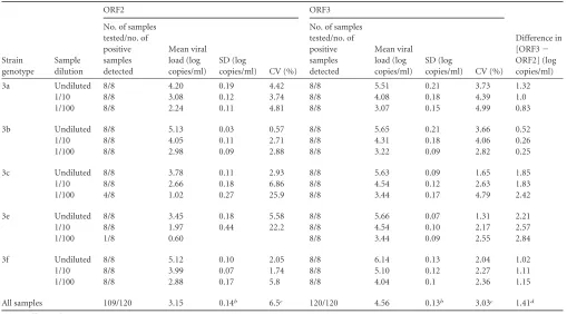
[image:2.585.39.548.76.358.2]However, only one of the eight 1/100-diluted genotype 3e
sam-ples was detected with the RT-PCR based on ORF2. Similarly, only
4 of the eight 1/100-diluted genotype 3c samples were detected
TABLE 1HEV RNA results for the panel of genotype 3a, 3b, 3c, 3e, and 3f strainsa
Strain genotype
Sample dilution
ORF2 ORF3
Difference in [ORF3⫺ ORF2] (log copies/ml) No. of samples
tested/no. of positive samples detected
Mean viral load (log copies/ml)
SD (log
copies/ml) CV (%)
No. of samples tested/no. of positive samples detected
Mean viral load (log copies/ml)
SD (log
copies/ml) CV (%) 3a Undiluted 8/8 4.20 0.19 4.42 8/8 5.51 0.21 3.73 1.32
1/10 8/8 3.08 0.12 3.74 8/8 4.08 0.18 4.39 1.0 1/100 8/8 2.24 0.11 4.81 8/8 3.07 0.15 4.99 0.83 3b Undiluted 8/8 5.13 0.03 0.57 8/8 5.65 0.21 3.66 0.52 1/10 8/8 4.05 0.11 2.71 8/8 4.31 0.18 4.06 0.26 1/100 8/8 2.98 0.09 2.88 8/8 3.22 0.09 2.82 0.25 3c Undiluted 8/8 3.78 0.11 2.93 8/8 5.63 0.09 1.65 1.85 1/10 8/8 2.66 0.18 6.86 8/8 4.54 0.12 2.63 1.83 1/100 4/8 1.02 0.27 25.9 8/8 3.44 0.17 4.79 2.42 3e Undiluted 8/8 3.45 0.18 5.58 8/8 5.66 0.07 1.31 2.21 1/10 8/8 1.97 0.44 22.2 8/8 4.54 0.10 2.17 2.57 1/100 1/8 0.60 8/8 3.44 0.09 2.55 2.84 3f Undiluted 8/8 5.12 0.10 2.05 8/8 6.14 0.13 2.04 1.02 1/10 8/8 3.99 0.07 1.74 8/8 5.10 0.12 2.27 1.11 1/100 8/8 2.88 0.17 5.8 8/8 4.04 0.1 2.36 1.15 All samples 109/120 3.15 0.14b 6.5c 120/120 4.56 0.13b 3.03c 1.41d
aCV, coefficient of variation. bMean standard deviation. cMean coefficient of variation. dMean difference.
on May 16, 2020 by guest
http://jcm.asm.org/
with the RT-PCR based on ORF2. All the other samples scored
positive with both assays (Table 1).
The virus loads in the 109 positive samples measured by each
assay are shown in Fig. 1. The ORF3 RT-PCR and ORF2 RT-PCR
results were linearly associated (
R
2⫽
0.52) and correlated (
⫽
0.69;
P
⬍
0.001) (Fig. 1). The ORF2 RT-PCR gave a mean HEV
RNA concentration of 3.15 log copies/ml, and the ORF3 RT-PCR
assay gave a mean HEV RNA concentration of 4.56 log copies/ml.
The mean deviation between the ORF3 and the ORF2 RT-PCR
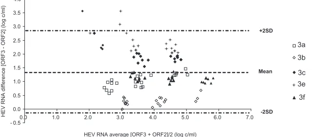
results was 1.41 log copies/ml. Bland-Altman analysis showed that
differences in virus load were independent of the concentration of
HEV RNA (Fig. 2). However, the average deviation varied with the
HEV subtype. The mean [ORF3
⫺
ORF2] differences were 2.54
log copies/ml for the genotype 3e samples, 2.03 log copies/ml for
the genotype 3c samples, 1.09 log copies/ml for the genotype 3f
samples, 1.05 log copies/ml for the genotype 3a samples, and 0.34
log copies/ml for the genotype 3b samples. HEV genetic
polymor-phisms may generate mismatches between HEV RNA and primers
or probes, which could contribute to underquantification by the
RT-PCR based on ORF2 (Fig. 3).
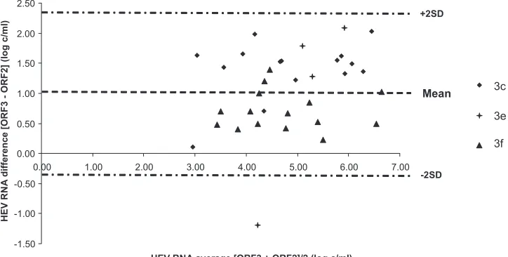
Clinical specimens.
We also tested 34 clinical samples with
both assays in order to eliminate any potential bias due to assaying
only the 5 reference strains. These samples included samples of
genotypes 3c (
n
⫽
15), 3e (
n
⫽
4), and 3f (
n
⫽
15), which are the
most prevalent genotype 3 subtypes found in Europe. The ORF3
RT-PCR and ORF2 RT-PCR results were correlated (
⫽
0.82;
P
⬍
0.001). The ORF3 RT-PCR gave a mean HEV RNA
concen-tration of 5.40 log copies/ml, and the ORF2 RT-PCR assay gave a
mean HEV concentration of 4.36 log copies/ml. The mean
devia-tion between the ORF3 and the ORF2 RT-PCR results was 1.04 log
copies/ml. Data for the Bland-Altman analysis are shown in Fig. 4.
Again, the average deviation varied with the HEV subtype. The
mean [ORF3
⫺
ORF2] differences were 1.41 log copies/ml for
genotype 3c, 0.96 log copies/ml for genotype 3e, and 0.70 log
cop-ies/ml for genotype 3f. The average deviation between the 3
sub-types was significantly different (
P
⫽
0.009). The bias between the
2 RT-PCRs was significantly greater for the genotype 3c samples
than for the genotype 3f samples (
P
⫽
0.007).
DISCUSSION
We have assessed the influence of HEV genotype 3 diversity on the
performances of two HEV RNA quantitative assays. The RT-PCR
assays were correlated, but we found substantial differences in the
quantities of RNA of the main genotype 3 subtypes detected by the
two real-time RT-PCRs.
Several protocols for real-time PCR targeting the ORF3 region
(13, 17, 40) or the ORF2 region (1, 16, 34, 47) have been developed
for the detection of HEV RNA over the past 10 years. The
perfor-FIG 1HEV RNA concentrations of the genotype 3a, 3b, 3c, 3e, and 3f reference strains measured by the ORF3 and ORF2 RT-PCRs.
FIG 2Bland-Altman plot for bias analysis between RT-PCRs based on ORF3 and ORF2 for the panel of genotype 3a, 3b, 3c, 3e, and 3f reference strains. SD, standard deviation.
on May 16, 2020 by guest
http://jcm.asm.org/
[image:3.585.136.451.67.237.2] [image:3.585.135.466.561.701.2]mances of assays based on the amplification of HEV RNA nucleic
acid were recently investigated by using a panel of
HEV-containing plasma samples (3). The panel was comprised of 22
HEV-positive plasma samples representing 10-fold serial
dilu-tions of HEV subtypes 3a, 3b, 3f, and 4c obtained from blood
donors. Only 2 of the 20 laboratories that tested the panel used an
RT-PCR protocol that targeted the ORF1 region of the HEV
ge-nome; the other methods targeted the ORF2 and ORF3 regions.
That study demonstrated that real-time RT-PCRs are more
sensi-tive than nested PCRs, but the sensitivities of the majority of the
assays differed enormously (100-fold to 1,000-fold), independent
of the virus strains (3). Among protocols targeting ORF3, we
se-lected one developed by Jothikumar et al., because it was the most
frequently used protocol in a recent evaluation performed by
Bay-lis et al. (17). Among protocols targeting ORF2, we selected a
method developed previously by Mansuy et al., because it was
previously used in our laboratory for HEV RNA detection and
quantification (26, 34).
The great genetic diversity of RNA viruses makes it very
diffi-cult to design appropriate primers and probes for use in the
de-velopment of molecular diagnostic assays. The performance of
quantitative assays for RNA viruses is influenced by their genetic
diversity (15, 23, 43). For the 34 clinical samples, the sequence
identity between the different subtypes in the ORF2 region ranged
from 83% to 90.4% (data not shown). Based on the genotype 3c,
3e, and 3f complete genome sequences available in the GenBank
database, the sequence identities between the different subtypes
ranged from 82.4% to 90% in the ORF2 region and ranged from
89.5% to 95.2% in the ORF3 region. This variability between
sub-types has prompted us to assess its influence on HEV RNA
quan-tification. Our sequence alignments have shown several
mis-matches, mainly for the primers and the probe targeting ORF2.
Nevertheless, the ORF2 assay showed a similar sensitivity in the
detection of genotype 3a and 3f standard preparations despite a
higher number of mismatches between the ORF2 primers and
probe and the 3a strain than between the ORF2 primers and probe
and the 3f strain. This may be because other critical parameters,
such as RNA conformation, could be similar for the two TaqMan
detection systems.
We evaluated the capacities of two different TaqMan real-time
RT-PCRs to detect and quantify HEV genotype 3 subtypes. Our
assays of serial 10-fold dilutions of genotype 3a, 3b, 3c, 3e, and 3f
reference samples indicated that the ORF2-based RT-PCR was less
FIG 3Alignment of reference sequences showing the positions of the primers and probes in the HEV ORF2 (A) and HEV ORF3 (B) regions. Nucleotides in gray indicate mismatches with the primer or probe. Numbers refer to the corresponding nucleotide positions of HEV (GenBank accession number
M73218).
FIG 4Bland-Altman plot for bias analysis between the RT-PCRs based on ORF3 and ORF2 for the 34 clinical samples. SD, standard deviation.
on May 16, 2020 by guest
http://jcm.asm.org/
[image:4.585.112.474.523.706.2]sensitive than the ORF3-based RT-PCR. The RT-PCR based on
ORF2 rarely detected the 100-fold-diluted genotype
3c and 3e samples, and our assays of several clinical plasma
samples showed that the RT-PCR based on ORF2 significantly
underestimated the HEV RNA concentration in genotype 3c
sam-ples compared to the genotype 3f samsam-ples. These data agree well
with findings described previously by Ward et al. (46), who
stud-ied the performances of 4 real-time RT-PCRs based on ORF3 and
ORF2 by testing Canadian swine genotype 3 samples. Those
au-thors compared the threshold cycle (
C
T) values obtained with the
real-time RT-PCR tests and found that the RT-PCR test based on
ORF3 was the most sensitive test for the detection of swine HEV
strains (46). Unfortunately, those researchers did not determine
the HEV subtypes, and their assays were only semiquantitative.
Our study comparison of two HEV RNA assays was focused on
HEV genotype 3, the most prevalent genotype in industrialized
countries. However, the primers and the probe for the RT-PCR
based on the ORF2 region, adapted from our previously reported
in-house protocol (26, 34), were designed in order to detect the 4
main genotypes of HEV. They were used to conduct several
pre-vious studies of HEV infections (19–21, 26, 32–34), and they allow
us to detect HEV genotype 1 and 4 infections (32). Similarly, we
have used the primers and the probes targeting the ORF3 region
designed previously by Jothikumar et al. (17). Those researchers
validated the capacity of these primers to detect HEV isolates
rep-resenting genotypes 1 to 4 with several samples. However, further
studies are needed to address whether the quantitative HEV RNA
assays are influenced by the HEV genetic diversity for all the
ge-notypes.
The extraction protocol can affect the sensitivities of molecular
tests (4, 14), but we used automated extraction with the Cobas
Ampliprep instrument to extract the HEV RNA used for both
assays. This automated extraction is more reliable, standardized,
reproducible, and time-saving than manual extraction for the
preparation of nucleic acids (24, 44). This procedure also limits
the risk of cross-contamination in the laboratory.
The diagnosis of an HEV infection in a patient requires
accu-rate and sensitive tools. Because serological tests may lack
sensi-tivity (9, 29), the detection of virus genomic RNA in serum or
stool samples by RT-PCR is a crucial marker of an acute or a
chronic HEV infection. The ubiquitous nature of HEV genotype 3
strains in domestic pigs and wild boars also raises public health
concerns for zoonotic infection through direct contacts with
in-fected animals (5, 7, 38) or the consumption of contaminated
animal products (6, 30, 45). In addition, genotype 3
HEV-contaminated coastal, drinking, irrigation, and sewage waters
could all be sources for human infections (35). This reinforces the
need for a highly sensitive RT-PCR protocol for the detection of
HEV in food or water samples. The development of sensitive
as-says may reveal that HEV is far more common in industrialized
countries than was previously thought (39). Additionally, an
un-derestimation of HEV RNA quantification could result in
false-negative results in treated immunocompromised patients (2, 19,
20) and thus may be the cause of a relapse if the treatment is
stopped too early.
In conclusion, our findings indicate that an RT-PCR protocol
based on ORF3 provides the most suitable tool for assaying the
HEV RNA of genotype 3 strains.
REFERENCES
1.Ahn JM, Rayamajhi N, Gyun Kang S, Sang Yoo H.2006. Comparison of real-time reverse transcriptase-polymerase chain reaction and nested or commercial reverse transcriptase-polymerase chain reaction for the detec-tion of hepatitis E virus particle in human serum. Diagn. Microbiol. Infect. Dis.56:269 –274.
2.Alric L, Bonnet D, Laurent G, Kamar N, Izopet J. 2010. Chronic hepatitis E virus infection: successful virologic response to pegylated interferon-alpha therapy. Ann. Intern. Med.153:135–136.
3.Baylis SA, Hanschmann KM, Blumel J, Nubling CM.2011. Standard-ization of hepatitis E virus (HEV) nucleic acid amplification technique-based assays: an initial study to evaluate a panel of HEV strains and inves-tigate laboratory performance. J. Clin. Microbiol.49:1234 –1239. 4.Bianchi S, Dal Vecchio A, Vilarino ML, Romalde JL.2011. Evaluation of
different RNA-extraction kits for sensitive detection of hepatitis A virus in strawberry samples. Food Microbiol.28:38 – 42.
5.Christensen PB, et al.2008. Time trend of the prevalence of hepatitis E antibodies among farmers and blood donors: a potential zoonosis in Den-mark. Clin. Infect. Dis.47:1026 –1031.
6.Colson P, et al.2010. Pig liver sausage as a source of hepatitis E virus transmission to humans. J. Infect. Dis.202:825– 834.
7.Colson P, Kaba M, Bernit E, Motte A, Tamalet C.2007. Hepatitis E associated with surgical training on pigs. Lancet370:935.
8.Dalton HR, Bendall R, Ijaz S, Banks M.2008. Hepatitis E: an emerging infection in developed countries. Lancet Infect. Dis.8:698 –709. 9.Drobeniuc J, et al.2010. Serologic assays specific to immunoglobulin M
antibodies against hepatitis E virus: pangenotypic evaluation of perfor-mances. Clin. Infect. Dis.51:e24 – e27.
10. Emerson SU, et al.2005. Hepevirus, p 853– 857.InFauquet CM, Mayo MA, Maniloff J, Desselberg U, Ball LA (ed), Virus taxonomy. Eighth re-port of the International Committee on Taxonomy of Viruses. Elsevier Academic Press, London, United Kingdom.
11. Emerson SU, Nguyen H, Torian U, Purcell RH.2006. ORF3 protein of hepatitis E virus is not required for replication, virion assembly, or infec-tion of hepatoma cells in vitro. J. Virol.80:10457–10464.
12. Emerson SU, et al.2010. Release of genotype 1 hepatitis E virus from cultured hepatoma and polarized intestinal cells depends on open reading frame 3 protein and requires an intact PXXP motif. J. Virol.84:9059 – 9069.
13. Enouf V, et al.2006. Validation of single real-time TaqMan PCR assay for the detection and quantitation of four major genotypes of hepatitis E virus in clinical specimens. J. Med. Virol.78:1076 –1082.
14. Gartner BC, Fischinger JM, Litwicki A, Roemer K, Mueller-Lantzsch N.
2004. Evaluation of a new automated, standardized generic nucleic acid extraction method (total nucleic acid isolation kit) used in combination with cytomegalovirus DNA quantification by COBAS AMPLICOR CMV MONITOR. J. Clin. Microbiol.42:3881–3882.
15. Gueudin M, et al.2007. Evaluation of the Roche Cobas TaqMan and Abbott RealTime extraction-quantification systems for HIV-1 subtypes. J. Acquir. Immune Defic. Syndr.44:500 –505.
16. Gyarmati P, et al.2007. Universal detection of hepatitis E virus by two real-time PCR assays: TaqMan and Primer-Probe Energy Transfer. J. Vi-rol. Methods146:226 –235.
17. Jothikumar N, Cromeans TL, Robertson BH, Meng XJ, Hill VR.2006. A broadly reactive one-step real-time RT-PCR assay for rapid and sensi-tive detection of hepatitis E virus. J. Virol. Methods131:65–71. 18. Kabrane-Lazizi Y, Meng XJ, Purcell RH, Emerson SU.1999. Evidence
that the genomic RNA of hepatitis E virus is capped. J. Virol.73:8848 – 8850.
19. Kamar N, et al.2010. Pegylated interferon-alpha for treating chronic hepatitis E virus infection after liver transplantation. Clin. Infect. Dis.
50:e30 – e33.
20. Kamar N, et al. 2010. Ribavirin therapy inhibits viral replication on patients with chronic hepatitis E virus infection. Gastroenterology139: 1612–1618.
21. Kamar N, et al.2008. Hepatitis E virus and chronic hepatitis in organ-transplant recipients. N. Engl. J. Med.358:811– 817.
22. Kar P, et al.2008. Does hepatitis E viral load and genotypes influence the final outcome of acute liver failure during pregnancy? Am. J. Gastroen-terol.103:2495–2501.
23. Klungthong C, et al.2010. The impact of primer and probe-template
on May 16, 2020 by guest
http://jcm.asm.org/
mismatches on the sensitivity of pandemic influenza A/H1N1/2009 virus detection by real-time RT-PCR. J. Clin. Virol.48:91–95.
24. Knepp JH, Geahr MA, Forman MS, Valsamakis A.2003. Comparison of automated and manual nucleic acid extraction methods for detection of enterovirus RNA. J. Clin. Microbiol.41:3532–3536.
25. Koonin EV, et al.1992. Computer-assisted assignment of functional domains in the nonstructural polyprotein of hepatitis E virus: delineation of an additional group of positive-strand RNA plant and animal viruses. Proc. Natl. Acad. Sci. U. S. A.89:8259 – 8263.
26. Legrand-Abravanel F, et al.2010. Characteristics of autochthonous hep-atitis E virus infection in solid-organ transplant recipients in France. J. Infect. Dis.202:835– 844.
27. Legrand-Abravanel F, et al.2011. Hepatitis E virus infection without reactivation in solid-organ transplant recipients, France. Emerg. Infect. Dis.17:30 –37.
28. Legrand-Abravanel F, et al.2009. Hepatitis E virus genotype 3 diversity, France. Emerg. Infect. Dis.15:110 –114.
29. Legrand-Abravanel F, et al.2009. Good performance of immunoglobulin M assays in diagnosing genotype 3 hepatitis E virus infections. Clin. Vac-cine Immunol.16:772–774.
30. Li TC, et al.2005. Hepatitis E virus transmission from wild boar meat. Emerg. Infect. Dis.11:1958 –1960.
31. Lu L, Li C, Hagedorn CH.2006. Phylogenetic analysis of global hepatitis E virus sequences: genetic diversity, subtypes and zoonosis. Rev. Med. Virol.16:5–36.
32. Mansuy JM, et al.2009. Acute hepatitis E in south-west France over a 5-year period. J. Clin. Virol.44:74 –77.
33. Mansuy JM, et al.2009. Molecular evidence of patient-to-patient trans-mission of hepatitis E virus in a hematology ward. Clin. Infect. Dis.48: 373–374.
34. Mansuy JM, et al.2004. Hepatitis E in the south west of France in indi-viduals who have never visited an endemic area. J. Med. Virol.74:419 – 424.
35. Meng XJ.2011. From barnyard to food table: the omnipresence of
hepa-titis E virus and risk for zoonotic infection and food safety. Virus Res.
161:23–30.
36. Meng XJ, Anderson DA, Arankalle VA. 2012. Hepeviridae. In King AMQ, Adams MJ, Carstens EB, Lefkowitz EJ (ed), Virus taxonomy: clas-sification and nomenclature of viruses. Ninth Report of the International Committee on Taxonomy of Viruses. Elsevier, London, United Kingdom. 37. Meng XJ, et al.1997. A novel virus in swine is closely related to the human
hepatitis E virus. Proc. Natl. Acad. Sci. U. S. A.94:9860 –9865.
38. Meng XJ, et al.2002. Prevalence of antibodies to hepatitis E virus in veterinarians working with swine and in normal blood donors in the United States and other countries. J. Clin. Microbiol.40:117–122. 39. Miyamura T.2011. Hepatitis E virus infection in developed countries.
Virus Res.161:40 – 46.
40. Orru G, et al.2004. Detection and quantitation of hepatitis E virus in human faeces by real-time quantitative PCR. J. Virol. Methods118:77– 82. 41. Purcell RH, Emerson SU.2008. Hepatitis E: an emerging awareness of an
old disease. J. Hepatol.48:494 –503.
42. Rutjes SA, et al.2009. Sources of hepatitis E virus genotype 3 in The Netherlands. Emerg. Infect. Dis.15:381–387.
43. Sarrazin C, et al.2006. Comparison of conventional PCR with real-time PCR and branched DNA-based assays for hepatitis C virus RNA quantifi-cation and clinical significance for genotypes 1 to 5. J. Clin. Microbiol.
44:729 –737.
44. Stelzl E, et al.2002. Evaluation of an automated sample preparation protocol for quantitative detection of hepatitis C virus RNA. J. Clin. Mi-crobiol.40:1447–1450.
45. Tei S, Kitajima N, Takahashi K, Mishiro S.2003. Zoonotic transmission of hepatitis E virus from deer to human beings. Lancet362:371–373. 46. Ward P, et al.2009. Comparative analysis of different TaqMan real-time
RT-PCR assays for the detection of swine hepatitis E virus and integration of feline calicivirus as internal control. J. Appl. Microbiol.106:1360 –1369. 47. Zhao C, et al.2007. Comparison of real-time fluorescent RT-PCR and conventional RT-PCR for the detection of hepatitis E virus genotypes prevalent in China. J. Med. Virol.79:1966 –1973.