0095-1137/08/$08.00⫹0 doi:10.1128/JCM.01212-08
Copyright © 2008, American Society for Microbiology. All Rights Reserved.
Analysis of Human Papillomavirus Type 16 (HPV16) DNA Load and
Physical State for Identification of HPV16-Infected Women with
High-Grade Lesions or Cervical Carcinoma
䌤
†
Mae
¨lle Saunier,
1Sylvain Monnier-Benoit,
1Fre
´de
´ric Mauny,
2,3Ve
´ronique Dalstein,
4Jenny Briolat,
4Didier Riethmuller,
1Bernadette Kantelip,
1,5Elisabeth Schwarz,
6Christiane Mougin,
1and Jean-Luc Pre
´tet
1*
EA 3181, IFR 133, Universite´ de Franche-Comte´, Rue Ambroise Pare´, 25000 Besanc¸on, France1; De´partement d’Informations Me´dicales (DIM),
Centre Hospitalier Universitaire, 25000 Besanc¸on, France2; UMR 6249 CNRS/Universite´ de Franche-Comte´ Chrono-environnement,
25030 Besanc¸on, France3; CHU Reims, Hoˆpital Maison Blanche, Laboratoire Pol Bouin, INSERM UMR-S 903, Universite´ de
Reims Champagne-Ardenne, UFR Me´decine, 51092 Reims, France4; Service d’Anatomopathologie, Hoˆpital Jean Minjoz,
25000 Besanc¸on, France5; and DKFZ F030, D-69120 Heidelberg, Germany6
Received 26 June 2008/Returned for modification 6 August 2008/Accepted 8 September 2008
Integration of human papillomavirus (HPV) DNA into the host cell genome is a frequent event in cervical carcinogenesis, even though this phenomenon does not seem to be mandatory for cervical cancer development. Our objective was to describe the load and physical state of HPV type 16 (HPV16) DNA in a series of cervical samples representative of the natural history of cervical cancer. We used a combination of three real-time PCR assays targeting E6, E2, and albumin genes to calculate HPV16 load (E6 and albumin) and the E2/E6 ratio as a surrogate of integration. This method was applied to 173 HPV16-positive cervical samples. Results show that
viral load increases with the lesion grade (from 102 HPV16 DNA copies per 103
cells in normal samples up to
56,354 copies per 103
cells in cancers), while E2/E6 ratio decreases (from 1 in normal samples down to 0.36 in
cancers). We propose that, according to this technique, an HPV16 viral load of higher than 22,000 copies/103
cells or an E2/E6 ratio of lower than 0.50 allows the identification of women with prevalent high-grade lesions or worse with a high specificity. In conclusion, both viral load and E2/E6 ratio, used in combination with an appropriate cutoff value, are suitable to screen women with prevalent cervical intraepithelial neoplasia grade 2 or 3 or cancer. Therefore, these assays would be useful in addition to routine HPV testing to more accurately identify women with (pre)cancerous lesions.
Cervical cancer is a worldwide health concern, with approx-imately 493,000 new cases and 274,000 deaths in 2002 (7, 38). The causality link between human papillomavirus (HPV) in-fection and this cancer has been well established by molecular and epidemiological studies (5, 52). HPVs belong to a family of circular double-stranded DNA viruses that are classified in terms of genus, species, and type (13). Papillomaviruses asso-ciated with cervical diseases are found mainly among the alpha genus. These viruses can be divided into low-risk HPV and high-risk HPV (HR-HPV), according to their oncogenic po-tentials (35).
In most cases, infection by HR-HPV is transient and cleared within 8 to 13 months (12, 15, 19, 54) and remains asymptom-atic. Induction of an efficient and specific immune response that eliminates the virus and/or the infected cells likely plays a pivotal role in the control of HPV infection. Thus, while HPV genital infection is the most common sexually transmitted in-fection, cervical cancer is a rare consequence of an HR-HPV infection (32). Cervical cancer takes decades on average to
arise and follows a progressive histopathological disease pat-tern. After an infection with an HR-HPV, cervical intraepithe-lial neoplasia grade 1 (CIN1) can arise. A high proportion of CIN1 regresses spontaneously if left untreated, but if HR-HPV infection persists, especially with HPV16 or HPV18 (23), CIN1 may progress to CIN2/3. Because these lesions are recognized as precancerous lesions, it is recommended to treat them gen-erally through surgical ablation. If not, CIN2/3 may persist or progress toward invasive carcinoma (30, 37). Pap smears, in-troduced in the 1950s, proved to be an efficient screening tool for cervical cancer prevention (45). It allows the identification of cytological abnormalities evocative of low-grade squamous intraepithelial lesions (LSIL), high-grade squamous intraepi-thelial lesions (HSIL), and cancer according to the Bethesda system. Some Pap smears are however difficult to categorize and are referred to as atypical squamous cells of undetermined significance (ASC-US) or atypical squamous cells that cannot exclude HSIL (ASC-H).
Several studies have shown that the persistence of the HR-HPV infection is necessary for the development and progression of lesions to CIN2/3 and/or invasive carcinoma (12, 53). More-over, studies showed that a high viral load was associated with an increased persistence of HPV infection and with an increased risk of developing CIN2/3 or cancer (12, 21, 27). Finally, an increasing viral load in persistently infected women was also shown to be associated with an increased cumulative prevalence of precancer-ous/cancerous lesions of the cervix (31, 51).
* Corresponding author. Mailing address: Laboratoire de Biologie Cellulaire et Mole´culaire, IFR 133, Universite´ de Franche-Comte´, Centre Hospitalier Universitaire, Boulevard A. Fleming, 25030 Besanc¸on cedex, France. Phone: 33.3.81.66.91.12. Fax: 33.3.81.66.83.42. E-mail: jean _luc.pretet@univ-fcomte.fr.
† This work is dedicated to the memory of Jean-Ge´rard Guillet.
䌤Published ahead of print on 17 September 2008.
3678
on May 16, 2020 by guest
http://jcm.asm.org/
Integration of the HPV genome in the host cell DNA rep-resents a key step in the progression of lesions, as it allows the continuous overexpression of E6 and E7. Recently, real-time PCR targeting E6 and E2 was shown to be reliable to study HPV16 DNA physical state (episomal or integrated) (1, 2, 8, 16, 25, 36, 40). As a whole, these studies showed that the proportion of lesions with integrated HPV16 increased with the grade of cervical lesion. However, in most studies, authors have limited their investigation to the description of (i) pure episomes, (ii) mixes of integrated and episomal forms, or (iii) pure integrated forms of HPV16 DNA in clinical specimens. Probably, the difficulties in retrieving a low frequency of inte-gration events due to the high load of episomal forms may have precluded a more detailed description of integration (2).
In this study, after a validation step, we applied E2 and E6 targeting real-time PCR to 173 HPV16-positive cervical sam-ples in order to describe the viral DNA physical state according to the lesion grade. Moreover, we wished to define cutoff values for viral load and integration that would allow the dis-crimination of prevalent high-grade lesions and/or cancer ver-sus low-grade lesions or normal epithelium.
MATERIALS AND METHODS
Sample collection.The study was retrospectively performed on 289 smears and 22 cervical cancer biopsy samples. One hundred ninety-four smears had been collected at the endo-exocervix junction with a cytobrush and conserved in STM (specimen transport medium; Digene, Gaithersburg, MD). These specimens had been denatured with sodium hydroxide for routine HR-HPV testing with the
HCII assay (Digene) and then stored at⫺20°C until further processing.
Ninety-five samples had been similarly collected but stored in PreservCyt (Cytyc, Mar-lborough, MA) or Cytoscreen (Seroa, Monaco, France) (liquid-based cytology transport medium) at 4°C until further processing for routine HR-HPV screen-ing with HCII.
Among the 289 smears, 151 samples harboring HPV16 were selected for viral load and integration analysis. Twenty-eight were samples within normal limits
Then, 400l of AL buffer (Qiagen, Courtaboeuf, France) and 40l proteinase
K (Qiagen) were added and the mix was digested overnight at 56°C. (ii) Four-milliliter portions of samples stored in PreservCyt or Cytoscreen medium were
centrifuged for 10 min at 2,500⫻g. The pellet was suspended in 400l
Tris-EDTA buffer (pH 8). Then, 400l of AL buffer (Qiagen) and 40l proteinase
K (Qiagen) were added and the mix was digested overnight at 56°C. All lysates were filtered through the column provided in the kit. After washes, DNA was
eluted with 80l of molecular biology-grade water.
DNA from the 22 paraffin-embedded cancer tissues was extracted from 1 to 16
10-m serial sections (according to the size of the specimen) by use of the
EXTRAcell kit (Bioline, Turin, Italy) according to the manufacturer’s instruc-tions. DNA from cultured cell lines was extracted by proteinase K digestion and subsequent phenol-chloroform extraction and served as the control.
Real-time PCR.Quantification of the human albumin gene and the HPV16 E6 and E2 genes was performed with a LightCycler 1.2 (Roche Diagnostics, Meylan, France) using TaqMan technology. Primers and probes used have been de-scribed elsewhere and their sequences are provided in Table 2 (26, 40, 41).
Albumin real-time PCR.The standard curve used for the albumin gene quan-tification was made with five serial dilutions (1:5) of calibrated human genomic DNA (Roche, Meylan, France) containing 82,500, 16,500, 3,300, 660, and 132
albumin gene copies. The PCR was performed in a final volume of 20l
containing 5 mM MgCl2, 1⫻fast start hybridization probe buffer containingTaq
polymerase (Roche Diagnostics), 100 nM TaqMan probe (Eurogentec, Seraing, Belgium), 200 mM each primer (MWG, Ebersberg, Germany), and 0.5 U uracil-DNA glycosylase (Roche Diagnostics).
E6 and E2 real-time PCR.Standard curves used to quantify E6 and E2 HPV16 copy number and to study the reproducibility of the E2 real-time PCR were made with six serial dilutions (1:10) of the pBR322-HPV16 plasmid containing
107
, 106
, 105
, 104
, 103
, and 102
HPV16 DNA copies diluted in 50 ng/l salmon
sperm DNA. The PCR targeting E6 has been previously described (41). Briefly,
the reaction was performed in a final volume of 20l containing 5 mM MgCl2,
1⫻fast start hybridization probe buffer (Roche Diagnostics), 50 nM TaqMan
probe (Eurogentec, Seraing, Belgium), 500 mM each primer (MWG, Ebersberg, Germany), and 0.5 U uracil-DNA glycosylase (Roche Diagnostics). The PCR targeting E2 has also been described in detail before (40). Briefly, the reaction
was performed in a final volume of 20l containing 4 mM MgCl2, 1⫻fast start
hybridization probe buffer, 170 nM TaqMan probe (Eurogentec, Seraing, Bel-gium), 500 mM each primer (MWG, Ebersberg, Germany), and 0.5 U uracil-DNA glycosylase (Roche Diagnostics).
Viral load and physical state determination.The viral load was determined using the HPV16 copy number (real-time PCR targeting HPV16 E6 open read-ing frame) normalized accordread-ing to the number of cells (real-time PCR targetread-ing the albumin gene). This normalization is important, as it limits bias linked to the number of cells harvested in smears and to the nature of the cervical samples for histology diagnosis.
b
[image:2.585.43.283.82.161.2]N, normal.
TABLE 2. Sequences of primers and probes used for viral load and E2/E6 ratio determination
Target Primer or probe Sequencea
E6 HPV16 Forward primer 5⬘-GAG-AAC-TGC-AAT-GTT-TCA-GGA-CC-3⬘
Reverse primer 5⬘-TGT-ATA-GTT-GTT-TGC-AGC-TCT-GTG-C-3⬘
Probe 3⬘BHQ-1-CAG-GAG-CGA-CCC-AGA-AAG-TTA-CCA-CAG-TT-FAM-5⬘
E2 HPV16 Forward primer 5⬘- AAC-GAA-GTA-TCC-TCT-CCT-GAA-ATT-ATT-AG-3⬘
Reverse primer 5⬘-CCA-AGG-CGA-CGG-CTT-TG-3⬘
Probe 3⬘BHQ-1-CAC-CCC-GCC-GCG-ACC-CAT-A-FAM-5⬘
Albumin Forward primer 5⬘-GCT-GTC-ATC-TCT-TGT-GGG-CTG-T-3⬘
Reverse primer 5⬘-ACT-CAT-GGG-AGC-TGC-TGG-TTC-3⬘
Probe 3⬘BHQ-1-GGA-GAG-ATT-TGT-GTG-GGC-ATG-ACA-GG-FAM-5⬘
aFAM, 6-carboxyfluorescein; BHQ-1, black hole quencher 1.
on May 16, 2020 by guest
http://jcm.asm.org/
[image:2.585.44.542.612.715.2]used in this study. Results are expressed as HPV16 copy number per 103
cells. The physical state of HPV16 was determined using real-time PCR targeting the E6 and E2 open reading frames of HPV16. Then, the E2/E6 ratio was calculated for each sample. To assess the validity of the method, E2/E6 ratios were deter-mined for mixes containing known E2 and E6 copy numbers. For this purpose, we set up a series made with mixes of pBR322-HPV16 plasmids (representing episomal forms) and SiHa cells DNA (representing integrated forms; two copies/ cell) with ratios varying from 0 to 1, with 0.1 increments.
Statistical analysis.Data are expressed as medians, ranges, and first and third quartiles. According to data set characteristics (continuous data but small sam-ples; non-Gaussian), differences between groups were assessed using the Kruskal-Wallis test. When differences were significant, post hoc pairwise com-parisons between groups were performed using the method described by Siegel and Castelane (47), which includes corrections for significance values to account for multiple comparisons. The level of significance was set to 0.05. Analyses were performed with the SYSTAT package (v 10.0), and StatXact (V5.0) was used to
compute Monte Carlo estimates of the exactPvalues.
RESULTS
HPV16 E2 real-time PCR is sensitive, specific, and
repro-ducible.First, we validated the E2 real-time PCR. Dilutions of
the pBR322-HPV16 plasmid were realized with cellular DNA from HaCat cells to obtain from 1 to 106E2 gene copies per
105 cells. The technique was performed six times
indepen-dently on all diluted samples. The detection limit reached 10 copies per 105cells.
The E2 real-time PCR was specific for HPV16, as no cross-reaction was observed for samples previously genotyped and harboring the phylogenetically related HPV31, HPV33, HPV52, HPV18, or HPV56 (data not shown).
The reproducibility of the E2 real-time PCR was assessed using standard curves obtained from 17 independent experi-ments. The coefficient of variation of crossing point (Cp) ob-tained at each concentration was less than 4%, showing that the technique was very reproducible (Fig. 1A).
The efficiency of real-time PCRs was also calculated from plasmid dilutions and from dilutions of clinical samples. Effi-ciency was equal to 1.9⫾0.1 and very close to that obtained with the real-time PCR targeting E6 (2⫾0.1).
E6 and E2 real-time PCR permit the physical state of
HPV16 DNA to be defined.Mixes of pBR322-HPV16 plasmid
and SiHa cell DNA representing E2/E6 ratios from 0 to 1 were subjected to real-time PCR targeting E2 and E6. In Fig. 1B, we
plotted experimental E2/E6 ratios (obtained from six indepen-dent experiments) against theoretical ratios. Overall, an excel-lent correlation (r⫽0.959;P⬍10⫺3) was observed. However,
we noted that the variability in experimental E2/E6 ratios was more important when ratios were higher than 0.8. One can propose that real-time PCR lacks the accuracy to retrieve high E2/E6 ratios. For this reason, we defined three classes of sam-ples regarding the physical state of HPV16 DNA: (i) E2/E6 values equaling 0 (representing fully integrated genomes), (ii) E2/E6 values between 0 and 0.8 (representing mixed ge-nomes), and (iii) E2/E6 values of more than 0.8 (mostly rep-resenting episomes).
After this validation step, we submitted the DNA from the 151 HPV16-positive smears and the 22 HPV16-positive cervi-cal carcinomas to the three real-time PCR assays (E6, E2, and albumin) to investigate the load and physical state of HPV16 DNA.
HPV16 viral load increases with the grade of the lesion.
Figure 2 represents the medians, ranges, and first and third quartiles of HPV16 viral loads per 103cells in the 151 smears
and in the 22 cervical cancer samples.
We observed that the median viral load increases with the lesion grade from 102 HPV16 DNA copies per 103 cells in
WNL to 270 in LSIL and to 20,974 in HSIL. Statistical analysis revealed differences in viral loads between WNL and HSIL (P⬍0.01) and between LSIL and HSIL (P⬍0.01). ASC-US samples showed a median viral load of 802 HPV16 DNA copies/103 cells. HPV16 loads were significantly higher in
ASC-US than in WNL and LSIL samples (P⬍0.01). However, they were lower than those noted for HSIL, but no significant difference was observed.
For the smears from women subsequently diagnosed with a CIN2/3 (median load of 7,270 copies/103 cells), we
ob-served HPV16 loads higher than those obob-served for the smears from women with CIN1 (median load of 94 copies/ 103cells;n ⫽14) or with normal cervixes (median load of
397 copies/103cells; n⫽ 4). However, no statistical
differ-ence was noted.
The highest HPV16 loads (median load of 59,963 copies/103
cells;n⫽22) were observed for biopsy samples from cervical FIG. 1. (A) Reproducibility of the E2 real-time PCR. Mean Cp and standard deviations from 17 standard curves are shown. Coefficients of variation (CV) for each Cp were lower than 4%. (B) Experimental versus theoretical E2/E6 ratios in a validation assay (six experiments). Coefficient of correlation value:r⫽0.959.
on May 16, 2020 by guest
http://jcm.asm.org/
[image:3.585.135.451.66.220.2]cancers. These loads were statistically different than those ob-served for the smears from women with CIN1 only.
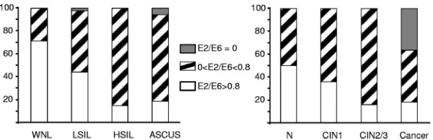
Physical state of HPV16 DNA is related to lesion grade.As
for cytology results, a majority of WNL (71%) presented E2/E6 values of more than 0.8, indicating that they harbored mostly episomal HPV16 genomes (Fig. 3). The remaining 29% of samples harbored both episomal and integrated HPV16 genomes. None of the samples belonging to this category pre-sented only integrated forms of HPV16 DNA (E2/E6 values equal to 0). In LSIL, the proportion of samples presenting E2/E6 values of⬎0.8 decreased to 44%, while the proportion of samples with mixed forms increased to 53%. In one smear, E2/E6 reached the value of 0.001, suggesting that most if not all HPV16 genomes were integrated. Unfortunately, the med-ical record was not available to confirm the final diagnosis. In HSIL, the proportion of specimens with E2/E6 values of more than 0.8 dropped to 15%, while 85% of these samples pre-sented integrated HPV16 DNA associated with episomes. Fi-nally, 76% of ASC-US samples harbored integrated HPV16 DNA (E2/E6 values between 0 and 0.8). Five percent of ASC-US (n⫽2) presented E2/E6 values of 0, indicating that HPV16 DNA was totally integrated. The remaining ASC-US specimens (19%) presented an E2/E6 value of ⬎0.8. As a whole, we observed that the proportion of samples with inte-grated HPV16 genomes increased with the lesion grade (P⬍
0.01; chi-square test).
Two smears from women with normal biopsy results pre-sented E2/E6 values of more than 0.8, whereas the two others presented both episomal and integrated HPV16 genomes. The proportion of smears with E2/E6 values of more than 0.8 decreased to 36% and to 16% for women with CIN1 and CIN2/3, respectively. The remaining samples presented both episomal and integrated HPV16 genomes.
For cancer biopsy samples, the proportion of specimens with E2/E6 values of more than 0.8 (18%) was close to the one observed for smears from women with a subsequent diagnosis of CIN2/3. In contrast, the proportion of samples with mixed forms decreased to 45% to the benefit of pure integrated HPV16 genomes, which were observed in 37% of cases.
We also noted that the proportion of smears with integrated HPV16 DNA increased with the lesion grade. However, this trend was not statistically significant (P ⬎ 0.05; chi-square test).
E2/E6 ratio decreases with lesion grade. The medians,
[image:4.585.137.451.67.237.2]ranges, and first and third quartiles of E2/E6 ratios for each category of samples are presented in Fig. 4. As a whole, the median of E2/E6 ratios decreased with the lesion severity (1 for WNL, 0.64 for LSIL, and 0.46 for HSIL). Whereas no significant difference was found between WNL and LSIL, the E2/E6 ratios were significantly lower for HSIL than for WNL and LSIL (P⬍0.01). The E2/E6 ratios for ASC-US (median of 0.55) were also significantly lower than for WNL (P⬍0.01). A FIG. 2. Box-and-whisker plots showing HPV16 load per 103cells (medians, first and third quartiles, and ranges) for cervical smears. Starting
material for cancers was fixed and paraffin-embedded tissues. N, normal. Symbols:**,Pvalue of⬍0.01 compared to WNL and LSIL;*,Pvalue of⬍0.05 compared to CIN1.
FIG. 3. Distribution of samples with E2/E6 ratio values equal to 0, between 0 and 0.8, and more than 0.8 for cervical smears. Further information and abbreviations are given in the legend for Fig. 2.
on May 16, 2020 by guest
http://jcm.asm.org/
[image:4.585.136.450.603.705.2]similar trend was observed when the 54 smears were classified according the subsequent histological data. Indeed, the median of E2/E6 decreased from 0.75 for smears from women with normal histologies to 0.58 for smears from women with CIN1 and 0.51 for smears from women with CIN2/3. No difference between E2/E6 ratios was observed.
The lowest E2/E6 ratio median (0.36) was noted for cancer samples. However, no statistical difference was observed in comparison to ratios calculated from smears.
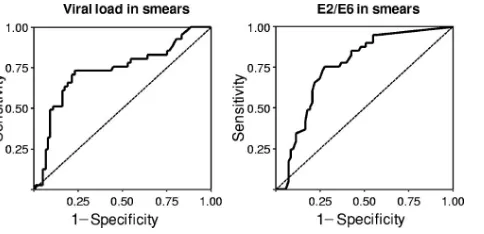
Appropriate cutoffs permit identification of women with
high-grade lesions.Figure 5 shows the receiver operating
char-acteristic (ROC) analysis results of viral load and E2/E6 ratios in discriminating WNL and LSIL samples from HSIL samples. For viral load, the cutoff with the highest positive likelihood ratio (LR⫹) (LR⫹of 5.1), was 21,907 copies/103cells. At this
threshold, HPV16 load allowed the identification of women with HSIL with a specificity of 90% and a sensitivity of 50%. We then used this cutoff to discriminate smears from women with normal cervixes or with CIN1 from smears from women with CIN2/3. The specificity and the sensitivity decrease to 83% and 40%, respectively, with the LR⫹dropping to 2.3 (area under the curve, 0.68; 95% confidence interval [95% CI], 0.52 to 0.85).
For the E2/E6 ratio, the cutoff with the highest LR⫹(LR⫹ of 3) was 0.52, with a specificity of 78% and a sensitivity of 66%. This cutoff was not used to discriminate smears of women with normal cervixes or with CIN1 from smears of women with CIN2/3, as the ROC analysis was not satisfactory (area under the curve, 0.66; 95% CI, 0.50 to 0.81).
DISCUSSION
In this study, we quantified HPV16 E6 and E2 DNA copy numbers by real-time PCR to analyze the load and physical state of the HPV16 genome in a series of 173 cervical samples. Several studies have reported the use of this methodology to characterize integration (1, 2, 8, 16, 25, 36, 40), even though some authors have highlighted limitations concerning the use of real-time PCR targeting E2 (2, 3, 29, 44). In order to pre-clude the erroneous interpretation of real-time PCR results, we validated our method with mixes of pBR322-HPV16 plas-mids (mimicking episomal forms) and SiHa cell DNA (two integrated HPV16 genomes per cell) representing E2/E6 ratios
from 0 to 1 with increments of 0.1. Moreover, we confirmed that E6 and E2 real-time PCR assays have almost equal effi-ciencies and that the calculation of E2/E6 ratios was accurate.
HPV16 DNA load analysis.First, we observed that HPV16
load increased with the lesion grade. An appropriate statistical analysis permitted us to retrieve significant differences between WNL and HSIL samples as well as between LSIL and HSIL samples. This trend was further confirmed when histology re-sults were taken into account. Thus, our data confirm previous observations suggesting that the higher the grade of the lesion, the higher is the viral load, even though statistical analyses were not always available (1, 9, 11, 18, 24, 25, 39, 50, 51, 55, 56). We would like to point out that a direct comparison between our results and those obtained in previous reports may be difficult because no standardized procedures were reported. (i) Original material differed widely. (ii) Different methods for HPV detection, more or less quantitative, were used: MY09/ MY11 PCR followed by type-specific dot blot hybridization (24), PCR followed by an enzyme-linked immunosorbent assay (56), conventional nested PCR (50), real-time PCR using SYBR green (11, 39), real-time PCR using the TaqMan tech-nology (9, 18, 25, 55), and consensus real-time PCR (1). (iii) Results were expressed by different means: copies per cell (1, 56), copies per ng of DNA (11, 25), copies per sample (39, 50), and qualitatively (18, 24). Thus, the value of viral load calcu-lated is likely to be dependent on the procedures used to quantify HPV DNA. This may explain the large range of values obtained for a given lesion grade in the different studies.
[image:5.585.301.543.67.184.2]We would like to stress the fact that the viral load values observed for invasive cancers were very high. This observation is in contrast with the results described by Cricca and cowork-ers, who noticed the lowest viral load for samples of invasive cancers (11). This can be attributed to the starting material and the number of samples analyzed (n⫽11). Moreover, smears evocative of invasive cancer may include not only cancer cells but also normal cells or cells from associated low-grade lesions, leading to an overall underestimation of the viral load (46). Here, we noticed that among cancer samples, those with the highest viral loads also presented the highest E2/E6 ratios. In contrast, cancer samples with most if not all HPV genomes integrated (E2/E6 values equal to 0) presented the lowest viral load values (data not shown). These observations are likely to FIG. 4. Box-and-whisker plots showing E2/E6 ratios (medians, first
and third quartiles, and ranges) for cervical smears. Further informa-tion and abbreviainforma-tions are given in the legend for Fig. 2.
FIG. 5. ROC analysis of viral load and E2/E6 ratio in discriminat-ing WNL and LSIL from HSIL. ROC curve characteristics were as follows: for viral load, the area under the curve was 0.728, the standard error was⫾0.05, and the 95% CI was 0.625 to 0.831; and for the E2/E6 ratio, the area under the curve was 0.760, the standard error was
⫾0.04, and the 95% CI was 0.671 to 0.850.
on May 16, 2020 by guest
http://jcm.asm.org/
[image:5.585.43.284.69.197.2]values equal to 0, representing fully integrated genomes; class II, with E2/E6 values between 0 and 0.8, representing mixes of episomal and integrated genomes; and class III, with E2/E6 values of more than 0.8, mostly representing episomes.
In our series, we noticed that the most frequently observed E2/E6 ratios were between 0 and 0.8. This means that the majority of samples harbored mixes of episomes and inte-grated forms of HPV16 genomes independently of the lesion grade. Our results also confirm that integration can be an early event, because 29% of the normal smears harbored a fraction of integrated HPV16 genomes. This is in accordance with previously published data (8, 11, 17, 25, 28, 40). Some authors do not agree with this observation (20, 49). But their methods (two-dimensional gel electrophoresis [20] and classic PCR [49]) were probably not sensitive enough to detect low-fre-quency integration events. We also observed that the propor-tion of samples with integrated forms increased with the lesion grade. This is consistent with the literature, with some varia-tions within each category of smears, however (1, 8, 11, 16, 25, 40, 49). For example, we found that 44% of LSIL (correspond-ing to 36% of smears from women with CIN1) harbored only episomal HPV16 genomes. This is close to the value (33.3%) we previously published on a series of CIN1 samples (8) but higher than the values from Kulmala et al. (15.4%) (25) and lower than the values from Cricca et al. (72.2%) (11), Tonon et al. (92%) (49), and Hudelist et al. (100%) (20). As for viral load, these discrepancies could be explained by the lack of standardized protocols to investigate integration and by defi-nitions of mixed forms of HPV16 genomes. As reported in a previous study, pure integrated forms (E2 values equal to 0) were rarely found for precancerous lesions (8). In the present study, 100% integration was observed for several cancer sam-ples, as already described (3, 10, 24, 40). This is consistent with the HPV-associated mechanism of carcinogenesis. Indeed, tu-mors grow from cellular clones selected for their proliferative advantages, which are driven by integration and continuous overexpression of E6 and E7 (33).
Quantitative analysis of E2/E6 ratios.We observed that the
median of E2/E6 ratios decreased with the increasing grade of the lesions. This indicates that the proportion of episomes decreases to the benefit of integrated forms of HPV16 ge-nomes in the higher-grade lesions. The methods used in this and other studies do not allow a determination of whether some cells harbor episomal forms and others integrated forms or if the same cells harbor both episomes and integrated forms. One possibility to address this question is to analyze the phys-ical state of the HPV16 genome at the single-cell level, for example after the isolation of single abnormal cells by laser microdissection followed by whole-genome amplification and HPV16 DNA analysis. Such experiments would give
interest-criminate normal plus LSIL versus HSIL or normal and CIN1 versus CIN2/3, since low-grade lesions spontaneously regress in most women within 3 years (32). Even though these lesions need to be monitored, they do not necessary need aggressive management as high-grade lesions do.
Using the ROC analysis, we have set optimal cutoffs at 21,907 copies/103 cells and at an E2/E6 ratio of 0.52 to
dis-criminate WNL plus LSIL from HSIL. These cutoffs allow the identification of women with prevalent high-grade lesion with a good specificity (90% for viral load and 78% for E2/E6). Moreover, we attempted to apply the viral load cutoff using a histology result as the end point. Interestingly, we were able to identify women with prevalent CIN2/3 with a specificity of 83%. E2/E6 ratios were not informative enough to discrimi-nate smears from women with normal cervixes or CIN1 from smears of women with CIN2/3. As expected, the sensitivities remained modest with these cutoffs, which were not designed for primary screening purposes.
We deliberately decided to exclude ASC-US samples from ROC analyses. Indeed, ASC-US can be suggestive of a wide range of histological diagnoses from a totally normal cervix to infiltrating carcinoma, as recently reported by Boulanger and Sevestre (CIN1 or condyloma in 20 to 54% of cases, CIN2 or CIN3 in 5 to 18% of cases, and invasive cancer in 0 to 4% of cases) (6). Thus, we noticed that HPV16 parameters (load, E2/E6 ratio) presented intermediate values, reflecting the di-versity of this category of samples.
In summary, we observed that the viral load and E2/E6 ratios were related to the lesion grade. Moreover, we con-firmed that integration is an early event in the course of natural HPV infection. Based on our results, we propose that among HR-HPV infected women, those with an HPV16 load of above 22,000 copies/103 cells or with an E2/E6 ratio below 0.5 are
likely to present a high-grade lesion. These cutoff values, how-ever, are relevant for HPV16 only, which is detected for 34 to 52% of patients with CIN2/3 and for 52 to 58% of cancers (4, 10, 34, 48) (62% of CIN2/3 and in 73% of cancers in France [42, 43]). Because other HR-HPVs (e.g., HPV18, HPV31, and HPV45) are also responsible for cervical lesions, relevant cut-offs should be determined for these genotypes. Now, prospec-tive cohort studies are necessary to evaluate the clinical rele-vance of these viral parameters. Moreover, there is an obvious need for standardized procedures, as our results may be de-pendent on the protocol we have used. Standardization would allow the comparison of viral load and integration results from different studies and laboratories. In this respect, we recently worked on the homogenization of techniques between differ-ent labs. Preliminary results indicate that it is possible to achieve very good inter-/intralaboratory and inter-/intraob-server reproducibility.
on May 16, 2020 by guest
http://jcm.asm.org/
ACKNOWLEDGMENTS
M. Saunier and S. Monnier-Benoit were recipients of a predoctoral scholarship from the Conseil Re´gional de Franche-Comte´ and J. Brio-lat was a recipient of a postdoctoral fellowship from the Canceropoˆle Grand-Est. Part of this work was supported by grants from La Ligue Contre le Cancer (Comite´ du Doubs) and from Institut National du Cancer.
We thank E. de Villiers (DKFZ, Germany) for kindly providing us with HPV16 plasmid and M. Ca˘pîlna and A. Petitjean for providing us with cervical samples. L. Gissmann is gratefully acknowledged for his advice regarding result interpretation.
REFERENCES
1.Andersson, S., H. Safari, M. Mints, I. Lewensohn-Fuchs, U. Gyllensten, and B. Johansson.2005. Type distribution, viral load and integration status of high-risk human papillomaviruses in pre-stages of cervical cancer (CIN).
Br. J. Cancer92:2195–2200.
2.Arias-Pulido, H., C. L. Peyton, N. E. Joste, H. Vargas, and C. M. Wheeler.
2006. Human papillomavirus type 16 integration in cervical carcinoma in situ
and in invasive cervical cancer. J. Clin. Microbiol.44:1755–1762.
3.Baker, C. C., W. C. Phelps, V. Lindgren, M. J. Braun, M. A. Gonda, and P. M. Howley.1987. Structural and transcriptional analysis of human
papil-lomavirus type 16 sequences in cervical carcinoma cell lines. J. Virol.61:
962–971.
4.Barbosa, M. S., D. R. Lowy, and J. T. Schiller.1989. Papillomavirus
polypep-tides E6 and E7 are zinc-binding proteins. J. Virol.63:1404–1407.
5.Bosch, F. X., M. M. Manos, N. Munoz, M. Sherman, A. M. Jansen, J. Peto, M. H. Schiffman, V. Moreno, R. Kurman, K. V. Shah, et al.1995. Prevalence of human papillomavirus in cervical cancer: a worldwide perspective. J. Natl.
Cancer Inst.87:796–802.
6.Boulanger, J. C., and H. Sevestre.2006. ASCUS: an update. Gynecol.
Ob-stet. Fertil.34:44–48. (In French.)
7.Bray, F., R. Sankila, J. Ferlay, and D. M. Parkin.2002. Estimates of cancer
incidence and mortality in Europe in 1995. Eur. J. Cancer38:99–166.
8.Briolat, J., V. Dalstein, M. Saunier, K. Joseph, S. Caudroy, J. L. Pretet, P. Birembaut, and C. Clavel.2007. HPV prevalence, viral load and physical state of HPV-16 in cervical smears of patients with different grades of CIN.
Int. J. Cancer121:2198–2204.
9.Carcopino, X., M. Henry, D. Benmoura, A. S. Fallabregues, H. Richet, L. Boubli, and C. Tamalet.2006. Determination of HPV type 16 and 18 viral load in cervical smears of women referred to colposcopy. J. Med. Virol.
78:1131–1140.
10.Clifford, G., S. Franceschi, M. Diaz, N. Munoz, and L. L. Villa.2006. HPV type-distribution in women with and without cervical neoplastic diseases.
Vaccine24(Suppl. 3):S26–S34.
11.Cricca, M., A. M. Morselli-Labate, S. Venturoli, S. Ambretti, G. A. Gen-tilomi, G. Gallinella, S. Costa, M. Musiani, and M. Zerbini.2007. Viral DNA load, physical status and E2/E6 ratio as markers to grade HPV16
positive women for high-grade cervical lesions. Gynecol. Oncol.106:549–
557.
12.Dalstein, V., D. Riethmuller, J. L. Pretet, K. Le Bail Carval, J. L. Sautiere, J. P. Carbillet, B. Kantelip, J. P. Schaal, and C. Mougin.2003. Persistence and load of high-risk HPV are predictors for development of high-grade
cervical lesions: a longitudinal French cohort study. Int. J. Cancer106:396–
403.
13.de Villiers, E. M., C. Fauquet, T. R. Broker, H. U. Bernard, and H. zur Hausen.2004. Classification of papillomaviruses. Virology324:17–27. 14.Doorbar, J.2005. The papillomavirus life cycle. J. Clin. Virol.32(Suppl.
1):S7–S15.
15.Franco, E. L., L. L. Villa, J. P. Sobrinho, J. M. Prado, M. C. Rousseau, M. Desy, and T. E. Rohan.1999. Epidemiology of acquisition and clearance of cervical human papillomavirus infection in women from a high-risk area for
cervical cancer. J. Infect. Dis.180:1415–1423.
16.Fujii, T., N. Masumoto, M. Saito, N. Hirao, S. Niimi, M. Mukai, A. Ono, S. Hayashi, K. Kubushiro, E. Sakai, K. Tsukazaki, and S. Nozawa.2005. Comparison between in situ hybridization and real-time PCR technique as a means of detecting the integrated form of human papillomavirus 16 in
cervical neoplasia. Diagn. Mol. Pathol.14:103–108.
17.Gallo, G., M. Bibbo, L. Bagella, A. Zamparelli, F. Sanseverino, M. R. Gio-vagnoli, A. Vecchione, and A. Giordano.2003. Study of viral integration of
HPV-16 in young patients with LSIL. J. Clin. Pathol.56:532–536.
18.Gravitt, P. E., M. B. Kovacic, R. Herrero, M. Schiffman, C. Bratti, A. Hildesheim, J. Morales, M. Alfaro, M. E. Sherman, S. Wacholder, A. C. Rodriguez, and R. D. Burk.2007. High load for most high risk human papillomavirus genotypes is associated with prevalent cervical cancer pre-cursors but only HPV16 load predicts the development of incident disease.
Int. J. Cancer121:2787–2793.
19.Ho, G. Y., R. D. Burk, S. Klein, A. S. Kadish, C. J. Chang, P. Palan, J. Basu, R. Tachezy, R. Lewis, and S. Romney.1995. Persistent genital human
pap-illomavirus infection as a risk factor for persistent cervical dysplasia. J. Natl.
Cancer Inst.87:1365–1371.
20.Hudelist, G., M. Manavi, K. I. Pischinger, T. Watkins-Riedel, C. F. Singer, E. Kubista, and K. F. Czerwenka.2004. Physical state and expression of HPV DNA in benign and dysplastic cervical tissue: different levels of viral
integration are correlated with lesion grade. Gynecol. Oncol.92:873–880.
21.Josefsson, A. M., P. K. Magnusson, N. Ylitalo, P. Sorensen, P. Qwarforth-Tubbin, P. K. Andersen, M. Melbye, H. O. Adami, and U. B. Gyllensten.
2000. Viral load of human papilloma virus 16 as a determinant for develop-ment of cervical carcinoma in situ: a nested case-control study. Lancet
355:2189–2193.
22.Kalantari, M., F. Karlsen, G. Kristensen, R. Holm, B. Hagmar, and B. Johansson.1998. Disruption of the E1 and E2 reading frames of HPV 16 in cervical carcinoma is associated with poor prognosis. Int. J. Gynecol. Pathol.
17:146–153.
23.Khan, M. J., P. E. Castle, A. T. Lorincz, S. Wacholder, M. Sherman, D. R. Scott, B. B. Rush, A. G. Glass, and M. Schiffman.2005. The elevated 10-year risk of cervical precancer and cancer in women with human papillomavirus (HPV) type 16 or 18 and the possible utility of type-specific HPV testing in
clinical practice. J. Natl. Cancer Inst.97:1072–1079.
24.Kovacic, M. B., P. E. Castle, R. Herrero, M. Schiffman, M. E. Sherman, S. Wacholder, A. C. Rodriguez, M. L. Hutchinson, M. C. Bratti, A. Hildesheim, J. Morales, M. Alfaro, and R. D. Burk.2006. Relationships of human pap-illomavirus type, qualitative viral load, and age with cytologic abnormality.
Cancer Res.66:10112–10119.
25.Kulmala, S. M., S. M. Syrjanen, U. B. Gyllensten, I. P. Shabalova, N. Petrovichev, P. Tosi, K. J. Syrjanen, and B. C. Johansson. 2006. Early integration of high copy HPV16 detectable in women with normal and low
grade cervical cytology and histology. J. Clin. Pathol.59:513–517.
26.Laurendeau, I., M. Bahuau, N. Vodovar, C. Larramendy, M. Olivi, I. Bieche, M. Vidaud, and D. Vidaud.1999. TaqMan PCR-based gene dosage assay for predictive testing in individuals from a cancer family with INK4 locus
hap-loinsufficiency. Clin. Chem.45:982–986.
27.Lillo, F. B., S. Lodini, D. Ferrari, C. Stayton, G. Taccagni, L. Galli, A. Lazzarin, and C. Uberti-Foppa.2005. Determination of human papilloma-virus (HPV) load and type in high-grade cervical lesions surgically resected from HIV-infected women during follow-up of HPV infection. Clin. Infect.
Dis.40:451–457.
28.Lukaszuk, K., J. Liss, I. Wozniak, J. Emerich, and C. Wojcikowski.2003. Human papillomavirus type 16 status in cervical carcinoma cell DNA
as-sayed by multiplex PCR. J. Clin. Microbiol.41:608–612.
29.Meissner, J. D.1999. Nucleotide sequences and further characterization of human papillomavirus DNA present in the CaSki, SiHa and HeLa cervical
carcinoma cell lines. J. Gen. Virol.80:1725–1733.
30.Melnikow, J., J. Nuovo, A. R. Willan, B. K. Chan, and L. P. Howell.1998. Natural history of cervical squamous intraepithelial lesions: a meta-analysis.
Obstet. Gynecol.92:727–735.
31.Monnier-Benoit, S., V. Dalstein, D. Riethmuller, N. Lalaoui, C. Mougin, and J. L. Pretet.2006. Dynamics of HPV16 DNA load reflect the natural history
of cervical HPV-associated lesions. J. Clin. Virol.35:270–277.
32.Moscicki, A. B., S. Shiboski, N. K. Hills, K. J. Powell, N. Jay, E. N. Hanson, S. Miller, K. L. Canjura-Clayton, S. Farhat, J. M. Broering, and T. M. Darragh.2004. Regression of low-grade squamous intra-epithelial lesions in
young women. Lancet364:1678–1683.
33.Munger, K., A. Baldwin, K. M. Edwards, H. Hayakawa, C. L. Nguyen, M. Owens, M. Grace, and K. Huh.2004. Mechanisms of human
papillomavirus-induced oncogenesis. J. Virol.78:11451–11460.
34.Munoz, N., F. X. Bosch, X. Castellsague, M. Diaz, S. de Sanjose, D. Ham-mouda, K. V. Shah, and C. J. Meijer.2004. Against which human papillo-mavirus types shall we vaccinate and screen? The international perspective.
Int. J. Cancer111:278–285.
35.Munoz, N., F. X. Bosch, S. de Sanjose, R. Herrero, X. Castellsague, K. V. Shah, P. J. Snijders, and C. J. Meijer.2003. Epidemiologic classification of human papillomavirus types associated with cervical cancer. N. Engl. J. Med.
348:518–527.
36.Nagao, S., M. Yoshinouchi, Y. Miyagi, A. Hongo, J. Kodama, S. Itoh, and T. Kudo.2002. Rapid and sensitive detection of physical status of human papillomavirus type 16 DNA by quantitative real-time PCR. J. Clin.
Micro-biol.40:863–867.
37.Ostor, A. G.1993. Natural history of cervical intraepithelial neoplasia: a
critical review. Int. J. Gynecol. Pathol.12:186–192.
38.Parkin, D. M.2001. Global cancer statistics in the year 2000. Lancet Oncol.
2:533–543.
39.Payan, C., A. Ducancelle, M. H. Aboubaker, J. Caer, M. Tapia, A. Chauvin, D. Peyronnet, E. Le Hen, Z. Arab, M. C. Legrand, A. Tran, E. Postec, F. Tourmen, M. Avenel, C. Malbois, M. A. De Brux, P. Descamps, and F. Lunel.
2007. Human papillomavirus quantification in urine and cervical samples by using the Mx4000 and LightCycler general real-time PCR systems. J. Clin.
Microbiol.45:897–901.
40.Peitsaro, P., B. Johansson, and S. Syrjanen.2002. Integrated human papil-lomavirus type 16 is frequently found in cervical cancer precursors as
on May 16, 2020 by guest
http://jcm.asm.org/
in France: EDITH study. Int. J. Cancer122:424–427.
44.Romanczuk, H., and P. M. Howley.1992. Disruption of either the E1 or the E2 regulatory gene of human papillomavirus type 16 increases viral
immor-talization capacity. Proc. Natl. Acad. Sci. USA89:3159–3163.
45.Shepherd, J. C., and R. A. Fried.1995. Preventing cervical cancer: the role
of the Bethesda system. Am. Fam. Physician51:434–440, 443–444.
46.Sherman, M. E., S. S. Wang, C. M. Wheeler, L. Rich, P. E. Gravitt, R. Tarone, and M. Schiffman.2003. Determinants of human papillomavirus load among women with histological cervical intraepithelial neoplasia 3: dominant impact of surrounding low-grade lesions. Cancer Epidemiol.
Bio-markers Prev.12:1038–1044.
47.Siegel, S., and N. J. Castelane.1988. Nonparametric statistics for behavioral sciences. McGraw-Hill, New York, NY.
48.Smith, J. S., L. Lindsay, B. Hoots, J. Keys, S. Franceschi, R. Winer, and G. M. Clifford.2007. Human papillomavirus type distribution in invasive cervical cancer and high-grade cervical lesions: a meta-analysis update. Int.
J. Cancer121:621–632.
49.Tonon, S. A., M. A. Picconi, P. D. Bos, J. B. Zinovich, J. Galuppo, L. V. Alonio, and A. R. Teyssie.2001. Physical status of the E2 human papilloma
papillomavirus is a necessary cause of invasive cervical cancer worldwide.
J. Pathol.189:12–19.
53.Wallin, K. L., F. Wiklund, T. Angstrom, F. Bergman, U. Stendahl, G. Wadell, G. Hallmans, and J. Dillner.1999. Type-specific persistence of human pap-illomavirus DNA before the development of invasive cervical cancer.
N. Engl. J. Med.341:1633–1638.
54.Woodman, C. B., S. Collins, H. Winter, A. Bailey, J. Ellis, P. Prior, M. Yates, T. P. Rollason, and L. S. Young.2001. Natural history of cervical human papillomavirus infection in young women: a longitudinal cohort study.
Lan-cet357:1831–1836.
55.Ylitalo, N., A. Josefsson, M. Melbye, P. Sorensen, M. Frisch, P. K. Andersen, P. Sparen, M. Gustafsson, P. Magnusson, J. Ponten, U. Gyllensten, and H. O. Adami.2000. A prospective study showing long-term infection with human papillomavirus 16 before the development of cervical carcinoma in
situ. Cancer Res.60:6027–6032.
56.Zerbini, M., S. Venturoli, M. Cricca, G. Gallinella, P. De Simone, S. Costa, D. Santini, and M. Musiani.2001. Distribution and viral load of type specific HPVs in different cervical lesions as detected by PCR-ELISA. J. Clin.
Pathol.54:377–380.