0095-1137/10/$12.00
doi:10.1128/JCM.01326-10
Copyright © 2010, American Society for Microbiology. All Rights Reserved.
Propionibacterium acnes
and
Staphylococcus epidermidis
Isolated from
Refractory Endodontic Lesions Are Opportunistic Pathogens
䌤
†
Sadia A. Niazi, Douglas Clarke, Thuy Do, Steven C. Gilbert,
Francesco Mannocci, and David Beighton*
Dental Institute, King’s College London, London, England
Received 30 June 2010/Returned for modification 2 August 2010/Accepted 16 August 2010
The predominant cultivable microbiota from 20 refractory endodontic lesions (9 with abscesses and 11
without abscesses) were determined, and
Propionibacterium acnes
and
Staphylococcus epidermidis
were among
the most predominant organisms. The number of species identified from lesions with abscesses (14.1
ⴞ
2.6)
was significantly greater (
P
< 0.001) than the number from lesions without abscesses (7.4
ⴞ
5.9). Comparison
of perioral isolates using repetitive extragenic palindromic PCR of the same species from the same subjects
demonstrated that the endodontic and skin populations were significantly different. The
P. acnes
isolates were
typed on the basis of
recA
gene sequence comparison, and only three types (types I, II, and III) were identified
among 125 isolates examined. However, we found that type I (type IA and IB) isolates were primarily isolated
from the skin, while types II and III were significantly more likely to be isolated from the endodontic lesions
(
P
< 10
ⴚ10). We found that the robustness of the
recA
phylotypes was not strong by comparing the partial gene
sequences of six putative virulence determinants, PAmce, PAp60, PA-25957, PA-5541, PA-21293, and PA-4687.
The resulting neighbor-joining trees were incongruent, and significant (phi test;
P
ⴝ
2.2
ⴛ
10
ⴚ7) evidence of
recombination was demonstrated, with significant phylogenetic heterogeneity being apparent within the
clus-ters.
P. acnes
and
S. epidermidis
isolated from refractory endodontic infections, with or without periapical
abscesses, are likely to be nosocomial infections.
Propionibacterium acnes
and coagulase-negative
staphylo-cocci, including
Staphylococcus epidermidis
, have been
identi-fied among the microflora of endodontic infections (8, 12, 40,
50, 52, 55, 56, 63, 64), but their importance as endodontic
pathogens has largely been ignored due to their nearly
univer-sal presence on the skin and the consequent likelihood of
sample contamination. However, there is now considerable
evidence that these organisms are increasingly isolated from
human infections, and so their association with endodontic
infections requires clarification.
P. acnes
, a non-spore-forming,
Gram-positive anaerobic or aerotolerant rod, is a member of
the resident microflora of the large intestine, conjunctiva, and
external ear canal (10, 15) and accounts for approximately half
of the total skin microbiota (59), predominating over other
pilosebaceous flora (16, 39). Although traditionally considered
to be relatively nonpathogenic, an increasing number of studies
have implicated
P. acnes
as an opportunistic pathogen
respon-sible for a wide range of infections and inflammatory
condi-tions. In addition to its well-established role in the
pathogen-esis of acnes vulgaris (16, 34), it has also been linked to
synovitis-acnes-pustulosis-hyperostosis-osteitis syndrome (44,
54), sarcoidosis (17), and prostate cancer (14). Recent studies
have revealed trauma and surgery as the predisposing factors
associated with numerous
P. acnes
infections, which include
brain abscesses (36), osteomyelitis after lumbar puncture (1),
discitis after surgery (23), spodylodiscitis following epidural
catheterization (22, 25), postoperative mediastinitis (19, 58),
endophthalmitis (7), and endocarditis (21). Furthermore, it is
also emerging as an important pathogen in infections related
to medical foreign-body implants, such as intraocular lenses
(65), ventroperitoneal shunts, orthopedic implants (9, 28, 51),
silicone implants (2), prosthetic heart valves (33, 42), and
pros-thetic joints (38, 61).
S. epidermidis
is part of the human skin microflora, where, as
a commensal organism, it usually exists in a benign relationship
with the host. Although it is ranked first in nosocomial and
implant-based infections, perhaps due to its ubiquity on the
skin, it is more likely to contaminate devices at the time of
insertion. As a recognized opportunistic pathogen, it is
respon-sible for nosocomial infections of indwelling medical devices
(43), such as peripheral or central intravenous catheters
(CVCs), prosthetic joints, vascular grafts, and central nervous
system shunts (46), and cardiac device infections, such as
pros-thetic valve endocarditis (PVE) (13), as well as ventricular
assist device driveline-related infections (4). Clearly, a
charac-teristic shared by both of these organisms is an association with
the infection of prosthetic devices.
The treatment of endodontic infections involves the
inser-tion of gutta-percha into the debrided and disinfected root
canal and the restoration of the tooth. This treatment may fail,
with the root canal becoming infected. A range of bacteria
have been isolated from such infected sites, including
P. acnes
and
S. epidermidis
, but these have generally been considered
contaminants. The objective of the present study was to
con-firm the endodontic origin of
P. acnes
and
S. epidermidis
iso-lates recovered from refractory endodontic infections. We
* Corresponding author. Mailing address: Department of
Microbi-ology, The Henry Wellcome Laboratories for Microbiology and
Sali-vary Research, KCL Dental Institute, Floor 17, Guys Tower, London
Bridge SE1 9RT, England. Phone: 2071887465. Fax:
44-0-2071887466. E-mail: [email protected].
† Supplemental material for this article may be found at http://jcm
.asm.org/.
䌤
Published ahead of print on 25 August 2010.
3859
on May 16, 2020 by guest
http://jcm.asm.org/
have used DNA fingerprinting of the
P. acnes
and
S.
epidermi-dis
isolates from endodontic lesions and samples from the
perioral skin of the same individual using repetitive extragenic
palindromic PCRs (REP-PCRs) (3). The
recA
phylotypes of
the
P. acnes
isolates were determined, as we expected to find
that the predominant phylotypes from the endodontic lesions
would be types II and III, which are almost exclusively
associ-ated with infections of implanted prostheses, while types IA
and IB are usually isolated from skin (26, 30, 31, 62). We also
investigated the phylogenetic status and robustness of the
recA
phylotypes by comparing the intrastrain relationships of partial
gene sequences of six putative virulence determinants, PAmce
(mammalian cell entry gene), PAp60, 25957, 5541,
PA-21293, and PA-4687 (20, 35). Here we have set out to
deter-mine if
P. acnes
and
S. epidermidis
isolated from refractory
endodontic lesions are contaminants arising during the sample
collection process or if they represent nosocomial infections.
MATERIALS AND METHODS
Sample collection.Patients recruited into the study received treatment in the Endodontic Department of the Dental Institute at Guys’ and St. Thomas’s Hospital, King’s College London. The selected teeth were single rooted with failed root canal treatment (refractory cases) with or without a periapical ab-scess. No communication between the abscess and the oral cavity or the skin surface was observed following clinical examination. All patients were medically stable and had no active, acute medical conditions, and they had not been prescribed antibiotics during the previous month. The sampled teeth had no root fracture or endoperiodontal lesion. All significant details about the history, clinical and radiographic signs, and symptoms about the involved tooth were recorded. The project was approved by the local ethics committee, and the patients gave their informed consent for their inclusion.
Twenty patients with refractory endodontic lesions (n⫽9 with periapical abscesses andn⫽11 without periapical abscesses) were investigated. After local anesthesia, the tooth to be treated was isolated with a rubber dam. The tooth and surrounding dam and clamp were cleaned with 30% (vol/vol) hydrogen peroxide and decontaminated with 2.5% sodium hypochlorite. After decontamination, the isolated tooth and surrounding dam were swabbed (LIP Limited, West York-shire, United Kingdom) to check for contamination.
The coronal restorations were removed, and the root filling was exposed. The coronal gutta-percha placed during the initial root canal treatment was removed using sterile Gates-Glidden drills, and the apical gutta-percha was removed with files. All of the material removed was transferred into 1 ml Tris-EDTA buffer (1.0 M Tris-HCl containing 0.1 M EDTA, pH 8.0, prepared in deionized water) (sample a). Radiographs were taken to ensure that all filling materials had been removed. An apex locator was used to determine the working length of the canal. Sterile saline solution was introduced into the canal, and the canal wall was filed up to the working length. The file samples were transferred into 1 ml Tris-EDTA buffer (sample b). The remaining root canal contents were absorbed onto paper points and transferred into 1 ml Tris-EDTA buffer (sample c). All samples were immediately transported on ice to the laboratory.
From 13 patients, perioral skin swab specimens (LIP Limited) were also taken for the isolation ofP. acnesandS. epidermidis.
Microbial analysis of samples.Each sample (samples a to c) was dispersed by vortexing with glass beads, diluted in fastidious anaerobic broth (Lab M, United Kingdom), and plated onto nonselective medium (fastidious anaerobic agar [FAA] supplemented with 5% [vol/vol] horse blood; Lab M) and also onto a range of selective media, including Rogosa agar (Oxoid, United Kingdom) (49) for lactobacilli, mupirocin-containing Trypticase phytone yeast (MTPY) agar (45) for bifidobacterium, Tryptone yeast cystine agar (TYC) agar (Lab M) (53) for streptococci, Veillonella agar (47, 48) and MacConkey agar (Oxoid, United Kingdom) for enteric bacteria, and CHROMagar (CHROMagar, France) for yeasts. The Rogosa, MTPY, and TYC agar media were incubated anaerobically for 3 days, the Veillonella agar was incubated anaerobically for 4 days, and FAA plates were incubated anaerobically for 7 days. The MacConkey agar and CHROMagar plates were incubated aerobically for 2 days at 37°C. After incu-bation, the colonies were counted and a predetermined number of colonies from each medium (n⫽ 300 maximum per patient) were randomly selected for identification.
The swabs taken from the prepared teeth prior to the removal of the resto-ration and the swabs from the perioral skin were plated directly onto FAA and incubated anaerobically for 7 days.
Identification of isolates.All randomly selected isolates were subcultured on FAA and grown for 24 h. Bacterial genomic DNA was extracted by boiling 100
l of a suspension of the cultured cells prepared in sterile distilled H2O for 10
min. The suspension was cooled on ice for 10 min and centrifuged at 13,000⫻
gfor 2 min, and the supernatant containing the genomic DNA was stored at
⫺20°C prior to analysis (38). A partial fragment of the 16S rRNA gene of all isolates was amplified using the following reaction mixture (final volume, 25l): either 0.3l of a 10-pmol/l concentration each 27F-YMa, 27F-YMb, 27F-YMc, and 27F-YMd forward primers (Integrated DNA Technology, United Kingdom) (18), 0.7l of 1492R reverse primer (concentration, 10 pmol/l), 22.5l of Reddymix buffer (Thermo Scientific, United Kingdom), and 0.6l DNA extract or 0.5 l of 9F forward primer (concentration, 10 pmol/l; MWG, United Kingdom), 0.5l of 907R reverse primer (concentration, 10 pmol/l; MWG), 23
l of Reddymix buffer (Thermo Scientific), and 1l DNA extract. The PCR products were cleaned with Microclean (Sigma, United Kingdom), in order to get ready for the sequence reaction. Amplicon sequencing was performed by using an ABI Prism BigDye Terminator sequencing kit (Applied Biosystems) with 30 cycles of denaturation at 96°C for 10 s, annealing at 50°C for 5 s, and extension at 60°C for 2 min. Sequencing reaction products were run on an ABI 3730xl sequencer Applied Biosystems). All DNA sequences were analyzed, trimmed, and aligned using BioEdit software (version 7.0.0; http://www.mbio .ncsu.edu/BioEdit/bioedit.html). The partial gene sequences were identified by a BLAST search of the NCBI database (http://0-www.ncbi.nlm.nih.gov.ilsprod.ilb .neu.edu/BLAST/), the Human Oral Microbiome database (http://www.homd .org/), or the Ribosome Database Project database http://rdp.cme.msu.edu/). Phylogenetic trees were constructed by the neighbor-joining (NJ) method, based on 16S rRNA gene sequence comparisons, using the MEGA (version 4.1) pro-gram (http://www.megasoftware.net/). Most isolates were identified using these sequences, but if isolates were identified asLactobacillusspp.,Veillonellaspp., or “Actinomyces naeslundii/A. viscosus,” these were more accurately identified by partial sequencing of thepheS(41),rpoB(5), andmetG(24) genes, respectively.
Phylotyping ofP. acnesisolates.Endodontic and skinP. acnesisolates iden-tified by partial 16S rRNA gene sequencing were typed by partialrecAgene sequencing. TheP. acnes recAgene was amplified using primer PAR-1 (positions
⫺96 to ⫺75; 5⬘-AGCTCGGTGGGGTTCTCTCATC-3⬘) and primer PAR-2 (positions⫹1105 to⫹1083; 5⬘-GCTTCCTCATACCACTGGTCATC-3⬘), which generated a 1,201-bp amplicon (38). The reaction mixture comprised 0.5l of PAR-1 (concentration, 10 pmol/l; Sigma), 0.5l of PAR-2 (concentration, 10 pmol/l; Sigma), 23l of Reddymix (Thermo Scientific), and 1l DNA extract in a 25-l final volume. The thermal cycling conditions included initial denatur-ation at 95°C for 3 min, denaturdenatur-ation at 95°C for 1 min, annealing at 55°C for 30 s, and extension at 72°C for 90 s, repeated for 35 cycles, and a final extension at 72°C for 10 min. The amplified products could be stored temporarily at 4°C. Amplified products were run on a 0.5% agarose gel and visualized under UV transillumination. Sequencing was performed as described above, and theP. acnes recAsequences were compared with GenBank sequences AY642055 (type IA), EU687255 (type IB), AY642061 (type II), and DQ672252 (type III). NJ trees were constructed using the Jukes-Cantor method with MEGA (version 4.1) software (www.megasoftware.net/).
Comparison of putativeP. acnesvirulence determinants.A number of genes have been associated with the virulence ofP. acnes(20, 35), including two invasion-associated proteins (PAmce and Pap60) and four predominant immu-noreactive surface proteins (PA-25957, PA-5541, PA-21293 and PA-4687), the first two of which have homology with M-like protein ofStreptococcus equiand have dermatan sulfate-binding activity, while the other proteins are similar to the product of theCorynebacterium diphtheriae htaAgene, which codes for an iron-regulated hemin binding protein. We used the previously reported primers to amplify and sequence internal fragments of PAmce and Pap60 (35) and designed primers to amplify and sequence fragments of each gene of these putative virulence determinants from a selection ofP. acnesstrains isolated in this study. PA5541 was amplified and sequenced using primers 5⬘-TCGACCTCAGCTTC AA-3⬘and 5⬘-AGGCGCTTGACGATAT-3⬘, PA25957 was amplified and se-quenced using primers 5⬘-CAATCGCAGCAATCAC-3⬘and 5⬘-CTCAGTCTT TGCGATC-3⬘, PA21693 was amplified and sequenced using primers 5⬘-CCGA GTTCTATGGCAA-3⬘and 5⬘-CCTGTTTGGTCATGGT-3⬘, while PA4687 was amplified and sequenced using primers 5⬘-GTATTGTTAGCCGTGC-3⬘ and 5⬘-ATCGTTTTGACCCTGC-3⬘. The amplicons sizes for the six genes used in the analyses were 783, 1,024, 491, 386, 464, and 473 bp, respectively.
The sequences of the same genes of the sequencedP. acnesstrains (strains J165 and SK137 [type IA], KPA171202 and SK187 [type IB], and J139 [type II])
on May 16, 2020 by guest
http://jcm.asm.org/
were downloaded from GenBank, and these were aligned with the sequences derived here using BioEdit and NJ trees for each gene and for the concatenated sequences of all the putative virulence genes that were constructed, using the Jukes-Cantor model, in MEGA (version 4.1). The concatenated sequences were tested for the presence of recombination using the phi test (11).
REP-PCR analysis ofP. acnesandS. epidermidis.Skin and endodonticP. acnes
andS. epidermidisisolates from the same individual (n⫽8 forS. epidermidisand
n⫽13 forP. acnes) were compared using REP-PCR (3). Primers REP1R-Dt (5⬘-555 NCG NCG NCA TCN GCC-3⬘) and REP2-Dt (5⬘-NCG NCT TAT CNG GCC TAC-3⬘) (Integrated DNA Technologies, Belgium) used in this study were described previously (3). The reaction mixture contained each of the four de-oxynucleoside triphosphates (dNTPs; Thermo Scientific) at a concentration of 125M, 150 ng of each primer, 3.75 mM MgCl2(Thermo Scientific), 2.5 U of
Taqpolymerase (Thermo Scientific), 20 mM (NH4)2SO4, 75 mM Tris-HCl (pH
9.0), 0.01% (wt/vol) Tween 80, and 3l of DNA solution made up to a final volume of 25l with sterile water. PCR amplification was performed in an automated thermocycler (Techne) with an initial denaturation (7 min at 95°C), followed by 32 cycles of denaturation (30 s at 94°C), annealing (1 min at 40°C), and extension (8 min at 65°C) and with a final extension (16 min at 65°C). The amplification products of REP-PCR (15l) were analyzed with a 2% Metasieve agarose gel (Flowgen, Staffordshire, England) containing Gel Red nucleic acid stain (10,000⫻in dimethyl sulfoxide; Cambridge Bioscience) and were separated electrophoretically on gels at 60 V for 5 h in 1⫻TBE (Tris-borate-EDTA) buffer. A molecular size marker (pGEM DNA markers; Promega) was included on all gels to facilitate comparison of tracks between gels. The gels were imaged (AlphaImager), and the resulting patterns were compared visually.
Biochemical characterization ofP. acnesisolates.P. acnesisolates from skin (n⫽29) and endodontic infections (n⫽34) and reference strainsP. acnestype IA (NCTC 737) andP. acnestype II (NCTC 10390) were characterized for their ability to ferment lactose,N-acetylglucosamine, erythritol, ribose, and sorbitol, according to a previously described method (6). The fermentation reactions were also performed in a Rapid ID 32A system, according to the manufacturer’s instructions (bioMe´rieux United Kingdom, Limited). -D-Fucosidase, -N -acetylgalactosaminidase, sialidase, ␣-L-fucosidase, -N-acetylglucosaminidase,
␣-glucosidase,-glucosidase,-galactosidase,␣-arabinosidase, and -galactosi-dase activities were determined using fluorogenic substrates, as described previ-ously (6). The strains were also tested for the production of lipase (32), DNase, nonspecific proteinase (37), elastase (29), lecithinase (60), and hyaluronidase (57).
Statistical analysis.Data distributions were compared using2tests, means
were compared using the Mann-Whitney U test in SPSSPC (version 16.0), and the analysis of the biochemical test results using canonical variates analysis was performed in the PAST-Palaeontological Statistics program (version 1.90; http: //folk.uio.no/ohammer/past/).
RESULTS
No organisms were recovered from the samples taken from
the sterilized tooth surfaces prior to removal of the restoration.
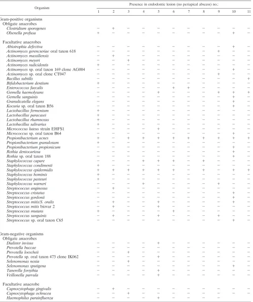
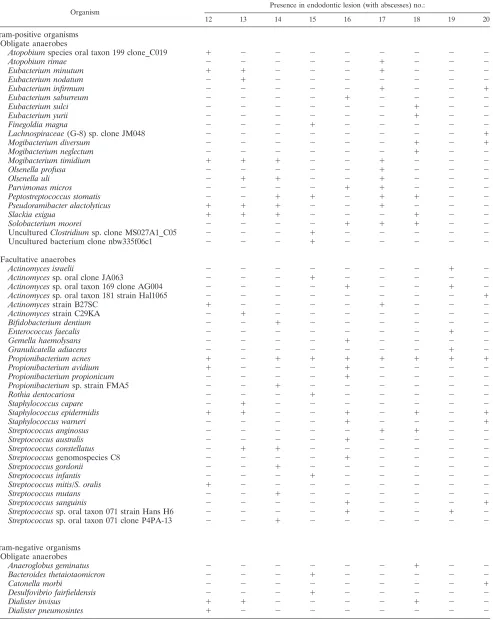
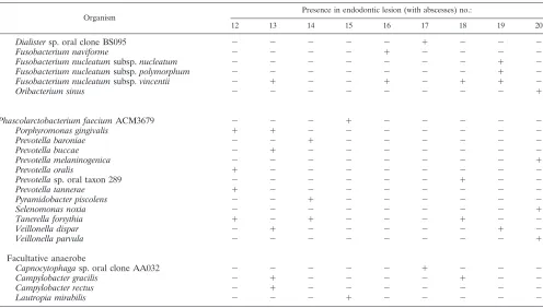
The range of organisms cultured from the 20 endodontic
le-sions is shown in Tables 1 and 2. The lele-sions without abscesses
(Table 1) were characterized by the presence primarily of
Gram-positive facultative anaerobic organisms, and these
or-ganisms were also present in the lesions with abscesses.
How-ever, the cultivable flora of the lesions with abscesses was more
complex, as these lesions also harbored obligate anaerobic
Gram-positive and Gram-negative organisms. The mean
num-ber of taxa from the lesions with periapical abscesses (Table 2)
was 14.1
⫾
2.6, and that from lesions without periapical
ab-scesses was 7.4
⫾
5.9 (
P
⬍
0.001).
P. acnes
was recovered from
10 of the 11 endodontic lesions without periapical abscesses
and from 8 of the 9 lesions with periapical abscesses (
P
⬎
0.1).
Other propionibacteria isolated from the lesions included
P.
avidium
,
P. granulosum
,
P. propionicum
, and
Propionibacterium
sp. strain FMA5. The median proportion of propionibacteria
recovered from the file samples in the teeth without abscesses
was 26.7% (range, not detected [ND] to 92%) compared to not
detected (range, ND to 76.7%) in teeth with abscesses (
P
⬍
0.01). Staphylococci, including
Staphylococcus caprae
,
S.
con-dimenti
,
S. epidermidis
,
S. hominis
,
S. pasteuri
and
S. warneri
,
were isolated from the endodontic lesions, with
S. epidermidis
being isolated from 5/9 endodontic lesions with abscesses and
from 9/11 of the lesions without abscesses (
P
⬎
0.1). The
median proportion of staphylococci recovered from the file
samples in the teeth without abscesses was 6.7% (range, ND to
73%) compared to not detected (range, ND to 13.3%) in teeth
with abscesses (
P
⬍
0.01).
Phylotyping of
P. acnes
isolates.
Partial
recA
sequences were
obtained for 65 skin and 60 endodontic
P. acnes
isolates; and
on the basis of sequence alignment, 3 distinct phylogenetic
lineages, type I, type II, and type III, were observed. The type
I isolates segregated into two distinct groups, into which the
sequences from known type IA and type IB sequences
clus-tered. When the distribution of the different
P. acnes
types was
considered (Table 3), the distribution of types recovered from
the skin was significantly different from that of all
P. acnes
isolates recovered from all endodontic lesions (
P
⬍
10
⫺10) and
from that of the
P. acnes
isolates recovered only from patients
from whom samples from skin were also taken (
P
⫽
2
⫻
10
⫺8).
Comparison of putative
P. acnes
virulence determinants.
Individual NJ trees were constructed for each of the partial
putative virulence gene sequences determined for the strains
isolated in this study, along with the corresponding sequences
derived from each of the 5 sequenced
P. acnes
genomes. In
PA-4687, we found that each of the type III isolates had
evi-dence of a 5-nucleotide (nt) deletion, such that the gene could
no longer be transcribed; these nucleotides were removed from
the sequences of the other phylotypes to enable the
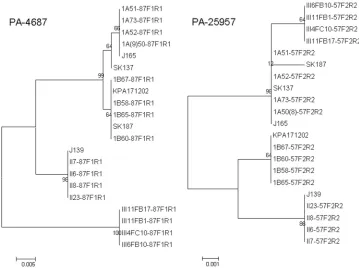
construc-tion of the NJ trees. The NJ trees for PA-25957 and PA-4687
are shown in Fig. 1 (the remaining trees are shown in Fig. 1S
in the supplemental material). From these it is apparent that
the type II and III isolates formed distinct clusters with each
gene. All the type I sequences for PA-5541, PA-4687, PAmce,
and PA-21693 formed either single clusters or clustered
to-gether but clustered as subclusters supported by bootstrap
values of
ⱕ
65. The type IA and IB strains formed distinct
discrete clusters with the partial sequences of PA-25957 and
Pap60, except that SK187 (type IB) was in the type IA cluster,
while the type IB and II strains were on the same lineage with
Pap60. The trees for the different genes were clearly
incongru-ent (Fig. 1).
We next constructed an NJ tree using the concatenated
sequences of the putative virulence determinants in order to
determine if the
recA
-based determination of phylotype was
replicated in this analysis. These data are shown in Fig. 2. In
the tree derived from the
recA
sequences, the 4 lineages are
well discriminated. However, in the trees derived from the
concatenated sequence data (3,616 bp), the type III isolates
formed a distinct lineage, as did the type II isolates, in which
two subbranches supported by significant bootstrap values of
⬎
95 were evident. The type IB isolates formed a single lineage
composed of all isolates except SK187, which was found in the
type IA lineage. The type IA lineage appeared to be diverse,
with SK187 and SK137 forming branches distinct from the
other 5 isolates in this lineage. There was statistical evidence of
recombination when the concatenated sequences were tested
(phi test,
P
⫽
2.2
⫻
10
⫺7).
on May 16, 2020 by guest
http://jcm.asm.org/
TABLE 1. Distribution of all 51 bacterial taxa identified among 638 isolates recovered from 11 refractory endodontic lesions without
periapical abscesses
Organism
Presence in endodontic lesion (no periapical abscess) no.:
1 2 3 4 5 6 7 8 9 10 11
Gram-positive organisms
Obligate anaerobes
Clostridium sporogenes
⫺
⫹
⫺
⫺
⫺
⫺
⫺
⫺
⫺
⫺
⫺
Olsenella profusa
⫹
⫺
⫺
⫺
⫺
⫺
⫺
⫺
⫺
⫹
⫺
Facultative anaerobes
Abiotrophia defectiva
⫺
⫺
⫺
⫺
⫺
⫺
⫺
⫺
⫺
⫹
⫺
Actinomyces gerencseriae
oral taxon 618
⫺
⫺
⫺
⫺
⫺
⫺
⫺
⫺
⫹
⫺
⫺
Actinomyces massiliensis
⫺
⫹
⫺
⫺
⫺
⫺
⫺
⫺
⫺
⫺
⫺
Actinomyces meyeri
⫺
⫺
⫹
⫺
⫺
⫺
⫺
⫺
⫺
⫺
⫺
Actinomyces radicidentis
⫹
⫺
⫺
⫺
⫺
⫺
⫺
⫺
⫺
⫺
⫺
Actinomyces
sp. oral taxon 169 clone AG004
⫺
⫺
⫺
⫺
⫺
⫺
⫺
⫺
⫺
⫹
⫺
Actinomyces
sp. oral clone CT047
⫺
⫺
⫺
⫺
⫺
⫺
⫺
⫺
⫹
⫺
⫺
Bacillus subtilis
⫺
⫺
⫺
⫺
⫺
⫺
⫺
⫺
⫺
⫺
⫹
Bifidobacterium dentium
⫹
⫺
⫺
⫺
⫺
⫺
⫺
⫺
⫺
⫺
⫺
Enterococcus faecalis
⫺
⫺
⫺
⫺
⫺
⫹
⫹
⫺
⫺
⫺
⫺
Gemella haemolysans
⫺
⫺
⫺
⫺
⫹
⫺
⫺
⫺
⫹
⫹
⫹
Gemella sanguinis
⫺
⫺
⫺
⫺
⫺
⫺
⫺
⫺
⫺
⫹
⫺
Granulicatella elegans
⫺
⫺
⫺
⫺
⫺
⫺
⫺
⫺
⫺
⫹
⫺
Kocuria
sp. oral taxon B56
⫺
⫺
⫺
⫺
⫺
⫺
⫺
⫺
⫺
⫹
⫺
Lactobacillus fermentum
⫹
⫺
⫺
⫺
⫺
⫺
⫺
⫺
⫺
⫺
⫺
Lactobacillus paracasei
⫹
⫺
⫺
⫺
⫺
⫺
⫺
⫺
⫺
⫺
⫺
Lactobacillus rhamnosus
⫹
⫺
⫺
⫺
⫺
⫺
⫺
⫺
⫺
⫺
⫺
Lactobacillus salivarius
⫹
⫺
⫺
⫺
⫺
⫺
⫺
⫺
⫺
⫺
⫺
Micrococcus luteus
strain EHFS1
⫺
⫺
⫺
⫺
⫹
⫺
⫺
⫺
⫺
⫺
⫺
Micrococcus
sp. oral taxon B64
⫺
⫺
⫺
⫺
⫺
⫺
⫺
⫺
⫺
⫹
⫺
Propionibacterium acnes
⫹
⫺
⫹
⫹
⫹
⫹
⫹
⫹
⫹
⫹
⫹
Propionibacterium granulosum
⫹
⫺
⫺
⫺
⫺
⫺
⫺
⫺
⫺
⫺
⫺
Propionibacterium propionicum
⫺
⫺
⫺
⫺
⫺
⫺
⫺
⫺
⫺
⫹
⫺
Rothia dentocariosa
⫺
⫺
⫺
⫺
⫺
⫺
⫺
⫺
⫺
⫹
⫺
Rothia
sp. oral taxon 188
⫺
⫺
⫺
⫺
⫺
⫺
⫺
⫺
⫺
⫹
⫺
Staphylococcus capare
⫺
⫺
⫺
⫹
⫹
⫹
⫺
⫹
⫺
⫺
⫺
Staphylococcus condimenti
⫹
⫺
⫺
⫺
⫺
⫺
⫺
⫺
⫺
⫺
⫺
Staphylococcus epidermidis
⫹
⫹
⫹
⫹
⫹
⫹
⫺
⫹
⫺
⫹
⫹
Staphylococcus hominis
⫹
⫺
⫺
⫺
⫺
⫺
⫺
⫺
⫺
⫺
⫺
Staphylococcus pasteuri
⫹
⫺
⫺
⫺
⫺
⫺
⫺
⫺
⫺
⫺
⫺
Staphylococcus warneri
⫺
⫺
⫺
⫹
⫺
⫺
⫺
⫺
⫹
⫺
⫺
Streptococcus anginosus
⫺
⫹
⫺
⫺
⫺
⫺
⫺
⫺
⫺
⫺
⫺
Streptococcus cristatus
⫺
⫺
⫺
⫺
⫺
⫺
⫺
⫺
⫺
⫹
⫺
Streptococcus gordonii
⫺
⫺
⫺
⫺
⫺
⫺
⫺
⫺
⫺
⫹
⫺
Streptococcus mitis
/
S. oralis
⫺
⫹
⫺
⫺
⫹
⫺
⫺
⫺
⫺
⫹
⫺
Streptococcus mitis
biovar 2
⫺
⫹
⫺
⫺
⫹
⫺
⫺
⫺
⫺
⫺
⫺
Streptococcus mutans
⫹
⫺
⫺
⫺
⫺
⫹
⫺
⫺
⫺
⫺
⫺
Streptococcus sanguinis
⫺
⫹
⫺
⫺
⫹
⫺
⫺
⫺
⫹
⫺
⫺
Streptococcus
sp. oral taxon C65
⫺
⫺
⫺
⫺
⫺
⫺
⫺
⫺
⫺
⫹
⫺
Gram-negative organisms
Obligate anaerobes
Dialister invisus
⫺
⫺
⫺
⫺
⫹
⫺
⫺
⫺
⫺
⫺
⫺
Prevotella buccae
⫺
⫺
⫺
⫺
⫺
⫺
⫺
⫺
⫺
⫺
⫺
Prevotella loescheii
⫺
⫺
⫹
⫺
⫺
⫺
⫺
⫺
⫺
⫺
⫺
Prevotella
sp. oral taxon 473 clone IK062
⫺
⫺
⫺
⫺
⫹
⫺
⫺
⫺
⫺
⫺
⫺
Selenomonas noxia
⫺
⫺
⫹
⫺
⫺
⫺
⫺
⫺
⫺
⫺
⫺
Selenomonas sputigena
⫺
⫺
⫺
⫺
⫺
⫺
⫺
⫺
⫺
⫹
⫺
Tanerella forsythia
⫺
⫺
⫺
⫺
⫹
⫺
⫺
⫺
⫺
⫺
⫺
Veillonella parvula
⫺
⫺
⫺
⫺
⫹
⫺
⫺
⫺
⫺
⫺
⫺
Facultative anaerobe
Capnocytophaga gingivalis
⫺
⫹
⫺
⫺
⫺
⫺
⫺
⫺
⫺
⫺
⫺
Capnocytophaga ochracea
⫺
⫺
⫹
⫺
⫺
⫺
⫺
⫺
⫺
⫺
⫺
Haemophilus parainfluenza
⫺
⫺
⫺
⫺
⫹
⫺
⫺
⫺
⫺
on May 16, 2020 by guest
http://jcm.asm.org/
TABLE 2. Distribution of all 77 bacterial taxa identified among 1,004 isolates recovered from 9 refractory endodontic
lesions with periapical abscesses
Organism Presence in endodontic lesion (with abscesses) no.:
12 13 14 15 16 17 18 19 20
Gram-positive organisms
Obligate anaerobes
Atopobium
species oral taxon 199 clone_C019
⫹
⫺
⫺
⫺
⫺
⫺
⫺
⫺
⫺
Atopobium rimae
⫺
⫺
⫺
⫺
⫺
⫹
⫺
⫺
⫺
Eubacterium minutum
⫹
⫹
⫺
⫺
⫺
⫹
⫺
⫺
⫺
Eubacterium nodatum
⫺
⫹
⫺
⫺
⫺
⫺
⫺
⫺
⫺
Eubacterium infirmum
⫺
⫺
⫺
⫺
⫺
⫹
⫺
⫺
⫹
Eubacterium saburreum
⫺
⫺
⫺
⫺
⫹
⫺
⫺
⫺
⫺
Eubacterium sulci
⫺
⫺
⫺
⫺
⫺
⫺
⫹
⫺
⫺
Eubacterium yurii
⫺
⫺
⫺
⫺
⫺
⫺
⫹
⫺
⫺
Finegoldia magna
⫺
⫺
⫺
⫹
⫺
⫺
⫺
⫺
⫺
Lachnospiraceae
(G-8) sp. clone JM048
⫺
⫺
⫺
⫺
⫺
⫺
⫺
⫺
⫹
Mogibacterium diversum
⫺
⫺
⫺
⫺
⫺
⫺
⫹
⫺
⫹
Mogibacterium neglectum
⫺
⫺
⫺
⫺
⫺
⫺
⫹
⫺
⫺
Mogibacterium timidium
⫹
⫹
⫹
⫺
⫺
⫹
⫺
⫺
⫺
Olsenella profusa
⫺
⫺
⫺
⫺
⫺
⫹
⫺
⫺
⫺
Olsenella uli
⫺
⫹
⫹
⫺
⫺
⫹
⫺
⫺
⫺
Parvimonas micros
⫺
⫺
⫺
⫺
⫹
⫹
⫺
⫺
⫺
Peptostreptococcus stomatis
⫺
⫺
⫹
⫹
⫺
⫹
⫹
⫺
⫺
Pseudoramibacter alactolyticus
⫹
⫹
⫹
⫺
⫺
⫹
⫺
⫺
⫺
Slackia exigua
⫹
⫹
⫹
⫺
⫺
⫺
⫹
⫺
⫺
Solobacterium moorei
⫺
⫺
⫺
⫺
⫹
⫹
⫹
⫺
⫺
Uncultured
Clostridium
sp. clone MS027A1_C05
⫺
⫺
⫺
⫹
⫺
⫺
⫺
⫺
⫺
Uncultured bacterium clone nbw335f06c1
⫺
⫺
⫺
⫹
⫺
⫺
⫺
⫺
⫺
Facultative anaerobes
Actinomyces israelii
⫺
⫺
⫺
⫺
⫺
⫺
⫺
⫹
⫺
Actinomyces
sp. oral clone JA063
⫺
⫺
⫺
⫹
⫺
⫺
⫺
⫺
⫺
Actinomyces
sp. oral taxon 169 clone AG004
⫺
⫺
⫺
⫺
⫹
⫺
⫺
⫹
⫺
Actinomyces
sp. oral taxon 181 strain Hal1065
⫺
⫺
⫺
⫺
⫺
⫺
⫺
⫺
⫹
Actinomyces
strain B27SC
⫹
⫺
⫺
⫺
⫺
⫹
⫺
⫺
⫺
Actinomyces
strain C29KA
⫺
⫹
⫺
⫺
⫺
⫺
⫺
⫺
⫺
Bifidobacterium dentium
⫺
⫺
⫹
⫺
⫺
⫺
⫺
⫺
⫺
Enterococcus faecalis
⫺
⫺
⫺
⫺
⫺
⫺
⫺
⫹
⫺
Gemella haemolysans
⫺
⫺
⫺
⫺
⫹
⫺
⫺
⫺
⫺
Granulicatella adiacens
⫺
⫺
⫺
⫺
⫺
⫺
⫺
⫹
⫺
Propionibacterium acnes
⫹
⫺
⫹
⫹
⫹
⫹
⫹
⫹
⫹
Propionibacterium avidium
⫹
⫺
⫺
⫺
⫹
⫺
⫺
⫺
⫺
Propionibacterium propionicum
⫺
⫺
⫺
⫺
⫹
⫺
⫺
⫺
⫺
Propionibacterium
sp. strain FMA5
⫺
⫺
⫹
⫺
⫺
⫺
⫺
⫺
⫺
Rothia dentocariosa
⫺
⫺
⫺
⫹
⫺
⫺
⫺
⫺
⫺
Staphylococcus capare
⫺
⫹
⫺
⫺
⫺
⫺
⫺
⫺
⫺
Staphylococcus epidermidis
⫹
⫹
⫺
⫺
⫹
⫺
⫹
⫺
⫹
Staphylococcus warneri
⫺
⫺
⫺
⫺
⫹
⫺
⫺
⫺
⫹
Streptococcus anginosus
⫺
⫺
⫺
⫺
⫺
⫹
⫹
⫺
⫺
Streptococcus australis
⫺
⫺
⫺
⫺
⫹
⫺
⫺
⫺
⫺
Streptococcus constellatus
⫺
⫹
⫹
⫺
⫺
⫺
⫺
⫺
⫺
Streptococcus
genomospecies C8
⫺
⫺
⫺
⫺
⫹
⫺
⫺
⫺
⫺
Streptococcus gordonii
⫺
⫺
⫹
⫺
⫺
⫺
⫺
⫺
⫺
Streptococcus infantis
⫺
⫺
⫺
⫹
⫺
⫺
⫺
⫺
⫺
Streptococcus mitis
/
S. oralis
⫹
⫺
⫺
⫺
⫺
⫺
⫺
⫺
⫺
Streptococcus mutans
⫺
⫺
⫹
⫺
⫺
⫺
⫺
⫺
⫺
Streptococcus sanguinis
⫺
⫺
⫺
⫺
⫹
⫺
⫺
⫺
⫹
Streptococcus
sp. oral taxon 071 strain Hans H6
⫺
⫺
⫺
⫺
⫹
⫺
⫺
⫹
⫺
Streptococcus
sp. oral taxon 071 clone P4PA-13
⫺
⫺
⫹
⫺
⫺
⫺
⫺
⫺
⫺
Gram-negative organisms
Obligate anaerobes
Anaeroglobus geminatus
⫺
⫺
⫺
⫺
⫺
⫺
⫹
⫺
⫺
Bacteroides thetaiotaomicron
⫺
⫺
⫺
⫹
⫺
⫺
⫺
⫺
⫺
Catonella morbi
⫺
⫺
⫺
⫺
⫺
⫺
⫺
⫺
⫹
Desulfovibrio fairfieldensis
⫺
⫺
⫺
⫹
⫺
⫺
⫺
⫺
⫺
Dialister invisus
⫹
⫹
⫺
⫺
⫺
⫺
⫹
⫺
⫺
Dialister pneumosintes
⫹
⫺
⫺
⫺
⫺
⫺
⫺
⫺
⫺
Continued on following page
on May 16, 2020 by guest
http://jcm.asm.org/
REP-PCR comparison of paired isolates.
When the
end-odontic and skin
P. acnes
isolates from the same individuals
(
n
⫽
13) were compared using REP-PCR, it was found that all
skin
P. acnes
isolates were distinct from all the endodontic
isolates except in 3 patients, for whom a REP-PCR pattern was
common to some of the
P. acnes
isolates from both the skin
and the endodontic lesion (
2⫽
56.05;
P
⬍
0.001). A typical
comparison of the REP-PCR patterns of skin and endodontic
isolates from the same person is shown in Fig. 3A. Similarly, in
the case of
S. epidermidis
, when the REP-PCR patterns of
endodontic and skin isolates from the same individual (
n
⫽
8)
were compared, all skin isolates were clearly distinct from the
endodontic ones (
2⫽
37.73;
P
⬍
0.001). A REP-PCR pattern
comparing skin and endodontic
S. epidermidis
isolates from the
same patient is shown in Fig. 3B.
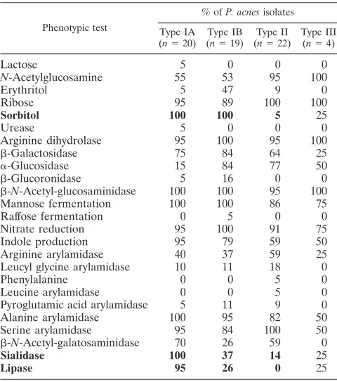
Phenotypic comparison of
P. acnes
phylotypes.
The
pheno-typic properties of the
P. acnes
phylotypes are shown in Table
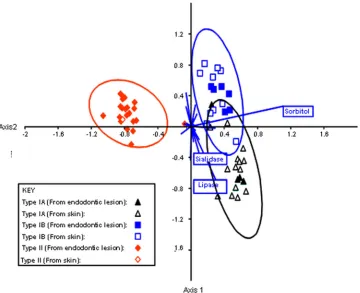
4. The phenotypic properties which differentiate between
P.
acnes
types IA, IB, and II were investigated using canonical
variates analysis (Fig. 4). Type III strains were not investigated
due to the small number of isolates. The ability to ferment
sorbitol and to produce sialidase and lipase activities
differen-tiated between types IA, IB, and II.
DISCUSSION
[image:6.585.45.541.79.361.2]We have used a culture-based approach to investigate the
microbiota of refractory root canal infections. With a
culture-based approach, it is likely that, overall, fewer taxa than are
detectable with 16S rRNA cloning or cloning combined with
culture approaches may be identified. Thus, in a study of 5
primary endodontic lesions in four patients, a mean of 20.2
taxa (range, 7 to 29) was identified using a combined cloning
and cultural approach (40), whereas a mean of 14.1 taxa was
identified from the refractory lesions with abscesses. We
inves-tigated refractory lesions which had previously been treated, so
DNA derived from dead bacteria may be present, which might
lead to an overestimation of the diversity of the microbiota if
a 16S rRNA cloning approach was used. However, as the
purpose of this study was not just to catalogue the taxa present
TABLE 2—
Continued
Organism
Presence in endodontic lesion (with abscesses) no.:
12 13 14 15 16 17 18 19 20
Dialister
sp. oral clone BS095
⫺
⫺
⫺
⫺
⫺
⫹
⫺
⫺
⫺
Fusobacterium naviforme
⫺
⫺
⫺
⫺
⫹
⫺
⫺
⫺
⫺
Fusobacterium nucleatum
subsp.
nucleatum
⫺
⫺
⫺
⫺
⫺
⫺
⫺
⫹
⫺
Fusobacterium nucleatum
subsp.
polymorphum
⫺
⫺
⫺
⫺
⫺
⫺
⫺
⫹
⫺
Fusobacterium nucleatum
subsp.
vincentii
⫺
⫹
⫺
⫺
⫹
⫺
⫹
⫹
⫺
Oribacterium sinus
⫺
⫺
⫺
⫺
⫺
⫺
⫺
⫺
⫹
Phascolarctobacterium faecium
ACM3679
⫺
⫺
⫺
⫹
⫺
⫺
⫺
⫺
⫺
Porphyromonas gingivalis
⫹
⫹
⫺
⫺
⫺
⫺
⫺
⫺
⫺
Prevotella baroniae
⫺
⫺
⫹
⫺
⫺
⫺
⫺
⫺
⫺
Prevotella buccae
⫺
⫹
⫺
⫺
⫺
⫺
⫺
⫺
⫺
Prevotella melaninogenica
⫺
⫺
⫺
⫺
⫺
⫺
⫺
⫺
⫹
Prevotella oralis
⫹
⫺
⫺
⫺
⫺
⫺
⫺
⫺
⫺
Prevotella
sp. oral taxon 289
⫺
⫺
⫺
⫺
⫺
⫺
⫹
⫺
⫺
Prevotella tannerae
⫹
⫺
⫺
⫺
⫺
⫺
⫺
⫺
⫺
Pyramidobacter piscolens
⫺
⫺
⫹
⫺
⫺
⫺
⫺
⫺
⫺
Selenomonas noxia
⫺
⫺
⫺
⫺
⫺
⫺
⫺
⫺
⫹
Tanerella forsythia
⫹
⫺
⫹
⫺
⫺
⫺
⫹
⫺
⫺
Veillonella dispar
⫺
⫹
⫺
⫺
⫺
⫺
⫺
⫹
⫺
Veillonella parvula
⫺
⫺
⫺
⫺
⫺
⫺
⫺
⫺
⫹
Facultative anaerobe
Capnocytophaga
sp. oral clone AA032
⫺
⫺
⫺
⫺
⫺
⫹
⫺
⫺
⫺
Campylobacter gracilis
⫺
⫹
⫺
⫺
⫺
⫺
⫹
⫺
⫺
Campylobacter rectus
⫺
⫹
⫺
⫺
⫺
⫺
⫺
⫺
⫺
Lautropia mirabilis
⫺
⫺
⫺
⫹
⫺
⫺
⫺
⫺
⫺
TABLE 3. Comparison of distribution of
P. acnes recA
phylotypes from all endodontic lesions and from the 13 paired endodontic samples
with the distribution of
P. acnes
phylotypes identified from the perioral samples
Isolate
No. of isolates
Pvalue (2test)
Type IA Type IB Type II Type III
P. acnes
from paired perioral lesions
36
26
3
0
P. acnes
from paired endodontic lesions
7
5
25
2
4
⫻
10
⫺8All endodontic
P. acnes
isolates
a9
12
35
4
⬍
10
⫺10a
n⫽20.
on May 16, 2020 by guest
http://jcm.asm.org/
[image:6.585.44.543.653.716.2]at a site but to investigate the genotypic and phenotypic
prop-erties of
P. acnes
and
S. epidermidis
, it was necessary to culture
the microbiota and to isolate these species.
Previous studies of the endodontic microbiota (2, 9, 28, 33,
38, 42, 51, 61) have recovered
P. acnes
and various
Staphylo-coccus
species, including
S. epidermidis
, but these organisms
are generally regarded as contaminants since they are skin
commensals. In the present study, we have used a
decontam-ination protocol to remove contaminating organisms from the
tooth surface prior to entering the diseased tooth, removed the
gutta-percha points, sampled the infected root dentine
border-ing the root canal, and isolated
P. acnes
and
S. epidermidis
. The
same species were also isolated from the perioral area and
compared using REP-PCR DNA fingerprinting. The
endodon-tic and skin isolates of both
P. acnes
and
S. epidermis
are
distinct and significantly different. These observations confirm
the endodontic origin of
P. acnes
and
S. epidermidis
and the
rejection of the hypothesis that these species recovered from
endodontic samples are contaminants. While we did not
inves-tigate further the other
Propionibacterium
or
Staphylococcus
species, it must also be presumed that these are also infecting
organisms. Foreign-body implants are one of the predisposing
factors associated with numerous
P. acnes
infections (1, 14, 22,
23).
P. acnes
infections are also associated with the presence of
foreign bodies in the root canals and may have been
intro-duced into the root canals at the time of obturation of the canal
with the gutta-percha or may have infected the canal
subse-quently, after failure. Similarly,
S. epidermidis
is associated with
infections of prostheses, and it is also significant that
S. caprae
,
which was isolated from 25% of patients and which is routinely
isolated from goats and may be used in cheese ripening, is also
associated with infections in patients with an indwelling
cath-eter or foreign materials, such as cardiac prostheses (27). This
supports the hypothesis that the gutta-percha in the root canal
may represent a foreign body susceptible to colonization by the
same range of organisms that infect foreign materials or
pros-theses.
[image:7.585.111.470.68.337.2]This hypothesis was supported when the
recA
phylotypes of
the isolates were considered. We confirmed the previous
ob-servations, as nucleotide sequencing of the
recA
housekeeping
gene of
P. acnes
isolates from both endodontic lesions and
samples from perioral skin revealed only 3 different
phyloge-netic lineages (37, 38). In agreement with these observations,
we found that
P. acnes
type I was the predominant phylotype
on skin, whereas types II and III have rarely been recovered
from samples from skin (37). In the present study,
P. acnes
type
II and type III were the prevalent phylotypes in the endodontic
infections, and the distribution of type II was similar to that
reported for type II isolates from failed prosthetic hip implants
(38), orthopedic implants (51), and radical prostatectomy
spec-imens from subjects with prostate cancer (14). Type III strains
have previously been recovered only from spine intervertabral
disc material and from a prosthetic hip joint removed during
revision arthroplasty. Here we failed to isolate any type III
strains from the skin, but 4 isolates were identified from the
infected root canals of 3 patients. The natural habitat of types
II and III is not known, but these phylotypes apparently exhibit
a tropism for inert foreign surfaces, or at least an ability to bind
to such materials. Although
P. acnes
isolates of the type II
phylotype were the most prevalent endodontic isolates in our
FIG. 1. Neighbor-joining trees comparing the taxonomic relationships of representatives of
P. acnes
types IA, IB, II, and III determined by
analysis of partial sequences of PA-4687 and PA-25957. Bootstrap values are indicated at corresponding nodes. J165 and SK137 are type IA, SK187
and KPA171202 are type IB, and J139 is type II by
recA
sequencing. The phylotype of the isolates recovered in this study is denoted by the first
letters of the strain name (1A [for type IA], 1B [for type IB], II, or III). Bars, 0.005 or 0.001 substitution per site.
on May 16, 2020 by guest
http://jcm.asm.org/
study, the virulence determinants have not been identified and
none of the differential phenotypic characteristics investigated
here appear to be relevant; and all isolates tested, irrespective
of their phylotype, possessed genes coding for each of the 6
putative virulence determinants. However, all of the type III
isolates have a 5-nt deletion in PA-4687, involved in iron
trans-port, suggesting that this gene did not yield a functional
prod-uct, which was supported by our inability to demonstrate
FIG. 2. Neighbor-joining trees comparing the taxonomic relationships of representatives of
P. acnes
types IA, IB, II, and III determined by analysis
of a partial
recA
sequence and the concatenated partial sequences of 6 putative virulence determinants. Bootstrap values are indicated at the
corresponding nodes. J165 and SK137 type are IA, SK187 and KPA171202 are type IB, and J139 is type II by
recA
sequencing. The phylotype of the
isolates recovered in this study is denoted by the first letters of the strain name (1A [for type IA], 1B [for type IB], II, or III). Bars, 0.002 or 0.0005
substitution per site.
[image:8.585.301.541.405.677.2]FIG. 3. REP-PCR patterns obtained from the
P. acnes
isolates of
patient 6 (A) and the
S. epidermidis
isolates of patient 13 (B).
(A) Lanes 1 and 7, molecular size marker (pGEM DNA markers;
Promega); lanes 2 and 3, skin isolates; lanes 4 to 6, isolates from the
endodontic sample. (B) Lane 7, molecular size marker; lanes 5 and 6,
isolates from the endodontic sample; lanes 1 to 4, skin isolates.
TABLE 4. Biochemical test results for 65
P. acnes
isolates,
including reference strains
P. acnes
type IA (NCTC 737)
and
P. acnes
type II (NCTC 10390)
aPhenotypic test
% ofP. acnesisolates Type IA
(n⫽20) Type IB (n⫽19)
Type II (n⫽22)
Type III (n⫽4)
Lactose
5
0
0
0
N
-Acetylglucosamine
55
53
95
100
Erythritol
5
47
9
0
Ribose
95
89
100
100
Sorbitol
100
100
5
25
Urease
5
0
0
0
Arginine dihydrolase
95
100
95
100

-Galactosidase
75
84
64
25
␣
-Glucosidase
15
84
77
50

-Glucoronidase
5
16
0
0

-
N
-Acetyl-glucosaminidase
100
100
95
100
Mannose fermentation
100
100
86
75
Raffose fermentation
0
5
0
0
Nitrate reduction
95
100
91
75
Indole production
95
79
59
50
Arginine arylamidase
40
37
59
25
Leucyl glycine arylamidase
10
11
18
0
Phenylalanine
0
0
5
0
Leucine arylamidase
0
0
5
0
Pyroglutamic acid arylamidase
5
11
9
0
Alanine arylamidase
100
95
82
50
Serine arylamidase
95
84
100
50

-
N
-Acetyl-galatosaminidase
70
26
59
0
Sialidase
100
37
14
25
Lipase
95
26
0
25
a
Major differential characteristics distinguishing between types IA, IB, and II are highlighted in boldface. Data from type III strains were not included in the analysis due to the small number of isolates tested. All isolates were positive for glycine arylamidase activity and negative for-fucosidase,␣-fucosidase, -glu-cosidase,␣-galactosidase,␣-arabinosidase, DNase, nonspecific proteinase, elas-tase, lecithinase, and hyaluronidase activities.