INTRODUCTION
Although many signaling factors and transcriptional regulators are essential for organ formation in vertebrates, little is known about how these multiple inputs are integrated to generate the specific locations and identities of particular organs. This is mainly because of the laborious nature of analysis of the cis-regulatory elements on which transcriptional and signaling inputs directly converge; enhancer searching by traditional genome walking is a slow and painstaking process. However, recent remarkable advances in genome analysis tools have revolutionized the process (Wasserman and Sandelin, 2004). Putative enhancers are predicted in silico within entire genomes as clusters of transcription factor-binding motifs and/or intergenic sequences conserved among related species. Especially in vertebrates, enhancer activities are often mapped to conserved non-coding elements associated with developmental regulatory genes (Woolfe et al., 2005).
From this perspective, we have investigated mechanisms of lens induction in Xenopus, where essential tools for genomics-based cis-regulatory analysis, i.e. the whole genome sequence (http://genome.jgi-psf.org/Xentr4/Xentr4.home.html) and a highly efficient transgenesis technique, have become recently available (Offield et al., 2000; Smith, 2005). The relatively large evolutionary distance between Xenopus tropicalisand mammalian genomes (350 million years) allows sequence comparison to highlight the most conserved elements involved in vertebrate genomic regulatory networks (Muller et al., 2002).
Embryological studies have shown that lens induction is a stepwise process that begins when a broad domain of the animal cap ectoderm acquires a lens-forming competence at mid-gastrula stages
(Grainger, 1992). This lens-competent field is subsequently narrowed down to the non-neural ectoderm surrounding the anterior margin of the newly formed neural plate. Accompanying this field-restriction process, a lens-forming bias is established in this region by planar signals provided by the adjacent anterior neural plate. This biased region corresponds to (or includes) the part of the non-neural ectoderm termed pre-placodal ectoderm, which will give rise to the lens, otic, olfactory and adenohypophyseal placodes at later stages. After neural tube formation, only the lateral part of the lens-biased ectoderm makes contact with the developing optic vesicle and at this stage determination occurs, followed several hours later by differentiation into lens tissue (lens placode and, subsequently, the lens vesicle).
This stepwise commitment process is likely to be mediated, at least in part, by several transcription factor genes that exhibit distinct but overlapping expression patterns during the course of lens-field specification (Ogino and Yasuda, 2000). Otx2, the earliest of these genes, exhibits expression from the end of gastrulation in the pre-placodal ectoderm and the adjacent anterior neural plate (Blitz and Cho, 1995; Zygar et al., 1998). Pax6and Six3show more-restricted expression in the lens-field within the pre-placodal ectoderm (Zhou et al., 2000; Zygar et al., 1998). After neural tube formation, their expression is followed by activation of lens-specific transcription factor genes such as MafB, L-mafand Pitx3, exclusively in the presumptive lens ectoderm (PLE) overlying the optic vesicle (Ishibashi and Yasuda, 2001; Pommereit et al., 2001).
The expression of Otx2, Pax6and Six3implies their involvement in the establishment of lens bias, and recent significant progress made by mouse genetic studies supports this view (Lang, 2004). However, little is known about how their activities are sequentially integrated to narrow down the lens-field, and how signaling from the optic vesicle is involved in this process. To address this question, we chose to study the regulation of a forkhead-family gene, Lens1, which is the earliest of the genes that are expressed primarily in the PLE overlying the optic vesicle (Kenyon et al., 1999), and which is
Convergence of a head-field selector Otx2 and Notch
signaling: a mechanism for lens specification
Hajime Ogino, Marilyn Fisher and Robert M. Grainger*Xenopusis ideal for systematic decoding of cis-regulatory networks because its evolutionary position among vertebrates allows one to combine comparative genomics with efficient transgenic technology in one system. Here, we have identified and analyzed the major enhancer of FoxE3(Lens1), a gene essential for lens formation that is activated in the presumptive lens ectoderm (PLE) when commitment to the lens fate occurs. Deletion and mutation analyses of the enhancer based on comparison of Xenopusand mammalian sequences and in vitro and in vivo binding assays identified two essential transcriptional regulators: Otx2, a homeodomain protein expressed broadly in head ectoderm including the PLE, and Su(H), a nuclear signal transducer of Notch signaling. A Notch ligand, Delta2, is expressed in the optic vesicle adjacent to the PLE, and inhibition of its activity led to loss, or severe reduction, of FoxE3expression followed by failure of placode formation. Ectopic activation of Notch signaling induced FoxE3 expression within head ectoderm expressing Otx2, and additional misexpression of Otx2in trunk ectoderm extended the Notch-induced FoxE3expression posteriorly. These data provide the first direct evidence of the involvement of Notch signaling in lens induction. The obligate integration of inputs of a field-selector (Otx2) and localized signaling (Notch) within target cis-regulatory elements might be a general mechanism of organ-field specification in vertebrates (as it is in Drosophila). This concept is also consistent with classical embryological studies of many organ systems involving a ‘multiple-step induction’.
KEY WORDS: Competence, Genomics, Induction, Lens, Notch, Xenopus Development 135, 249-258 (2008) doi:10.1242/dev.009548
Department of Biology, University of Virginia, Charlottesville, VA 22904, USA.
*Author for correspondence (e-mail: rmg9p@virginia.edu)
Accepted 23 October 2007
D
E
V
E
LO
P
M
E
N
activated at the time when commitment to the lens fate occurs (Grainger, 1992). Synteny analysis using Metazome 1.1 (http://www.metazome.net/) showed that theX. tropicalis Lens1 locus is orthologous to mouse Foxe3and we therefore refer to it as Xenopus FoxE3in this study. Analyses of mouse mutants have shown that Foxe3is essential for lens epithelial proliferation and lens vesicle closure (Blixt et al., 2000; Brownell et al., 2000).
Using in vivo and in silico approaches, we identify and characterize an enhancer of X. tropicalis FoxE3responsible for its PLE-specific expression, and demonstrate that Xenopus-mammalian genome comparison is a powerful strategy for prediction and for further detailed analysis of vertebrate cis-regulatory elements. A ‘co-transgenesis’ assay, in which separate enhancer and reporter element constructs are co-injected, was also developed to facilitate the rapid survey of possible enhancer activities of the predicted cis-regulatory elements. Our analysis has led to the first recognition that Notch signaling (Lai, 2004) is a lens-inducing signal, and revealed a role for Otx2 acting in concert with Notch signaling to specify the lens-field. The data presented here reveal one of the first molecular mechanisms found to underlie stepwise determination of the lens, as well as suggesting a general mechanism for how organ progenitor cells are segregated within a broader ‘zone of responsiveness’ during vertebrate development.
MATERIALS AND METHODS Plasmid constructs
pBSSK+EGFP was generated by introducing an EGFP-poly(A) cassette excised from pEGFP-1 (Clontech) into pBluescript SK+ (Stratagene).
–10.6kGFP was generated by introducing the FoxE3 promoter region
(–10,632 to +118) isolated from a X. tropicalisgenomic DNA library (a gift
from Dr Richard Harland, University of California, Berkeley, CA) into pBSSK+EGFP. This promoter sequence is followed by an open reading
frame whose nucleotide sequence has 93% identity with that of X. laevis
FoxE3(Lens1) (Kenyon et al., 1999) in X. tropicalisgenome assembly 4.1 (scaffold 1: 2902082-2903173, http://genome.jgi-psf.org/Xentr4/
Xentr4.home.html). We assigned the transcription start site (+1) to the 5⬘end
of the putative 5⬘untranslated region (UTR) (scaffold 1: 2901906-2902081),
which was predicted from its homology to the 5⬘UTR of X. laevis FoxE3
(94% identity), to number the flanking sequence. GFP was generated by
introducing the chicken -actinbasal promoter (–55 to +53) excised from
pLuc (Ogino and Yasuda, 1998) into pBSSK+EGFP. A series of
base-substitution mutant constructs (mt1-mt9) were generated from Xt462-GFP
using the Quikchange Site-Directed Mutagenesis Kit (Stratagene) with the following primers and their complements (mutated sequences are underlined):
mt1, 5⬘CAAGGAGAGTGAAATGAGAGGTACCATGTTTTCATC ATC
-CG-3⬘;
mt2, 5⬘GAGTGAAATGAGATAATCCATCGATTCATCATCCGTAGG
-CC-3⬘;
mt3, 5⬘CTCTTTTCACAAGCCATGGTACGTACTTTATTAGGCTGA
-GC-3⬘;
mt4, 5⬘CATGGGCCGTACTTTATTAGGTACCGCAGTTCTGGGCC
-TGTAAG-3⬘;
mt5, 5⬘ATGCAGAATGGCAGAAACCGGTAGGCCCAGTACATTTT
-CC-3⬘;
mt6, 5⬘CAACATCAGATTTTCCTACATCTAGAGTGCAGAAATCCC
-ACAC-3⬘;
mt7, 5⬘CTACAGATAGAGTGCAGAAATCTAGAACATGTCCAAATC
-TGTTAACATC-3⬘;
mt8, 5⬘GCAGAAATCCCACACATGTGGCCATCTGTTAACATCTGA
-CATG-3⬘;
mt9, 5⬘
-CCACACATGTCCAAATCGATTAACATCTGACATGAAGTC-3⬘.
Otx-Su(H)-GFP, mtOtx-Su(H)-GFP and Otx-mtSu(H)-GFP were
generated by introducing the following double-stranded oligonucleotides
into GFP (Otx- and Su(H)-binding sequences are underlined; linker
sequences are shown in lowercase):
Otx-Su(H), 5⬘ctagaGGGATTAGAGTTCCCACACGGGATTAAATTTC
-CCACGGAGGATTAGGGTTCCCACAAGGGATTAGATTTCCCACg-3⬘;
mtOtx-Su(H), 5⬘ctagaGGCGGTAGAGTTCCCACACGGCGGTAAAT
TTCCCACGGAGCGGTAGGGTTCCCACAAGGCGGTAGATTTCCC
-ACg-3⬘;
Otx-mtSu(H), 5⬘ctagaGGGATTAGAGTTCGGTCACGGGATTAAAT
TTCGGTCGGAGGATTAGGGTTCGGTCAAGGGATTAGATTTCGG
-TCg-3⬘.
Details of other reporter constructs are given in the Results.
pGEM-XFoxE3 was generated by cloning the coding sequence of X. laevis FoxE3
into the pGEM-T Easy vector (Promega). pCS2+GR-Otx2-En was generated by introducing the coding sequence of a GR ligand-binding domain isolated
from pCS2+GR-Su(H)VP16 (Rones et al., 2000) into the 5⬘end of the Otx2
coding sequence of pCS2+Otx2-En (Gammill and Sive, 2001).
Xenopustransgenesis
Transgenic embryos were generated by a sperm nuclear transplantation method (Kroll and Amaya, 1996), and their GFP expression was detected by in situ hybridization (Sive et al., 2000) for maximum sensitivity. The fraction of embryos that developed normally until scoring stages (stages 22-24) (Nieuwkoop and Faber, 1967) varied between 10-20% of total injected embryos depending on egg quality. However, the frequency of reporter gene expression within the group of normal embryos was fairly constant. In the
case of three independent assays using Xt462-GFP, the average fraction of
embryos with PLE-specific GFP expression in a normal group was 21±1.7%. This construct was injected in parallel with a series of its mutant constructs (mt1-mt9) as a control to monitor transgenesis efficiency. For
co-transgenesis, the 462 bp enhancer fragment of Xenopus FoxE3, amplified
by PCR, was mixed with a -actinpromoter-GFP cassette excised from
GFP in a molar ratio of 4:1 and directly used for transgenesis.
In situ hybridization and RNA injections
In situ hybridization analyses of Delta1, Delta2, Otx2, ␥1-crystallinand Rx
were performed as described (Blitz and Cho, 1995; Chitnis et al., 1995; Jen et al., 1997; Mathers et al., 1997; Offield et al., 2000). The antisense probes for GFP, FoxE3, Serrate1and Notch2were generated using pBSSK+EGFP, pGEM-XFoxE3, pBS-XSerrate1 (a gift from Dr C. Kintner, The Salk Institute for Biological Studies, La Jolla, CA) and an EST clone (NCBI accession BX855333), respectively.
Capped mRNAs for microinjections were transcribed from pCS2+GR-Su(H)VP16, pCS2+GR-Su(H)DBM (Rones et al., 2000), pCS2+NICD (Chitnis et al., 1995), pCS2+XOtx2-GR (Gammill and Sive, 1997), pCS2+GR-Otx2-En, pCS2+XOtx2 (Blitz and Cho, 1995), pCS2+XDelta1,
pCS2+XDelta1Stu(Chitnis et al., 1995), pCS2+XDelta2, pCS2+XDelta2Tr
(Jen et al., 1997), pCS2+EGFP and pCS2+nlacZ. For lacZ staining,
magenta-gal was used as the substrate (Rones et al., 2000).
RESULTS
Identification of the FoxE3cis-regulatory element
by both classic promoter deletion assays and in silico analysis
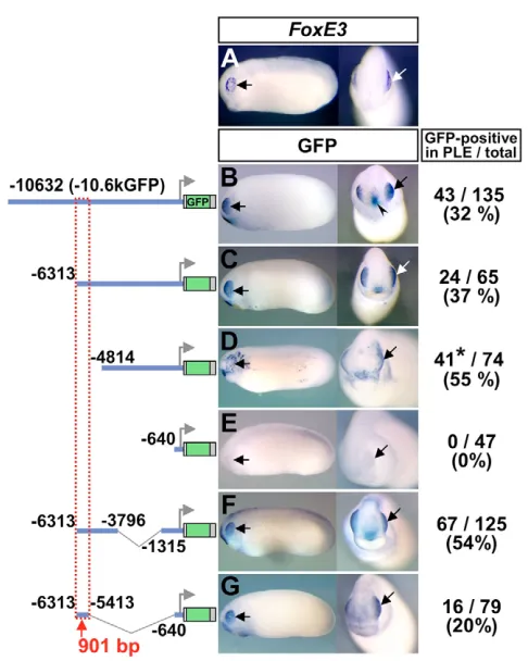
PLE-specific expression of FoxE3in early tailbud embryos of X. tropicalis(Fig. 1A) was indistinguishable from the previously reported FoxE3expression in X. laevis(Kenyon et al., 1999). This expression was closely recapitulated by GFP expression in transgenic X. laevisembryos generated with a reporter construct carrying a 10.6 kb upstream sequence of X. tropicalis FoxE3(Fig. 1B, –10.6kGFP; see Fig. S1 in the supplementary material). Sequences responsible for PLE-specific expression were narrowed down by a series of promoter deletion assays (Fig. 1C-G), which identified a 901 bp element (–6313 to –5413) as the one that essentially recapitulates the activity of the –10.6 kb promoter when linked to the basal promoter region (–640 to +118) (Fig. 1G).
We also approached the same question from a bioinformatic perspective. To examine a possible relationship between conserved non-coding elements distributed around vertebrate FoxE3loci and
D
E
V
E
LO
P
M
E
N
the functionally identified 901 bp enhancer, we aligned a ~60 kb sequence of the human FOXE3locus with orthologous mouse, chicken and X. tropicalis sequences using a genome alignment program, MultiPipmaker (http://pipmaker.bx.psu.edu/pipmaker/) (Fig. 2A). In this alignment, the percent identity plot (pip) shows both the position in the human sequence and the degree of similarity for each aligning segment between the human and other sequences. The pip in the human-mouse alignment (Fig. 2A, top row) indicates extensive sequence conservation between these two species. However, the pip in the human-chicken and the human-Xenopus alignments (Fig. 2A, second and third rows) indicates that only one region is conserved in all four species besides the coding region (Fig. 2A, red box). This conserved region corresponds to the 462 bp sequence between –6159 and –5698 in theX. tropicalis FoxE3 locus, and is included in the 901 bp element (–6313 to –5413) described above.
[image:3.612.52.295.55.359.2]Sequence conservation in the 462 bp element was further analyzed by phylogenetic footprinting (Fig. 2B). The evolutionary distance from Xenopusto human resolves the 462 bp element into Fig. 1. In vivo deletion analysis identifies a 901 bp enhancer that directs PLE-specific expression of FoxE3.(A) FoxE3expression in
[image:3.612.319.554.61.531.2]X. tropicalisembryos (stage 23) detected by in situ hybridization. (B-G) GFP expression detected by in situ hybridization in representative transgenic embryos (stages 22-24) generated with the reporter constructs shown to the left. White and black arrows in A-G indicate the PLE. The arrowhead in B indicates ectopic GFP expression in the presumptive oral ectoderm. Numbers of embryos with GFP expression in the PLE and the total number of normally (or near normally) developing embryos injected with each construct are indicated to the right, along with the percentage of GFP-positive cases. The 901 bp element necessary for PLE-specific expression is boxed with a dotted red line. *The expression in D was positive in the PLE but very spotty and broad, as shown in the left-hand panel.
Fig. 2. In silico analysis of FoxE3cis-regulatory elements. (A) Genomic sequence of the human FOXE3locus (–29.0 to +30.4 kb) aligned with its orthologous mouse, chicken and X. tropicalissequences using MultiPipMaker. The aligned sequences were downloaded from the VISTA Browser (http://genome.lbl.gov/vista/index.shtml) or from the
Xenopus tropicalisv4.1 genome site (http://genome.jgi-psf.org/ Xentr4/Xentr4.home.html). A non-coding region conserved from human to Xenopusis boxed with a dotted red line. Black arrows indicate exons of human FOXE3and its neighboring gene, FOXD2, and their orientations. Coding and untranslated sequences are shaded with light blue and light cyan, respectively. The scale at the bottom of the alignment indicates relative positions in the human FOXE3locus. (B) The conserved non-coding element of Xenopus FoxE3(–6159 to –5698) identified by MultiPipMaker is aligned with its orthologous chicken, mouse and human sequences using ClustalW
(http://www.ebi.ac.uk/clustalw/). Sequences conserved in at least three species are shaded in gray. Putative transcription factor-binding motifs mapped in conserved sequences are boxed in different colors. The three gray boxes indicate conserved sequences that do not match any known
binding motifs.
D
E
V
E
LO
P
M
E
N
discontinuous stretches of conserved sequences of 6-11 bp, each of which may predict transcription factor-binding motifs. Eight of these stretches are identical or similar to known transcription factor-binding motifs and include target sequences of three signaling pathways: a Smad1-binding motif for BMP signaling (Kusanagi et al., 2000); a Su(H) (also known as CBF1/RBP-J/Lag-1)-binding motif for Notch signaling (Tun et al., 1994); and a Tcf3/Lef1-binding motif for canonical Wnt signaling (Eastman and Grosschedl, 1999). Other predicted motifs are two Otx-binding motifs (Gan et al., 1995), two Fox motifs (Kaufmann et al., 1995) and a GATA motif (Ko and Engel, 1993).
Identification of transcription factor-binding motifs essential for PLE-specific expression
The 901 bp enhancer identified by the deletion analysis contains not only the conserved 462 bp, but also surrounding non-conserved sequences. Transgenic embryos generated with a construct in which the conserved 462 bp sequence alone was linked to a heterologous basal promoter (chicken -actin) exhibited GFP expression in the PLE similar to the embryos carrying the 901 bp enhancer construct (Xt462-GFP in Fig. 3A, compare with Fig. 1G), whereas the construct with the -actin promoter alone (GFP) did not drive any detectable expression in any embryos (Fig. 3B). Interestingly, PLE-specific expression was also detected when the 462 bp element amplified by PCR was co-injected with the GFP cassette (Fig. 3C), an approach taken because this transgenic method is known to produce a concatemer of transgenic inserts. This novel, cloning-free ‘co-transgenesis’ strategy is a powerful tool for the rapid survey of enhancer activities of conserved non-coding elements that are widely distributed in vertebrate genomes (see Discussion).
Transgenic embryos generated with a construct in which the 462 bp Xenopuselement was replaced with the orthologous 423 bp element of mouse Foxe3 (–3529 to –3107) used for the phylogenetic footprinting (Fig. 2B) exhibited GFP expression that was indistinguishable from that driven by the Xenopus element (compare Fig. S2A with B in the supplementary material), suggesting that the sum of the discontinuous stretches of short conserved sequences identified by the phylogenetic footprinting is sufficient to account for the expression. To evaluate the role of each short conserved sequence, we introduced base-substitution mutations individually into all of the eight putative transcription factor-binding motifs mapped there, and into one of the conserved motifs with no similarity to known transcription factor-binding motifs (indicated as Factor X motif in Fig. 2B). The mutant constructs were generated from Xt462-GFP, which drove PLE-specific expression in 21% of the generated embryos in transgenic assays as described (Fig. 3A and ‘wt’ in Fig. 3D). None of the mutations led to additional ectopic expression, but the percentage of embryos with PLE-specific expression was decreased to different extents (Fig. 3D). The most striking result was obtained with the construct carrying a mutation in the Su(H) motif (Fig. 3D, mt7), which completely abolished the expression in all cases except one (n=70). Even in this one positive case, the expression was very faint (not shown). The mutation of the 3⬘-most Otx motif and the mutation of the unknown Factor X motif decreased the positive cases to 6% and 8%, respectively (Fig. 3D, mt5 and mt4). Mutation of the 5⬘-most Fox motif, Smad1 motif, or GATA motif, somewhat decreased the positive cases, to ~12% (Fig. 3D. mt2, mt3 and mt6). Mutation of the 5⬘Otx motif, Tcf3/Lef1 motif, or 3⬘ Fox motif did not significantly reduce the percentage of
[image:4.612.317.562.61.368.2]positive cases (Fig. 3D, mt1, mt8 and mt9). By 2 test (http://www.graphpad.com/quickcalcs/chisquared1.cfm), the percentage of positive cases in the wild type and the Su(H), 3⬘ -most Otx, or unknown Factor X mutant constructs are significantly different (P<0.0001, P=0.0006 and P=0.0018, respectively), whereas the differences observed in other cases are not significant (P>0.05). These results show that the Su(H), 3⬘ -most Otx, and unknown Factor X motifs are essential for the enhancer activity, whereas other motifs might serve to boost its level and/or are involved in the regulation at different developmental stages.
Fig. 3. Mapping of regulatory motifs essential for PLE-specific activity of the FoxE3enhancer. (A-C) Representative transgenic embryos (stages 22-24) generated with the GFP reporter constructs shown on the left. Black arrows indicate the PLE. Numbers of embryos with GFP expression in the PLE and the total number of normally (or near normally) developing embryos injected with each construct are indicated on the right-hand side with percentages of the GFP-positive cases. The white line in A indicates the plane of the transverse section shown in the inset. The black arrow in the inset indicates GFP expression in the PLE overlying the optic vesicle. The embryo shown in C was generated by co-transgenesis, i.e. co-injection of the 462 bp enhancer of Xenopus FoxE3amplified by PCR along with the GFP cassette. (D) Identification of transcription factor-binding motifs essential for PLE-specific expression by mutation analysis. wt is the construct used in Fig. 3A (Xt462-GFP). mt1-mt9 were generated from wt/Xt462-GFP by introducing a base-substitution mutation (cross) into each of the conserved transcription factor-binding motifs. The bar chart shows the percentage of the embryos that showed GFP expression in the PLE among total developed embryos injected with the constructs shown on the left. Actual numbers of GFP-positive cases and total numbers of scored embryos are indicated in parentheses.
D
E
V
E
LO
P
M
E
N
Regulation of FoxE3expression and lens placode formation by Notch signaling
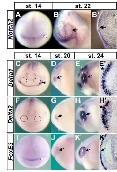
Gel retardation assays showed direct binding of XenopusSu(H) protein to the putative Su(H) motif identified in the FoxE3 enhancer in vitro, and chromatin immunoprecipitation experiments confirmed in vivo binding of the Su(H) protein to the enhancer (see Fig. S3A and Fig. S4A in the supplementary material). Su(H) is ubiquitously expressed and activates transcription only when it forms a nuclear complex with the intracellular domain of Notch receptor (NICD) that is translocated from the cytoplasm upon activation by a ligand (Fig. 7A) (Lai, 2004). We found that a Xenopus homolog of mammalian Notch2, newly identified in this study, is expressed during the course of lens formation in the pre-placodal ectoderm, PLE and developing lens vesicle (Fig. 4A-B⬘and see Fig. S5 in the supplementary material).
The Notch-ligand genes, Delta1(Chitnis et al., 1995), Delta2(Jen et al., 1997) and Serrate1(Kiyota et al., 2001) have been identified in Xenopus. To assess the involvement of Notch ligands in FoxE3 regulation, their expression was compared with that ofFoxE3during the course of lens-field formation. In neural plate-stage embryos, neither Delta1nor Delta2is expressed in the presumptive retina fields that will give rise to optic vesicles (Fig. 4C,F) (Chitnis et al., 1995). Interestingly, Delta1expression in the anterior neural ridge is adjacent to FoxE3 expression in the pre-placodal ectoderm at this stage (Bourguignon et al., 1998; Kenyon et al., 1999) (compare Fig. 4C with I), where it may act as an early signal involved in FoxE3 expression. Accompanying neural tube formation, both Delta genes exhibit strong upregulation in the developing optic vesicle (Fig. 4D-E⬘,G-H⬘), which is followed by FoxE3expression in the PLE (Fig. 4J-K⬘) (Kenyon et al., 1999). The cells expressing Delta genes are located in the outermost region of the optic vesicle and make contact with the overlying PLE cells expressing FoxE3(Fig. 4E⬘,H⬘,K⬘). Serrate1is not expressed in these tissues at the neurula or early tailbud stages, but later is expressed in the developing lens placode as reported (not shown) (Kiyota et al., 2001).
To examine possible roles of the Delta genes in FoxE3 regulation, we blocked Delta1 and Delta2 activities using their dominant-negative forms, Delta1Stuand Delta2Tr, respectively (Chitnis et al., 1995; Jen et al., 1997). mRNA encoding either Delta1Stu, Delta2Tr or GFP was injected along with a lineage tracer, nlacZ mRNA (nuclear lacZ, 50 pg), into one dorsal blastomere of eight-cell stage X. laevisembryos. The injected embryos were fixed at the early tailbud stages (stages 22-24), and stained for lacZexpression to trace distribution of the injected mRNAs. Only the embryos that showed lacZstaining in the optic vesicle were subjected to in situ hybridization with a FoxE3 probe. Control injections using GFP mRNA did not have any significant effects on FoxE3expression (n=55). Embryos injected with Delta1StumRNA exhibited loss or severe reduction of FoxE3 expression on the injected side (100%, n=22), but this phenotype appeared to be associated with head abnormalities caused by the expression of this construct (not shown). Although the effect seen here is consistent with a role for Delta1 in lens formation, we did not study Delta1 further because of the complexity in interpreting the cause of lens defects in light of the head abnormalities seen in these experiments. By contrast, injection of Delta2TrmRNA led to a very specific phenotype: FoxE3 expression was lost or severely reduced (75%, n=32, Fig. 5A) in the PLE overlying the optic vesicle stained for lacZ, but the optic vesicle itself, which was marked by expression of the retina-specific homeobox gene Rx (Mathers et al., 1997), did not show any detectable abnormalities (100%, n=37, Fig. 5B).
In addition to FoxE3, we examined expression of a lens differentiation marker, ␥1-crystallin(Offield et al., 2000), by in situ hybridization, to investigate late phenotypes of embryos expressing Delta2Tr. At late tailbud stages (stages 29/30), the lens placode of the uninjected sides showed clear ␥1-crystallinexpression (Fig. 5D), but the ␥1-crystallin-positive cells on the injected sides formed a tiny cell mass or were absent (71%, n=42, Fig. 5C,I). Expression analysis of Rxshowed that the optic vesicle of the injected sides still had no significant defects, at least through these stages (100%, n=47, Fig. 5F-H). The downregulation of ␥1-crystallinby Delta2Trwas rescued by co-injection of mRNA encoding wild-type Delta2 (75%, n=67, Fig. 5E,I), but not Delta1 (n=38, Fig. 5I), indicating the specific activity of Delta2 for lens induction. These results show that Delta2 activity in the optic vesicle is necessary for FoxE3expression in the PLE and for subsequent lens placode formation.
[image:5.612.337.528.59.339.2]To examine how the responses to Notch signaling might directly impinge on FoxE3 expression in lens cells, we used a construct encoding an inducible dominant-negative form of Su(H), GR-Su(H)DBM (Rones et al., 2000). This construct was generated by fusing the human glucocorticoid receptor ligand-binding domain (GR) to a modified version of XenopusSu(H), which contains a Fig. 4. Comparative expression analysis of Notch signaling components and FoxE3in X. laevisembryos, showing that Notch2and FoxE3are expressed in PLE, while Delta1and Delta2 are expressed in presumptive retina.Expression of Notch2(A-B⬘),
Delta1(C-E⬘), Delta2(F-H⬘), and FoxE3(I-K⬘) was detected by in situ hybridization from neural plate stages to early tailbud stages. Regions circled with black dotted lines in C and F are the approximate
presumptive retina fields, where neither Delta1nor Delta2is expressed. Arrows in B,B⬘, D-E⬘, G-H⬘and J-K⬘indicate expression of Notch2,
Delta1, Delta2and FoxE3, respectively. The black arrowhead in C indicates Delta1 expression in the anterior neural ridge, and white arrowheads in A and I indicate Notch2and FoxE3expression in the pre-placodal ectoderm, respectively. White lines in E,H,K indicate the planes of transverse eye sections shown in E⬘,H⬘,K⬘.
D
E
V
E
LO
P
M
E
N
point mutation in its DNA-binding domain. This GR-Su(H)DBM protein, which inhibits Notch signaling in response to dexamethasone (Dex) by sequestering NICD from endogenous Su(H), allowed us to circumvent possible head defects that could be caused by constitutive inhibition of Notch signaling. mRNA encoding GR-Su(H)DBM was injected along with nlacZmRNA into one dorsal blastomere of four-cell stage X. laevisembryos. The injected embryos were cultured in the absence of Dex until stages 15-16, and then maintained with Dex (10 M) either present (induced) or absent (uninduced) until fixation at early tailbud stages (stages 22-24). This time period was chosen to yield functional GR-Su(H)DBM protein at the time when endogenous FoxE3expression is upregulated in the PLE following neural tube closure. The fixed embryos were subjected to lacZstaining to select embryos in which expression was targeted to the anterior ectoderm including the PLE. As observed in embryos injected with Delta2Tr mRNA, FoxE3 expression was lost or severely reduced on injected sides of the Dex-treated embryos (55%, n=33, Fig. 5J). Downregulation of FoxE3 was not observed on uninjected sides of any of these embryos (Fig. 5K) or on injected sides of any sibling embryos untreated with Dex (n=65, Fig. 5L), indicating that Dex itself had no effect on FoxE3 expression and the FoxE3 downregulation in the Dex-treated embryos depended on the activation of GR-Su(H)DBM by hormone treatment.
Effects of ectopic activation of Notch signaling on FoxE3 expression were examined using an inducible, active form of Su(H), GR-Su(H)VP16 [Xenopus Su(H) fused to GR and to a
VP16 activation domain], which mimics Notch pathway activation in response to Dex (Rones et al., 2000). Similar to the experiments with GR-Su(H)DBM, GR-Su(H)VP16 was expressed by injecting its mRNA, and the resulting embryos were cultured with or without Dex until fixation at stages 22-24. Ninety-five percent of the injected embryos treated with Dex exhibited ectopic FoxE3 expression in the anterior region of the injected sides stained with lacZ(n=55, Fig. 5M). In contrast to the extended lacZstaining, the ectopic FoxE3expression was spotty and localized in a domain of the ectoderm overlying the anterior brain and that surrounding the cement gland. Ectopic FoxE3 expression was not observed on the uninjected sides of any of these embryos (Fig. 5N) or on the injected sides of any sibling embryo untreated with Dex (n=69, Fig. 5O).
[image:6.612.54.478.58.289.2]The activation and inhibition of Notch signaling using the active and dominant-negative forms of Su(H), respectively, induced upregulation and downregulation of FoxE3. These results demonstrate the essential role of Notch signaling in PLE-specific FoxE3expression, and suggest that Notch signaling in the PLE is likely to be activated by Delta2 expressed in the adjacent optic vesicle. Interestingly, the ectopic FoxE3expression induced by the active form of Su(H) was regionally restricted to part of the anterior ectoderm, which suggests pre-localization there of a factor enabling responsiveness to Notch signaling. In addition, this restricted ectopic expression is consistent with a role for Notch signaling as a cue to turn on FoxE3at the right place within this competent domain.
Fig. 5. Effects of manipulation of Notch signaling on FoxE3expression and subsequent lens placode formation.(A,B) Frontal view of
Xenopusembryos injected with mRNA encoding Delta2Tr(500 pg), fixed at stage 23, and subjected to lacZstaining (magenta) and in situ hybridization with FoxE3or Rxprobe (purple or deep purple staining). White and black arrowheads in A-H indicate in situ hybridization signals on injected and uninjected sides of embryos, respectively. (C,D,F,G) The injected and uninjected sides of embryos injected with Delta2TrmRNA, fixed at stages 29/30, and hybridized with ␥1-crystallinor Rxprobe. (H) A transverse head section of the embryo shown in F,G. (E) The injected side of an embryo injected with both Delta2TrmRNA (500 pg) and wild-type Delta2 mRNA (500 pg), fixed at stage 29, and hybridized with ␥1-crystallinprobe. (I) Summary of Delta2TrmRNA injection experiments. GFP mRNA (1000 pg) was injected as a control. (J,K) The injected and uninjected sides, respectively, of an embryo injected with GR-Su(H)DBM mRNA (1000 pg) and induced with Dex. Arrows in J-O indicate endogenous FoxE3
expression in the PLE. (L) The injected side of an embryo injected with GR-Su(H)DBM but not induced with Dex. (M,N) The injected and uninjected sides, respectively, of an embryo injected with GR-Su(H)VP16 mRNA (1000 pg) and induced with Dex. Black and white arrowheads in M indicate ectopic FoxE3 expression in the ectoderm overlying the anterior brain and that surrounding the cement gland, respectively. (O) The injected side of an embryo injected with GR-Su(H)VP16 but not induced with Dex.
D
E
V
E
LO
P
M
E
N
Otx2 confers the ability to activate FoxE3in response to Notch signaling
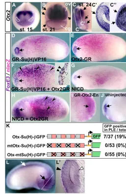
We examined Otx2, as a candidate factor responsible for the regional responsiveness to Notch signaling, for two reasons. First, we identified a putative Otx motif as among the most essential of the transcription factor-binding motifs in the FoxE3enhancer (Fig. 3D, mt5). Second, during the time window chosen for the activation of GR-Su(H)VP16 (from the neural plate to early tailbud stages), Otx2 shows diffuse expression in the head ectoderm, including not only the PLE, but also the surrounding region where the ectopic FoxE3 expression was observed (Fig. 6A-C⬙, compare Fig. 5M and Fig. 6C) (Zygar et al., 1998).
Gel retardation assays showed direct binding in vitro of Xenopus Otx2 protein to the putative Otx motif identified in the FoxE3 enhancer, and chromatin immunoprecipitation experiments confirmed in vivo binding of endogenous Otx2 protein to the enhancer (see Fig. S3B and Fig. S4B in the supplementary material). To test whether Otx2 misexpression confers the ability to respond to Notch signaling in vivo, we used an inducible form of Xenopus Otx2, GR-fused Otx2 (Otx2-GR), whose activity can be controlled by Dex treatment (Gammill and Sive, 1997). This construct allows
us to circumvent severe gastrulation defects and spina bifida caused by misexpression of wild-type Otx2 in Xenopusembryos (Blitz and Cho, 1995). mRNAs encoding GR-Su(H)VP16 and Otx2-GR were injected separately or together into a ventral blastomere of four-cell stage embryos to target expression in the trunk region instead of the head ectoderm expressing endogenous Otx2. All injected embryos were treated with Dex at stages 15-16, fixed at stages 22-24, and subjected to lacZstaining and in situ hybridization with the FoxE3 probe.
[image:7.612.298.555.58.453.2]None of embryos misexpressing either GR-Su(H)VP16 or Otx2-GR exhibited ectopic FoxE3expression in the trunk region, where the lacZstaining indicated broad distribution of the injected mRNAs (n=66 and n=52, respectively; Fig. 6D,E). This indicates that neither Notch signaling nor Otx2 activity is sufficient for FoxE3activation. However, when embryos were co-injected with GR-Su(H)VP16 and Otx2-GR mRNAs, 91% (n=54) exhibited striking FoxE3 expression in the trunk region throughout the lacZ-stained region (Fig. 6F, black arrowheads). This trunk expression was restricted to the ectoderm layer, and was absent from the underlying neural and mesodermal tissues (Fig. 6F, inset). Ectopic FoxE3 expression was also detected in embryos that were induced with Dex in the presence of a protein Fig. 6. Otx2 confers the ability to activate FoxE3in
response to Notch signaling.(A-C⬙) Expression of Otx2in the anterior ectoderm detected by in situ hybridization. At the neural plate stages, Otx2is expressed in the anterior ectoderm including the presumptive lens-fields, which are circled with white dotted lines in A. Arrows in B indicate broad expression in the ectoderm that overlies the optic vesicles (ov) and surrounds the cement gland primordium (cg). Arrows in C indicate the border of ectodermal Otx2expression. White lines in C indicate the planes of transverse sections shown in C⬘and C⬙. Arrows in C⬘and C⬙, respectively, indicate expression in the PLE overlying the optic vesicle and in the ectoderm overlying the forebrain. (D-H) Notch-Otx2 combination activates FoxE3
in the trunk ectoderm. Xenopusembryos injected with the mRNAs indicated in each panel were induced with Dex, and then subjected to lacZstaining and in situ hybridization with
FoxE3probe. Ectopic FoxE3expression was not detected in embryos injected with mRNA encoding GR-Su(H)VP16 (1000 pg) (D), Otx2-GR (250 pg) (E), or NICD (1000 pg) (G), whereas it was detected in embryos injected with both GR-Su(H)VP16 (750 pg) and Otx2-GR (250 pg) (F), or both NICD (750 pg) and Otx2-GR (250 pg) (H). Arrowheads in F and H indicate ectopic FoxE3expression. Arrows indicate endogenous
FoxE3expression in the PLE. The white line in F indicates the plane of the transverse section shown in the inset. Black arrowheads in the inset indicate the overlap of FoxE3
expression and nuclear lacZstaining in the ectodermal cells, and white arrowheads indicate cells in the underlying mesoderm layer showing nuclear lacZstaining but no FoxE3
expression. (I,J) The injected and uninjected sides, respectively, of an embryo injected with mRNA encoding GR-Otx2-En (250 pg), induced with Dex from stage 18, and then subjected to lacZstaining and in situ hybridization with FoxE3probe at stage 22. Arrows indicate the PLE. (K) Transgenic experiments using Otx-Su(H) reporter constructs. Numbers of embryos with GFP expression in the PLE and the total number of normally (or near normally) developing embryos injected with the
constructs shown on the left are indicated on the right-hand side with percentages of the GFP-positive cases. Gray and red boxes indicate Otx- and Su(H)-binding motifs, respectively, in
the constructs, and crosses indicate base-substitution mutations introduced there. (L,L⬘) A representative transgenic embryo generated with Otx-Su(H)-GFP. Black and white arrows indicate GFP expression in the eye and spinal cord, respectively. The white line indicates the plane of the transverse eye section shown in L⬘. Black and white arrowheads indicate GFP expression in the PLE and optic vesicle, respectively.
D
E
V
E
LO
P
M
E
N
synthesis inhibitor, cycloheximide, verifying a direct effect of GR-Su(H)VP16 and Otx2-GR on the FoxE3 promoter (see Fig. S6 in the supplementary material).
The trunk expression was unlikely to result from an increase in the total amount of misexpressed transcription factors, as the total amount of mRNAs co-injected in this experiment (750 pg of GR-Su(H)VP16 and 250 pg for Otx2-GR) was kept the same as that for the misexpression of GR-Su(H)VP16 alone (1000 pg). The trunk expression was also observed when wild-type XenopusOtx2, instead of Otx2-GR, was misexpressed in combination with GR-Su(H)VP16 (95%, n=22, not shown). This indicates that the ectopic expression was not associated with the GR ligand-binding domain fused to Otx2, although many of the Otx2-injected embryos exhibited a spina bifida phenotype that is characteristic of misexpression of wild-type Otx2.
The Otx2-dependent activation of FoxE3was also observed when NICD was misexpressed instead of GR-Su(H)VP16 to activate Notch signaling: misexpression of NICD alone in the trunk ectoderm did not induce ectopic FoxE3expression in any injected embryos (n=33, Fig. 6G), but the combination of NICD and Otx2-GR did (49%, n=45; Fig. 6H, black arrowheads). This showed that endogenous Su(H) activates FoxE3 as effectively as the artificial construct GR-Su(H)VP16, if Notch is activated.
In addition to these gain-of-function experiments, we designed loss-of-function experiments for Otx2 to verify that its activity is required for FoxE3expression in the PLE. Since constitutive loss of Otx2 activity impairs anterior neural fate determination (Gammill and Sive, 2001), we chose to inject mRNA encoding an inducible dominant-negative form of Otx2, GR-Otx2-En. We generated this construct by fusing a coding sequence of GR to a dominant-negative
form of Otx2, Otx2-En (XenopusOtx2 fused to the minimal repressor domain of Engrailed), which has been shown previously to specifically block Otx2 function (Gammill and Sive, 2001). The injected embryos induced with Dex from stage 18 onward did not exhibit any detectable head abnormalities (Fig. 6I,J), suggesting that anterior neural defects were circumvented by the use of this inducible construct. Loss, or significant reduction, of FoxE3expression in the PLE was detected on their injected sides by in situ hybridization (58%, n=33, Fig. 6I), but not in their uninjected sides (Fig. 6J) nor in the injected sides of any sibling embryos untreated with Dex (n=25, not shown).
These experiments demonstrate that Otx2 enables FoxE3 activation in response to Notch signaling. To test whether Otx2 and Notch inputs are sufficient to direct PLE-specific expression, we generated a reporter construct carrying four copies of a pair of consensus Otx- and Su(H)-binding motifs in front of the -actin promoter-GFP cassette [Otx-Su(H)-GFP]. Transgenic embryos injected with this Otx-Su(H) reporter exhibited GFP expression not only in the PLE, but also in the optic vesicle and spinal cord (Fig. 6K-L⬘), although the expression in the PLE did not appear so strong as that driven by theFoxE3enhancer. This eye-specific expression was not detected when transgenic embryos were generated with reporter constructs in which either the Otx or Su(H) motifs were mutated [mtOtx-Su(H)-GFP and Otx-mtSu(H)-GFP, respectively; Fig. 6K], or when a reporter construct that carried eight copies of the Su(H) motifs and no copy of the Otx motif was used (not shown). These reporter assays suggest that both Otx2 and Notch inputs are necessary and sufficient to drive expression in PLE, but additional inputs are required for boosting the expression level in the PLE and for repressing expression in neural tissues, thereby directing the more defined expression of the FoxE3enhancer.
DISCUSSION
A model of FoxE3activation
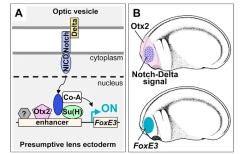
We have presented gain-of-function and loss-of-function strategies demonstrating that Otx2 activity and Notch signaling directly converge on the FoxE3enhancer to direct PLE-specific expression (Fig. 7A). These are the first data directly showing involvement of Notch signaling in lens induction. Based on these results, we propose a stepwise model of FoxE3activation (Fig. 7B): (1) Otx2 regionally enables responsiveness to Notch signaling within head ectoderm including PLE; (2) Delta2 signaling from the optic vesicle locally activates Notch signaling in the overlying PLE to turn on FoxE3 expression within this broader competent region. The results of the transgenic assay using the Otx-Su(H) reporter suggest that other transcription factors might contribute to boosting and refining the PLE-specific expression in combination with Otx2 and Su(H). An unknown factor that binds to the Factor X motif (Fig. 2B and Fig. 3D), and FGF, BMP and ZEB2 (SIP1) signaling, the inhibition of which causes a reduction in Foxe3expression in mouse embryos, might be involved in this process (Faber et al., 2001; Yoshimoto et al., 2005).
[image:8.612.52.293.59.213.2]Because the dominant-negative form of Delta1 induced head defects in embryos, we could not examine possible roles for Delta1 in FoxE3 regulation and subsequent lens formation. However, neural tube formation is accompanied by a dynamic change in Delta1expression from the anterior neural ridge to the optic vesicle (Fig. 4C-E⬘), which might be responsible for the shift of FoxE3 expression from the pre-placodal ectoderm to the PLE (Fig. 4I-K⬘). FoxE3expression in the PLE of embryos expressing the dominant-negative form of Delta2 was severely reduced, but remained in some cases, which might be due to some contribution of Delta1 to FoxE3 regulation.
Fig. 7. A model of FoxE3activation in Xenopus.(A) Integration of Otx2 and Notch inputs on theFoxE3enhancer. Otx2 and Su(H) are proposed to bind to theFoxE3enhancer prior to receiving Notch signaling, but remain in a quiescent state. In response to stimuli from Delta ligands, the Notch intracellular domain (NICD) is cleaved from its extracellular domain and translocated into the nucleus. Although this model shows a direct interaction between membrane-bound Notch and Delta, we do not know the exact mechanism by which this signaling may occur in this system (see Discussion). In the nucleus, NICD forms a complex with Su(H) and activates transcription by recruiting a co-activator (Co-A). Otx2 synergistically stimulates this transcription. An unidentified factor(s) (indicated with a question mark) may contribute to boosting and refining the PLE-specific expression. (B) Otx2 is broadly expressed in the head ectoderm that includes PLE, while localized Notch signaling is provided to the lens-field from the underlying optic vesicle (upper). FoxE3 is expressed in the PLE, which is the region where there is both Otx2expression and Notch signaling (beneath).
D
E
V
E
LO
P
M
E
N
As in Xenopus, Otx2is expressed in the PLE of mouse embryos, and the lens placode of Otx2heterozygous mutant mice fail to form a normal lens vesicle on an Otx1homozygous mutant background (Martinez-Morales et al., 2001). deltaC, a zebrafish Notch-ligand gene that has the highest sequence similarity to Xenopus Delta2, is expressed in the developing optic vesicle in the same manner as Xenopus Delta2 (Smithers et al., 2000), whereas a mammalian homolog of Delta2/deltaChas not been identified yet. Interestingly, Jag1, a mammalian homolog of Xenopus Serrate1, is expressed in the optic vesicle of rat embryos (Bao and Cepko, 1997), and is deleted in the mouse mutant coloboma (Cm), whose lens fails to detach from the ectoderm as in the Foxe3mutant mouse, dysgenetic lens (dyl) (Blixt et al., 2000; Brownell et al., 2000; Theiler and Varnum, 1981; Xue et al., 1999). Hence the role of Delta2/deltaCin the lens induction of lower vertebrates might be taken over by Jag1 in mammals. Regarding Notch receptors, mammalian Notch2and Notch3are expressed in the developing lens (Lindsell et al., 1996), but their expression at earlier stages has not been characterized in detail. In mammalian embryos, cells in the optic vesicle and lens ectoderm are physically separated by a space, but are connected by cytoplasmic extensions (McAvoy, 1980), which may permit direct contact for Notch signaling. It is also possible that Notch ligands might have secreted forms that are involved in Notch signaling (Qi et al., 1999).
Otx2-Notch interactions in lens determination programs, and their analogy with
selector-signaling system inDrosophila
The data presented here have significant implications for molecular mechanisms underlying the stepwise determination of the lens. Otx2 expression in head ectoderm might constitute a part of the lens-forming bias suggested by embryological studies, and Notch signaling is likely to be one of the inducing signals provided from the optic vesicle to turn on the lens-specification programs in this competent/biased ectoderm. Unlike FoxE3, expression of a lens differentiation marker, ␥1-crystallin, was not induced in the trunk ectoderm by misexpression of Otx2-GR and GR-Su(H)VP16 (not shown), suggesting that the Otx2-Notch combination is not sufficient to activate the whole lens-differentiation program. However, severe reduction or loss of ␥1-crystallin-positive lens placode cells in embryos expressing the dominant-negative Delta2 suggests a crucial role for Notch signaling in lens specification. The lens differentiation programs are presumably turned on when a set of all terminal regulators, such as FoxE3and L-maf, is activated in the PLE by different, but possibly overlapping, mechanisms.
Genetic studies in mouse have shown that Pax6lies upstream of Mab21l1, and thatMab21l1lies upstream of Foxe3(Yamada et al., 2003). We found that the combination of Otx2 and Notch signaling induced ectopic FoxE3expression in the trunk ectoderm without activating Pax6(not shown). These findings imply that the Pax6-Mab21l1pathway controls FoxE3expression indirectly through regulation of Otx2 and/or Notch signaling. Notch2might be a downstream target of Pax6, as the broad expression of Notch2in the head ectoderm is, as development proceeds, gradually localized to the lens and olfactory fields expressing Pax6(Fig. 4A-B⬘and see Fig. S5 in the supplementary material).
Interestingly, the combinatorial mechanism of FoxE3 regulation is similar to the selector-signaling system in Drosophila, in which selective gene activation by signals for cell fate specification is achieved by obligate integration of both inputs of field-specific transcription factors (selectors) and signal-activated transcription factors at the level of their target
cis-regulatory elements (Guss et al., 2001). Although this system has not been previously examined in vertebrate development, our study suggests that the same mechanism underlies the ‘multiple-step induction’ of the lens. As classic embryological studies demonstrate similar stages in many vertebrate organ systems (Gurdon, 1992), a selector-signaling system might be broadly used in vertebrates for specifying a variety of organ and tissue identities by reiteratively using a limited number of signaling pathways. While the events we are studying in this paper occur during the bias and specification phases of lens induction, the same principle could apply earlier, during the competence period, when a broadly expressed selector gene may contribute to the period of competence and converge with an early lens-inducing signal.
Xenomics (Xenopusgenomics) for analysis of
genomic regulatory networks for development The results of our classic-style deletion analysis are in close agreement with those from the comparative analysis of human and Xenopusgenomes, which demonstrates the effectiveness of using the Xenopusgenome for in silico prediction of conserved regulatory elements in vertebrates. The conserved enhancer of mouse Foxe3 identified in our study (–3529 to –3107) is included in the lens element that was independently identified by Kondoh’s group by deletion analysis in transgenic mice (–4.40 to –2.63 kb) (Yoshimoto et al., 2005), showing that Xenopus and mouse assays give consistent results.
An important challenge in the post-genomic era is to untangle the complex wiring of gene regulatory networks controlling development, growth and differentiation. As shown in the pioneering study of the gene regulatory network for sea urchin endomesoderm specification (Davidson et al., 2002), this type of study requires a high-throughput assay system for comprehensive analysis of cis-regulatory elements. The mammalian-Xenopus genome comparison and an approach developed in the course of this study – co-transgenesis – which rapidly tests enhancer activities by co-injection of PCR products along with the basal promoter-GFP cassette, will allow Xenopusto serve as a vertebrate model system that fulfils this requirement.
We acknowledge Ms Renee Aloise and Dr Lyle Zimmermann for their early
work attempting to clone the Xenopus FoxE3promoter. We thank Drs Milan
Jamrich, Mark Mercola, Chris Kintner, Ira L. Blitz, Ken W. Y. Cho, Hazel Sive and David Turner for kindly providing plasmids; Mr William B. McConnell and Ms Hong Jin for their expert technical assistance in this study; and other members of the Grainger laboratory for ongoing helpful discussions. This work was supported in part by a postdoctoral fellowship for research abroad from the Japan Society for the Promotion of Science to H.O. and by grants RR13221, EY06675, EY10283 and EY17400 to R.M.G. from the NIH.
Supplementary material
Supplementary material for this article is available at http://dev.biologists.org/cgi/content/full/135/2/249/DC1
References
Bao, Z. Z. and Cepko, C. L.(1997). The expression and function of Notch pathway genes in the developing rat eye. J. Neurosci. 17, 1425-1434.
Blitz, I. L. and Cho, K. W.(1995). Anterior neurectoderm is progressively induced
during gastrulation: the role of the Xenopushomeobox gene orthodenticle.
Development121, 993-1004.
Blixt, A., Mahlapuu, M., Aitola, M., Pelto-Huikko, M., Enerback, S. and Carlsson, P.(2000). A forkheadgene, FoxE3, is essential for lens epithelial proliferation and closure of the lens vesicle. Genes Dev.14, 245-254.
Bourguignon, C., Li, J. and Papalopulu, N.(1998). XBF-1, a winged helix transcription factor with dual activity, has a role in positioning neurogenesis in Xenopuscompetent ectoderm. Development125, 4889-4900.
Brownell, I., Dirksen, M. and Jamrich, M.(2000). Forkhead Foxe3maps to the dysgenetic lenslocus and is critical in lens development and differentiation.
Genesis27, 81-93.
D
E
V
E
LO
P
M
E
N
Chitnis, A., Henrique, D., Lewis, J., Ish-Horowicz, D. and Kintner, C.(1995).
Primary neurogenesis in Xenopusembryos regulated by a homologue of the
Drosophilaneurogenic gene Delta. Nature375, 761-766.
Davidson, E. H., Rast, J. P., Oliveri, P., Ransick, A., Calestani, C., Yuh, C. H., Minokawa, T., Amore, G., Hinman, V., Arenas-Mena, C. et al.(2002). A
genomic regulatory network for development. Science295, 1669-1678.
Eastman, Q. and Grosschedl, R.(1999). Regulation of LEF-1/TCF transcription factors by Wnt and other signals.Curr. Opin. Cell Biol. 11, 233-240.
Faber, S. C., Dimanlig, P., Makarenkova, H. P., Shirke, S., Ko, K. and Lang, R. A.(2001). Fgf receptor signaling plays a role in lens induction. Development
128, 4425-4438.
Gammill, L. S. and Sive, H.(1997). Identification of otx2target genes and
restrictions in ectodermal competence during Xenopuscement gland formation.
Development124, 471-481.
Gammill, L. S. and Sive, H.(2001). otx2expression in the ectoderm activates
anterior neural determination and is required for Xenopuscement gland
formation. Dev. Biol. 240, 223-236.
Gan, L., Mao, C. A., Wikramanayake, A., Angerer, L. M., Angerer, R. C. and Klein, W. H.(1995). An orthodenticle-related protein from Strongylocentrotus purpuratus. Dev. Biol. 167, 517-528.
Grainger, R. M.(1992). Embryonic lens induction: shedding light on vertebrate tissue determination. Trends Genet.8, 349-355.
Gurdon, J. B.(1992). The generation of diversity and pattern in animal
development. Cell68, 185-199.
Guss, K. A., Nelson, C. E., Hudson, A., Kraus, M. E. and Carroll, S. B.(2001). Control of a genetic regulatory network by a selector gene. Science292, 1164-1167.
Ishibashi, S. and Yasuda, K.(2001). Distinct roles of mafgenes during Xenopus
lens development. Mech. Dev.101, 155-166.
Jen, W. C., Wettstein, D., Turner, D., Chitnis, A. and Kintner, C.(1997). The Notch ligand, X-Delta-2, mediates segmentation of the paraxial mesoderm in Xenopusembryos. Development124, 1169-1178.
Kaufmann, E., Muller, D. and Knochel, W.(1995). DNA recognition site analysis of Xenopuswinged helix proteins. J. Mol. Biol. 248, 239-254.
Kenyon, K. L., Moody, S. A. and Jamrich, M.(1999). A novel fork headgene
mediates early steps during Xenopuslens formation. Development126,
5107-5116.
Kiyota, T., Jono, H., Kuriyama, S., Hasegawa, K., Miyatani, S. and Kinoshita, T.(2001). X-Serrate-1is involved in primary neurogenesis in Xenopus laevisin a
complementary manner with X-Delta-1. Dev. Genes Evol.211, 367-376.
Ko, L. J. and Engel, J. D.(1993). DNA-binding specificities of the GATA transcription factor family. Mol. Cell. Biol. 13, 4011-4022.
Kroll, K. L. and Amaya, E.(1996). Transgenic Xenopusembryos from sperm nuclear transplantations reveal FGF signaling requirements during gastrulation. Development122, 3173-3183.
Kusanagi, K., Inoue, H., Ishidou, Y., Mishima, H. K., Kawabata, M. and Miyazono, K.(2000). Characterization of a bone morphogenetic protein-responsive Smad-binding element. Mol. Biol. Cell 11, 555-565.
Lai, E. C.(2004). Notch signaling: control of cell communication and cell fate. Development131, 965-973.
Lang, R. A.(2004). Pathways regulating lens induction in the mouse. Int. J. Dev. Biol. 48, 783-791.
Lindsell, C. E., Boulter, J., diSibio, G., Gossler, A. and Weinmaster, G.(1996).
Expression patterns of Jagged, Delta1, Notch1, Notch2, and Notch3genes
identify ligand-receptor pairs that may function in neural development. Mol. Cell. Neurosci.8, 14-27.
Martinez-Morales, J. R., Signore, M., Acampora, D., Simeone, A. and Bovolenta, P.(2001). Otxgenes are required for tissue specification in the
developing eye. Development128, 2019-2030.
Mathers, P. H., Grinberg, A., Mahon, K. A. and Jamrich, M.(1997). The Rx
homeobox gene is essential for vertebrate eye development. Nature387,
603-607.
McAvoy, J. W.(1980). Cytoplasmic processes interconnect lens placode and optic vesicle during eye morphogenesis. Exp. Eye Res.31, 527-534.
Muller, F., Blader, P. and Strahle, U.(2002). Search for enhancers: teleost models in comparative genomic and transgenic analysis of cis regulatory elements. BioEssays 24, 564-572.
Nieuwkoop, P. D. and Faber, J.(1967). Normal Table of Xenopus laevis. Amsterdam: North-Holland.
Offield, M. F., Hirsch, N. and Grainger, R. M.(2000). The development of Xenopus tropicalistransgenic lines and their use in studying lens developmental
timing in living embryos. Development127, 1789-1797.
Ogino, H. and Yasuda, K.(1998). Induction of lens differentiation by activation of a bZIP transcription factor, L-Maf. Science280, 115-118.
Ogino, H. and Yasuda, K.(2000). Sequential activation of transcription factors in lens induction. Dev. Growth Differ.42, 437-448.
Pommereit, D., Pieler, T. and Hollemann, T.(2001). Xpitx3: a member of the Rieg/Pitxgene family expressed during pituitary and lens formation in Xenopus laevis. Mech. Dev.102, 255-257.
Qi, H., Rand, M. D., Wu, X., Sestan, N., Wang, W., Rakic, P., Xu, T. and Artavanis-Tsakonas, S.(1999). Processing of the notch ligand delta by the metalloprotease Kuzbanian. Science283, 91-94.
Rones, M. S., McLaughlin, K. A., Raffin, M. and Mercola, M.(2000). Serrate and Notch specify cell fates in the heart field by suppressing cardiomyogenesis. Development127, 3865-3876.
Sive, H., Grainger, R. and Harland, R.(2000). Early Development of Xenopus laevis – A Laboratory Manual. Cold Spring Harbor: Cold Spring Harbor Laboratory Press.
Smith, J. C.(2005). Xenopusgenetics and genomics. Mech. Dev.122, 259-262.
Smithers, L., Haddon, C., Jiang, Y. J. and Lewis, J.(2000). Sequence and embryonic expression of deltaCin the zebrafish. Mech. Dev.90, 119-123.
Theiler, K. and Varnum, D. S.(1981). Development of coloboma(Cm/+), a mutation with anterior lens adhesion. Anat. Embryol. 162, 121-126.
Tun, T., Hamaguchi, Y., Matsunami, N., Furukawa, T., Honjo, T. and Kawaichi, M.(1994). Recognition sequence of a highly conserved DNA binding protein RBP-J. Nucleic Acids Res. 22, 965-971.
Wasserman, W. W. and Sandelin, A.(2004). Applied bioinformatics for the identification of regulatory elements.Nat. Rev. Genet.5, 276-287.
Woolfe, A., Goodson, M., Goode, D. K., Snell, P., McEwen, G. K., Vavouri, T., Smith, S. F., North, P., Callaway, H., Kelly, K. et al.(2005). Highly conserved non-coding sequences are associated with vertebrate development. PLoS Biol. 3, e7.
Xue, Y., Gao, X., Lindsell, C. E., Norton, C. R., Chang, B., Hicks, C., Gendron-Maguire, M., Rand, E. B., Weinmaster, G. and Gridley, T.(1999). Embryonic lethality and vascular defects in mice lacking the Notch ligand Jagged1. Hum. Mol. Genet.8, 723-730.
Yamada, R., Mizutani-Koseki, Y., Hasegawa, T., Osumi, N., Koseki, H. and Takahashi, N.(2003). Cell-autonomous involvement of Mab21l1is essential for
lens placode development. Development130, 1759-1770.
Yoshimoto, A., Saigou, Y., Higashi, Y. and Kondoh, H.(2005). Regulation of ocular lens development by Smad-interacting protein 1 involving Foxe3
activation. Development132, 4437-4448.
Zhou, X., Hollemann, T., Pieler, T. and Gruss, P.(2000). Cloning and
expression of xSix3, the Xenopushomologue of murine Six3. Mech. Dev.91,
327-330.
Zygar, C. A., Cook, T. L. and Grainger, R. M., Jr(1998). Gene activation during early stages of lens induction in Xenopus. Development125, 3509-3519.