1199
Introduction
Ascidians are invertebrate chordates in which the notochord and the dorsal neural tube are present during larval stages, when embryos exhibit a tadpole body form (reviewed by Satoh, 1994). Ascidian embryos develop with a fixed cleavage pattern, a small number of cells and well-described cell lineage.
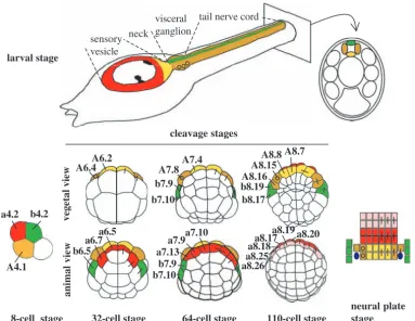
We have been using these simple embryos to study development and patterning of the central nervous system (CNS). The ascidian larval CNS consists, along the anterior-posterior axis, of a sensory vesicle, neck, visceral ganglion and tail nerve cord (Fig. 1) (for reviews, see Lemaire et al., 2002; Meinertzhagen and Okamura, 2001; Meinertzhagen et al., 2004). The entire CNS consists of only around 330 cells. The simplicity of the ascidian neural tube is most apparent at the level of the tail nerve cord, which is only four cells in cross section, one dorsal (b-line), two lateral and one ventral (A-line). Despite this comparative simplicity, the ascidian CNS retains many features in common with its vertebrate counterpart. The expression of many genes along the dorsal-ventral and anterior-posterior axes of the CNS is conserved between ascidian and vertebrate embryos. Orthologues of Otx, Pax2/5/8and Hox genes are differentially expressed along the anterior-posterior axis, while genes encoding Hedgehog and HNF3β are expressed in the ventral neural tube and Snail, Pax3/7, BMP2/4and Msxare expressed in the lateral or dorsal
part of the neural tube (Aniello et al., 1999; Corbo et al., 1997a; Hudson and Lemaire, 2001; Imai et al., 2002; Miya et al., 1997; Takatori et al., 2002; Wada et al., 1997; Wada et al., 1998; Wada and Saiga, 1999a; Wada and Saiga, 1999b). Patterning of both the ascidian and vertebrate CNSs along the anterior-posterior and dorsal-ventral axes starts during gastrulation when the CNS exists as a neural plate.
The cell lineages of the ascidian CNS are described in Fig. 1. The bilaterally symmetrical 8-cell stage embryo consists of the founder cells of four lineages: vegetal cells named A4.1 (A-line) and B4.1 (B-line) and animal cells named a4.2 (a-line) and b4.2 (b-line) (Conklin, 1905). The embryo continues to cleave in a bilaterally symmetrical manner and thus each blastomere name refers to a pair. The ascidian CNS derives from three of the four founder lineages, the a-, b- and A-lineages, which contribute to different parts of the CNS (Fig. 1) (Cole and Meinertzhagen, 2004; Nicol and Meinertzhagen, 1988a; Nicol and Meinertzhagen, 1988b; Nishida, 1987; Taniguchi and Nishida, 2004). The a-line (red and pink blastomeres in Fig. 1) gives rise to the anterior part of the sensory vesicle, including the characteristic pigmented cells. The posterior sensory vesicle and the ventral and lateral parts of the visceral ganglion and tail nerve cord come from the A-line neural A-lineages (yellow and tan blastomeres in Fig. 1), and the dorsal-most cell of the CNS from the posterior sensory
Ascidians are invertebrate chordates with a simple larval tadpole form containing a notochord and an overlying dorsal neural tube. As in vertebrates, the neural tube of ascidian larvae displays positional differences along the rostral-caudal and dorsal-ventral axes in terms of neuronal cell types generated, morphology and gene expression. However, how these differences are established in this simple chordate remains largely unknown. In this study, we show that a single blastomere named b6.5, which is situated in a lateral position in the 32-cell-stage embryo, is a source of signal(s) required for patterning across the medial-lateral axis (future ventral-dorsal axis) of the neural plate. We identify this signal as a Cionahomologue of Nodal, Ci-Nodal. Transcriptional activation of Ci-Nodal in b6.5 depends upon vegetally derived Ci-FGF9/16/20. Using three distinct reagents to inhibit Nodal signals, we show
that Nodal signalling is required for neural plate patterning across the medial-lateral axis and that, in the absence of this signal, the caudal-lateral part of the neural plate adopts a medial-like fate. Secondary muscle fate is similarly affected. We conclude that specification of the lateral neural plate is initiated by signalling sources laterally flanking the neural plate and involves a cell-fate choice between lateral and medial neural fates, with Nodal signalling promoting lateral fate. This role for Nodal signalling during ascidian neural plate patterning contrasts with that in vertebrates, where it is implicated in promoting a medial neural fate, the floor plate.
Key words: FGF, Nodal, ALK4/5/7, Ciona, Neural patterning, Secondary muscle, Ascidian, Tunicate
Summary
Patterning across the ascidian neural plate by lateral Nodal
signalling sources
Clare Hudson and Hitoyoshi Yasuo
Unité de Biologie du Développement (UMR7009), CNRS/UPMC, Station Zoologique, Observatoire Océanologique, 06230 Villefranche-sur-mer, France
e-mail: clare.hudson@obs-vlfr.fr and yasuo@obs-vlfr.fr
Accepted 5 January 2005
Development 132, 1199-1210
Published by The Company of Biologists 2005 doi:10.1242/dev.01688
Research article
De
vesicle to the tail nerve cord comes from the b-line cells (green blastomeres in Fig. 1). In this study, we have focused mainly on patterning of the A-line neural lineages. The four A-line neural cells (A7.4 and A7.8) emerge at the 64-cell stage with the medially positioned A7.4 precursors (yellow in Fig.1) giving rise to the posterior sensory vesicle and the ventral part of the visceral ganglion and tail nerve cord and the laterally positioned A7.8 blastomere lineage (tan in Fig. 1) giving rise to the lateral parts of the visceral ganglion and tail nerve cord. The mechanisms underlying the specification of neural fate in ascidian embryos have begun to emerge. The onset of acquisition of neural fate in the a- and b-lineages involves induction by one of the fibroblast growth factor family members, Ci-FGF9/16/20 (Bertrand et al., 2003). In the absence of induction by Ci-FGF9/16/20, these cells adopt an epidermal fate (Bertrand et al., 2003). By contrast, neural fate in the A-line is specified following a cell-fate decision between notochord and neural fates in which the neural fate is adopted in a cell-autonomous manner (Minokawa et al., 2001).
While the specification of neural fate in ascidians is reasonably well understood, very little is known about how the neural lineages become patterned. The Ras/MEK/ERK/Ets signalling pathway, which is activated downstream of FGF-like signalling, has been implicated in posteriorisation of the neural tube (Akanuma and Nishida, 2003; Hudson et al., 2003). In embryos in which this signalling pathway is inhibited, markers of posterior neural fate are lost and a greater number of A-line cells express markers of the anterior CNS.
In this study, using blastomere ablation experiments and analysis of the FGF and Nodal signalling pathways, we addressed how the ascidian neural plate becomes patterned across the medial-lateral (future ventral-dorsal) axis, mainly focusing on the A-line neural lineages.
Materials and methods
mRNA injection constructs and morpholinos
Ci-tALK4/5/7, containing the extracellular and transmembrane domains was PCR amplified from Ci-ALK4/5/7cDNA (cieg008p16) with the primers 5′ -GGGATCCACCATGAACTGTTTATC-AATTCTATTCC-3′ and 5′ -GGGATCCTTAGTCCAAGAGATC-TTGCATGT-3′(BamHI sites are underlined) and cloned into pRN3 (Lemaire et al., 1995). Similarly, the coding region of Ci-NodalcDNA (cicl090l02) was PCR amplified and cloned into pRN3. mRNA was synthesised using the mMESSAGE mMACHINE kit (Ambion). Nodal-MO (5′-GCGATATTAAACATAGAAATCATAT-3′) was purchased from GeneTools LLC. FGF9/16/20-MO was a gift from Patrick Lemaire (Bertrand et al., 2003).
Embryo culture and manipulation
Embryo culture and cytochalasin (Sigma) and U0126 (Calbiochem) treatments have been described previously (Hudson et al., 2003). SB431542 was purchased from Tocris and used at a concentration of 5µmol/l, which was the lowest concentration giving robust inhibition of Ci-Snail and Ci-Delta2 expression at the early gastrula stage. SB431542 was added to embryos at the 16-cell stage, just prior to the onset of Ci-Nodalexpression at the 32-cell stage, until fixation, except for embryos in Fig. 6E,F, where SB431542 was washed away at the mid-gastrula stage when Ci-Nodalexpression is no longer detectable in b-lineages. Unfertilised eggs were microinjected as described previously (Hudson et al., 2003). The concentration chosen for injection of mRNA or morpholino was the lowest concentration giving a consistent larval phenotype and effect on Ci-Delta2and Ci-Snail expression at early gastrula stage. This was 0.25µg/µl for Ci-tALK4/5/7 mRNA, 0.125µg/µl for Ci-Nodal mRNA and 0.4-0.5 mmol/l for Nodal morpholino. Ci-FGF9/16/20 morpholino was injected at 0.25 mmol/l. Blastomere ablation was carried out by microinjecting the blastomere with water until it burst.
we
i
v
l
a
mi
n
a
we
i
v
l
a
te
g
e
v
8-cell stage
cleavage stages
neural plate stage larval stage
A6.2 A6.4
b6.5
a6.7a6.5
A7.8 A7.4
b7.9 b7.10
a7.13
a7.9a7.10 b7.9 b7.10
b8.19 b8.17 A8.16
A8.15 A8.8 A8.7
a8.20
a8.19
a8.18a8.17
aa8.268.25
a4.2 b4.2
A4.1
32-cell stage 64-cell stage 110-cell stage
sensory vesicle
neck
visceral ganglion
[image:2.612.42.425.72.367.2]tail nerve cord
Fig. 1. Cell lineages of the ascidian larval CNS. Cell lineages are indicated as follows: the a-line is coloured red (anterior sensory vesicle precursors) or pink (anterior epidermis and
pharynx/neurohypothesis precursors); b-line is green and A-line yellow at the 8-cell stage and light yellow (medial cells) or tan (lateral cells) from the 32-cell stage. Bars connecting two blastomeres on the right side of the drawings indicate sister cell relationship. Small circles in the lateral-A-line precursors on the left side indicate that these blastomeres give rise to the motoneurons; the final position of the motoneurons in the visceral ganglion is also indicated on the drawing of the larvae. At the neural plate stage, only the neural plate is shown and the dark blue ovals represent the secondary muscle lineage.
De
In situ hybridisation and probes
In situ hybridisation was carried out as described, except that nuclei were labelled with Hoechst for Fig. 5 (Hudson and Lemaire, 2001). Dig-probes were synthesised from the following cDNA clones: Ci-Snail(Corbo et al., 1997a); Ci-HB9/MNX, Ci-ETR, Ci-Otx(Hudson et al., 2003); Ci-FGF9/16/20(Bertrand et al., 2003); and from the following cDNAs derived from the Kyoto Gene Collection Plates (e.g. Satou et al., 2002a; Satou et al., 2002b): Ci-Actin(ciad013d07); Ci-ChAT(ciad094n13) (Takamura et al., 2002); Ci-Chordin(cicl016e09); Ci-Delta2 (cieg005o22); Ci-FGF8/17/18 (citb002j04); Ci-HES-b (ciad039g19); Ci-Lefty(cicl007p08) and Ci-Nodal(cicl090l02).
Results
The b6.5 blastomere is a signalling source required to pattern the A-line neural lineages
It has previously been shown that the MEK signalling pathway, which acts downstream of FGF-like signalling, is required during the 32-cell stage to pattern the A-line neural cells (Hudson et al., 2003). In neurula-stage embryos in which MEK signalling is inhibited prior to the 32-cell stage, Ci-Otx expression is expanded from medially positioned A-line neural precursors (A7.4 descendants) into the lateral (A7.8) cell descendants. Thus, MEK signalling is implicated in defining the lateral fate of the A7.8 lineage. There is no obvious difference in the levels of activation of ERK, a MAP kinase activated downstream of FGF/MEK signalling, between the medial (A6.2) and lateral (A6.4) A-line neural precursors at the 32-cell stage (Hudson et al., 2003). Therefore, it is possible that the MEK signalling pathway acts indirectly on the A-line
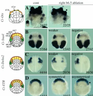
neural precursors as a result of its activation in neighbouring cells. As well as its broad activation in vegetal cells, ERK activation is also observed in the animal cells, a6.5 and b6.5 (Hudson et al., 2003). The b6.5 blastomere is a good candidate for the source of a secondary signal, as it is positioned adjacent to the lateral A-line neural precursors at this stage and during subsequent stages (Fig. 1).
To address whether signals from b6.5 are required to pattern the adjacent A-line neural blastomeres, we ablated the b6.5 blastomere on the right side at the 32-cell stage, leaving the left side as a control. We then analysed expression of Ci-Otx at the neurula stage in embryos that had been treated with cytochalasin B from the 64-cell stage to facilitate analysis (Fig. 2A). In the presence of cytochalasin B, cytokinesis is blocked, but blastomeres continue to express marker genes consistent with the principal fates that they would adopt during normal development. In unoperated embryos, Ci-Otxis expressed in the A7.4 blastomere but not in the A7.8 blastomere. However, following ablation of b6.5, expression of Ci-Otx was also observed in the A7.8 blastomere on the right side. Thus, ablation of the b6.5 blastomere affected patterning of the A-line neural A-lineages.
[image:3.612.241.562.408.740.2]Expression of Ci-Snailand Ci-Delta2is one of the earliest known signs of a molecular difference between the lateral and medial A-line neural lineages. At the early gastrula stage, Ci-Snailis expressed in the A8.15 and A8.16 blastomeres (Fig. 2B) (Corbo et al., 1997a; Wada and Saiga, 1999b). The A8.15/A8.16 blastomeres arise from the A7.8 blastomere and give rise to the lateral neural tube and secondary muscle
Fig. 2.Ablation of the right side b6.5 blastomere affects A-line neural patterning. Experimental conditions are shown at the top of the panels and the marker used on the left of the panels. All embryos are in vegetal pole view. Schematic representations of embryos are shown on the left, with black dots indicating cells that express the marker and a cross indicating ablated cell descendants (in green). Lineages are shown with the same colour code as in Fig. 1. Numbers are: number of embryos with similar expression as the image/total number of embryos analysed. In all cases, the expression on the left side of the experimental embryos was positive. (A) Ci-Otx: open arrowheads indicate A7.4 blastomeres and the filled arrowhead indicates an A7.8 blastomere expressing Ci-Otx. Other expression domains are the a-line neural lineage and anterior epidermis. The insert is a control cleaving embryo. (B) Ci-Snail: 3/43 of ablated embryos showed equal expression, presumably due to failure of ablation. (C) Ci-Delta2: 1/37 of b6.5 ablated embryos showed equal expression on both sides, including b-line, probably due to failure of ablation, and was not included in the analysis. In some embryos (middle and left panel), expression can also be detected in the trunk lateral cell precursor situated between the b-line cells and the lateral A-line neural precursors. (D) Ci-ETR: among the b6.5 ablated embryos showing
symmetrical expression, 5/10 had strong expression in A8.16 on both sides and 5 had weak expression on both sides. Arrowhead indicates stronger expression of Ci-ETRin A8.16 on the ablated side.
De
lineages (Fig. 1). Ci-Delta2is also expressed in the A8.15/8.16 blastomeres as well as in the b6.5 descendents, namely b8.20, b8.18 (epidermis precursors), b8.19, b8.17 (tail nerve cord, secondary muscle and tail endoderm precursors) (Fig. 2C). When the b6.5 blastomere was ablated on the right side, expression of Ci-Snailand Ci-Delta2in right A8.15/A8.16 was completely abolished in 63% and 44% of cases, respectively (Fig. 2B,C). The remaining embryos showed a severe downregulation of gene expression (Fig. 2B,C). Weak expression in these embryos may be due to some signal already having passed between b6.5 and the A-line neural lineage before ablation was carried out. Consistent with this, when the mother blastomere of b6.5, the b5.3 blastomere, was ablated on the right side of 16-cell-stage embryos, Ci-Delta2and Ci-Snailexpression was abolished in A8.15/A8.16 in 88% (29/33) and 94% (30/32) of cases, respectively. Following b6.5 ablation, the A-line neural lineages continued to express Ci-ETR,a general neural marker, on the ablated side, indicating that these cells have not lost their neural fate (Fig. 2D). Ci-ETR starts to be expressed in all eight A-line neural lineages at the beginning of gastrulation. During the early gastrula stages, however, Ci-ETRexpression in the A8.16 blastomere becomes downregulated. This may be due to the fact that the A8.16 lineage is induced to give rise to secondary muscle as well as neural fate by an as-yet-unknown mechanism (Meedel et al., 1987; Meedel et al., 2002; Nishida, 1990). In 41% of embryos in which the b6.5 blastomere was ablated on the right side, stronger expression of Ci-ETRin A8.16 was maintained on the ablated side, suggesting that b6.5 may also be required for the downregulation of Ci-ETRexpression in A8.16 (Fig. 2D).
Taken together, these results show that the b6.5 blastomere is required to signal to the lateral A7.8-line cells to instruct them to undertake a different developmental programme to that of the medial A7.4-line cells. We next investigated the nature of this instructive signal, which should originate from the b6.5 blastomere and be dependent upon MEK signalling.
Ci-Nodalexpression is induced in b6.5 blastomeres
by Ci-FGF9/16/20
Activation of ERK is seen in the b6.5 blastomere during the late 32-cell stage (Hudson et al., 2003; Nishida, 2003). We have found that a Ciona homologue of the TGFβ family member,Ci-Nodal,starts to be expressed in this blastomere at the same stage (Fig. 3A). At the 32-cell stage, Ci-Nodal expression is also transiently observed in the vegetal cells A6.1, A6.3 and B6.1 (Fig. 3A). Expression of Ci-Nodalin b6.5 is maintained in its descendants during the following cell divisions until mid-gastrula stages (Fig. 3B) (C.H. and H.Y., unpublished) (Imai et al., 2004; Morokuma et al., 2002). Thus, Ci-Nodalis expressed in the b6.5 blastomere and at the right time, making it a good candidate for the signal required to pattern the A-line neural lineages.
A-line neural patterning is MEK-dependent. Therefore, we expect that the molecule required for this patterning event would depend upon MEK signalling in some way. To address whether the transcriptional activation of Ci-Nodal in b6.5 requires MEK signalling, embryos were treated with an inhibitor of MEK, U0126, from the 16-cell stage. Following this treatment, the expression of Ci-Nodal in b6.5 was completely suppressed, whereas the vegetal expression of Ci-Nodalwas reduced but not eliminated (Fig. 3A).
Ci-FGF9/16/20 is broadly expressed in the vegetal hemisphere of embryos from the 16-cell stage and has recently been shown to be required for Ci-Otxexpression in a6.5 and b6.5 at the 32-cell stage (Bertrand et al., 2003). We therefore addressed whether Ci-FGF9/16/20 is required for Ci-Nodal expression in b6.5. In embryos injected with antisense morpholino oligonucleotides against Ci-FGF9/16/20 (Ci-FGF9/16/20-MO), Ci-Nodal expression was no longer observed in the b6.5 blastomere (Fig. 3A). As with U0126 treatment, Ci-FGF9/16/20-MO injection reduced, but did not eliminate, Ci-Nodalexpression in the vegetal cells (Fig. 3A).
Finally, if Ci-Nodal in b6.5 is the signal that patterns the A-line neural cells, Ci-Nodal expression should become independent of MEK signalling by the end of the 32-cell stage, that is, when patterning of the A-line neural lineages with respect to Ci-Otx expression becomes independent of MEK. To test this we treated embryos with U0126 from the early 32-cell stage or from the late 32-cell stage and analysed expression of Ci-Nodal at the early gastrula stage, when Ci-Nodal was expressed in the four b6.5 derivatives b8.20, b8.19, b9.18 and b9.17 (Fig. 3B). We found that Ci-Nodalexpression became independent of MEK activity by the end of the 32-cell stage (Fig. 3B).
These results indicate that Ci-FGF9/16/20 signalling is required for activation of Ci-Nodal expression in the b6.5 blastomere at the 32-cell stage. Ci-Nodal is thus an excellent candidate for the signal responsible for patterning of the A-line neural lineages.
Nodal signals are required for patterning of the A-line neural A-lineage, but not for specification of neural fate
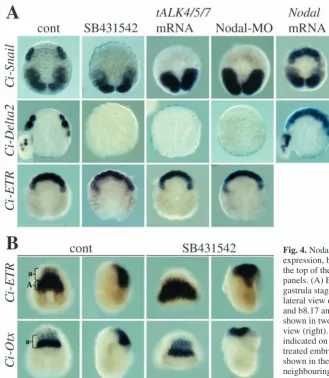
In order to investigate the role of Nodal during neural patterning, three methods were chosen to inhibit Nodal signalling. Firstly, we used a pharmacological inhibitor, SB431542, which blocks the TGFβ type I receptors ALK4, ALK5 and ALK7, for Activin and Nodal ligands, without inhibiting other ALK family members that bind to BMP ligands (Inman et al., 2002). In the Ciona genome, one potential Nodal receptor is apparent, named Ci-TGFβ-receptor Ic, which appears to represent vertebrate ALK4, -5and -7and thus in this study we refer to it as Ci-ALK4/5/7(Hino et al., 2003). As a second method to inhibit Nodal signalling, we constructed a truncated version of Ci-ALK4/5/7in which the cytoplasmic domain was removed. This truncation of the type I receptors of the TGFβligand superfamily has been shown in other systems to act as a dominant negative (Chang et al., 1997; Suzuki et al., 1994). Finally, we injected an antisense morpholino oligonucleotide against Ci-Nodal(Ci-Nodal-MO) to knock down the Nodal ligand. Analysis of the Cionagenome has revealed the presence of a single Nodalgene (Hino et al., 2003).
Using these three reagents to inhibit Nodal signalling, we looked at expression of Ci-Snail and Ci-Delta2 at the early gastrula stage. Inhibition of Nodal signalling by all three methods abolished Ci-Snail and Ci-Delta2 expression in A8.15/A8.16 (Fig. 4A; Table 1). Expression of Ci-Delta2 in the b-line cells was also lost, suggesting that Ci-Nodal may also play a role in b-line fate (Fig. 4A; Table 1). Expression of Ci-Snail in the primary muscle lineages was not affected. In contrast to inhibition of Nodal, ectopic activation of Nodal
De
signalling by injecting Ci-Nodal mRNA into eggs had the opposite effect, such that Ci-Delta2 and Ci-Snail were expressed in up to all eight A-line neural cells in 59% (16/27 in all eight cells) and 53% (19/36 in all eight cells) of cases, respectively (Fig. 4A).
Despite the loss of Ci-Snail and Ci-Delta2 expression following inhibition of Nodal signalling, A8.15/A8.16 still expressed Ci-ETR, suggesting that they still adopt a neural fate
(Fig. 4A; Table 1). Consistent with the observation that Nodal signalling is not required for specification of neural fate, the neural plate still formed in embryos in which Nodal signalling was inhibited. Ci-ETR and Ci-Otx, expression of which normally covers a large part of the neural plate, were still expressed in SB431542-treated embryos (Fig. 4B). Although it was not possible to count the exact number of cells expressing these genes, the size of the neural plate appeared similar in size to that of control embryos. The position of the neural plate was altered in SB431542-treated embryos, so that it remained ‘on top’ of the embryo owing to defects in gastrulation movements associated with inhibition of Nodal signalling.
These results show that Nodal signalling is required for the lateral A-line neural precursors to initiate a molecular programme different from that of the medial precursors, but is not required for the formation or maintenance of generic neural fate.
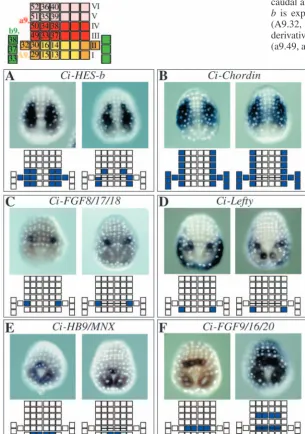
[image:5.612.277.561.74.213.2]A collection of neural plate markers to investigate neural patterning The neural plate of ascidian embryos exhibits a highly regular organisation of cell arrangement and cell division pattern. At the mid-gastrula stage, before the neural plate starts to roll up, the neural plate
Fig. 3. Ci-Nodalexpression in the b6.5 blastomere depends on MEK and Ci-FGF9/16/20. Orientation and treatment of the embryo are shown on the bottom and top of each panel, respectively. (A) Expression of Ci-Nodalat the 32-cell stage. Expression in vegetal cells is variable. For the U0126 experiments, 18/20 control embryos had expression of Ci-Nodalin b6.5 and 16/20 in one or more vegetal blastomeres (average number of cells=3.9), whereas U0126-treated embryos expressed Ci-Nodalin b6.5 in 0/34 of cases and in one or more vegetal cells in 20/34 cases (average number of cells=2.3). For the FGF9/16/20-MO experiments, 26/29 control embryos had expression in b6.5 and 20/29 in one or more vegetal cells (average number of cells=2.2) whereas in FGF9/16/20-MO injected embryos 0/30 of cases expressed Ci-Nodalin b6.5 and 11/30 of cases in one or more vegetal
cells (average number of cells=0.9). (B) Expression of Ci-Nodalat the early gastrula stage. 46/46 control embryos were positive; 0/46 embryos treated with U0126 at the early 32-cell stage were positive; and 40/50 embryos treated with U0126 from the late 32-cell stage were positive.
Fig. 4. Nodal signalling is required for lateral neural marker gene expression, but not for neural fate. Embryo treatment is shown at the top of the panels and the marker analysed to the left of the panels. (A) Expression of Ci-Snail, Ci-Delta2and Ci-ETRat the gastrula stage. All embryos are of vegetal pole view. Inserts show lateral view of Ci-Delta2expression in b-line cells (weak in b8.19 and b8.17 and strong in b8.20 and b8.18). (B) Each embryo is shown in two orientations, a neural plate view (left) and a lateral view (right). A-line and a-line parts of the neural plate are indicated on the control embryos. Neural plate cells of SB431542-treated embryos were not always as well aligned as the embryos shown in the picture for Ci-Otxstaining. Sometimes, cells in neighbouring rows intercalated with each other, which resulted in wider rows of 1-2 cells in depth.
De
[image:5.612.42.371.365.743.2]consists of six rows of a total of 44 neural cells aligned as shown in Fig. 5 (top). A-line neural lineages contribute to the first two caudal rows, while a-line neural lineages form the remaining four rows. Three cells of the b-line neural lineages flank each side of the neural plate. In order to investigate in more detail the consequences of Nodal inhibition on neural
[image:6.612.42.348.304.739.2]plate patterning, we first conducted a small-scale in situ hybridisation screen to collect genes expressed in a distinct manner in the neural plate. Six markers collected during the course of the screening are shown in Fig. 5. Expression patterns of the markers are shown in embryos in which the neural plate consists of six rows of cells and the following stage when the cells in row II of the neural plate have divided along the rostral-caudal axis, resulting in a neural plate of seven rows. Ci-HES-b is expressed in the lateral cells of the A-line neural plate (A9.32, A9.30, A9.29), including more medial A7.4 derivatives (A9.15, A9.16) and row III a-line neural plate (a9.49, a9.33) (Fig. 5A). Ci-Chordinis expressed in the lateral-most A- and a-line blastomeres of all six rows of the neural plate (A9.32, A9.30, A9.29, a9.49, a9.50, a9.51, a9.52), as well as the b-line neural cells, epidermal cells bordering the neural plate and weakly in the notochord (Fig. 5B). More specific markers of the lateral A-line neural lineage are Ci-Lefty,which is expressed in A9.29, and Ci-FGF8/17/18, which is expressed in A9.30 (Fig. 5C,D). Ci-Lefty is also expressed in the posterior epidermis with variable intensity. Markers of medial A-line neural cells are Ci-HB9/MNX and Ci-FGF9/16/20. Ci-HB9/MNX is expressed in A9.15 and A9.13 at the six-row neural plate stage, becoming weaker in A9.15 at the seven-row neural plate stage (Fig. 5E). During tailbud stages, expression of Ci-HB9/MNX reappears in the CNS and is found in the motoneurons, which are a lateral A-line neural cell derivative (see below). Ci-FGF9/16/20 is
Table 1. Expression of neural markers at the early gastrula stage
Control SB431542 tALK4/5/7 Nodal-MO
A b n e A b n e A b n e A b n e
Ci-Snail 99 – 331 10 0 – 120 4 0 – 33 3 0 – 39 3
Ci-Delta2 98 97 219 8 6 8 84 3 5 10 40 3 3 3 30 2
Ci-ETR 7.1 – 194 7 7.7 – 78 3 7.5 – 24 2 7.7 – 27 2
A, expression in A8.15/A8.16 blastomeres (Ci-Snailand Ci-Delta2), or the entire A-line neural lineage (Ci-ETR). b, expression in b6.5 descendants (Ci-Delta2only).
Numbers in bold indicate percentage of embryos showing expression in the neural lineages (Ci-Snailand Ci-Delta2), or the average number of A-line neural cells showing expression (Ci-ETR) following the treatment indicated at the top.
n, number of embryos analysed. e, number of independent experiments.
Fig. 5. A collection of neural plate markers. At the top of the panels is a schematic drawing of the neural plate when it consists of six rows of cells, with each square representing a neural plate cell. Names of each cell are indicated, and should be prefixed with an ‘a9.’ for a-line cells, ‘A9.’ for A-a-line cells and ‘b9.’ for b-a-line cells. I-VI indicate the row number, with Row I the closest to the blastopore, which is at the most caudal position of the neural plate. Colour scheme used is as in Fig. 1. (A-F) For each marker, two stages are shown; when the neural plate has six rows of cells (left) and when the neural plate has seven rows of cells (right). Nuclei are labelled to allow easy identification of individual cells. A schematic drawing of the neural plate with the blastomeres expressing each marker coloured in blue is presented below each embryo.
De
expressed in A9.16 and A9.14 at the six-row neural plate stage and also in a9.34 and a9.38 at the seven-row neural plate stage (Fig. 5F). Both these latter genes are also expressed in the primary muscle lineages. Using these markers, we investigated the effects on the patterning of the neural plate when Nodal signalling was perturbed.
Lateral neural tissue and secondary muscle markers are not expressed following Nodal inhibition
We tested expression of all markers in embryos treated with
SB431542 and Nodal-MO injection and examples of both are shown in Figs 6 and 7. Some markers were also tested in embryos injected with Ci-tALK4/5/7mRNA (Table 2; Fig. 7B). In all cases, equivalent results were obtained (Figs 6, 7; Table 2).
[image:7.612.326.558.189.536.2]Markers of the lateral neural plate, Ci-Chordin,Ci-HES-b, Ci-FGF8/17/18 and Ci-Lefty, were suppressed following Nodal inhibition (Fig. 6A-D; Table 2). While we have concentrated on the patterning of the A-line neural plate in this
[image:7.612.43.291.191.616.2]Fig. 6. Expression of marker genes of lateral neural plate and secondary muscle in embryos in which Nodal signalling has been inhibited. Experimental conditions are shown at the top of the panels and the marker analysed is on the left of the panels. (A-D) Neural plate views of embryos at the late gastrula stage. (A) Expression of Ci-Chordinin the notochord can be seen in control and manipulated embryos. (E,F) Embryos at the early tailbud stage are shown in dorsal (far left) and lateral view. Treated embryos are shown in a lateral view. (G) Panel on the far left is a control cleaving embryo at the neurula stage; the rest are treated with cytochalasin from the 76-cell stage. Arrowheads point to the secondary muscle lineage (A8.16) in the control panel.
Fig. 7.Expression of marker genes of the medial neural plate in embryos in which Nodal signalling has been inhibited.
(A) Experimental conditions are shown at the top of the panels and the marker analysed is indicated on the left of the panels. Neural plate views are shown for all embryos except cytochalasin B-treated embryos (lower panels). Insert shows a control cleaving embryo for Ci-Otxexpression. Open arrowheads indicate A7.8 blastomeres. In all panels, filled arrowheads indicate ectopic marker expression. Ci-Otxexpression was observed in one or both A7.8 blastomeres in 0/80 control embryos, 64/67 SB431542-treated embryos, 13/15 Ci-tALK4/5/7injected embryos and 18/20 Ci-Nodal-MO injected embryos. (B) The marker analysed is indicated at the top of each graph. Graphs show the percentage of embryos (y-axis) that express the marker genes in 0-8 neural plate cells (x-axis). Control embryos (n=more than 100) are indicated by red bars; SB431542-treated embryos (n=82 for Ci-HB9/MNXand n=59 for Ci-FGF9/16/20) by blue horizontal striped bars; Ci-tALK4/5/7 injected embryos (n=37 for Ci-HB9/MNXand n=11 for Ci-FGF9/16/20) by blue diagonal striped bars; and Ci-Nodal-MO-injected embryos (n=24 for Ci-HB9/MNXand n=31 for Ci-FGF9/16/20) by filled blue bars.
De
study, we also observed that expression of Ci-HES-band Ci-Chordinin the lateral a-line neural lineage and expression of Ci-Chordinin the b-line neural lineages were also lost (Fig. 6A-D). This suggests that Nodal signalling may be required for the entire lateral neural plate to form.
We also addressed whether a specific neuronal type derived from the lateral neural plate was generated in the absence of Nodal signals. Motoneurons of ascidian embryos originate from the A8.15 lineage (Fig. 1) (Cole and Meinertzhagen, 2004). Interestingly, it has been observed that motoneurons may still form in isolated A4.1 explants (Okada et al., 1997). We used two motoneuron markers, Ci-ChAT, which encodes cholinergic acetyltransferase (Takamura et al., 2002; Yoshida et al., 2004), and the Ciona homologue of HB9 and MNR2 genes, which play a crucial role in motoneuron specification in other systems, and of which there is one representative in the Ciona genome, Ci-HB9/MNX (Broihier and Skeath, 2002; Odden et al., 2002; Shirasaki and Pfaff, 2002; Wada et al., 2003). Expression of these motoneuron markers was completely dependent on an intact Nodal signalling pathway (Fig. 6E,F; Table 2). Therefore, the formation of these specific neuronal cell types also depends upon Nodal signalling.
The A8.15/A8.16 blastomeres generate the secondary muscle lineage as well as the lateral neural plate. Formation of the so-called secondary muscle requires inductive interactions, unlike the autonomously determined primary muscle (Meedel et al., 1987; Meedel et al., 2002; Nishida, 1990). At the 110-cell stage, the A7.8 blastomere divides to generate A8.15 (neural fate) and A8.16 (neural and muscle fates) (Fig. 1). Finally, A8.16 divides into muscle (A9.31) and neural (A9.32) precursors during neural plate stages (Fig. 1) (Cole and Meinertzhagen, 2004; Nicol and Meinertzhagen 1998a; Nicol and Meinertzhagen 1998b; Nishida, 1990). As the A9.31 secondary muscle comes from the same lineage as the lateral neural plate, we investigated whether the secondary muscle lineage was also affected by Nodal inhibition. In embryos that had been cleavage arrested after the 76-cell stage when A7.8 had divided into A8.15 and A8.16, expression of Ci-Actin could be detected in the secondary muscle lineage (A8.16), which remained in a different position to the primary muscle lineage and could thus be easily distinguished (Fig. 6G, arrowheads). Formation of secondary muscle was inhibited in embryos in which Nodal signalling was blocked (Fig. 6G; Table 2). This suggests that the entire lineage of the A8.15/A8.16 blastomere, which gives rise to lateral neural tube
and secondary muscle, requires Nodal signals for its fate specification.
Medial A-line neural plate fates expand following Nodal inhibition
We next addressed whether the inhibition of Nodal causes a general disruption of neural plate patterning, or affects only the lateral neural plate fates. We analysed expression of Ci-HB9/MNXand Ci-FGF9/16/20, which are expressed in distinct sets of medial A-line cells of the neural plate (Figs 5, 7). In Nodal-inhibited embryos, these markers continued to be expressed in the correct row of neural plate cells, but their expression was expanded laterally to a maximum of eight cells in total (Fig. 7A,B). We also analysed Ci-Otx expression at neurula stages in embryos cleavage-arrested in cytochalasin B from the 64-cell stage. Under these conditions, Ci-Otx was expressed only in the medial A7.4 blastomeres of otherwise unmanipulated embryos. When Nodal signalling was blocked, however, Ci-Otx was also expressed in the lateral A7.8 cells (Fig. 7A). Altogether, this suggests that, within the A-line neural lineages, Nodal signalling specifically determines lateral cell fates, and that, in the absence of Nodal, the lateral cells adopt a medial-cell-like fate.
Discussion
Ci-FGF9/16/20 induction of Ci-Nodal in b6.5 is required for patterning of the neural plate along the medial-lateral axis
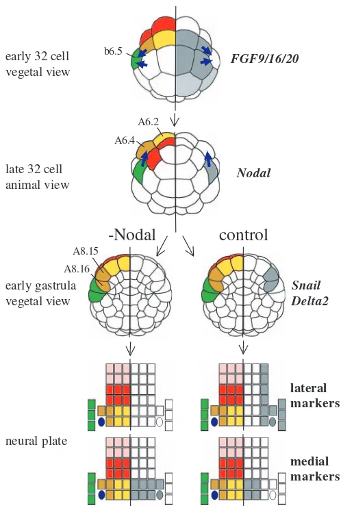
[image:8.612.39.560.84.179.2]A summary of the main conclusions of this study can be found in Fig. 8. Firstly, we revealed by cell ablation that the b6.5 blastomere acts as a signalling source and induces the lateral A-line neural blastomeres to adopt a lateral character and express genes such as Ci-Snail and Ci-Delta2 at the early gastrula stage. We then showed that Ci-FGF9/16/20 from vegetal blastomeres is responsible for the MEK-dependent activation of Ci-Nodal in b6.5 at the 32-cell stage. Furthermore, we showed that inhibition of Nodal signalling, using three distinct reagents, mimicks the effects of b6.5 ablation and resulted in a loss of the lateral neural markers Ci-Snail and Ci-Delta2 at the early gastrula stage. Conversely, when Nodal signals were ectopically activated by injection of Ci-Nodal mRNA, medial neural cells expressed lateral markers. By neural plate stages, in embryos in which Nodal
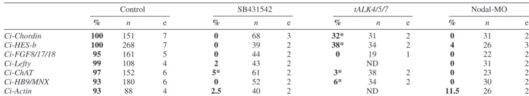
Table 2. Expression of lateral neural markers depends upon Nodal signalling
Control SB431542 tALK4/5/7 Nodal-MO
% n e % n e % n e % n e
Ci-Chordin 100 151 7 0 68 3 32* 31 2 0 31 2
Ci-HES-b 100 268 7 0 39 2 38* 34 2 4 26 3
Ci-FGF8/17/18 95 161 5 0 44 2 0 19 1 0 22 2
Ci-Lefty 99 108 4 2 43 2 ND 0 31 2
Ci-ChAT 97 152 6 5* 61 2 3* 38 2 0 23 2
Ci-HB9/MNX 93 180 6 0 52 2 6* 34 2 0 30 2
Ci-Actin 93 88 4 2.5 40 2 ND 11.5 26 2
Numbers in bold indicate percentage (%) of embryos showing expression in the lateral neural plate (Ci-Chordin,Ci-HES-b,Ci-FGF8/17/18 andCi-Lefty), presumptive motoneurones (Ci-ChATand Ci-HB9/MNX) or secondary muscle lineage (Ci-Actin) following the treatment indicated at the top.
*, most of these embryos showed reduced expression.
n, number of embryos analysed. e, number of independent experiments.
De
signalling had been disrupted, markers of the lateral A-line neural plate were inhibited and instead these cells expressed markers of medial fate. These results suggest that the A-line neural plate cells make a cell fate choice between lateral and medial neural fates and that this cell fate choice is mediated by Nodal signals from the b6.5 blastomere.
The present study identified the b6.5 blastomere as a Nodal signalling source, dependent on the specific activation of the Ci-Nodalgene in this blastomere by Ci-FGF9/16/20 signalling from vegetal blastomeres. However, ERK1/2 activation is observed in both b6.5 and a6.5 blastomeres, and an FGF-response element of the Ci-Otxgene is able to drive expression of a reporter gene in both lineages (Bertrand et al., 2003; Hudson et al., 2003). We propose that the transcriptional activation of Ci-Nodal in b6.5 but not in a6.5 is due to a difference in competence between a- and b-line blastomeres to respond to FGF-signalling, as has been previously observed
during the formation of anterior neural fates and sensory neurons (Hudson and Lemaire, 2001; Ohtsuka et al., 2001). Activation of Ci-Nodal specifically in b6.5 may be the first read-out of this difference in competence. Localised activation of Ci-Nodalin this manner creates a lateral Ci-Nodal signalling source, which acts to pattern the A-line neural lineages.
Our observations suggest that the action of Ci-Nodal on the lateral A-line neural precursors probably occurs directly, rather than via activation of other signalling pathways in surrounding cells. Firstly, the cell ablation studies indicate that the signal from b6.5 is occurring during the 32-cell stage, when these two cell types are in direct contact. Secondly, injection of Ci-Nodal mRNA revealed that the medial A-line neural precursors are capable of responding to Nodal and expressing lateral markers at the early gastrula stage. This suggests that, during normal embryogenesis, the medial A-line neural precursors do not express lateral markers because they are not in contact with b6.5 and therefore do not receive a Nodal signal. Furthermore, it is unlikely that neural plate patterning defects are a consequence of a general perturbation of cell fate specification, because treatment of embryos with SB431542 does not affect expression of markers for primary notochord [Ci-Bra at the 64-and 110-cell stages (Corbo et al., 1997b); Ci-Chordinin Fig. 6A], primary muscle (Ci-Snailin Fig. 4 and Ci-Actinin Fig. 6), or endoderm [Ci-Titf1at the early gastrula (Ristoratore et al., 1999)] (C.H. and H.Y., unpublished).
In addition to mediating A-line neural patterning, Nodal signals were also required for expression of lateral neural fate markers in the a- and b-line neural lineages, as seen by the loss of Ci-Delta2(b-line), Ci-Chordin(a- and b-line) and Ci-HES-b(a-line) expression following Nodal inhibition (Figs 4, 5, 8). This suggests that Nodal signalling is required for lateral patterning of the entire neural plate.
Distinct mechanisms control anterior-posterior and medial-lateral patterning of the neural plate
We have found that Nodal signalling is involved in medial-lateral patterning of the neural plate but not in anterior-posterior patterning. Ci-HB9/MNX, Ci-FGF9/16/20 and Ci-Otx are differentially expressed along the anterior-posterior axis of the neural plate in row I, row II and rows III-VI, respectively. In embryos in which Nodal signalling was blocked, the expression domains of these genes remained in their correct anterior-posterior positions (Fig. 4B, Fig. 7). Therefore, the mechanisms leading to anterior-posterior and medial-lateral patterning of the neural plate are separable. There are some indications that the MEK signalling pathway may play a role in anterior-posterior patterning of the neural plate in addition to its role in medial-lateral patterning via activation of Ci-Nodal. For example, Ci-HB9/MNX is not expressed in the neural plate of embryos treated with an inhibitor of MEK, while this gene continues to be expressed in the correct row of cells following Nodal inhibition (Hudson et al., 2003) (present study).
Nodal signalling and secondary muscle formation In ascidians, muscle cells originate from three lineages. The primary lineage derives from the B-line and is specified cell-autonomously (e.g. Nishida and Sawada, 2001). The secondary lineage arises from the b6.5- and A8.16-lineages (Nishida, 1987; Nishida, 1990). In this study, we observed that the FGF9/16/20
early 32 cell vegetal view
late 32 cell animal view
early gastrula vegetal view
Nodal
Snail
Delta2
-Nodal
lateral markers
medial markers
control
neural plate
b6.5
A6.4 A6.2
A8.16 A8.15
Fig. 8.A summary of the results. Embryonic stage is indicated on the left of the schematic drawings. On the left half of each drawing, the neural lineages are indicated using the same colour code as in Fig. 1. On the right half of each drawing, blastomeres expressing the markers indicated on the far right are shown in grey. From the early gastrula stage, drawings are shown in two columns; those on the right indicate control embryos and those on the left represent embryos in which Nodal signalling has been inhibited. Thick blue arrows represent signalling between blastomeres.
De
[image:9.612.42.291.71.445.2]formation of the A8.16-derived secondary muscle lineage, like lateral neural fates, depended on Nodal signals. In contrast to the primary lineage, the secondary lineage is known to require cellular interactions for fate specification; isolated A4.1 blastomeres of the 8-cell stage embryo do not form muscle fate unless recombined or co-isolated with animal blastomeres (Meedel et al., 1987; Meedel et al., 2002; Nishida, 1990). In addition, it has been shown in Halocynthiathat FGF/Ras/MEK signalling is required between the 32- and the 64-cell stage for secondary muscle cell formation (Kim and Nishida, 2001). We propose that the role of FGF signalling in secondary muscle formation is indirect, via the activation of Ci-Nodal in b6.5 blastomeres. The A8.16 blastomere forms following cell division of A7.8 in the medial-lateral direction and remains in contact with the Ci-Nodalexpressing b6.5 descendants. A8.15, the sister blastomere of A8.16, is positioned more medially, such that it is no longer in contact with the b6.5 descendants. The A8.15 blastomere gives rise to only neural fates, whereas the A8.16 blastomere gives rise to neural and secondary muscle fates. It is possible that a short exposure to Nodal signals results in lateral neural fate, the fate taken by A8.15, and a longer exposure is required for induction of muscle fate in the A8.16 lineage. Consistent with this idea, in Halocynthia, A8.16 blastomeres isolated early in their cell cycle do not form muscle, whereas some of those isolated late in their cell cycle can develop autonomously into muscle (Nishida, 1990).
Nodal signalling during development of ascidians and vertebrates
During development of vertebrate embryos, Nodal signals are involved in formation of the anterior-posterior and left-right axes and specification of the endoderm and mesoderm germ layers (Schier and Shen, 2000; Whitman, 2001; Bertocchini and Stern, 2002; Perea-Gomez et al., 2002). Its role in left-right axial patterning appears to be conserved between ascidians and vertebrates (Morokuma et al., 2002). However, we did not observe profound effects following inhibition of Nodal signals on expression of marker genes for endoderm and mesoderm. It is possible that, due to the rapid development of ascidian embryos, the role of Nodal in endoderm and mesoderm has been bypassed by the recruitment of maternal determinants to specify the major tissue types. Alternatively, since Nodal signalling is not involved in germ layer specification in echinoderms, this role may be a vertebrate invention (Duboc et al., 2004). Nodal signalling is required for secondary muscle induction in Ciona, suggesting that at least some mesoderm cell-types are dependent on Nodal signalling. In addition, Ci-Nodal is expressed in the vegetal cells at the 32-cell stage, albeit transiently, and embryos do not gastrulate correctly following inhibition of Nodal signalling. Therefore, there are likely to be further roles for Nodal signalling during ascidian development that remain to be understood.
The vertebrate neural tube is patterned across the dorsal-ventral (lateral-medial neural plate) axis, by signals from tissues adjacent to, as well as from within, the neural tube. A variety of signalling molecules has been identified in this patterning event. Formation of the floor plate, the ventral-most part of the spinal cord, involves Nodal and SHH signalling pathways (for reviews, see Appel, 2000; Lewis and Eisen, 2003; Strahle et al., 2004). In zebrafish embryos, Nodal signalling is required for specification of floor plate precursors.
In mouse and chick embryos, however, SHH signalling appears to play a more pivotal role during floor plate specification, with the role for Nodal much less clear, although recently, roles for Nodal have begun to emerge in these vertebrates as well (Lewis and Eisen, 2003; Strahle et al., 2004). Subsequent to floor plate formation, SHH signalling from the floor plate and notochord patterns the ventral part of spinal cord to specify distinct neuronal precursors. In contrast, the dorsal part of the neural tube is patterned by BMP signalling from the laterally (future dorsally) situated epidermis that borders the neural plate and later from the roof plate (reviewed by Altmann and Hemmati-Brivanlou, 2001; Lee and Jessell, 1999).
In this study, we uncovered a number of differences in the role of Nodal signalling in ascidian neural patterning compared with vertebrates. Firstly, in ascidians, Nodal ligands emanate from cells laterally flanking the neural plate, not from axial tissues as in vertebrates. Secondly, Nodal signals are required for lateral neural fates, but not for medial (future ventral) fates. Finally, Nodal signals in ascidians restrict medial neural fates by promoting lateral fates. It may be that in the ancestral chordate Nodal was involved in both dorsal and ventral neural fate specification. In this case, one could postulate that the role of Nodal during induction of lateral fates has been lost (or perhaps overlooked) in vertebrate lineages, and the role of Nodal during formation of ventral fates has been lost in ascidians. Alternatively, the role of Nodal signalling in dorsal or ventral patterning may have been recruited independently in vertebrate and invertebrate chordates, respectively. It will be important to look again in vertebrates to see if the role of Nodal signalling during dorsal patterning of the neural tube has been overlooked owing to the severe developmental defects that occur earlier during germ layer formation upon Nodal inhibition.
Despite this apparent difference in the role of Nodal signalling during CNS patterning, other aspects of dorsal-ventral neural tube patterning may be conserved. For example, an ascidian homologue of hedgehog, Ci-hh2, is expressed in the ventral-most cell of the tail nerve cord from the early tail bud stage and BMP2/4is expressed in the borders of the neural plate (Miya et al., 1997; Takatori et al., 2002). Furthermore, BMP2/4-Chordin antagonism is required for formation of the pigment cells, a dorsal cell fate derived from the a-line lateral neural plate, implicating this pathway in dorsal patterning (Darras and Nishida, 2001). Future work should study the relationship between Nodal, SHH and BMP2/4 signalling pathways. It will also be important to determine whether the medial neural fate of the ascidian neural plate is an induced fate or specified as a default fate of the A-line neural lineages. Whatever details are uncovered, it is already clear that the distinct manner in which Nodal signals are involved in neural patterning in vertebrates and ascidians implies a certain degree of evolutionary plasticity in the mechanisms used to generate a conserved structure such as the chordate neural tube.
We would like to thank V. Bertrand and P. Lemaire (Ci-FGF9/16/20-MO), and S. Fujiwara (Ci-Snail) for kindly providing tools and N. Satoh and the Japanese In Situ Consortium for kindly sharing the Gene Collection plates with the ascidian community. Thank you to the service ‘Modèle Biologique’ (Roscoff Marine Biology Station) for adult Ciona intestinalis and the staff of the UMR7009 for their help and support. We thank V. Bertrand, E. Houliston, L. Kodjabachian, P. Lemaire, T. Lepage, Y. Ohtsuka, A.
De
Pasini and U. Rothbacher for helpful discussions and comments on the manuscript. This work is supported by the Centre National de la Recherche Scientifique (CNRS), including an ATIP grant, the Université Paris VI and the Association Française contre les Myopathies (AFM, grant no. 9960).
References
Akanuma, T. and Nishida, H. (2003). Ets-mediated brain induction in embryos of the ascidian Halocynthia roretzi. Dev. Genes Evol. 214, 1-9.
Altmann, C. R. and Hemmati-Brivanlou, A. (2001). Neural patterning in the vertebrate embryo. Int. Rev. Cytol. 203, 447-482.
Aniello, F., Locascio, A., Villani, M. G., Di Gregorio, A., Fucci, L. and Branno, M. (1999). Identification and developmental expression of Ci-msxb: a novel homologue of Drosophila msh gene in Ciona intestinalis.
Mech. Dev. 88, 123-126.
Appel, B. (2000). Zebrafish neural induction and patterning. Dev. Dyn. 219, 155-168.
Bertocchini, F. and Stern, C. D. (2002). The hypoblast of the chick embryo positions the primitive streak by antagonizing nodal signaling. Dev.Cell3, 735-744.
Bertrand, V., Hudson, C., Caillol, D., Popovici, C. and Lemaire, P. (2003). Neural tissue in ascidian embryos is induced by FGF9/16/20, acting via a combination of maternal GATA and Ets transcription factors. Cell115, 615-627.
Broihier, H. T. and Skeath, J. B. (2002). Drosophila homeodomain protein dHb9 directs neuronal fate via crossrepressive and cell-nonautonomous mechanisms. Neuron35, 39-50.
Chang, C., Wilson, P. A., Mathews, L. S. and Hemmati-Brivanlou, A.
(1997). A Xenopus type I activin receptor mediates mesodermal but not neural specification during embryogenesis. Development124, 827-837.
Cole, A. G. and Meinertzhagen, I. A. (2004). The central nervous system of the ascidian larva: mitotic history of cells forming the neural tube in late embryonic Ciona intestinalis. Dev. Biol. 271, 239-262.
Conklin, E. G. (1905). The organisation and cell lineage of the ascidian egg.
J. Acad. Natl. Sci. (Philadelphia)13, 1-119.
Corbo, J. C., Erives, A., Di Gregorio, A., Chang, A. and Levine, M.
(1997a). Dorsoventral patterning of the vertebrate neural tube is conserved in a protochordate. Development124, 2335-2344.
Corbo, J. C., Levine, M. and Zeller, R. W. (1997b). Characterization of a notochord-specific enhancer from the Brachyury promoter region of the ascidian, Ciona intestinalis. Development124, 589-602.
Darras, S. and Nishida, H. (2001). The BMP/CHORDIN antagonism controls sensory pigment cell specification and differentiation in the ascidian embryo. Dev. Biol. 236, 271-288.
Duboc, V., Rottinger, E., Besnardeau, L. and Lepage, T. (2004). Nodal and BMP2/4 signaling organizes the oral-aboral axis of the sea urchin embryo.
Dev. Cell6, 397-410.
Hino, K., Satou, Y., Yagi, K. and Satoh, N. (2003). A genomewide survey of developmentally relevant genes in Ciona intestinalis. VI. Genes for Wnt, TGFbeta, Hedgehog and JAK/STAT signaling pathways. Dev. Genes Evol. 213, 264-272.
Hudson, C. and Lemaire, P. (2001). Induction of anterior neural fates in the ascidian Ciona intestinalis. Mech. Dev. 100, 189-203.
Hudson, C., Darras, S., Caillol, D., Yasuo, H. and Lemaire, P. (2003). A conserved role for the MEK signalling pathway in neural tissue specification and posteriorisation in the invertebrate chordate, the ascidian Ciona intestinalis. Development130, 147-159.
Imai, K. S., Satoh, N. and Satou, Y. (2002). Region specific gene expressions in the central nervous system of the ascidian embryo. Gene Expr. Patterns 2, 319-321.
Imai, K. S., Hino, K., Yagi, K., Satoh, N. and Satou, Y. (2004). Gene expression profiles of transcription factors and signaling molecules in the ascidian embryo: towards a comprehensive understanding of gene networks.
Development131, 4047-4058.
Inman, G. J., Nicolas, F. J., Callahan, J. F., Harling, J. D., Gaster, L. M., Reith, A. D., Laping, N. J. and Hill, C. S. (2002). SB-431542 is a potent and specific inhibitor of transforming growth factor-beta superfamily type I activin receptor-like kinase (ALK) receptors ALK4, ALK5, and ALK7. Mol. Pharmacol. 62, 65-74.
Kim, G. J. and Nishida, H. (2001). Role of the FGF and MEK signaling pathway in the ascidian embryo. Dev. Growth Differ. 43, 521-533.
Lee, K. J. and Jessell, T. M. (1999). The specification of dorsal cell fates in the vertebrate central nervous system. Annu. Rev. Neurosci. 22, 261-294.
Lemaire, P., Garrett, N. and Gurdon, J. B. (1995). Expression cloning of Siamois, a Xenopus homeobox gene expressed in dorsal-vegetal cells of blastulae and able to induce a complete secondary axis. Cell81, 85-94.
Lemaire, P., Bertrand, V. and Hudson, C. (2002). Early steps in the formation of neural tissue in ascidian embryos. Dev. Biol. 252, 151-169.
Lewis, K. E. and Eisen, J. S. (2003). From cells to circuits: development of the zebrafish spinal cord. Prog. Neurobiol. 69, 419-449.
Meedel, T. H., Crowther, R. J. and Whittaker, J. R. (1987). Determinative properties of muscle lineages in ascidian embryos. Development100, 245-260.
Meedel, T. H., Lee, J. J. and Whittaker, J. R. (2002). Muscle development and lineage-specific expression of CiMDF, the MyoD-family gene of Ciona intestinalis. Dev. Biol. 241, 238-246.
Meinertzhagen, I. A. and Okamura, Y. (2001). The larval ascidian nervous system: the chordate brain from its small beginnings. Trends Neurosci. 24, 401-410.
Meinertzhagen, I. A., Lemaire, P. and Okamura, Y. (2004). The neurobiology of the ascidian tadpole larva: recent developments in an ancient chordate. Annu.Rev.Neurosci. 27, 453-485.
Minokawa, T., Yagi, K., Makabe, K. W. and Nishida, H. (2001). Binary specification of nerve cord and notochord cell fates in ascidian embryos.
Development128, 2007-2017.
Miya, T., Morita, K., Suzuki, A., Ueno, N. and Satoh, N. (1997). Functional analysis of an ascidian homologue of vertebrate Bmp-2/Bmp-4 suggests its role in the inhibition of neural fate specification. Development124, 5149-5159.
Morokuma, J., Ueno, M., Kawanishi, H., Saiga, H. and Nishida, H. (2002). HrNodal, the ascidian nodal-related gene, is expressed in the left side of the epidermis, and lies upstream of HrPitx. Dev. Genes Evol. 212, 439-446.
Nicol, D. and Meinertzhagen, I. A. (1988a). Development of the central nervous system of the larva of the ascidian, Ciona intestinalis L. I. The early lineages of the neural plate. Dev. Biol. 130, 721-736.
Nicol, D. and Meinertzhagen, I. A. (1988b). Development of the central nervous system of the larva of the ascidian, Ciona intestinalis L. II. Neural plate morphogenesis and cell lineages during neurulation. Dev. Biol. 130, 737-766.
Nishida, H. (1987). Cell lineage analysis in ascidian embryos by intracellular injection of a tracer enzyme. III. Up to the tissue restricted stage. Dev. Biol. 121, 526-541.
Nishida, H. (1990). Determinative mechanisms in secondary muscle lineages of ascidian embryos: development of muscle-specific features in isolated muscle progenitor cells. Development108, 559-568.
Nishida, H. (2003). Spatio-temporal pattern of MAP kinase activation in embryos of the ascidian Halocynthia roretzi. Dev. Growth Differ. 45, 27-37.
Nishida, H. and Sawada, K. (2001). macho-1 encodes a localized mRNA in ascidian eggs that specifies muscle fate during embryogenesis. Nature409, 724-729.
Odden, J. P., Holbrook, S. and Doe, C. Q. (2002). Drosophila HB9 is expressed in a subset of motoneurons and interneurons, where it regulates gene expression and axon pathfinding. J. Neurosci. 22, 9143-9149.
Ohtsuka, Y., Obinata, T. and Okamura, Y. (2001). Induction of ascidian peripheral neuron by vegetal blastomeres. Dev. Biol. 239, 107-117.
Okada, T., Hirano, H., Takahashi, K. and Okamura, Y. (1997). Distinct neuronal lineages of the ascidian embryo revealed by expression of a sodium channel gene. Dev. Biol. 190, 257-272.
Perea-Gomez, A., Vella, F. D., Shawlot, W., Oulad-Abdelghani, M., Chazaud, C., Meno, C., Pfister, V., Chen, L., Robertson, E., Hamada, H. et al. (2002). Nodal antagonists in the anterior visceral endoderm prevent the formation of multiple primitive streaks. Dev. Cell3, 745-756.
Ristoratore, F., Spagnuolo, A., Aniello, F., Branno, M., Fabbrini, F. and Di Lauro, R. (1999). Expression and functional analysis of Cititf1, an ascidian NK-2 class gene, suggest its role in endoderm development. Development 126, 5149-5159.
Satoh, N. (1994). Developmental Biology of Ascidians. Cambridge, UK: Cambridge University Press.
Satou, Y., Takatori, N., Fujiwara, S., Nishikata, T., Saiga, H., Kusakabe, T., Shin-i, T., Kohara, Y. and Satoh, N. (2002a). Ciona intestinalis cDNA projects: expressed sequence tag analyses and gene expression profiles during embryogenesis. Gene287, 83-96.
Satou, Y., Yamada, L., Mochizuki, Y., Takatori, N., Kawashima, T., Sasaki, A., Hamaguchi, M., Awazu, S., Yagi, K., Sasakura, Y. et al. (2002b). A
De
cDNA resource from the basal chordate Ciona intestinalis. Genesis33, 153-154.
Schier, A. F. and Shen, M. M. (2000). Nodal signalling in vertebrate development. Nature403, 385-389.
Shirasaki, R. and Pfaff, S. L. (2002). Transcriptional codes and the control of neuronal identity. Annu. Rev. Neurosci. 25, 251-281.
Strahle, U., Lam, C. S., Ertzer, R. and Rastegar, S. (2004). Vertebrate floor-plate specification: variations on common themes. Trends Genet. 20, 155-162.
Suzuki, A., Thies, R. S., Yamaji, N., Song, J. J., Wozney, J. M., Murakami, K. and Ueno, N. (1994). A truncated bone morphogenetic protein receptor affects dorsal-ventral patterning in the early Xenopus embryo. Proc. Natl. Acad. Sci. USA 91, 10255-10259.
Takamura, K., Egawa, T., Ohnishi, S., Okada, T. and Fukuoka, T. (2002). Developmental expression of ascidian neurotransmitter synthesis genes. I. Choline acetyltransferase and acetylcholine transporter genes. Dev. Genes Evol. 212, 50-53.
Takatori, N., Satou, Y. and Satoh, N. (2002). Expression of hedgehog genes in Ciona intestinalis embryos. Mech. Dev. 116, 235-238.
Taniguchi, K. and Nishida, H. (2004). Tracing cell fate in brain formation during embryogenesis of the ascidian Halocynthia roretzi. Dev. Growth Differ. 46, 163-180.
Wada, S. and Saiga, H. (1999a). Vegetal cell fate specification and anterior neuroectoderm formation by Hroth, the ascidian homologue of orthodenticle/otx. Mech. Dev. 82, 67-77.
Wada, S. and Saiga, H. (1999b). Cloning and embryonic expression of Hrsna, a snail family gene of the ascidian Halocynthia roretzi: implication in the origins of mechanisms for mesoderm specification and body axis formation in chordates. Dev. Growth Differ. 41, 9-18.
Wada, H., Holland, P. W., Sato, S., Yamamoto, H. and Satoh, N. (1997). Neural tube is partially dorsalized by overexpression of HrPax-37: the ascidian homologue of Pax-3 and Pax-7. Dev. Biol. 187, 240-252.
Wada, H., Saiga, H., Satoh, N. and Holland, P. W. (1998). Tripartite organization of the ancestral chordate brain and the antiquity of placodes: insights from ascidian Pax-2/5/8, Hox and Otx genes. Development125, 1113-1122.
Wada, S., Tokuoka, M., Shoguchi, E., Kobayashi, K., Di Gregorio, A., Spagnuolo, A., Branno, M., Kohara, Y., Rokhsar, D., Levine, M. et al.
(2003). A genomewide survey of developmentally relevant genes in Ciona intestinalis. II. Genes for homeobox transcription factors. Dev. Genes Evol.
213, 222-234.
Whitman, M. (2001). Nodal signaling in early vertebrate embryos: themes and variations. Dev. Cell1, 605-617.
Yoshida, R., Sakurai, D., Horie, T., Kawakami, I., Tsuda, M. and Kusakabe, T. (2004). Identification of neuron-specific promoters in Ciona intestinalis. Genesis39, 130-140.