R E S E A R C H A R T I C L E
Open Access
Antioxidant potential, cytotoxic activity and
total phenolic content of
Alpinia pahangensis
rhizomes
Chung-Weng Phang, Sri Nurestri Abd Malek
*and Halijah Ibrahim
Abstract
Background:Alpinia pahangensis, a wild ginger distributed in the lowlands of Pahang, Malaysia, is used by the locals to treat flatulence. In this study, the antioxidant and cytotoxic activities of the crude aqueous methanol and fractionated extracts ofAlpinia pahangensisagainst five different cancer and one normal cell lines were investigated. The total phenolic content of each extract and its fractions were also quantified. This is the first report on the antioxidant and cytotoxic activities ofAlpinia pahangensisextract.
Methods:In the current study, the crude methanol and fractionated extract of the rhizomes ofAlpinia pahangensis were investigated for their antioxidant activity using four different assays namely, the DPPH scavenging activity, superoxide anion scavenging,β-carotene bleaching and reducing power assays whilst their phenolic contents were measured by the Folin-Ciocalteu’s method.
In vitroneutral red cytotoxicity assay was employed to evaluate the cytotoxic activity against five different cancer cell lines, colon cancer (HCT 116 and HT-29), cervical cancer (Ca Ski), breast cancer (MCF7) and lung cancer (A549) cell lines, and one normal cell line (MRC-5). The extract that showed high cytotoxic activity was further investigated for its chemical constituents by GC-MS (gas chromatography–mass spectrometry) analysis.
Results:The ethyl acetate fraction showed the strongest DPPH radical scavenging (0.35 ± 0.094 mg/ml) and SOD activities (51.77 ± 4.9%) whilst the methanol extract showed the highest reducing power and also the strongest antioxidant activity in theβ-carotene bleaching assays in comparison to other fractions. The highest phenolic content was found in the ethyl acetate fraction, followed by the crude methanol extract, hexane and water fractions. The results showed a positive correlation between total phenolic content with DPPH radical scavenging capacities and SOD activities. The hexane fraction showed potent cytotoxic effect against KB, Ca Ski and HCT 116 cell lines with IC50of 5.8 ± 0.1 and 9.1 ± 2.0 ug/ml, respectively. The major components of hexane fraction analysed
by GC-MS analysis were mostly methyl esters.
Conclusions:The current study suggests that the methanol extract and ethyl acetate fraction of A. pahangensis is a potential source of natural antioxidant for protective as well as prevention of life-threatening diseases. The hexane fraction of A. pahangensis may have the potential to be developed into therapeutic option for treating cancer.
Keywords: Alpinia pahangensis, Antioxidant, Phenolic content, Cytotoxic activity
* Correspondence:srimalek@um.edu.my
Institute of Biological Sciences, Faculty of Science, University of Malaya, Kuala Lumpur, 50603, Malaysia
Background
Alpiniaspecies, from the Zingiberaceae family have been extensively studied for their chemical and biological properties [1]. Based on the ethnobotanical studies, many species of this genus have been used in traditional medicine, and in the preparations and flavorings of food
in many Asian countries. Among the Alpinia species,
the rhizomes of Alpinia galanga have been widely used
as spice and in the treatment of stomachic in China and Thailand; coughs, asthma, bronchitis, headache, inflam-mation, rheumatoid arthritis and colic in Malaysia [2,3]. One wild species, Alpinia muticahas been used to treat stomach upset by natives and it has also been reported to show good antioxidant and cytotoxic anticancer properties [4]. A few novel compounds isolated from the species of Alpinia had been found to have cancer, anti-inflammatory, anti-spasmodic, anti-ulcerogenic, neuropro-tective, analgesic, hepatoprotective and cardioprotective properties [5]. Due to its wide therapeutic values, it is of great interest to conduct more studies on the unexplored species from this genus which may possess medicinal properties, yet has not been fully studied. In this
en-deavour, Alpinia pahangensis was thus selected for
investigation.
Alpinia pahangensis, a wild ginger, occcuring less common in the genus, is a perennial plant distributed in
the lowlands of Pahang, Malaysia. The rhizomes of A.
pahangensis have been used by tribal natives for relie-ving flatulence. However, there is a limited study on the biological activity of theA. pahangensis. A recent report by Awang et al. [6] showed that the essential oil
extracted from the rhizomes and leaves of Alpinia
pahangensis had good antimicrobial activity against Staphylococcus aureusstrains and selected fungi.
Free radicals produced in our body due to aerobic respiration and substrate oxidation, can cause oxidative stress which may contribute to the development of several diseases including cancer, Alzheimer’s disease, aging, diabetes, Parkinson disease and atherosclerosis [7-13]. Overproduction of free radicals in our bodies may be increasing due to pollution and other external factors, and their removal by our antioxidant systems may be lower than before due to a number of factors related to our lifestyle among others. Oxidative stress causes serious damage to important cellular macromole-cules such as protein and DNA. However, the produc-tion of free radicals can be balanced by antioxidant actions of endogenous enzymes as well as natural and synthetic antioxidants [14,15]. Antioxidants exert its action through several mechanisms including preven-tion of chain initiapreven-tion, chelating of transipreven-tion metal ion catalysts, decomposition of peroxidases, preven-tion of continued hydrogen abstracpreven-tion and radical scavenging [16].
These deleterious effects of free radicals have drawn the attentions of scientists to the importance of antioxi-dants in prevention and treatment of diseases [17]. Thus, there has been increasing interest in finding natural diet-derived antioxidant to prevent oxidative damage [18,19]. Thus many studies have been carried out on natural sources to unravel the components which possess antioxidant properties and with low cytotoxicities [20]. Natural antioxidants are generally more desirable for consumption than the synthetic one such as butylated hydroxyanisole (BHA) which was reported to be carcino-genic to humans [21]. Recently, many studies have been carried out on the antioxidant properties of phenolic compounds which have aroused increasing interest in the isolation of such compounds present in the plants [22].
Cancer is a genetic disease, which is mainly driven by genetic instability, including changes in oncogenes and tumor suppressor genes which leads to the expression of abnormal proteins involved in the stimulation of cell proliferation and survival [23,24]. A large body of evidences have shown that free radicals have been impli-cated in the development of cancer in humans [25,26]. One example of the free radicals, is the hydroxyl radical which can cause genetic mutation by forming adduct with guanine to form hydroxylated bases of DNA (8 hy-droxyl-2′-deoxyguanosine) causing transversions of GC (guanine-cytosine) to TA (thymine-adenine) [27,28]. Epi-demiologic studies have also shown that cancer may be due to several factors such as exposure to environmental carcinogenic agents, lifestyle (tobacco and alcohol consumption), nutritional habit and infectious agents [29-32]. These factors can initiate and promote carcino-genesis which may progress to cancer.
To the best of our knowledge, there is no antioxidant and cytotoxic investigation on extracts of this species. Thus, this paper reports the antioxidant and cytotoxic activities of the crude aqueous methanol and fraction-ated extracts of the rhizomes ofAlpinia pahangensisand to determine the phenolic content. This study also aims to correlate the phenolic content of the crude and fractionated extracts with its antioxidant properties. The active extract was further subjected to gas chromatog-raphy–mass spectrometry (GC-MS) analysis for identifi-cation of the components present in the extract.
Methods Sample collection
Reagents and chemicals
Butylated hydroxyanisole (BHA), ascorbic acid, Folin-Ciocalteu’s phenol reagent, β-carotene, linoleic acid, Tween 80, gallic acid and 2,2-diphenyl-1-picrylhydrazyl (DPPH), potassium ferricyanide were acquired from Sigma-Aldrich. Methanol, hexane, ethyl acetate and trichloroacetic acid were obtained from Merck. All solvents were purchased in analytical grade.
Human cell line and culture medium
The cell lines were purchased from the American Tissue Culture Collection (ATCC, USA). The human cell lines used were nasopharyngeal epidermoid carcinoma cell line (KB), cervical carcinoma cell line (Ca Ski), colon adeno-carcinoma cell line (HT-29), colon adeno-carcinoma cell line (HCT 116), lung adenocarcinoma epithelial cell line (A549), hormone-dependent breast carcinoma cell line (MCF7) and non-cancer human fibroblast cell line (MRC-5). The cells were propagated using the following growth media: RPMI (Sigma) for MCF7, Ca Ski, HT-29 cell lines,
McCOY’s (Sigma) for HCT 116 cell line, and EMEM
(Sigma) for MRC5 and KB cell lines, supplemented with
10% foetal bovine serum (PAA Lab, Austria), 100 μg/ml
penicillin/streptomycin (PAA Lab, Austria) and 50μg/ml of fungizone (PAA Lab, Austria). The cells were given new media every 2 to 3 days until 90% confluency. The viability of the cells was checked before and after treatment by the tryphan blue exclusion dye method. Frozen cell stocks were stored in liquid nitrogen (−196°C) prior to use.
Extraction and fractionation
The dried, ground rhizomes ofAlpinia pahangensis(200 g) were soaked in 80% aqueous methanol (3 L) for 3 days at room temperature. The solvent-containing extract was then filtered and the filtrate obtained was evaporated using a rotary evaporator at 40°C under vacuum to give the crude methanol extract (31.19 g, 15.60% based on the weight of dried, ground rhizomes). The crude methanol extract (31.19 g) was extracted with hexane (500 mL) and repeated three times (each time using 500 mL of hex-ane). The hexane-containing extracts were combined
and concentrated in vacuo to give the hexane fraction
(1.87 g, 6.00%). The hexane insoluble residue was
fur-ther partitioned using ethyl acetate and water
(500:500 mL) to give the ethyl acetate fraction (2.70 g, 8.66%) and the water fraction (24.43 g, 78.33%). The yield of crude methanol extract was calculated based on the weight of the dried, ground rhizomes whereas the yields of the fractions were calculated based on the weight of the crude methanol extract.
Determination of total phenolic content
The total phenolic content was determined according to the Folin-Ciocalteu method as described by Phang et al
[33]. The crude methanolic extract, hexane fraction, ethyl acetate fraction and positive control (BHA and ascorbic acid) were dissolved in methanol while water fraction was dissolved in distilled water. The total phenolic content (mg/ g of plant extract) in the crude aqueous methanol extract and its fractions expressed in gallic acid equivalents (GAE). Mean values were calculated from three measurement.
DPPH radical scavenging assay
The DPPH radical scavenging activity was determined using the method as described by Phang et al. [33]. An aliquot of extract of different concentrations were mixed with 0.8% of DPPH solution (0.02 mL) in methanol. Reaction mixtures were mixed well and incubated at room temperature for 30 minutes. Absorbance was read at 520 nm using spectrophotometer (UV-2450 Shimadzu). Methanol was used as blank and DPPH solution without addition of extract was used as control. BHA and ascorbic acid were used as standards. The percentage inhibition activity was calculated as [(A0−A1)/A0] × 100, where A0
was the absorbance of the control, and A1was the
absorb-ance of the extract/standard. The IC50 value was
deter-mined by interpolation from non-linear regression of plot of percentage of inhibiton against the concentration of ex-tracts, which is defined as the amount of extract needed to scavenge 50% of DPPH radicals.
β-carotene bleaching assay
The antioxidant activity of the extract was determined according to the method of Phang et al. [33]. A reagent mixture was prepared containing ofβ-carotene (0.2 mg/ml in chloroform), linoleic acid (0.02 ml) and Tween 80 (0.2 ml). The reagent mixture was then transferred into a round bottom flask and the chloroform was removed using rotary evaporator. Oxygenated water (50 ml) was then added into the flask and shaken vigorously. Aliquots (5 ml) of the emulsion were transferred into test tubes containing 0.2 ml of extracts with different concentrations (4, 8, 16 and 20 mg/ml). After the emulsion was added into each test tube, the absorbance at zero time was measured im-mediately at 470 nm using a spectrophotometer (Genesys). The test tubes were then incubated at 50°C and the absorb-ance of each tube was measured again at time intervals of 20 minutes for 2 hours. The blank is the flask that is devoid ofβ-carotene whilst methanol is used as negative control. BHA was used as positive control.
The degradation rate ofβ-carotene (R) was calculated according to the equation below based on that described by Al-Saikhan et al. [34]:
R¼1n Að 0=AtÞ t
where ln is natural logarithm, A0 is absorbance at
80, 100 or 120 minutes. The antioxidant activity (%) was calculated in terms of percentage inhibition rela-tive to the control, using the equation below:
Antioxidant activityð Þ ¼% Rcontrol−Rsample
Rcontrol
100%
Reducing power assay
The reducing power was determined by the method of Murugan and lyer [35]. Different concentration of ex-tracts (1, 0.5, 0.25, 0.125, 0.0625, 0.03125, 0.015625 mg/ml) dissolved in 1.0 mL of methanol, were mixed with
200 μL of 0.2 M phosphate buffer (pH 6.6) and
200 μL of 1% (w/v) solution of potassium
ferri-cyanide. The mixture was incubated at 50°C for 30
mi-nutes. Then, 200 μL of 10% (w/v) trichloroacetic acid
solution was added after the mixture had cooled
down. Aliquot of the upper layer (200 μL) was
trans-ferred to a 96 well plate and 20 μL of 0.1% (w/v)
solution of ferric chloride was added. Absorbance of the reaction mixture was read at 620 nm in a plate reader (BioTek). Mean values from three measure-ment were taken. BHA and ascorbic acid were used as standards and the reaction mixture with methanol instead of the extract was used as (negative) control. The total reducing activity was determined by using formula:
Total reducing activityð Þ ¼% 1−ðAc=AtÞ 100
Where:
Ac= Absorbance of control (reaction mixture with
methanol instead of extract). At= Absorbance with extracts/standards.
Superoxide anion scavenging activity assay
The enzymatic antioxidant activity of the extract was determined using the SOD assay Kit-WST purchased from Sigma-Aldrich. The concentration of the extract/ fractions and standards used was 5 mg/ml. This assay was done using 96 wells microtiter plate. Sample solu-tion (20 μl) was added to sample well and blank 2 well, and 20μl of ddH2O (doubled distilled water) was added
to blank 1 and blank 3 wells. WST working solution (20μl) was then added to each well and 20μl of enzyme working solution was added to the sample well and the blank 1 well. The resultant mixtures were then mixed thoroughly. The plate was then incubated at 37°C for 20 min.
After incubation, the absorbance was read at 450 nm using an Elisa microplate reader. The superoxide anion
scavenging activity was calculated according to the following equation:
SOD activity inhibiton rateð ;%Þ
¼ f ðAblank1–Ablank3Þ– Asample–Ablank2
=ðAblank1–Ablank3Þg 100
Where Ablank1, Ablank2, Ablank3 and Asample are
absor-bances of blank1, blank2, blank3, and sample wells. One unit of SOD activity was defined as the amount of enzyme having a 50% inhibitory effect on WST-1. The experiment was conducted in triplicates.
In vitroneutral red cytotoxicity assay
The Neutral Red cytotoxicity assay used was based on the method described by Borenfreund and Puerner [36] with some modifications. Briefly, confluent cells were detached from the flask by incubating in 1 ml of 0.25% Trypsin-EDTA solution and were then seeded into ster-ile 96 wells microtiter plates (Nunc) at a density of 1 × 10⁴ cells per well. The cells were allowed to attach for
24 hours in a humidified 5% CO2incubator at 37°C and
maintained with growth medium. After 24 hours, the cells were treated with different concentration range of extracts (1, 10, 50, 100 ug/ml) for 72 hours. Doxorubicin was used as the positive control. The wells containing untreated cells were used as the negative control. At the end of the incubation period, the cells were incubated
with media containing 50 μg/ml of Neutral Red for
3 hours. After 3 hours, the absorbance of dye eluted from viable cells was measured at 540 nm using a spectropho-tometer Elisa plate reader (Molecular Devices EMax). The assay was carried out in triplicates. The concentration of extract which causes 50% inhibition or cell death is the 1C50. IC50 value for each extract was extrapolated from
the graph plotted using the OD values obtained. The percentage of inhibition of each of the test samples was calculated according to the following formula:
%of inhibition¼ODcontrol−ODsample ODcontrol
100%
Where ODcontrol: Absorbance of negative control and
ODsample: Absorbance of sample.
Identification of the components
total ion chromatogram obtained was autointegrated by Chemstation, and the components were identified by comparison with the accompanying mass spectral data-base (NIST 05 Mass Spectral Library).
Statistical analysis
Data are expressed as mean ± SD of triplicates. Analysis of variance was used to determine any significant differences between groups using STATGRAPHICS Plus software (version 3.0, Statistical Graphics Corp., Princeton, NJ, USA). Statistical significance was accepted at p < 0.05. Duncan’s multiple range tests (DMRT) were used to deter-mine the significant differences between groups.
Results and discussion
Amount of phenolic compounds inAlpinia pahangensis extract
Phenolic compounds are secondary metabolites that are derived from the pentose phosphate, shikimate and phenylpropanoid pathways in plants [37]. Phenolic compounds have been recognized to possess high anti-oxidant properties. The antianti-oxidant activity of phenolic compounds is mainly due to their redox properties which allow them to act as radical scavengers, metal chelators, reducing agents, hydrogen donors, and singlet oxygen quenchers [38,39]. Thus, it is essential to evaluate the effect of the total phenolic content on the antioxidant activity of the extract and its fractions. Selec-tion of solvents for extracSelec-tion and fracSelec-tionaSelec-tion is import-ant in order to obtain desirable phenolic constituents. In general, aqueous alcohol (80% methanol and 70% ethanol) are the most preferred solvents to extract phenolic compounds from plants especially herbs [40,41].
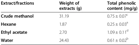
Table 1 shows the yield of extracts/fractions and their respective total phenolic content. The highest amount of phenolic compounds (p < 0.05) was found in the ethyl acetate fraction which was 1.09 ± 0.11 mg of GAEs/g extract, followed by the crude methanol extract (0.75 ± 0.07 mg of GAEs/g extract), water fraction (0.61 ± 0.02 mg of GAEs/g extract) and hexane fraction (0.25 ± 0.03 mg of GAEs/g extract). This result suggested that extraction
using polar solvents resulted in a higher content of phenolic compounds than those using solvent with low polarity.
Determination of DPPH radical scavenging activity
This method has been widely used to evaluate the rad-ical scavenging ability of the plant extracts as it is simple and highly sensitive. DPPH, a nitrogen-centered radical with a maximum absorption at 520 nm accepts an elec-tron from an antioxidant which acts as a hydrogen donor. The scavenging activity of the extract was moni-tored based on the amount of DPPH radicals remaining in the test sample using a spectrophotometer. In our study, the highest scavenging effect was observed in the ethyl acetate fraction with an IC50of 0.349 ± 0.009 mg/ml.
This is followed by the crude methanol extract (0.579 ± 0.017 mg/ml), water fraction (0.999 ± 0.038 mg/ml) and hexane fraction (2.677 ± 0.094 mg/ml). However, BHA and ascorbic acid exhibited better scavenging ability than the ethyl acetate fraction. Table 2 shows the IC50values of
the crude extract and its fractions as compared to the standards, BHA and ascorbic acid.
Determination of reducing power
[image:5.595.304.539.602.699.2]Figure 1 shows the reductive ability of the crude and fractionated extracts of the rhizomes of A. pahangensis in comparison to BHA and ascorbic acid. Reductive ability was measured by the reduction of ferricyanide complex/Fe3+to the ferrous form (Fe2+) in the presence of antioxidant (reductant). The Fe2+ formation produce Perl’s Prussian blue and can be monitored at absorbance of 620 nm by a spectrophotometer. The reductive cap-ability of the extracts and the standard compounds increased in the following order: water < hexane < ethyl acetate < methanol < BHA < ascorbic acid. The reducing power of the extract increased with the increase in con-centration of the extract until it reaches a certain level and then become constant. Basically, reducing power is
Table 1 Extraction yields and content of phenolic compounds in the crude and fractionated extracts of
Alpinia pahangensis
Extract/fractions Weight of extracts (g)
Total phenolic content (mg/g)
Crude methanol 31.19 0.75 ± 0.07c
Hexane 1.87 0.25 ± 0.03a
Ethyl acetate 2.70 1.09 ± 0.11d
Water 24.43 0.61 ± 0.02b
Values expressed are mean ± SD of triplicate measurements. Means with different letters (a-d) in the same column are significantly different (p < 0.05). mg/g: mg of gallic acid equivalents/g of extract or fractions.
Table 2 Radical scavenging activity of the crude and fractionated extracts ofAlpinia pahangensisagainst DPPH radical
Extracts and standards IC50values (mg/ml)
Crude methanol 0.579 ± 0.017c
Hexane 2.677 ± 0.094e
Ethyl acetate 0.349 ± 0.009b
Water 0.999 ± 0.038d
Ascorbic acid 0.015 ± 0.600a
BHA 0.013 ± 0.600a
IC50values expressed are mean ± standard deviation of triplicate
[image:5.595.55.293.630.708.2]associated with the presence of reductones that break the free radical chain by donating a hydrogen atom [42].
β-carotene linoleate model system
Bleaching of β-carotene of the crude and fractionated
extracts was assessed by theβ-carotene-linoleate method based on Cheung et al. In this system, linoleic acid under-goes oxidation and produce hydroperoxides at 50°C in the presence of oxygen. In the absence of antioxidants, the hydroperoxides formed upon abstraction of a hydrogen atom from one of its diallylic methylene groups reacts with unsaturated β-carotene molecules to form a stable radical. As a result,β-carotene becomes oxidized and loses its chromophore (orange color) in the system [43,44]. However, the presence of antioxidants can hinder the ex-tent ofβ-carotene bleaching by neutralizing the linoleate-free radical and other linoleate-free radicals formed in the system [45]. Therefore, the antioxidant activity was measured based on reduction of the orange color which was the amount ofβ-carotene present in the testing solution. The level of bleaching of color of a test solution was monitored
at 470 nm. The antioxidant activities of the extracts varied significantly with different concentration of extracts (p < 0.05, Table 3). The antioxidant activity of the extract and its fractions and the standard compound increased in the following order: hexane fraction < water fraction < ethyl acetate fraction < methanol extract < BHA at every concentration. The antioxidant activity of the extract and its fractions increased with an increasing concentration of the extract as shown in Table 3. The crude methanol extract exhibited 81.21 ± 0.9% of antioxi-dant activity at 20 mg/ml, which was comparable to that of BHA at 4 mg/ml (81.51 ± 0.67%, Table 3).
Superoxide anion scavenging activity
The superoxide anion scavenging ability of the extracts was determined using SOD assay kit-WST. Superoxide dismutase (SOD) is an enzymatic antioxidant that can scavenge superoxide anion radical (O2-) by catalyzing the
dismutation of the superoxide anion into hydrogen peroxide and molecular oxygen. This assay is based on the measurement of superoxide dismutase inhibition ac-tivity. In this assay, the superoxide anion reduce WST-1 (2-(4-iodophenyl)-3-(4-nitrophenyl)-5-(2.4-disulfophenyl)-2H-tetrazolium) to generate the water-soluble formazan dye in the testing solution, which is measured spectro-photometrically at 450 nm. In the presence of an enzym-atic antioxidant, the reduction of WST-1 can be inhibited by neutralizing O2-. Thus, the SOD activity can be
quanti-fied by measuring the decrease in the color development at 450 nm. The results in Table 4 show that the ethyl acetate fraction exhibited the highest superoxide anion scavenging ability with inhibition rate of 51.74 ± 4.9% among all extracts and fractions. This is followed by hexane fraction (32.21 ± 6.5%), methanol extract (29.32 ± 4.5%) and water fraction (18.06 ± 4.6%).
In summary, the crude and fractionated extracts of
rhizomes of Alpinia pahangensis showed varying
anti-oxidant properties in the entire in vitro antioxidant
assays. The ethyl acetate fraction showed the greatest free radical quenching activity and superoxide anion scavenging activity associated with the highest amount of phenolic content. Thus, this shows that the phenolic
0 10 20 30 40 50 60 70 80 90 100
15.625 31.25 62.5 125 250 500 1000
Total reducing activity
(%)
Concentration (µg/ml)
[image:6.595.57.293.88.236.2]PM PH PE PW BHA AA
Figure 1The reducing power of the crude and fractionated extracts and the standards at various concentrations.The concentrations of the crude and fractionated extracts were 1000, 500, 250, 125, 62.5, 31.25 and 15.625μg/ml. Butylated hydroxyanisole and ascorbic acid were used as the standards. Values expressed are means ± standard deviation of triplicate measurements. PM., crude methanol extract, PH., hexane fraction, PE., ethyl acetate fraction, PW., water fraction, BHA., butylated hydroxyanisole, AA., ascorbic acid.
Table 3 Antioxidant activity (%) of crude and fractionated extracts at various concentrations assayed byβ-carotene bleaching assay
Concentrations (mg/ml) Antioxidant activity of crude methanol extract and its fractions
Methanol extract Hexane fraction Ethyl acetate fraction Water fraction BHA
4 66.06 ±0.63dw 23.92 ±4.46aw 59.64 ± 1.44cw 50.03 ± 2.67bw 81.51 ± 0.67ew
8 74.73 ±1.84cx 42.17 ± 3.41ax 72.35 ± 2.30cx 57.54 ± 1.83bx 84.50 ± 0.21dx
16 77.84 ±0.41cy 52.62 ± 2.21ay 72.87 ± 1.73bx 71.74 ± 1.80by 89.54 ± 0.27dy
20 81.21 ±0.91cz 56.80 ± 2.70ay 74.51 ± 0.53bx 74.79 ± 1.56by 92.92 ± 0.23dz
[image:6.595.58.539.625.708.2]content was positively correlated with DPPH radical scavenging activity and superoxide anion scavenging activity. As phenolic compounds have redox properties, this result is hardly surprising. The radical scavenging activity is usually related to the presence of hydroxyl substituents in aromatic rings, which contribute to their hydrogen donating activity [46]. Thus, the radical scav-enging efficiency of the ethyl acetate extracts from Alpinia pahangensismight have been contributed by the phenolic constituents. However, the crude methanol extract possessed the strongest reducing activity against ferric ions and the highest antioxidant activity in the β -carotene bleaching assay. Lipid-soluble components are present in the crude methanol extract for the antioxi-dant activity shown in these two assays.
It was also observed that use of polar solvents like aque-ous methanol and ethyl acetate resulted in the extraction of components showing good antioxidant activity whereas extracts obtained from non-polar solvents and water showed weak antioxidant activity. A wide variety of
phen-olic constituents has been reported in Alpinia species
including flavonoids, tannins and some terpenoids. Thus, it is of interest to identify the compounds responsible for the antioxidant activity. Therefore, chemical isolation on the methanol or ethyl acetate fraction needs to be conducted to isolate the active components.
In vitroneutral red cytotoxicity assay
In vitro cytotoxicity assays are widely used for drug delivery to evaluate the biological effects of chemicals on mammalian cells. Many currently available assays mea-sure cytotoxicity based on alterations of plasma per-meability and the leakage of components into the supernatant or the uptake of dyes, by viable cells [47]. In this study, quantification of number of viable cells in the culture was based on the ability of the viable cells to uptake neutral red which was incorporated into the lyso-somes of the cells. Acidified ethanol solution was then used to extract the dye from the viable cells and the absorbance of the solubilized dye was then measured [48]. According to US NCI plant screening program, the extract that gave IC50 of 20 μg/ml or less is considered
[image:7.595.59.291.111.195.2] [image:7.595.58.539.620.716.2]active whilst it is 4 μg/ml or less for pure compound
[49,50].
Based on the result of the cytotoxic activity of the crude methanol and fractionated extracts (hexane, ethyl acetate and water) ofAlpinia pahangensis(Table 5), the hexane fraction showed the highest cytotoxic activity with IC50 less than 20μg/ml against KB, A549, Ca Ski,
HCT 116 and HT-29 with the exception of MCF7. It also showed remarkable cytotoxic effect towards KB and HCT 116 with IC50value of 5.8 ± 0.1 and 9.1 ± 2.0μg/ml
respectively. However, it also showed cytotoxic effect against the normal cell, MRC-5 with IC50value of 17.3 ±
0.5μg/ml. This was followed by the ethyl acetate fraction which showed strong cytotoxic activity against KB and HCT 16 with IC50values of 10.2 ± 0.3 μg/ml and 19.9 ±
1.1 μg/ml respectively and moderate cytotoxic effect
against other cell lines. However, the crude methanol and water fraction did not show cytotoxic effect against all cancer cell lines tested (IC50more than 100μg/ml). In this
study, doxorubicin, a drug used for cancer chemotherapy as positive. Doxorubicin showed much higher cytotoxic activity against all the cancer cell lines tested in compari-son to the hexane fraction and ethyl acetate fraction. Doxorubicin not only showed excellent cytotoxic effect against the cancer cell lines but it also showed strong cytotoxic effect against the normal cell line, MRC-5, with
Table 4 Inhibiton rate (SOD activity) of the crude and fractionated extracts
Extract/fractions Inhibiton rate (%)
Methanol 29.32 ± 4.5b
Hexane 32.21 ± 6.5b
Ethyl acetate 51.74 ± 4.9c
Water 18.06 ± 4.6a
BHA 70.19 ± 2.9d
Each value is expressed as mean ± standard deviation of triplicate measurements. Means with different letters (a-d) in the same column are significantly different (p < 0.05). Data are expressed as percentage of inhibition of superoxide radicals.
Table 5 IC50(μg/ml) values of crude and fractionated extracts ofAlpinia pahangensisagainst selected cell lines
Extract/ fraction Inhibition concentration, IC50(μg/ml)
KB MCF7 A549 Ca Ski HCT 116 HT-29 MRC-5
Methanol >100 >100 >100 >100 >100 >100 >100
Hexane 5.8 ± 0.1 22.0 ± 1.3 16.6 ± 0.2 18.4 ±0.2 9.1 ± 2.0 16.9 ± 0.5 17.3 ± 0.5
Ethyl acetate 10.2 ± 0.3 50.3 ± 0.3 25.9 ± 1.4 35.3 ± 2.0 19.9 ± 1.1 38.4 ± 0.4 44.0 ± 3.0
Water >100 >100 >100 >100 >100 >100 >100
Doxorubicin 0.46 ± 0.02 0.088 ± 0.01 1.01 ± 0.02 0.31 ± 0.10 0.41 ± 0.07 0.56 ± 0.05 0.69 ± 0.05
Each value is expressed as mean ± standand deviation of triplicate measurements.
an IC50value of 0.69 ± 0.05 ug/ml which is much higher
than the hexane fraction.
In the GCMS analysis, the hexane fraction showed the presence of a major component, methyl palmitate (13.67%), and minor components such as methyl oleate (7.10%), methyl stearate (2.35%), 1,2-dimethyldecahydro-naphthalene (3.05%), 1,5-dimethyldecahydro1,2-dimethyldecahydro-naphthalene (2.98%), 3,3,7,11-tetramethyl-tricyclo[6.3.0.0(2,4)] underc-8-ene (2.40%), 5-phenylundecane (0.82%) 6-phenyldode-cane (0.87%) and 5-phenyldode6-phenyldode-cane (1.35%). Sri Nurestri et al.[51] reported that a mixture of methyl palmitate, methyl oleate and methyl stearate showed strong cytotoxic effect against Ca Ski, A549, as well as the normal cell line,
MRC-5, with IC50 values less than 20 ug/ml. Methyl
palmitate was also reported to exert cytotoxic effect on T-cell leukemia T-cell line (Molt-4) with an IC50value of 2.28
ug/ml whilst methyl stearate showed cytotoxicity to acute promyeloblastic leukemia cell line (HL-60) and Molt-4 cell line with IC50 values of 3.08 and 4.65μg/ml respectively
[52]. In view of the above report, it is highly probable that the toxicity shown by the hexane fraction maybe partly due to the presence of methyl palmitate, methyl oleate and methyl stearate. The cytotoxic effect might be contributed by one or a combination of two or more of these components. Cytotoxic agents may cause necrosis in cells whereby cells lose membrane integrity leading to cell lysis or induce apoptosis cell death by activating an or-dered series of biochemical events [53,54].
Conclusions
This work describes for the first time the in vitro anti-oxidant and cytotoxic activity of the rhizomes ofAlpinia pahangensis. The rhizomes showed good antioxidant capacity when evaluated against 5 antioxidant assays. The ethyl acetate fraction showed good DPPH radical scavenging and superoxide anion scavenging activities whilst the crude methanol extract possessed excellent re-ducing power ability almost comparable to that of the
standards BHA and ascorbic acid, and good β-carotene
bleaching activity. In contrast, the hexane extract showed good antiproliferative activity against KB and Ca Ski cell lines but weak antioxidant activity. It can thus
be concluded that the rhizomes of Alpinia pahangensis
have the potential to be used as chemopreventive and chemotherapeutic agent and consumption of these rhi-zomes may provide some health benefits. Further inves-tigation on the underlying mechanism responsible for the biological activities should be attempted.
Abbreviations
DPPH:2,2-diphenyl-1-picrylhydrazyl; SOD: Superoxide dismutase; GC-MS: Gas chromatography–mass spectrometry; DNA: Deoxyribonucleic acid; GC: Guanine-cytosine; TA: Thymine-adenine; GAE: Gallic acid equivalents; BHA: Butylated hydroxyanisole; WST-1: 2-(4-iodophenyl)-3-(4-nitrophenyl)-5-(2,4-disulfophenyl)-2H-tetrazolium.
Competing interests
The authors declare that they have no competing interests.
Authors’contributions
CWP was responsible for conducting the experiments, data analysis and interpretation, and preparing the manuscript. SNAM was responsible for providing the grants, conception of ideas, identification of components, and revising the manuscript. HI was responsible for providing grants, conception of ideas, collection and identification of plants, and revising the manuscript. All authors read and approved the final manuscript.
Acknowledgements
The author wish to acknowledge the Ministry of Science, Technology and Innovation (MOSTI) and the University of Malaya (UM) for financial assistance received through the following grants: MOSTI 12-02-03-2070 and PPP PS319/2010A.
Received: 10 May 2013 Accepted: 23 September 2013 Published: 1 October 2013
References
1. Victório PC:Therapeutic value of the genusAlpinia, Zingiberaceae.
Rev Bras Farmacogn2011,21:194–201.
2. Matsuda H, Pongpiriyadacha Y, Morikawa T, Och M, Yoshikawa M: Gastroprotective effects of phenylpropanoids from the rhizomes of
Alpinia galangain rats: structural requirements and mode of action.
Eur J Pharmacol2003,471:59–67.
3. Burkill IH:A Dictionary of the Economic Products of the Malay Peninsula.
London: Crown Agent; 1966.
4. Malek SN, Phang CW, Ibrahim H, Norhanom W, Sim KS:Phytochemical and cytotoxic investigations ofAlpinia muticarhizomes.Molecules2011, 16:583–589.
5. Ghosh S, Rangan L:Alpinia: the gold mine of future therapeutics.
3 Biotech2013,3:1–13.
6. Awang K, Ibrahim H, Rosmy Syamsir D, Mohtar M, Mat Ali R, Azah Mohamad Ali N:Chemical constituents and antimicrobial activity of the leaf and rhizome oils ofAlpinia pahangensisRidl., an endemic wild ginger from peninsular Malaysia.Chem Biodivers2011,8:668–673. 7. Paz-Elizur T, Sevilya Z, Leitner-Dagan Y, Elinger D, Roisman LC, Livneh Z:
DNA repair oxidative DNA damage in human carcinogenesis: potential application for cancer risk assessment and prevention.Cancer Lett2008, 266:60–72.
8. Moreira P, Smith MA, Zhu X, Honda K, Lee HG, Aliev G, Perry G:Since oxidative damage is a key phenomenon in Alzheimer’s disease, treatment with antioxidants seems to be a promising approach for slowing disease progression. Oxidative damage and Alzheimer’s disease: are antioxidant therapies useful?Drug News Perspect2005,18:13–19. 9. Liu J, Mori A:Oxidative damage hypothesis of stress-associated aging
acceleration: neuroprotective effects of natural and nutritional antioxidants.Res Commun Biol Psychol Psychiat Neurosci2006,31:103–119. 10. Naito Y, Uchiyama K, Yoshikawa T:Oxidative stress involvement in
diabetic nephropathy and its prevention by astaxanthin.Oxid Stress Disease2006,21:235–242.
11. Jain SK:Superoxide dismutase overexpression and cellular oxidative damage in diabetes. A commentary overexpression of mitochondrial superoxide dismutase in mice protects the retina from diabetes.Free Rad Biol Med2006,41:1187–1190.
12. Beal MF:Mitochondria, oxidative damage, and inflammation in Parkinson’s disease.Ann N Y Acad Sci2003,991:120–131. 13. Heinecke JW:Mechanisms of oxidative damage of low density
lipoprotein in human atherosclerosis.Curr Opin Lipidol1997,8:268–274. 14. Halliwell B:How to characterize a biological antioxidant.Free Rad Res
Commun1990,9:1–32.
15. Halliwell B:Antioxidants: the basic- what they are and how to evaluate them.Adv Pharmacol1997,38:3–20.
16. Volka M, Rhodes CJ, Moncol J, Izakovic M, Mazur M:Free radicals, metals and antioxidants in oxidative stress-induced cancer.Chem Biol Interact
2006,160:1–40.
18. Moure A, Cruz JM, Franco D, Dominguez JM, Sineiro J, Dominguez H, Nunez M, Parajo JC:Natural antioxidants from residual sources.
Food Chem2001,72:145–171.
19. Madsen HL, Bertelsen G:Spices as antioxidants.Trends Food Sci Technol
1995,6:271–277.
20. Hazra B, Biswas S, Mandal N:Antioxidant and free radical scavenging activity ofSpondias pinnata.BMC Complement Altern Med2008,8:63. 21. Ani V, Naidu KA:Antioxidant potential of bitter cumin (Centratherum
anthelminticum(L.) Kuntze) seeds inin vitromodels.BMC Complement Altern2011,11:40.
22. Rohman A, Riyanto S, Yuniarti N, Saputra WR, Utami R, Mulatsih W: Antioxidant activity, total phenolic and total flavonoid of extracts and fractions of red fruit (Pandanus conoidenusLam).Int Food Res J2010, 17:97–106.
23. Kleinsmith LJ:Principles of Cancer Biology.San Fransisco: Pearson Benjamin Cummings; 2006.
24. Herceg Z, Hainaut P:Genetic and epigenetic alterations as biomarkers for cancer detection, diagnosis and prognosis.Mol Oncol2007,1:26–41. 25. Reuter S, Gupta SC, Chaturvedi MM, Aggarwal BB:Oxidative stress,
inflammation, and cancer: how are they linked?Free Radic Biol Med2010, 49:1603–1616.
26. Rahman K:Studies on free radicals, antioxidants, and co-factors.
Clin Interv Aging2007,2(2):219–236.
27. Lombardi V, Valko L, Stolc S, Valko M, Ondrejickova O, Horakova L, Placek J, Troncone A:Free radicals in rabbit spinal cord ischemia: electron spin resonance spectroscopy and correlation with SOD activity.Cell Mol Neurobiol1998,18:399–412.
28. Lunec J, Holloway KA, Cooke MS, Faux S, Griffiths HR, Evans MD:Urinary 8-oxo-2′-deoxyguanosine: redox regulation of DNA repair in vivo?
Free Rad Biol Med2002,33:875–885.
29. Sugimura T:Multi-step carcinogenesis: a 1992 perspective.Science1992, 258:603–607.
30. Go VLW, Butrum RR, Wong DA:Diet, nutrition and cancer prevention: the postgenomic.Era J Nutr2003,133:3830–3836.
31. Murthy NS, Mathew A:Cancer epidemiology,prevention and control.
Curr Sci2004,86:518–527.
32. Floyd RA, Carney JM:Free radical damage to protein and DNA: mechanisms involved and relevant observations on brain undergoing oxidative stress.Ann Neurol1992,32:22–27.
33. Phang CW, Malek SNA, Ibrahim H, Wahab NA:Antioxidant properties of crude and fractionated extracts ofAlpinia muticarhizomes and their total phenolic content.Afr J Pharm Pharacol2011,5:842–852. 34. Al-Saikhan MS, Howard LR, Miller JRJC:Antioxidant activity and total
phenolics in different genotypes of potato (Solanum tuberosum, L.).
J Food Sci1995,60:341–343.
35. Murugan K, Lyer VV:Antioxidant and antiproliferative activities of Marine Algae,Gracilaria edulisandEnteromorpha lingulata, from Chennai Coast.
Int J Cancer Res2012,8:15–26.
36. Borenfreund E, Puerner JA:A simple quantitative procedure using monolayer cultures for cytotoxicity assays (HTD/ NR-90).J Tissue Culture Meth1984,9:7–9.
37. Randhir R, Lin YT, Shetty K:Phenolics, their antioxidant and antimicrobial activity in dark germinated sprouts in response to peptide and phytochemical elicitors.Asia Pac J Clin Nutr2004,13:295–307. 38. Rice-Evans CA, Miller NJ, Paganga G:Antioxidant properties of phenolic
compounds.Trends Plant Sci1997,2:152–159.
39. Proestos C, Boziaris IS, Nychas GJE, Komaitis M:Analysis of flavonoids and phenolics acids in Greek aromatic plants: investigation of their antioxidant capacity and antimicrobial activity.Food Chem2006, 95:664–671.
40. Yu L, Haley S, Perret J, Harris M, Wilson J, Qian M:Free radical scavenging properties of wheat extracts.J Agric Food Chem2002,50:1619–1624. 41. Apak R, Güçlü K, Demirata B, Ozyürek M, Celik SE, Bektașoglu B, Berker K,
Ozyurt D:omparative evaluation of various total antioxidant capacity assays applied to phenolic compounds with the CUPRAC assay.
Molecules2007,12:14696–1547.
42. Duh PD:Antioxidant activity of burdock (Arctium lappaLinne): its scavenging effect on free radical and active oxygen.J Am Oil Chem Soc
1998,75:455–461.
43. Kumaran A, Karunakaran RJ:Antioxidant and free radical scavenging of an aqueous extract of Coleus aromaticus.Food Chem2006,97:109–114.
44. Moon JK, Shibamoto T:Antioxidant assays for plant and food components.J Agric Food Chem2009,57:1655–1666.
45. Jayaprakasha GK, Singh RP, Sakariah KK:Antioxidant activity of grape seed (Vitis vinifera) extracts on peroxidation models in vitro.Food Chem2001, 73:285–290.
46. Brand-Williams W, Cuvelier ME, Berset C:Use of free radical method to evaluate antioxidant activity.Lebensm-Wiss Technol1995,28:25–30. 47. Chiang LC, Chiang W, Chang MY, Ng LT, Lin CC:Antileukimic activity
selected natural products in Taiwan.Am J Chin Med2003,31:37–46. 48. Repetto G, del Peso A, Zurita JL:Neutral red uptake assay for the
estimation of cell viability/cytotoxicity.Nat Protoc2008,3:1125–1131. 49. Lee CC, Houghton P:Cytotoxicity of plants from Malaysia and Thailand
used traditionally to treat cancer.J Ethanopharmacol2005,100:237–243. 50. Boik J:Natural compounds in cancer therapy.Minnesota, USA: Oregon
Medical Press; 2001.
51. Sri Nurestri AM, Sim KS, Norhanom AW, Hashim Y:Phytochemical and cytotoxic investigations of Pereskia grandifolia Haw. (Cactaceae) leaves.
J Biol Sci2009,9:488–493.
52. Takeara R, Jimenez PC, Wilke DV, Odorico de Moraes M, Pessoa C, Peporine Lopes N, Lopes JLC, Monteiro da Cruz Lotufo T, Costa Lotufo LV: Antileukemic effects ofDidemnum psammatodes(Tunicata: Ascidiacea) constituents.Comp Biochem Physiol A Mol Integr Physiol2008,
151:363––369.
53. Miret S, De Groene EM, Klaffke W:Comparison of in vitro assays of cellular toxicity in the human hepatic cell line HepG2.J Biomol Screen
2006,11:184–193.
54. Syed Abd Rahman SN, Abdul Wahab N, Abd Malek SN:In vitromorphological assessment of apoptosis induced by antiproliferative constituents from the rhizomes ofCurcuma zedoria. Evid Based Complement Alternat Med2013, 2013:14.
doi:10.1186/1472-6882-13-243
Cite this article as:Phanget al.:Antioxidant potential, cytotoxic activity and total phenolic content ofAlpinia pahangensisrhizomes.
BMC Complementary and Alternative Medicine201313:243.
Submit your next manuscript to BioMed Central and take full advantage of:
• Convenient online submission
• Thorough peer review
• No space constraints or color figure charges
• Immediate publication on acceptance
• Inclusion in PubMed, CAS, Scopus and Google Scholar
• Research which is freely available for redistribution