SEGMENTAL INTERCHANGE I N M I C E
P. C . ROLLER
Institute of Animal Genetics, University of Edinburgh, Edinburgh, Scotland
Received November 4, 1943
INTRODUCTION
H R E E lines, A, B and T, showing reduced fertility, were producedin
T
mice by X-rays. Breeding experiments have shown that this X-ray-in- duced low fertility behaves as a dominant character which is most plausibly interpreted as due to structural rearrangement in the chromosomes. The sug- gestion that inherited reduced fertility was due to segmental interchange (reciprocal translocation) between two non-homologous chromosomes was first put forward by BELLING (1925) to explain some results obtained in species crosses of Stizolobium. The genetical implications of both spontaneous and induced segmental interchange have been analysed cytologkally and geneti- cally in various plants-namely, Datura (BLAKESLEE 1929), Zea (BURNHAM1930, 1932, 1g34), Pisum (SANSOME 1931, 1933; SUTTON 1g35), and in Dro- sophila (DOBZHANSKY and STURTEVANT 1931). I n Zea and Pisum the inter- change heterozygote has been referred to as “semi-sterile” due to the fact that in these plants nearly all the interchanges are reported to be associated with 50 percent sterility.
SNELL (1933) was the first to report X-ray-induced “semi-sterility” in mice. This was followed by the discovery that reduced litter size of the interchange heterozygote is due to zygotic lethality (SNELL, BODEMANN, and HOLLENDER 1934; SNELL and PICKEN 1935; BRENNEKE 1937). The manifestation and be- haviour of X-ray-induced semi-sterility in mice has been most extensively studied by SNELL (1935) and HERTWIG (1935, 1938, 1940) in breeding experi- ments. The presence of a characteristic chromosome configuration during the meiotic division in the descendants of X-rayed mice showing low fertility was detected by KOLLER and AUERBACH (1941) and was assumed to be brought about by the association of two pairs of non-homologous chromosomes due to segmental interchange. The present paper contains a more detailed description of the cytological analysis of interchange heterozygotes in mice and discusses the causal relationship between low fertility and chromosome behaviour.
MATERIAL A N D METHOD
The method of inducing and detecting reduced fertility or semi-sterility was the same as that used previously by SNELL (1g35), A number of males from stocks kept in this Institute had their testes exposed to X-ray doses varying between approximately 750 and goo r. From three of these males semi-sterile lines were obtained, indicating the production of segmental inter- change by the radiation. Average litter size in the stocks from which the males were taken, and from which later on normal animals were chosen for fertility tests, was 7.7. The three interchange lines A, B, and T were kept going by selecting in every generation a certain number of semi-sterile animals (male or
248 P. C. KOLLER
female), either by subjecting them to breeding tests or, in the case of males, by cytological examination of one testis, leaving the other in position and func- tioning. Once it had been shown that the results of the two methods were in good agreement, the cytological test was preferred because it saved labour and time.
Small pieces of testis were fixed in modified Minouchi’s solution (z percent osmic acid, I percent chromic acid, 3 percent potassium bichromate, and dis- tilled water, in the ratio 2:4:4:10). Sections were cut a t 16-I& thickness and stained with gentian violet, Squash and smear preparations were also made; they were fixed in acetic alcohol (3: I) stained with lacmoid, orcein, or
Feulgen’s basic fuchsin (DARLINGTON and LA COUR 1942).
Living spermatozoa of normal and interchange heterozygotes were taken from the epididymis and studied by dark field illumination.
CHROMOSOME MORPHOLOGY I N THE MOUSE
The accounts given by various authors concerning the mitotic chromosomes in the mouse are in agreement that the diploid number is 40 and that the chromosomes are “rod-shaped” (cf. CREW and KOLLER 1932; MAKINO 1941).
Improved methods of fixation and staining, however, showed that the appar- ently ‘rod-shaped” chromosomes are composed of one long and one very short arm, which indicates that the centromere is not terminal. In some chromosomes its subterminal position was clearly seen (fig. I). The structure and shape of
FIG. I . The diploid chromo- some complement in the
spermatogonium of the
mouse. Squash preparation,
stained with orcein. X 1,200.
G
H
IPLATE 1.-A. Trivalent, simulating a “triple chiasma” in a primary spermatocyte of line R.
X ~,roo.-R. Ring-of-four association in line T, showing nondisjunctional co-orientation of chro-
SEGMENTAL INTERCHANGE IN MICE 249
CHROMOSOME BEHAVIOUR AND SEGMENTAL INTERCHANGE
Line T
The chromosomal basis of reduced fertility was most extensively studied in line T both cytologically and by breeding experiments. The number of first
. . . .... ...
.... . . .
...
...
. . . ;:.'
A
...,:
"
..,.w
..i
O d . I
. .
:...
., ;
.
e a *Q
':.. i...:' .. ..
;
)&$c .. ..&&$&
;
.
i :
. '. ! i . . . . . . . . . . . . . . ... .. B ... ... ...
O b J . .
... c ...
'. A ... ... ... .... ...
...
......-
-._. ... '?.A
...'.: gt@Q@ '' ! \
i
wmfi
: i
J
))I F,fi\\.
\
d 9 Q U
;
;i.. &*&a ;
.... .... ....
1.
... ...
,. .. .. .
A
: . \ I
: ,
;' ,
w
&fl*w
;.
:;,.
.... ... ... . . .
. .
. .
w
.,.:'... D
:' H
,..'
\.
. . . ...\..
.... E . . . ... . . . . . . . .... ... . . . _.. ., ...' J
: C G
'.:
.:&
... .... . . . . . . ... . . . ... ...
-*;
*
G FFIG. 2. Chromosome behaviour in the primary spermatocytes of interchange line T. The
unequal sex bivalent and the chromosomes involved in the segmental interchange are drawn
solid black. Description of figures in text. XI,OOO.
generation offspring of semi-sterile T's tested was 26; 1 1 were found to be
fertile and 15 semi-sterile-that is, about half of the viable offspring of semi- sterile parents carry the interchange. Similar results were reported on a much larger scale by SNELL (1935) and Hertwig (1940). The chromosome behaviour was studied in the primary spermatocytes of eight semi-sterile males. I n many cells besides the 18 bivalents, including the unequal XY chromosome pair, a large ring representing an association-of-four was clearly seen (Plate
IB,
text fig. 2B). As a result of reciprocal interchange of non-homologous chromosome250 P. C. KOLLER
result of five crossovers, one of which has taken place in an interstitial segment -that is, in the chromosome region between a centromere and the point of exchange,-while the four others are formed in the interchange segments and in the short arms. This configuration should be distinguished from the true figure-of-eight in which six chromosomes are associated (SANSOME 1933).
A cross-shaped configuration was also encountered (fig. 2C), and it was as- sumed that the association of the four chromosomes is the result of three chiasmata, one in an interstitial segment and one in each of the two inter- changed chromosome arms. I n a few primary spermatocytes the four chromo- somes formed a chain; this configuration is also due to three chiasmata, formed in three chromosome arms.
Primary spermatocytes were also found with 2 0 bivalents. While the unequal
XY bivalent was easily recognized, no unequal bivalents were identified (fig. 2F). I n a few cells a chromosome configuration of an unusual shape was ob- served; it may represent the interchanged chromosomes forming a “bivalent” (fig. zE). The new chromosome types could not be identified in the diploid complement, which suggests that the interchanged segments themselves are of similar size.
The anaphase segregation of chromosomes from an association-of-four is determined by their co-orientation during metaphase. This may be either ( I )
disjunctional-that is, adjacent members move to the opposite poles (conver- gent or zigzag arrangement), ( 2 ) non-disjunctional (fig. zD), or (3) chromatid
non-disjunctional (which results from the figure -of-eight). The non-disjunc- tional (linear, parallel, or discordant) co-orientation necessarily leads to the formation of genetically unbalanced gametes (DARLINGTON 1937). Chromatid non-disjunction or half disjunction leads to the production of half balanced, half unbalanced gametes. The frequencies of the first two kinds of chromosome co-orientation in the ring-of-four associations of line T were found to be very nearly the same.
Besides the two-by-two non-disjunctional arrangements, numerical non-dis- junction, by which three chromosomes went to one nucleus and one to the other, was observed (fig. 2H, G ) . The relative frequencies of these two types
could not be determined.
The analysis of the type and the frequency of the various multi-chromosomal associations strongly suggest that, in semi-sterile line T, two non-homologous chromosomes of medium size interchanged relatively short segments of similar lengths.
The total number of primary spermatocytes analysed in the eight males of line T was 2 2 9 (86, 31, 2 5 , 23, 20, 17, 15, and 1 2 in the different animals).
The data obtained show that in the different males the frequency of the same chromosome associations and consequently the frequency of the disjunctional and non-disjunctional gametes is very similar.
. Line B
SEGMENTAL INTERCHANGE IN MICE 2.51
countered in primary spermatocytes. The unpaired chromosomes could easily be identified by their size and shape and by the number of bodies present in the primary spermatocytes (fig. 3F, H). It was found to be 2 2 (18"+4') or 21
(1g11+21). In some cells only two univalents were seen; the other two chromo-
somes forming an unequal pair could not be identified (fig. 3G). The failure of pairing frequently observed in the primary spermatocytes is attributed to the interchanged chromosomes.
FIG. 3. Chromosome behaviour in the primary spermatocytes of interchange line B. XI,OOO.
252 P. C. KOLLER
... .... .... ... ...
.... ....
... ...
... .. ..
*
"?... ..
".:
;
810
. :
. . . . . .
. .
. .
. . . . . .
ULV
;;. .
...
.... ...
0
j ' . , .. ..: ...
I '
... :.' E F
... ...
D
....
. . .
I
FIG. 4. Chromosome behaviour in the primary spermatocytes of interchange line A. X 1,000.
I n line B, chains-of-four chromosomes were not seen; an association-of-three chromosomes was observed in about 7 percent of the primary spermatocytes analysed (fig. 3E; Plate IA, H). Unequal bivalents were also identified (Plate
IC,
E ) ; they differ from the XY (Plate 11) and may be easily distinguished. Evidence of numerical non-disjunction was also obtained (fig. 31).The types and frequencies of various chromosome associations indicate that the chromosome pairs involved in the interchange of line B are of medium size and that the interchanged segments differ greatly in length. Due to this differ- ence, the unequal bivalents could easily be distinguished.
Chromosome behaviour was analysed and found to be similar in five semi- sterile males. The total number of primary spermatocytes studied was 156
Line A
It was found that in the primary spermatocytes of line A the chain-of-four chromosome association showing linear or non-disjunctional co-orientation was
SEGMENTAL INTERCHANGE I N MICE 253
TABLE I
Litter size of interchange heterozygotes in lines A , B , and T.
NOTES NO. OF YOUNG I N MEAN LIT-
LINE SEX GENERATION
SUCCESSIVE LITTERS TER SIZE
dcf
A
9 9
dl3
9 9
c f c f
'r
9 9
Fz
Fz Fz Fa (TA8:2)
FI
F3
2 , o*, o*, o* o*, o*, o*, o*
o*, o*
31 4, I, 3, 5r 6
2 , 3, 2, 2, I, 2
3, 2 , 4, 3, 5, 4 2 , 3, o*, o*, o*, o*
2 , o*, o*, o*, o*
39 59 6, 3 5, 6, 3, 4 5, 3
4, 3, 5
2 . 6 2 . 8
4.5 4 . 0 4.3 3 . 7 3 . 3
2 . 0
3 . 5
4 , 3
4 . 5
4 . 0
4 . 0 4 . 0
4 . 1
4 . 2
2.7
CA.** 2 V.P.t C.A. z V.P.
2 V.P.
C.A. C.A. C.A. C.A. C.A. C.A. C.A. I V.P.
C.A. C.A. 2 V.P.
*
** C.A. = Cytologically analysed.
t
V.P. =Vaginal plugs observed.o=No litter from mating with separate females.
254 P. C. KOLLER
The analysis of chromosome association suggests that one medium and one short chromosome pair are probably involved in the interchange of line A. The interchanged segments are unequal. It is believed that one of the breaks oc- curred in the long, the other in the short arm of two non-homologous chromo- somes. The frequent failure of pairing indicates that the unequal interchanged segments are relatively small, their length is below or near the minimum length which conditions chiasma formation.
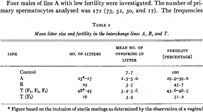
Four males of line A with low fertility were investigated. The number of pri- mary spermatocytes analysed was 1 7 2 (73, 5 2 , 30, and 17). The frequencies
TABLE 2
Mean litter size and fertility i n the interchange lines A , B, and T . LINE
MEAN NO. OF
NO. OF LITTERS OFFSPRING IN FERTILITY
(PERCENTAGE) LITTER
Control 7.7 I O 0
A 23*-17 2.3-3.0 29 f 9-39.0
B 29 3.5 45.7
T @I, Fz, Fa) 48*-45 3.4-3.6 43.6-46.5
T (FJ 16 3.9 51.2
* Figure based on the inclusion
of sterile matings as determined by the observation of a vaginal Plug.of the various chromosome configurations were similar in all the males except one.
Line A was found to be the most sterile. On account of the sterility of several males it was a t first thought that the sex chromosomes might have been in- volved in the in terchange. However, breeding tests and cytological analysis have shown that this is not the case. The sex bivalent (Plate 11) differs from the unequal bivalent (Plate I D ) formed by the interchanged chromosomes.
DIFFERENTIAL FERTILITY BETWEEN INTERCHANGE LINES
Breeding experiments have shown that the three interchange lines differ significantly in fertility, measured by the litter size. The results are sum- marized in tables I and 2. Animals producmg only one litter are not included.
SEGMENTAL INTERCHANGE I N MICE 255 ing the degree of fertility in the different lines. (a) The size of the litters was not always recorded on the day of birth. On account of early depletion in certain litters, this may have reduced litter size slightly in both control and inter- change lines. (b) Animals of the interchange lines which had in their first litter six or more young were not further tested; they were considered t o be normal. When a surplus of animalswas available, even first litters of five were takenasa basis for discarding an animal. This procedure will tend to reduce the figure for litter size in lines B and T, in which litters with five or six young were not infrequent. The breeding data from line A has presumably not been influenced by this source of error. I n this line animals with a first litter of five were not discarded without further test on accountof the small number of animals avail- able for carrying on the interchange. Furthermore, litters in line A with more than four young were very rare and occurred only from the third generation onward. (c) Some males in line A and in the Fz of line T produced either no lit- ter a t all, or one or two litters with one or two young, although they were kept with different females (renewed from time to time) for several weeks. I n some instances, the presence of vaginal plugs was observed, indicating that mating had taken place but failed to produce a litter. These matings can probably be regarded as litters of size 0. Separate-totals are calculated with these matings
included. (d) The mean size of litters (7.7) in controls was computed from a random sample of litters produced during the first months of the experiment by animals from stock; the same stocks were used for outcrosses. The fertile sibs of semi-sterile animals were not usually kept for breeding; data based on their performance comprise mainly first and second litters and are therefore less reliable controls than those gained from the stocks.
AS will be seen from the data summarized in table 2, line A showed about 30-39 percent fertility, line B about 46 percent fertility, line T about 44-46 per- cent fertility. Line A is probably significantly less than 50 percent fertile. I n the case of lines B and T, the existence of a significant departure from 50 per cent fertility is uncertain. It is of interest to consider the cause of the different fertil- ity of the three lines.
Random two-by-two separation of the four members of the translocation ring gives six types of gametes in equal numbers. The proportion of the geneti- cally balanced and unbalanced gametes ( 2 : 4) in interchange heterozygotes is determined by the metaphase arrangements of translocation rings and may also be influenced by the formation of associations-of-three and univalents. Thus BRINK and COOPER (1933) found that 50 percent fertility in an inter- change line (semi-sterile-I) of maize is associated with an equal frequency of non-disjunctional and disjunctional co-orientation of chromosomes in the ring- of-four. The two largest chromosome pairs are involved in this interchange.
256 P. C. ROLLER
pole and only nine a t the other. The resulting n + ~ pollen is capable of func- tioning, as indicated by the fact that 21 chromosome plants are occasionally
produced. The n- I pollen presumably aborts and may account entirely or in
part for the sterility in excess of 50 percent. Two of the shorter chromosomes are involved.
The importance of the length of the interchange segments as well as t h a t of the chromosomes in determining co-orientation was proved by BROWN (1940)
and PIPKIN (1940) in Drosophila; they corroborated the findings of DOBZHAN-
SKY (1933) that the frequency of non-disjunction is negatively correlated with
the length of the interchange segment. The number of chiasmata in an associa- tion-of-four is another factor which influences co-orientation. BEADLE (1932)
reported in maize that fertility of an interchange heterozygote is increased from 43 percent to 48 percent when the ring-of-four association is replaced by the chain-of-four. A similar observation was made by SAX and ANDERSON
(1933) in Tradescantia. Furthermore, the different fertility of interchange
heterozygotes in Datura, Zea, Pisum, and Campanula indicates that the posi- tion and terminalisation of chiasmata also play a part in determining chromo- some co-orientation in the multivalent ( GAIRDNER and DARLINGTON 1930). I n Datura and Campanula, also Oenothera, terminalisation is complete and the percentage of disjunction high, whereas in Zea and Pisum with 50 percent of non-disjunction, interstitial chiasmata are present a t metaphase. Chromosomes with a median centromere form a disjunctional ring-of-four of the zigzag type more often than do chromosomes with a subterminal centromere. Thus the fer- tility of interchange heterozygotes is high in Datura, Oenothera, and Campa- nula, and much lower in Tradescantia and Rhoeo. According to BERGNER, SATINA, and BLAKESLEE (1933), most of the translocation heterozygotes are fully fertile in Datura. THOMPSON and HUTCHESON (1942) reported 95-90 per- cent fertility in diploid wheat. SAX and ANDERSON (1933) observed a very high frequency of non-disjunctional co-orientation (80 percent) in the ring-of-twelve of Rhoeo. I n mice the fertility of the interchange line A is less than that ob- served in B and T, which indicates clearly that the structure and behaviour of the chromosomes involved in the interchanges differ in these lines and deter- mine the different frequencies of balanced gametes.
SEGMENTAL INTERCHANGE IN MICE 2 5 7
T A B L E 3
Fertility of the interchange lines estimated by the chromosome behaviour.
PERCENTAGE INTERCHANGE LINES
CHROMOSOME CONYIG. OF BALANCED
GAMETES A n T
Disjunct. Non-disj. Ring-of-four Disjunct. Non-disj. Chain-of-four Normal Abnormal Figure-of-Eight Association-of-three Unequal Bivalent Univalent Non-analysed
Total no. primary spermatocytes
Expected (cytological)
(Observed (breeding)
Fertility (percentage)
1
I 0 0
0
I o 0
0 5 0 0 33-3 5 0 25 2 5 2 I3 0 8** 2 I O 14 17 73 23.5
29 * 9-39
8 8
4 16
0 I O
0 I
7* I 2
4 t 3
* Some are probably disjunction ring-of-four.
** See fig. 4C.
t
See fig. 3C.centage of these cells is 23.2 percent, 41.1 percent and 31.3 percent in line A, B, and T, respectively, which is very high and shows the great difficulty involved in an accurate cytological analysis.
A fairly satisfactory agreement between expected (cytological) and observed (breeding) fertility was found in semi-sterile line T ; the fertilities are 48.3 and 43.6-46.5 percent, respectively. The four Ft interchange heterozygote males analysed had a similar fertility calculated by chromosome behaviour.
I n the case of line B the figures are 44.2 percent fertility (expected cytologi- cal) and 45.7 percent fertility (observed breeding). If allowance is made for chromosome configurations classed as figures-of-eight but perhaps actually dis- junctional rings-of-four, the cytological fertility would be higher.
Line A has the most sterile interchange; fertility calculated from observed litter size is about 39 percent of that shown by the controls, or if sterile matings are included in the calculation, about 29.9 percent. This agrees well with the expected fertility of 23.5 percent calculated from cytological observations.
2 5 8 P. C. ROLLER
lated with the frequency of disjunctional co-orientation of chromosomes within the translocation ring and may also be influenced by differences in the fre- quency of associations-of-three and of univalents.
The three interchange lines show that radiation induces segmental inter- changes which differ and that the differences may be detected not only by breed- ing but also by cytological analysis. There is evidence that in the similar experi- ments of HERTWIG (1940) different interchanges were produced by radiation. Thus she found that litter size is 4.05 in line 746 and 2.74 in line 621; in line 623
the embryos die late, about ten days after fertilization. HERTWIG suggests that
TABLE 4
Fertility of interchange heterozygotes i n successive generations of line T .
NO. OF INTERCHANGE
MEAN
HETEROZYGOTES NO. OF FERTILITY
GENERATION LITTER
LITTERS (PERCENTAGE)
SIZE
MALE FEMALE
Fi I 6 2.0 25 - 9
F3 4 2 16 3.9 51.2
Fa 3 4 23 3.7 48.6
this line carries a “relatively small translocation” producing little unbalance in the gametes and allowing the embryos to develop to an advanced stage It is probable that differences observed by her between the semi-sterile lines, in- volving litter size and time of zygotic lethality, are expressions of differences involving the interchanged chromosomes.
DIFFERENTIAL FERTILITY WITHIN INTERCHANGE LINES
I n the third generation of the interchange heterozygotes of line T, a sudden increase was observed in fertility (table 4). It may be seen that while the F1 male (progenitor of line T) exhibits a very low fertility (25.9 percent), in his offspring and descendants it has risen to 48.6-51.2 percent. Owing to the fact that, of the three F2 males, two not only had a very small litter size, but also failed to produce litters with several females known to be fertile, the fertility of F2 interchange heterozygotes is probably much less than 48.6 per cent. The litter size in F3 may perhaps be considered to be most truly characteristic of in- terchange T, since other induced aberrations, if present, probably would have segregated out. Cytological analysis of the F1 and F2 males did not show any additional irregularities, except perhaps in one F2 male in which a few sperma- tocytes were seen with univalents.
SEGMENTAL INTERCHANGE IN MICE 2 5 9
Differential fertility was also encountered in line A. Three Fz males when mated to fertile females failed to produce offspring. Vaginal plugs indicated that matings had taken place. An FB male was apparently more fertile. The loss of additional induced irregularities is perhaps responsible.
Similar cases of differential fertility within an interchange line have been described by other authors. CLARKE and ANDERSON (1935) found greater steril- ity in the earlier generations of an interchange in maize than in later genera- tions. Completely sterile individuals were reported by HERTWIG (1940) amongst descendants of the semi-sterile male No. 733. They disappeared in the third generation. It is very probable that these mice were carrying additional chromosome changes or gene mutations induced by the radiation.
INTERCHANGE HOMOZYGOTES AND MULTIPLE INTERCHANGE HETEROZYGOTES
Several sisterx brother matings of semi-steriles were attempted in order to produce interchange homozygotes in lines B and T. It was found that the num- ber of offspring in this cross,as expected,fell below the mean number character- istic of lines T and B when outcrossed. Cytological analysis of 2 6 males in line
T and 2 2 in line B has shown either normal chromosome behaviour or, besides
the 18 bivalents, an association-of-four indicating the presence of an inter- change in heterozygous condition. Some of these males which show 2 0 bivalents
in every primary spermatocyte may have been homozygous for the inter- change. Several such males were outcrossed, and the absence of quadrivalents suggests that they were normal. It is not improbable that the failure to detect interchange homozygotes in lines T and B may be due to the fact that the in- terchanges are lethal when homozygous. Owing to various difficulties arising from the war, the breeding experiments as originally planned could not be carried to completion, and the data obtained are not sufficient to draw a defi- nite conclusion. I n similar experiments HERTWIG (1940) reported two certain and one uncertain interchange homozygotes, which indicates that interchanges in mice a s in Drosophila are not necessarily lethal when homozygous.
Another experiment was planned t o produce multiple interchange heterozy- gotes by combining interchanges A and T in one individual. This could have been detected cytologically'by the presence of either two rings-of-four or a ring-of-six if one of the chromosomes involved in the interchanges was com- mon. The cytological analysis of offspring of 1 2 matings did not indicate the
presence of these interchanges in combination. It was noticed by breeding tests that a great reduction was induced in fertility by crossing the two interchange heterozygotes together, the litter size falling below two. When two different interchanges are crossed, the expected ratio of viable to inviable zygotes is ap- proximately I : 3.
FACTORS DETERMINING THE PREQUENCY O F UNBALANCED GAMETES
2 60 P. C. HOLLER
non-disjunctional or genetically unbalanced gametes, and consequently the degree of sterility of an interchange heterozygote, may be classified as follows:
(I) length (a) of the chromosome, (b) of the interchanged segments, (c) of the
interstitial segment; ( 2 ) position (a) of the centromere, (b) of. the chiasma; (3)
number of chiasmata; and (4) degree of terminalisation.
The expression of sterility is the result of an interaction of these different fac- tors. The variations in chromosome structure and behaviour underlie the dif- ference in sterility found not only between the interchange heterozygotes of the different species, but between individuals carrying the same interchange, examples of which have been found in mice.
ZYGOTIC AND GAMETIC LETHALITY I N INTERCHANGE HETEROZYGOTES
I n mice reduced fertility of the interchange lines is due to zygotic lethality. The motility and morphology of living spermatozoa in normals and inter- change heterozygotes were studied by means of dark field illumination. A small portion (about 5-7 percent) of immobile spermatozoa was seen in both normals and interchange heterozygotes, due apparently to environmental factors. The genetically unbalanced spermatozoa of the latter could not be detected by this means. A more extensive comparative study of living spermatozoa of different interchanges would be very desirable in view of the fact that chromosome or genic unbalance may lead to the immobility of the sperm. That either genic 01
numerical unbalance may lead to gametic lethality in animals was shown by DETLEFSEN (1912). He reported the presence of immobile spermatozoa in the interspecific hybrids of Cavia rufescens XC. porcellus. The proportion of im- mobile sperm decreases in the successive backcross generations, due apparently to a reduction in the chromosome unbalance. There is evidence that in the in- terchange line A the differentiation of some spermatids into spermatozoa is arrested. These may represent the numerically unbalanced gametes. The elimination of unbalanced gametes a t this early stage would, of course, raise the fertility as estimated by litter size.
The investigations of SNELL, BODEMANN, and HOLLENDER (1934), SNELL and PICKEN (1935), BRENNEKE (1937), and HERTWIG (1940) have shown that reduced litter size is brought about either (I) by the elimination of fertilised
eggs before or after implantatian or ( 2 ) by the death of the embryo during early
or late development. When estimating sterility of interchanges in animals, it should be realized that eggs fertilised with unbalanced sperm may be elimi- nated before implantation, hence the number of "uterine moles" or dead em- bryos cannot be relied upon as the measure of reduction in fertility, for which reason in the present study this method was deliberately avoided. The stage in which lethality comes into operation is determined primarily by the degree of unbalance. It is not impossible that it may lead to the inactivity of sperma- tozoa or even arrest the differentiation of spermatids into spermatozoa.
SEGMENTAL INTERCHANGE IN MICE 261 in the first or second cleavage division suggest that the difference in the be- haviour of non-disjunctional gametes in animals and plants is not so discon- tinuous as it appears to be. The occurrence of greater gametic lethality in plants, nevertheless, is to be expected because of the fact that they have a gametophyte stage, between meiosis and gamete production, during which two or three mitotic divisions occur. It is here, in plants, that unbalance usually leads to breakdown.
HERITABLE LOW FERTILITY I N DOMESTIC ANIMALS
The most significant genetical implication of segmental interchange is re- duced fertility; because of this it is usually selected against in nature. However, instances are known, mostly in plants-namely, Oenothera-where heterozy- gotes are the rule. Amongst animals the best known interchange found in a free living population was reported by CAROTHERS (1931). Interchange, be- cause it facilitates the establishment of a balanced lethal mechanism, can be maintained in nature and may lead to isolation and formation of incipient spe- cies. Comparative cytological analysis in closely related species (Drosophila, Datura, Crepis, etc.) has shown that interchange actually must have played a part in species differentiation. It may be fixed somewhat more easily in ani- mal than in plant populations according to the mathematical calculation of
While an interchange, unless it is associated with a balanced lethal mecha- nism, is usually eliminated by natural selection, artificial selection in animals under domestication may favour its maintenance, especially if the chromo- somes involved carry genes favoured in breeding. It is interesting to note that in domestic animals (cattle, horses) reduced fertility, often approaching com- plete sterility, is very common and is attributed to various causes-genic, physiological, nutritional, etc. I n view of the fact that structural changes occur in the chromosomes of higher organisms, e.g., inversion in man (KOLLER 1937) and in the pig (CREW and KOLLER 1g3g)-it is not improbable that in- herited reduced fertility in some of the highly infertile domestic animals may also be due to structural changes. It would be of great interest to analyse cyto- logically domestic animals known to be transmitting reduced fertility in order to determine whether or not this is due to segmental interchange.
The cytological analysis given in the present paper shows that radiation can induce heritable semi-sterility or reduced fertility in organisms higher than plants or insects. Nowadays, when radiation is extensively employed in radio- therapy, engineering, physics and chemistry, man is exposed to radiation in various ways. There can no longer be any doubt that the chromosomes of the germinal cells in man if not protected from X-rays may also undergo the same structural changes as in mice. The potential consequence is semi-sterility or re- duced fertility in the descendants of the individual irradiated.
WRIGHT (I 941).
SUMMARY
262 P. C. ROLLER
segmental interchange between two pairs of non-homologous chromosomes, was identified during the first meiotic division in each of the lines.
The approximate fertility of the three lines estimated by breeding tests was found to be 30-39 percent in
A,
46 percent inB,
44-46 percent in T (51 percent in the Fa generation of T).Cytological analysis has shown that differences in fertility are correlated with corresponding differences in the frequencies of non-disjunctional co-ori- entation of chromosomes in the ring-of-four. Fertility estimated on cytological analysis is expected to be about: 2 4 percent in A, 44+ percent in
B,
and 48percent in T.
Differential fertility observed in successive generations within lines A and T
is attributed to either minor structural changes or gene mutations brought about by the radiation responsible for the segmental interchanges.
The various factors on which depends the frequency of genetically balanced gametes-that is, the fertility of an interchange heterozygote-are analysed.
It is suggested that in some instances, inherited low fertility, common in domestic animals, may be due to segmental interchange, the maintenance of which may be favoured by artificial selection.
ACKNOWLEDGMENTS
The author is greatly indebted to PROFESSOR H. J. MULLER for suggesting the problem and for the help he gave in the initial stages of the experiment. The tedious breeding experiments were carried out by Dr. C. A. AUERBACH whose help is gratefully acknowledged. Thanks are also due to DR. G. D. SNELL for the help given in the preparation of the manuscript The investiga- tions were aided by a grant from the British Empire Cancer Campaign.
LITERATURE CITED
BEADLE, G. W., 1932 The relation of crossing over to chromosome association in Zea-Euchlaena
BELLING, J., I925 A unique result in certain species crosses. Z. i. A. V. 39: 286-288.
BERGNER, A. D., S. SATINA, and A. F. BLAKESLEE, 1933 Prime types of Datura. Proc. Nat.
BLAKESLEE, A. F., 1929 Cryptic types in Datura due to chromosomal interchange and their BRENNEKE, H., 1937 Strahlenschiidigung von Mause und Rattensperm, beobachtet an der BRINK, R. A., and D. C. COOPER, 1933 The association of semistede-r in maize with two link-
BROWN, SUCIIE M., 1940 The relation between chiasma formation and disjunction. Univ. Texas BURNHAY, C. R., 1930 Genetical and cytological studies of semi-sterility and related phenomena
in maize. Proc. Nat. Acad. Sci. 12: 269-277.
1932 An interchange in maize giving low sterility and chain configurations. Proc. Nat. Acad. Sci. 18: 434-40.
1934 Cytogenetic studies of an interchange between chromosomes 8 and 9 in maize. Genetics
19: 430-447.
CAROTHERS, E., 1931 The maturation divisionsgnd segregation of heteromorphic homologous chromosomes in Acrididae (Orthoptera). J. Morph. 28: 445-521.
hybrids. Genetics 17: 481-501.
Acad. Sci. rg: 103-115.
geographical distribution. J. Hered. 20: 177-190. Friihentwicklung der Eier. Strahlentherapie 60: 73-103. age groups. Genetics 16: 595-628.
SEGMENTAL INTERCHANGE I N MICE 263
CLARKE, A. E., and E. G. ANDERSON, 1935 A chromosomal interchange in maize without ring formation, Amer. J. Bot. 22: 711-716.
CREW, F. A. E., and P. C. KOLLER, 1932 The sex incidence of chiasma frequency and genetical crossing-over in the mouse. J. Genet. 26: 359-383.
CREW, F. A. E., and P. C. ROLLER, 1939 Cytogenetical analysis of the chromosomes in the pig. Proc. Roy. Soc. Edinb. 59: 163-175.
DARLINGTON, C. D., 1937
DARLINGTON, C. D., and L. T. LA COUR, 1942 Unwin & Co.
DETLEFSEN, J., 1912 3: 261-265. DOBZHANSKY, TA., 1933
over and disjunction of chromosomes. 2. i. A. V. 64: 269-309. 1941
Translocation between the second and third chromosomes of Drosophila and their bearing on Oenothera problems. Carnegie Instn. Wash. Pub. No. 421 : 25-59.
ijber Sterilitatserscheinungen bei rontgenbestrahlten Mausen und deren Nachkommenschaft. 2. Vererbl. 70: 517-523.
1938 Untershiede in der Entwicklungsfahigkeit von Fl-Mausen nach Rontgenhestrahlung
von Spermatogonien, fertigen und unfertigen Spermatozoen. Biol. Zbl. 58: 273-301.
1940 Vererbbare Semisterilittit bei Mausen nach Rontgenbestrahlung verursacht durch
reziproke Chromosomentranslokationen. Z. i. A. V. 79: 1-29.
GAIRDNER, A. E., and C. D. DARLINGTON, I930 Ring formation in diploid and polyploid &m-
panula persicifolh. Genetica 13: I 13-150.
KOLLER, P. C., 1937 The genetical and mechanical properties of sex chromosomes. 111. Man. Proc. Roy. Soc. Edinb. 57: 194-214.
KOLLER, P. C., and C. A. AUERBACH, 1941 ChromosJme breakage and sterility in the mouse. Nature 148: 501.
MAKINO, S., 1941 Studies on the Murine chromosomes. I. Cytological investigations of mice in- cluded in the genus Mus. J. Fac. Sci. Hokkaido Imp. Univ., Ser. vi Zool. 7: 305-380. PIPKIN, S. B., 1940 Segregation and crossing over in a 2, 3 translocation in Drosophila melano-
gaster. Univ. Texas Pub. No. 4032: 73-125. SANSOME, E. R., 1931
1933
SAX, K., and E. ANDERSON, 1933 netics 18: 53-67.
SNELL, G. D., 1933 1935
SNELL, G. D., E. BODEMANN, and W. HOLLENDER, 1934 A translocation in the house mouse and its effect on development. J. Exp. 2001.67: 93-104.
SNELL, G. D., and D. L. PICKEN, 1935 Abnormal development in the house mouse caused by chromosome unbalance. J. Genet. 31 : 213--235.
SUTTON, E., 1935 Half-disjunction in an association of four chromosomes in Pisum sativum. Ann. Bot. 49: 689-698.
THOMPSON, W. P., and I. HUTCHESON, 1942 Chromosome behaviour and fertility in diploid wheat with translocation complexes of four and six chromosomes. Canad. J. Res.20: 267-281. WRIGHT, S., 1941 On the probability of fixation of reciprocal translocations. Amer. Nat. 75:
Recent advances in cytology. 2nd ed. London: Churchill & Co. The handling of chromosomes. London: Allen-
The fertility of hybrids in a mammalian species-cross. Amer. Breed. Mag.
Studies on chromsome conjugation. 11. The relation between crossing- Genetics and origin of species. 2nd ed. London: Oxford Univ. Press.
DOBZHANSKY, TH., and A. H. STURTEVANT, 1931
HERTWIG, P., 1935
Chromosome association in Pisum. J. Genet. 25: 34-54. Segmental interchange in Pisum. 11. Cytologia 5: 15-30.
Segmental interchange in chromosomes of Tradescantia. Ge- Genetic changes in mice induced by X-Ays. Amer. Nat. 67: 24-31. The induction by X-rays of hereditary changes in mice. Genetics 20: 545-567.