Copyright 0 1988 by the Genetics Society of America
Conservative Intrachromosomal Recombination Between
Inverted Repeats in Mouse Cells: Association Between
Reciprocal Exchange and Gene Conversion
Roni J. Bollag
and
R.
Michael Liskay
Departments of Human Genetics and Therapeutic Radiology, Yale University School of Medicine, New Haven, Connecticut 0651 0
Manuscript received October 10, 1987
Revised copy accepted February 4, 1988
ABSTRACT
Recombination in mammalian cells is thought to involve both reciprocal and nonreciprocal modes
of exchange, although rigorous proof is lacking due to the inability to recover all products of an
exchange. To investigate further the relationship between these modes of exchange, we have analyzed intrachromosomal recombination between duplicated herpes simplex virus thymidine
kinase (HSV t k ) mutant alleles arranged as inverted repeats in cultured mouse L cells. In crosses between inverted repeats, a single intrachromatid reciprocal exchange leads to inversion of the
sequence between the crossover sites and recovery of both genes involved in the event. The majority
of recombinant products do not display such inversion and are thus consistent with a nonreciprocal mode of recombination (gene conversion). The remaining products display the sequence inversion predicted for intrachromatid reciprocal exchange. In light of the fact that intrachromatid exchanges occur, the rarity of intrachromatid double reciprocal exchanges strengthens the interpretation that the majority of events in this and previous investigations involve gene conversion. Furthermore, in accord with prediction, one-third of the reciprocal recombinants (inversions) display associated gene conversion. This association suggests that reciprocal and nonreciprocal modes of exchange are mechanistically related in mammalian cells. Finally, the occurrence of inversion recombinants suggests that intrachromosomal recombination can be a conservative (nondestructive) process.
I
NTRACHROMOSOMAL recombination between closely linked repeated sequences in cultured mammalian cells has been studied using a variety of substrates (LISKAY and STACHELEK 1983, 1986; LIN and STERNBERG 1984; LISKAY, STACHELEKand LETSOU1984; SMITH and BERG 1984; STRINGER et al. 1985; SUBRAMANI and RUBNITZ 1985; RUBNITZ and SUBRA-
MANI 1986). With substrates containing pairs of full-
length mutant alleles (LISKAY and STACHELEK 1983; LISKAY, STACHELEK and LETSOU 1984; SMITH and
BERG 1984; SUBRAMANI and RUBNITZ 1985), recom- bination can generate two types of products: one in which flanking markers are exchanged and one in which markers remain in the parental configuration. The former product can be interpreted as the result of a classical reciprocal crossover, while the latter resembles gene conversion, a nonreciprocal form of information transfer. These recombination events may involve two kinds of interactions: intrachroma- tid, in which all DNA transactions are intramolecular; or sister chromatid, in which events are precipitated by unequal pairing between replicated DNA duplexes.
Studies from this laboratory have involved exten- sive analysis of recombination between a single pair of full-length direct repeats (LISKAY, STACHELEK and LETSOU 1984; LETSOU and LISKAY 1986). A majority
Genetics 119: 161-169 (May, 1988).
of these events (85%) do not perturb the external marker configuration and thus resemble gene con- version, while the remainder involve reciprocal flank- ing marker exchange. However, the nature of the interaction leading to recombination cannot be de- termined for recombinants recovered in these studies. A simple crossover between genes on the same mol- ecule (intrachromatid) generates a product identical to that resulting from a crossover between unequally paired genes on sister chromatids. This product is a single reconstructed wild-type gene with the sequence between the points of exchange deleted. Likewise, conversion of one of the two alleles without flanking marker exchange leads to identical recombinants
regardless of the nature of the interaction. An ad- ditional ambiguity is introduced by the possibility that double reciprocal exchange between sister chro- matids following unequal pairing could produce re- combinant products that are indistinguishable from single gene conversion events. It seems unlikely that double exchange events between sister chromatids are the most prevalent recombination events; how- ever, we cannot exclude this explanation because of the difficulty in recovering all products of a recom- bination event.
162 R. J. Bollag and R. M. Liskay
f r o m one of two interacting DNA molecules is re- placed by corresponding information from its part- ner, with no change in the donor molecule. Rigorous
proof for gene conversion is only possible in fungi, where meiotic analysis allows the recovery of all DNA
strands involved in a recombination event. A common
observation in such studies is that nonreciprocal
recombination events (gene conversions) are often
associated with reciprocal flanking marker exchange
(for reviews see FOGEL et al. 1979; ORR-WEAVER a n d
SZOSTAK 1985). Such an association is a common
feature of recombination models (e.g., MESELSON a n d
R A D D I N G 1975; SZOSTAK et al. 1983). Exceptions to this theme come from studies of meiotic recombina- tion in Ascobolus (ROSSIGNOL et al. 1984), Drosophila (CARPENTER 1984), between repeated genes in yeast
(KLEIN and PETES 1981; KLAR a n d STRATHERN 1984;
KLEIN 1984; JACKSON and FINK 1981, 1985), and
d u r i n g mitotic growth in yeast (ROMAN a n d FABRE
1983), each study having demonstrated that the two types of recombination can be dissociated.
To expand the scope of our studies in mammalian
cells and to better simulate meiotic analyses in fungi, we have studied recombination between full-length genes arranged as inverted repeats. Intrachromatid crossing over between inverted repeats is distinguish-
able from unequal sister chromatid exchange by
flanking marker analysis. Intrachromatid crossing
over leads to the recovery of both genes with an
inversion of the sequence between the points of
exchange; whereas products of a single unequal sister
chromatid crossover are aberrant and not likely to
be recovered. The two products of an intrachromatid
event are thus available o n a single chromsome for
analysis (analogous to half-tetrad analysis in yeast). From these studies we have been able to: (1) obtain stronger evidence for nonreciprocal recombination than was possible using direct repeats, (2) establish a frequent association between gene conversion and reciprocal exchange, (3) determine that reciprocal events can occur either within a chromatid o r between sister chromatids, and (4) provide strong evidence
for a conservative mechanism of recombination for
chromosomal sequences in mouse cells.
MATERIALS AND METHODS
Cell culture and generation of experimental lines:
Thymidine kinase-deficient ( t k - ) mouse L cells were cul- tured at 37” with 5% C o n in Dulbecco’s modified Eagle media (DMEM) supplemented with 12% fetal bovine serum or with 2% fetal and 10% newborn bovine sera (Sigma). Cell lines were derived by introducing ClaI-linearized pJS- 4 into nuclei by either of two methods: by calcium phos- phate/DNA coprecipitation as described previously (LISKAY, STACHELEK and LETSOL 1984) or by direct microinjection (CAPECCHI 1980). Transformants were selected with 400 kg/ml G-418 sulfate (Geneticin, Gibco), subcloned, and
tested for stability as described previously (LISKAY, LETSOU
and STACHELEK 1987).
Plasmid description: Plasmid pJS-4 is identical to pJS-
3 described previously (LISKAY, STACHELEK and LETSOU
1984), except that the 2.5-kb HSV tk gene inserted at a BamHI site is flipped so that complementary sequences in the 2.5- and 2.0-kb segments are oriented in opposite directions (see Figure 1, “parent”). Note that the HSV tk
DNA sequences are identical to those described previously, but sizes are reported to reflect more precisely the lengths
based on the known tk gene sequence (WAGNER, SHARP and
SUMMERS 1981 and W. C. SUMMERS, personal communica-
tion). The regions in which these sequences overlap are entirely homologous, except where XhoI linker insertions interrupt the coding sequence at positions 735 ( t k 2 6 ) and 1220 ( t k 8 ) , according to the numbering system of WAGNER, SHARP and SUMMERS (1981). Mutations t k 2 6 and tk8 are stable and have not been observed to revert spontaneously
(frequencies less than LISKAY, STAcHELEKand LETSOU
1984).
Southern transfer hybridization techniques: Cellular DNA was isolated and purified as previously described (LISKAY and EVANS 1980). Restriction enzymes were pur- chased from New England Biolabs and digestions were performed as recommended by the supplier. Southern transfer hybridization was performed essentially as previ- ously described (LISKAY and STACHELEK 1983). Briefly, DNA restriction fragments (8 pg/lane) were separated by elec- trophoresis on 0.8% agarose gels (Sigma), denatured and transferred to nitrocellulose filters (Schleicher and Schuell). Filters were hybridized to lo7 cpm of denatured HSV tk
probe prepared by nick translation of the 2.5-kb BamHI fragment of p S 4 with [a-32P]dCTP to a specific activity in excess of 10 cpm/kg using an Amersham nick translation kit.
Identification of low-copy parent lines: Copy number of plasmid integrations in parental lines was based on band intensities and on the number of unique junction fragments determined by hybridization techniques. DNAs were di- gested separately with Hind111 or with BamHI, liberating one gene on a fragment of predicted length and the other gene on a higher molecular weight junction fragment representing a cellular restriction site adjacent to the spe- cific plasmid integration site. Copy numbers were verified by hybridization analysis of recombinants, in which only the wild-type gene is expected to become resistant to digestion with XhoI (for further discussion see LISKAY, LETSOU and STACHELEK 1987).
Recombination analysis: Recombination rates were de- termined by performing Luria-Delbruck fluctuation anal-
yses on colonies arising in HAT media ( M hypoxan-
thine, 2 X M aminopterin, 1.5 X M thymidine;
SZYBALSKI, SZYBALSKA and RAGNI 1962). For each parent line at least ten independent subcultures, each derived from a small number of progenitor cells, were expanded in nonselective media and plated into HAT media (3 X
IO6 cells per 100-mm dish; at least 6 X lo6 cells per
subculture). After 12 to 16 days under selection, surviving colonies were fixed with methanol, stained and counted. Rates based on HAT-resistant (HAT‘) colonies from in- dependent subcultures were calculated as previously de- scribed (LISKAY, LETSOU and STACHELEK 1987). In addition to subcultures for fluctuation analyses, smaller scale sub-
cultures were grown in parallel and 0.5-1 x lo6 cells
plated into HAT media. Single HAT‘ segregants were
harvested from each subculture for hybridization analysis, thus ensuring that each recombinant analyzed arose by an independent event.
Recombining Inverted Repeats 163
TABLE 1
Rates of recornbination between inverted repeats
Parent line Copy no. Rate
1.2 x 10⁻⁶ 1.6 X 1O“j
0.5 x
1.3 x
Mean 1.2 x 10⁻⁶
Rates of recombination in four lines harboring inverted dupli- cations of mutant HSV tk alleles. Four parent lines were subjected to Luria-Delbriick fluctuation analyses as described in MATERIALS AND METHODS. The rate represents eventdduplicatiodcell division.
T o compensate for increased copy number, the overall rate for line 4 was divided by four (for rationale, see LETWU and LISKAY 1987).
TABLE 2
Analysis of inverted-repeat recombinants
Conversion Reciprocal
Parent line tk8 tk26 Associated Simple
1 56 22 1” 4
2 46 22 l b 2
3 22 10 2“ 2
Totals 124
4
4,F
% (n = 190) 94% 6%
Tabulation of recombinant products in three single-copy parent lines. Identifications were made by molecular hybridization anal- yses and classification according to Figure 1 . One recombinant not included in the table was consistent with either symmetric heter- oduplex or with double reciprocal intrachromatid exchange (“dou- ble mutant” depicted in Figure 1; see text). Three additional recombinants were not easily interpreted by our analyses and are not included.
“Reciprocal
+
conversion tu.” “Reciprocal+
separated conversion.”tk26.”
‘ “Reciprocal
+
conversion tk8” and “reciprocal+
conversionRESULTS
Rates of recombination: Four parent lines were generated which stably maintained duplicated mutant
HSV tk genes arranged as inverted repeats (see Figure 1, “parent”). The basic recombination substrate was
identical to that used in previous studies of direct repeats (LISKAY, STACHELEK and LETSOU 1984), ex- cept that the 2.5-kb fragment carrying the tk8 allele flanked by BamHI restriction sites was in the reversed configuration. These parent cell lines were subjected to Luria-Delbriick fluctuation analyses to determine rates of recombination between inverted repeats. The rates of recombination, shown in Table 1, displayed limited variation among these four parental lines. This is in accord with previous studies from our laboratory (LISKAY, LETSOU and STACHELEK 198’7;
parent 7
(C) H’
. 1.5 0.5 N E 0
1U.g
-
B i 8.e:
1 5 1 0
H
.... lk26 H
....
8’ ti8
-
1-
LBE(;sconverrlon lk8 .-- N E 0
tk26 H 8 wt 123
conversion lk26 - . + P N E 0 C -.. 55
lk8
e
doublermw ...”-<
-
x N E 0ti tk8 lk26 lH
... 1
B
w
t
-v
mcIPr0-I
-*
wt 6 0 3 N H.
lk26lk8e
...*
rscLpmcal+ 6 H
conversion I C 8 ”’ fH wt ilk26 ... 2
NF”*
converslon fk26 . ‘ . F i .
8 H
wt 03N “e .’.
’
mcipoce4 +
mnvenion
... ti ...
,
3
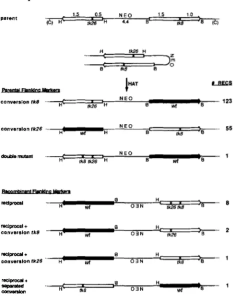
H iksFIGURE 1 .-Schematic representations of recombination sub- strate and recombination products selected in HAT media. Indi- cated are restriction sites for enzymes Hind111 (H), BamHI (B) and ClaI (C) in the original pJS-4 plasmid used to generate the cell lines in this study. Amowheadr represent directions of transcrip- tion of HSV tk sequences to emphasize the inverted nature of the duplication construct. The tk alleles flank the dominant selectable marker (NE0 = resistance to G-418) used to generate cell lines. Alleles tk8 and tk26 are indicated at the site ( X ) of the XhoI linker insertion mutations. Among recombinants, wild-type genes, which no longer contain X h I linker insertions, are shaded. Recombinants are separated into two categories according to flanking marker configurations (positions of HindIII and BamHI sites). Products with parental flanking markers have unaltered junction fragments (established as in Figure 2) with one tk fragment flanked by HindIII sites and one fragment flanked by BamHI sites, as in the “parent.” Reciprocal recombinants have recombinant flanking markers (each tk fragment is flanked by Hind111 and BamHI sites) resulting from inversion of interstitial sequences, which is depicted by inverting NEO. Conversion products are identified by the loss of the XhoI restriction site marking a given allele (e.g., allele tR8 is lost in “conversion tk8”). Number of recombinants (#RECS) cor- respond to data tabulated in Table 2.
LETSOU and LISKAY 1986, 1987), indicating that the recombination rate is intrinsic to a given construct and not heavily influenced by the site of integration in the Ltk- cell genome. The rates for inverted repeats observed in this study (mean rate =
1.2
xl o p 6 ) were slightly lower than rates observed for the same pair of alleles arranged as direct repeats (mean rate = 3.6 X LISKAY, S T A C H E L E K ~ ~ ~ LETSOU
1984; LETSOU and LISKAY 1986).
164 R. J. Bollag a n d R. M. Liskay
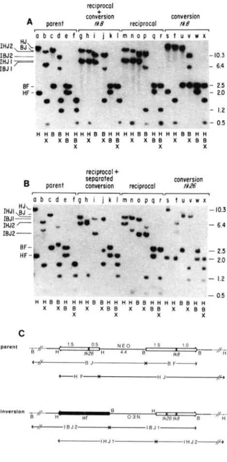
FIGURE 2.-Analysis of inverted-repeat recombinants. A, Mo-
lecular hybridization analysis of representative recombinants from parent line 1. DNAs from the H A T parent line and from the indicated HAT‘ recombinants were digested with the enzymes indicated below each lane, separated by electrophoresis through agarose and probed with HSV fk-specific probe as described in MATERIALS ASD METHODS. Restriction enzyme abbreviations are as in Figure 1. Examination of junction fragments provides flanking
marker phenotype. The parent and conversion recombinants
retain the same junction fragments (HJ for HindIII digestion, lanes a and s; BJ for BamHI digestion, lanes c and u). Reciprocal recombinants possess novel junctions as predicted for inversion (IHJl and IHJ2 for HindIII digestion, lanes g and m; IBJl and IBJ2 for EamHI digestion, lanes i and 0 ; see interpretation of
junctions in Figure 2C). Examination of resistance toXhoI digestion indicates wild-type tk sequence and sites of sequence correction. In “conversion fk8” (lanes s-x) the EamHI fragment (BF, lanes u-
x; sequences contained in HJ, lanes s, t) is resistant to digestion by XhoI, indicating that the f k 8 allele has been corrected to wild type, whereas the HindIII fragment (HF, lanes s and w; sequences contained in BJ, lane u) is sensitive to XhoI digestion, indicating the presence of only the fk26 mutation by comparison with the “parent.” In inversion recombinants [lanes g-r; depicted for simple inversion in (C)], wild-type sequences resistant to XhoI digestion are contained in the 2.5-kb fragment flanked by Hind111 and BamHI sites (IHJl, IBJ2 and BF). The accompanying gene is sensitive to XhoI. In the case of simple “reciprocal” recombinants the accompanying gene (e.g., on fragment IBJl in lane 0) contains
both alleles, t k 8 and fk26, and cuts twice with XhoI. There are three resultant bands in lanes n and r, but two of these comigrate on the gel at 0.5 kb (doublet; actual sizes 528 bp and 485 bp). By contrast in the “reciprocal
+
conversion fk8” the accompanying gene retains only the fk26 allele and cuts only once to give identical fragments (lane I) as for the “conversion fk8” (lane x). Marker sizes at the right are given in kb. Fragment designations at the left areas described in (C). E , Molecular hybridization analysis of repre- sentative recombinants from parent line 2. The restriction diges- tions for each lane are the same as those in (A). Junction fragments are of different sizes for parent line 2 than for parent line 1 indicating a different site of integration in the genome. Flanking marker analysis is the same as in Figure 2A. In “conversion tk26” (lanes s-x) the 2.0-kb HF fragment has been corrected to wild type and does not cleave with XhoI (lanes s, t, w and x), while the 2.5-kb BF fragment retains only the fk8 allele. Analysis of the simple “reciprocal” (lanes m-r) is the same as in (A). The recom- binant labeled “reciprocal
+
separated conversion” (lanes g-I) is an inversion recombinant based on flanking marker analysis, but in contrast to the simple “reciprocal” discussed in A, the wild-type gene is contained in the 2.0-kb fragment flanked by Hind111 andBamHI sites (IHJ2, IBJl and HF). The accompanying gene
possesses the t k 8 allele and cleaves once with XhoI to generate fragments (lane I) of sizes identical to those obtained with conver- sion fk26 (lane x). For further discussion see the text. C, Interpre- tation of junction fragments for molecular hybridization analyses. Allele representations and notations are as described for Figure 1. Double bars represent unknown sequence between known plasmid sequences and the indicated cellular restriction sites, which vary among cell lines due to different sites of integration into the genome. Junction fragments are abbreviated as follows: HJ =
HindIII junction; BJ = BamHI junction; IHJ = inversion Hind111
junction; IBJ = inversionBamHIjunction; BF = EamHl fragment;
Recombining Inverted Repeats 165
repeats, independent HAT‘ segregants were ana-
lyzed by molecular hybridization techniques. In all cases the functional or wild-type tk gene could be identified by the diagnostic loss of a XhoI restriction site which marks the site of the linker insertion mutation. T h e majority of recombination products analyzed (178 of 190 or 94%; see Table 2) retained the accompanying gene in the parental configuration without gain of the XhoI mutation from the recon- structed wild type gene, as expected for gene con- version. Segregants which exhibited the 2.5-kb BamHI fragment as wild type and retained the ac- companying 2.0-kb HindIII fragment with the tk26 mutation (as depicted in Figure 1) are classified as conversions of the tk8 mutation in Table 2. Hybrid- ization analysis of such a recombinant from parent line 1 is presented in Figure 2A, lanes s-x. Similarly, recombinants which had the 2.0-kb HindIII fragment as wild type and retained the tk8 mutation in the accompanying 2.5-kb gene are classified as conver- sions of the tk26 mutation (see Figure 1 and lanes s- x in Figure 2B).
A reproducible observation is that conversions of tk8 occur roughly twice as often as conversions of tk26 (1 24 conversions tk8 to 54 conversions tk26; see Table 2). This discrepancy is consistent among all inverted repeat lines studied, but at present we have no explanation for this observation.
A second class of recombinants (12 of 190 or 6%),
tabulated in Table 2 as reciprocal events, were con- sistent with an intrachromatid reciprocal exchange in that the 2.5-kb and 2.0-kb HSV tk-containing segments were each flanked by one HindIII site and one BamHI site; ie., the flanking markers were exchanged. Individual BamHI or HindIII digests of these recombinants demonstrated that junctions of novel sizes were now associated with each known restriction site (IBJ1 and IBJ2 in the case of BamHI, IHJl and IHJ2 in the case of HindIII; see Figures 2A and 2B, lanes g-r and Figure 2C). Further restriction hybridization analyses on a number of these recombinants confirmed that these products were the result of an intrachromosomal reciprocal exchange between the two genes generating an in- version of chromosomal sequences between the cross- over sites. These reciprocal exchanges most likely represent intrachromatid events, since single recip- rocal exchanges involving unequally paired sister chromatids should generate unstable, aberrant
chromsomes in which only the wild type gene has recombinant flanking markers (as depicted in Figure
3).
Reciprocal exchange associated with gene con- version: A subset of the reciprocal class of recombi- nants showing inversion of internal sequences also provided evidence for a nonreciprocal aspect to the recombination event. A single crossover in the inter-
. .
. .
: :
. .
1
ti
e
’
tk8’
s
.
tk26 Ik8?
+
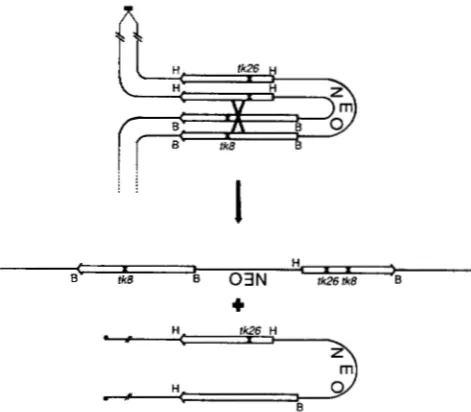
FIGURE 3.-Consequences of reciprocal exchange between in- verted genes on unequally paired sister chromatids. Allele repre- sentations and notations are as described for Figure 1. Duplicated centromeres (filled boxes) are depicted in one of two possible orientations. As drawn, the wild-type gene would be generated on a dicentric chromatid with all centromere-proximal sequences duplicated and all sequences distal to the repeats deleted. In the reversed centromere configuration (not depicted), the wild-type gene would be generated on the acentric chromatid with all centromere-distal sequences duplicated and proximal sequences deleted. Neither chromatid is expected to segregate properly at mitosis. These products should be distinguishable from inversion recombinants, since only one allele displays recombinant flanking markers (H and B sites as indicated). As expected no such recombinants were recovered.
Val between the two mutations without gene conver- sion at either mutant site would be expected to produce a viable wild type sequence on a 2.5-kb fragment flanked by HindIII and BamHI sites. T h e accompanying 2.0-kb gene, also flanked by HindIII and BamHI sites, would retain both mutations. Such products are termed simple reciprocal exchanges and are diagramed as “reciprocal” in Figure 1. However,
4
of 12 inversion products retained only one mutation in the nonfunctional gene, consistent with a nonre- ciprocal process at the other mutant site associated with the crossover. In two recombinants, the tk8mutation originally on the 2.5-kb fragment was lost, and in another case the tk26 mutation originally on the 2.0-kb fragment was lost. An additional recom- binant in the latter class was observed in line
4,
but is not included in Table 2, since the complexity of line 4 (4 copies of the duplication) made accurate analysis of all recombinants difficult. These products, diagramed in Figure 1, are most easily explained by a reciprocal exchange with associated conversion of the tk8 mutation (“reciprocal+
conversion tk8”;molecular analysis presented in Figure 2A, lanes g-
166 R. J. Bollag and R. M. Liskay tk26 mutation (“reciprocal
+
conversion t u b ” ) ,respectively.
Simple crossovers and the associated events de- scribed above resulted in the wild type gene on a 2.5- kb fragment. In both these latter events, a gene conversion tract is presumed to have occurred adja- cent to the crossover. One product which involved sequence inversion was recovered in which the 2.0-
kb fragment was resistant to XhoI digestion and thus wild type (“reciprocal
+
separated conversion” in Figures 1 and 2B). T h e accompanying 2.5-kb frag- ment retained only the tk8 mutation. Such a product is most easily explained by a conversion event at tk26separated from the crossover site as discussed below. Similar types of products have also been observed in experiments designed to recover recombinants be- tween directly repeated genes (R. J. BOLLAG, J. L. STACHELEK and R. M . LISKAY, unpublished results).
DISCUSSION
T h e results of this investigation show that homol- ogous recombination between inverted repeats in mouse cells occurs at a rate of approximately 1.2 x
This rate, albeit slightly reduced, is not mark- edly different from the rate of 3.6 X l o p 6 observed with the same gene pair arranged as direct repeats
(LISKAY, STACHELEK and LETSOU 1984; LETSOU and
LISKAY 1986). The large majority of products seen here (94%) are consistent with a nonreciprocal re- combination process in that flanking marker ex- change (via inversion) was not seen. T h e remaining products can be explained by a reciprocal process (crossover) leading to flanking marker exchange and sequence inversion. One third of these crossovers were accompanied by gene conversion at one of the two mutant sites.
Among products of recombination between in- verted repeats, approximately 6% (12 of 190) were consistent with intrachromatid reciprocal exchange, while none were consistent with unequal sister chro- matid reciprocal exchange. This was not a surprising observation, since reciprocal exchange following un- equal pairing of sister chromatids would lead in the case of inverted repeats to mitotically unstable chro- mosomes with duplications and deficiencies, as de- picted in Figure 3. Studies of direct repeats suggest that 15% of all recombination events are reciprocal in nature, while the remaining events resemble gene conversion (LISKAY, STACHELEK and LETSOU 1984). In those studies, products of both sister chromatid and intrachromatid events could be recovered. T h e inability to recover products of sister chromatid events between inverted repeats may account for the reduced proportion of reciprocal exchanges observed in the present study as compared to those observed between direct repeats.
FIGURE 4.-Model for antiparallel pairing of sister chromatids across the region of the inverted repeats. Alleles are depicted as in Figure 1. Loops represent 4.4-kb sequences between tk alleles which are not in register in this pairing configuration. This mode
of interaction as an alternative to intrachromatid exchange would require a conversion tract (or double crossovers) spanning the stippled region to generate the observed majority class of inversion recombinants (“simple reciprocal” in Table 2).
Although the simplest explanation for the inversion class of recombinants is a single intrachromatid cross- over, there exists the formal possibility that these recombinants could arise following antiparallel mis- pairing of sister chromatids in the region of the duplication, such that both alleles on both chromatids are paired (depicted in Figure 4). Recombination following such pairing could be executed in either of two ways: by a gene conversion involving all four genes and including the 4.4 kb of nonhomology between the alleles or by double crossovers with one exchange in each gene pair. Both of these explana- tions require correction of a large nonhomology
(it?.,
4.4 kb). Meiotic conversions of large deletions and insertions occur in yeast and models have been de- tailed to account for these ( e . g . , R A D D I N G 1979). In recent yeast studies such intrachromosomal conver- sions spanning large nonhomologies have been pro- posed to account for “plasmid conversion” between duplicated ADE8 alleles during meiosis (MALONEYand FOGEL 1987) and for mitotic rearrangements involving repeated delta sequences at the SUP4 locus
(ROTHSTEIN, HELMS and ROSENBERG 1987). However, recent results from our laboratory suggest that in mammalian cells, removal of insertions exceeding 1 kb occurs at least two orders of magnitude less efficiently than correction of small insertions of 1-8 bp (LETSOU and LISKAY 1987). Thus, we believe that the complex pairing configuration shown in Figure 4 is not likely to account for the observed inversions.
Recombining Inverted Repeats 167
could have involved double exchanges between sister chromatids. We have no prior evidence for intrach- romatid interactions in mammalian cells. Others have described recombination products between direct re- peats consistent with unequal sister chromatid ex- changes (SMITH and BERG 1984; STRINGERd al. 1985).
The results of this study combined with those re- ported previously (LISKAY, STACHELEK and LETSOU
1984) suggest that both intrachromatid and sister chromatid interactions contribute to intrachromoso- mal recombination. If double exchange events rather than gene conversions are responsible for the major class of recombinants, then we should have frequently observed recombinants in which intrachromatid dou- ble exchanges resulted in a double mutant gene accompanying the wild type recombinant without sequence inversion. Among over 190 products ex- amined, only one recombinant consistent with this interpretation was obtained (diagramed as “double mutant” in Figure 1). We prefer to explain this single event by symmetric heteroduplex covering the two mutations with independent correction on each chro- matid, rather than as a double reciprocal intrachro- matid exchange. Furthermore, events consistent with intrachromatid double exchanges were rarely (pos- sibly one in greater than 200 total events) observed in previous studies with direct-repeat constructs (LIS-
KAY, STACHELEK and LETSOU 1984; LISKAY and
STACHELEK 1986; LISKAY, LETSOU and STACHELEK 1987; LETSOU and LISKAY 1987). Therefore, we con- clude that the class of recombinants exhibiting no flanking marker exchange represent nonreciprocal events.
Further evidence for nonreciprocality in the present recombinant analysis is provided by the con- comitant loss of one of the original mutations among one third of recombinants showing inversion (4 of
12 events). These products are most easily explained by a reciprocal exchange leading to inversion accom- panied by nonreciprocal transfer of information at one of the two mutant sites. Since the overall fre- quency of all recombination events is approximately it is unlikely that two independent events would be picked up in a single segregant. Therefore, we favor the notion that the two processes are associated and likely represent alternative resolutions to a single recombination event, as has been postulated for meiotic recombination in fungi (for a review see ORR- WEAVER and SZOSTAK 1985). If reciprocal exchange is always accompanied by gene conversion proceeding by heteroduplex formation at one of the two sites, and if correction at the mismatched site is unbiased
( L e . , restoration is as likely as conversion), then we would predict that one third of the total reciprocal events should display associated conversion. Our results are in accord with this prediction. Although our sample size is small and our knowledge of average
conversion tract lengths in this system is limited
(LISKAY and STACHELEK 1986), we conclude that most and possibly all reciprocal exchanges between re- peated sequences in mouse cells are associated with nonreciprocal information transfer.
Among the inversions with associated conversion is included a recombinant depicted in Figures 1 and
2B as “reciprocal
+
separated conversion.” This recombinant is difficult to explain by a single event involving conversion contiguous to the site of crossing over. Rather, our interpretation is that conversion occurred at the site of the tk26 allele but the event was resolved as a crossover distal to the tk8 allele. Similar reciprocal recombinants with separated con- version tracts have been observed in fungi, and possible explanations are discussed by WHITEHOUSE (1982) and by ORR-WEAVER and SZOSTAK (1985).Studies by AYARES et al. (1985), in which extrachro- mosomal reciprocal recombinations between auton- omously replicating plasmids generated dimers whereby both interacting molecules could be exam- ined, yielded similar evidence for conversion associ- ated with reciprocal exchange. However, extrachro- mosomal recombination frequencies are sufficiently high that such products may actually represent mul- tiple events.
168 R. J. Bollag and R. M. Liskay
is unlikely. Because reciprocal events that are not necessarily conservative (between direct repeats)
occur at only a slightly increased frequency over reciprocal events that are obligatorily conservative (between inverted repeats), we conclude that in con- trast to extrachromosomal events, intrachromosomal recombination proceeds by a conservative mecha- nism. Earlier studies in our lab have suggested that extra- and intrachromosomal recombination differ mechanistically in other respects, such as a differ- ential sensitivity to base-pair mismatch (WALDMAN
and LISKAY 1987).
It is of interest to compare these results with those of similar studies in yeast and bacteria. Specifically, the proportions of reciprocal and gene conversion recombinants in these studies are similar to propor- tions of such events between duplicated HIS4 alleles during mitotic recombination in yeast (JACKSON and
FINK 1981). However, during meiotic recombination between repeated genes there is a marked deficit of conversions associated with reciprocal exchange
(KLEIN and PETES 198 1 ; KLAR and STRATHERN 1984; KLEIN 1984; JACKSON and FINK 1985). The similarity in frequencies of reciprocal recombination between inverted repeats (this study) and between direct re- peats (LISKAY, STACHELEK and LETSOU 1984; LETSOU
and LISKAY 1986) apparently does not hold in Sal-
monella typhimurium where inversion recombination occurs at lower frequencies than recombination be- tween direct repeats (SEGALL and ROTH 1986).
This report presents results similar to those of
WILLIS and KLEIN (1987) who, working with yeast, directly selected intrachromosomal inversions. These authors found that the inversions (i.e., reciprocal exchanges) are frequently associated with gene con- version at one or more scorable sites within the repeats. Similarly, BORTS and HABER (1987) and SYM- INGTON and PETES (1988) often found gene conver-
sion in the vicinity of reciprocal exchanges during meiotic recombination in yeast. Association between gene conversion and reciprocal exchange is a com- mon (FOGEL et al. 1979; ORR-WEAVER and SZOSTAK
1985) though not universal (KLEIN and PETES 198 1;
KLEIN 1984; JACKSON and FINK 198 1 , 1985; ROMAN
and FABRE 1983; ROSSIGNOL et al. 1984) feature of fungal recombination. The frequent association has suggested that the two types of events are mechan- istically related. This relationship is incorporated into the current models for recombination (MESELSON and
RADDINC 1975; SZOSTAK et al. 1983). Our study sug-
gests that chromosomal recombination in mammalian cells shares this fundamental aspect of the mechanism with fungal recombination.
In summary, our studies using closely linked in- verted repeats in mouse cells allow us to formulate several conclusions concerning intrachromosomal re- combination. First, recombination between inverted
repeats is similar in rate and products to recombi- nation between direct repeats. Second, reciprocal exchanges can involve either intrachromatid or sis- ter chromatid interactions. Third, reciprocal and nonreciprocal exchanges are frequently associated and are therefore likely to share a common mecha- nism. Finally our results argue that intrachromosomal recombination is a conservative process.
The authors thank A. WALDMAN, A. GODWIX, J. STACHELEK, and W. BOLLAG for helpful comments during the course of these studies, J. STACHELEK for excellent technical assistance, M. SEID-
MAN for a stimulating discussion on conservative us. nonconserva- tive recombination, and C. RADDING for valuable criticisms of the manuscript. This investigation was supported by Public Health Service grant R01 GM 32741 to R.M.L. R.J.B. was supported by a National Research Service Award (GM07499) to Yale University. R.M.L. is a Leukemia Society of America Scholar.
LITERATURE CITED
AYARES, D., J. SPESCER, F. SCHWARTZ, B. MORSE and R. KUCHER- LAPATI, 1985 Homologous recombination between autono-
mously replicating plasmids in mammalian cells. Genetics 11 1:
BORTS, R. H., and J. E. HABER, 1987 Meiotic recombination in yeast: alteration by multiple heterozygosities. Science 237:
CAPECCHI, M. R., 1980 High efficiency transformation by direct microinjection of DNA into cultured mammalian cells. Cell
22: 479-488.
CARPENTER, A. T. C., 1984 Meiotic roles of crossing-over and of gene conversion. Cold Spring Harbor Symp. Quant. Biol. 49:
CHAKRABARTI, S., and M. M. SEIDMAS, 1986 Intramolecular recombination between transfected repeated sequences in mammalian cells is nonconservative. Mol. Cell. Biol. 6 : 2520- 2526.
FOGEL, S., R. MORTIMER, K. LusxAKand F. TAVARES, 1979 Meiotic gene conversion: a signal of the basic recombination event in yeast. Cold Spring Harbor Symp. Quant. Biol. 43: 1325-1341. JACKSOS, J. A,, and G. R. FINK, 1981 Gene conversion between
duplicated genetic elements in yeast. Nature 292: 306-31 1. JACKSON, J. A., and G. R. FINK, 1985 Meioic recombination
between duplicated genetic elements in Saccharomyces cerevisiae.
Genetics 109: 303-332.
KLAR, A. J. S., and J. N. STRATHERN, 1984 Resolution of recom- bination intermediates generated during yeast mating type switching. Nature 310: 744-748.
KLEIN, H. L., 1984 Lack of association between intrachromosomal gene conversion and reciprocal exchange. Nature 310: 748- 753.
KLEIN, H. L., and T. D. PETES, 1981 Intrachromosomal gene conversion in yeast. Nature 289: 144-148.
LETSOU, A,, and R. M. LISKAY, 1986 Intrachromosomal recom- bination in mammalian cells. pp. 383-409. In: Gene Transfer,
Edited by R. KUCHERLAPATI. Plenum, New York.
LETSOU, A., and R. M. LISKAY, 1987 Effect of the molecular nature of mutation on the efficiency of intrachromosomal gene conversion in mouse cells. Genetics 117: 759-769. LIS, F.-L., and N. STERSBERG, 1984 Homologous recombination
between overlapping thymidine kinase gene fragments stably inserted into a mouse cell genome. Mol. Cell. Biol. 4: 852- 861.
LISKAY, R. M., and R. J. E v ~ s s , 1980 Inactive X chromosome 375-388.
1459-1465.
Recombining Inverted Repeats 169
DNA does not function in DNA-mediated cell transformation for the hypoxanthine phosphoribosyltransferase gene. Proc. Natl. Acad. Sci. USA 77: 4895-4898.
LISKAY, R. M., and J. L. STACHELEK, 1983 Evidence for intra- chromosomal gene conversion in cultured mouse cells. Cell
LISKAY, R. M., and J. L. STACHELEK, 1986 Information transfer between duplicated chromosomal sequences in mammalian cells involves contiguous regions of DNA. Proc. Natl. Acad. Sci. USA 83: 1802-1806.
LISKAY, R. M., A. LETSOU and J. L. STACHELEK, 1987 Homology requirement for efficient gene conversion between duplicated chromosomal sequences in mammalian cells. Genetics 115: 35: 157-165.
161-167.
LISKAY, R. M.,J. L. STACHELEKand A. LETSOU, 1984 Homologous recombination between repeated chromosomal sequences in mouse cells. Cold Spring Harbor Symp. Quant. Biol. 49: 183- 189.
MALOSEY, D. H., and S. FOGEL, 1987 Gene conversion, unequal crossing-over and mispairing at a non-tandem duplication during meiosis of Saccharomyces cerevisiae. Curr. Genet. 12: 1- 7.
MESELSOS, M. S., and C. M. RADDISG, 1975 A general model for genetic recombination. Proc. Natl. Acad. Sci. USA 72: 358- 361.
ORR-WEAVER, T. L., and J. W. SZOSTAK, 1985 Fungal recombi- nation. Microbiol. Rev. 49: 33-58.
RADDISG, C. M., 1979 T h e mechanism of conversion of deletions and insertions. Cold Spring Harbor Symp. Quant. Biol. 43:
ROMAS, H., and F. FABRE, 1983 Gene conversion and associated reciprocal recombination are separable events in vegetative cells of Saccharomyces cerevisiae. Proc. Natl. Acad. Sci. USA 80:
ROSSIGSOL, J.-L., A. NICOLAS, H. HAMZA and T. LASGIS, 1984 Origins of gene conversion and reciprocal exchange in
Ascobolus. Cold Spring Harbor Symp. Quant. Biol. 49: 13-21. ROTHSTEIS, R., C. HELMS and N. ROSESBERG, 1987 Concerted deletions and inversions are caused by mitotic recombination between delta sequences in Saccharomyces cerevisiae. Mol. Cell. Biol. 7: 1198-1207.
RCBSITZ, J., and S. SUBUMASI, 1986 Extrachromosomal and 1315-1316.
6912-6916.
chromosomal gene conversion in mammalian cells. Mol. Cell. Biol. 6: 1608-1614.
SEIDMAS, M. M., 1987 Intermolecular homologous recombination between transfected sequences in mammalian cells is primarily nonconservative. Mol. Cell. Biol. 7: 3561-3565.
SEGALL, A. M., and J. R. ROTH, 1986 Restriction on the formation of spontaneous inversion rearrangements in Salmonella typhi- murium. Genetics 113: s14.
SMITH, A. J. H., and P. BERG, 1984 Homologous recombination between defective neo genes in mouse 3T6 cells. Cold Spring Harbor Symp. Quant. Biol. 49: 171-181.
STRINGER, J. R., R. M. KUHN, J. L. NEWMAS and J. C. MEADE, 1985 Unequal homologous recombination between tandemly arranged sequences stably incorporated into cultured rat cells. Mol. Cell. Biol. 5 : 2613-2622.
SUBRAMMAXI, S., and J. R~BSITZ, 1985 Recombination events after transient infection and stable integration of DNA into mouse cells. Mol. Cell. Biol. 5 : 659-666.
SYMINGTOS, L. S., and T. D. PETES, 1988 Expansions and con- tractions of the genetic map relative to the physical map of yeast chromosome ZZZ. Mol. Cell. Biol. 8: 595-604.
SZOSTAK, J. W., T. L. ORR-WEAVER, R. J. ROTHSTEIS and F. W. STAHL, 1983 T h e double-strand-break repair model for re- combination. Cell 33: 25-35.
SZYBALSKI, W., E. H. SZYBALSKA and G. RAGSI, 1962 Genetic studies with human cell lines. Natl. Cancer Inst. Monogr. 7:
WAGNER, M. J., J. A. SHARPand W. C. SUMMERS, 1981 Nucleotide sequence of the thymidine kinase gene of herpes simplex virus type 1 . Proc. Natl. Acad. Sci. USA 78: 1441-1445.
WALDMAS, A. S., and R. M. LISKAY, 1987 Differential effects of base-pair mismatch on intrachromosomal versus extrachro- mosomal recombination in mouse cells. Proc. Natl. Acad. Sci. USA 84: 5340-5344.
WHITEHOUSE, H. L. K., 1982 Genetic Recombination: Understanding
the Mechanism. John Wiley & Sons, New York.
WILLIS, K. K., and H. L. KLEIS, 1987 Intrachromosomal recom- bination in Saccharomyces cerevisiae: reciprocal exchange in an inverted repeat and associated gene conversion. Genetics 117: 633-643.
75-88.