Copyright1999 by the Genetics Society of America
In Vivo
Analysis of the Domains of Yeast Rvs167p Suggests Rvs167p Function
Is Mediated Through Multiple Protein Interactions
Karen Colwill,* Deborah Field,*
,†Lynda Moore,* James Friesen*
,†and Brenda Andrews*
*Department of Molecular and Medical Genetics and†Banting and Best Department of Medical Research, University of Toronto, Toronto, Ontario M5S 1A8, Canada
Manuscript received December 15, 1998 Accepted for publication March 18, 1999
ABSTRACT
Morphological changes during cell division in the yeast Saccharomyces cerevisiae are controlled by cell-cycle regulators. The Pcl-Pho85p kinase complex has been implicated in the regulation of the actin cytoskeleton at least in part through Rvs167p. Rvs167p consists of three domains called BAR, GPA, and SH3. Using a two-hybrid assay, we demonstrated that each region of Rvs167p participates in protein-protein interactions: the BAR domain bound the BAR domain of another Rvs167p protein-protein and that of Rvs161p, the GPA region bound Pcl2p, and the SH3 domain bound Abp1p. We identified Rvs167p as a Las17p/Bee1p-interacting protein in a two-hybrid screen and showed that Las17p/Bee1p bound the SH3 domain of Rvs167p. We tested the extent to which the Rvs167p protein domains rescued phenotypes associated with deletion of RVS167: salt sensitivity, random budding, and endocytosis and sporulation defects. The BAR domain was sufficient for full or partial rescue of all rvs167 mutant phenotypes tested but not required for the sporulation defect for which the SH3 domain was also sufficient. Overexpression of Rvs167p inhibits cell growth. The BAR domain was essential for this inhibition and the SH3 domain had only a minor effect. Rvs167p may link the cell cycle regulator Pcl-Pho85p kinase and the actin cytoskeleton. We propose that Rvs167p is activated by phosphorylation in its GPA region by the Pcl-Pho85p kinase. Upon activation, Rvs167p enters a multiprotein complex, making critical contacts in its BAR domain and redundant or minor contacts with its SH3 domain.
T
HE commitment to enter a new cell division cycle Measday1998). Pho85p is activated by association with 10 different Pho85p cyclins (Pcls) that are divided into occurs during the G1phase in most eukaryotic cells(Pardee1989;Cross1995). In the budding yeast Sac- two subfamilies: PHO80 and PCL1,2 (Measday et al.
1997). Members of the PHO80 subfamily (PHO80, PCL6,
charomyces cerevisiae, the point of commitment to
cellu-lar division is termed Start and is analogous to the re- PCL7, PCL8, and PCL10) are required to regulate
phos-phate and glycogen metabolism (Andrewsand
Meas-striction point in mammalian cells (Planas-Silvaand
Weinberg1997). Passage through Start leads to mor- day1998). Several observations suggest that the PCL1,2 subfamily (PCL1, PCL2, CLG1, PCL5, and PCL9) regu-phological changes in the yeast cell that allow polarized
growth toward the budding daughter cell (Lew and lates early cell-cycle events. First, the Pcl1,2-Pho85p ki-nase is essential for cell-cycle progression in the absence
Reed1995). The actin cytoskeleton becomes polarized
toward the new bud in late G1and is believed to direct of Cln1p and Cln2p, suggesting a role for the
Pcl1,2-Pho85p kinases at Start (Espinozaet al. 1994;Measday
growth and secretion toward the bud (Lewand Reed
1995). et al. 1994). Second, expression of three members of
the PCL1,2 subfamily (PCL1, PCL2, PCL9) is cell-cycle Two cyclin-dependent kinase (CDK) complexes,
Cln-Cdc28p and Pcl-Pho85p, have been implicated in signal- regulated with peak transcript levels occurring in G1
(AndrewsandMeasday1998). Third, yeast strains lack-ing cell-cycle position to the actin cytoskeleton.
Im-proper expression of Cln-Cdc28p complexes, the master ing the entire PCL1,2 subfamily display morphological defects that include elongated buds, delocalized actin cell-cycle activators in yeast, alters the timing of bud
emergence, suggesting that Cln-Cdc28p complexes are patches, and abnormal (random) budding in diploids, involved in regulating this process (Lewet al. 1992;Lew implying that the PCL1,2 subfamily may regulate actin andReed1993;Nasmyth1993;Nigg1995). The Pcl- polarization during the cell cycle (Measdayet al. 1997;
Pho85p kinase has roles in both cellular metabolism Leeet al. 1998;Tennysonet al. 1998).
and in cell-cycle regulation (reviewed inAndrewsand Recent experiments suggest that Pcl1,2-Pho85p ki-nases contribute to actin regulation at least in part through the Rvs167 protein. RVS167 and a similar gene,
Corresponding author: Brenda Andrews, Department of Molecular and RVS161, were first identified in a screen for mutations
Medical Genetics, University of Toronto, Rm. 4285, Medical Sciences
that caused reduced viability upon starvation (Crouzet
Bldg., 1 Kings College Circle, Toronto, Ontario M5S 1A8, Canada.
E-mail: [email protected] et al. 1991; Bauer et al. 1993). Deletion of either of
these genes leads to phenotypes similar to those seen of Rvs167p and Rvs161p has also been shown by copre-cipitation from yeast extracts (Navarro et al. 1997).
in actin mutants (Ayscough and Drubin1996;
Bot-stein et al. 1997): (1) loss of viability and unusual cell Thus, it appears that Rvs167p is capable of forming several protein contacts and may participate in multiple morphology in poor growth or salt-containing media
(Crouzetet al. 1991;Baueret al. 1993;Leeet al. 1998); protein complexes involved in regulating the actin cy-toskeleton.
(2) delocalized actin patches under suboptimal growth
conditions (Baueret al. 1993;Sivadonet al. 1995;Lee Two mammalian genes, Amphiphysin I and II, are similar to RVS167 (WiggeandMcMahon1998). Both
et al. 1998); (3) random budding in diploids (Baueret
al. 1993;Sivadonet al. 1995;Leeet al. 1998); (4) endo- Amphiphysin I and II are involved in endocytosis of synaptic vesicles at nerve termini (Wigge and
McMa-cytic defects (Munnet al. 1995;Leeet al. 1998); and (5)
sporulation defects for rvs161 (Desfargeset al. 1993). hon1998). Amphiphysin I is required for cytoskeleton rearrangement during neurite outgrowth (Mundiglet
Yeast deleted for RVS161, but not RVS167, also displays
cell fusion defects (Dorer et al. 1997; Brizzio et al. al. 1998). Thus, Rvs167p and the Amphiphysin family
have similar functions. Interestingly, Amphiphysin I is 1998). Deletion of PHO85 or several members of the
PCL1,2 subfamily shows similar defects to rvs and actin an autoantigen in Stiff-Man Syndrome associated with breast cancer, and Amphiphysin I levels are often ele-mutants, suggesting that Pcl-Pho85p, Rvs167p, and actin
lie in the same pathway (Leeet al. 1998). vated in breast cancer tissue (Davidet al. 1994;Floyd
et al. 1998). A splice variant of Amphiphysin II, BinI,
Biochemical evidence also supports the hypothesis
that the Pcl-Pho85p kinase regulates Rvs167p. Rvs167p negatively regulates Myc and is lacking in several human tumors, suggesting it is a tumor suppressor (Sakamuro
was identified as a Pcl-binding protein in two
indepen-dent two-hybrid screens using Pcl2p and Pcl9p as bait et al. 1996). Thus, members of the Amphiphysin/RVS
family appear to be important for proper cell-cycle con-(Lee et al. 1998). Pcl2p and Rvs167p also interact in
an affinity chromatography assay in vitro and can be trol.
The structural similarity between the Amphiphysins coimmunoprecipitated from yeast extracts (Lee et al.
1998). In vitro, Rvs167p is a substrate of the Pcl2-Pho85p and the Rvs proteins is primarily in their N termini where they have a common domain, BAR (also known kinase and, in vivo, phosphoforms of Rvs167p are
re-duced in pho85 and multiple pcl mutants, suggesting as Rvs and Domain A; Bauer et al. 1993; David et al.
1994;Sakamuro et al. 1996). Rvs161p consists only of
that Rvs167p is an in vivo substrate of the Pcl-Pho85p
kinase (Lee et al. 1998). the BAR domain. There are two predicted coiled-coil motifs within this domain (Navarroet al. 1997). At the
The Rvs proteins likely form part of a multiprotein
complex that functions to regulate actin polarization. C termini of Rvs167p and the Amphiphysins, there is an SH3 domain, a protein module well defined for binding A mutant allele of RVS161, identified in a screen for
endocytosis mutants, shows noncomplementation with proline-rich sequences (PawsonandScott1997). The Rvs167p SH3 domain shows a two-hybrid interaction an actin mutant strain, suggesting that Rvs161p and
actin interact (Munn et al. 1995). In addition, rvs161 with actin and binds Abp1p (Amberget al. 1995;Lila
andDrubin1997). The middle portion of Rvs167p and and rvs167 mutations are synthetically lethal with a set
of actin mutations (BretonandAigle1998). Deletion the Amphiphysin proteins is not conserved. In Rvs167p, the central portion of the molecule consists of a GPA of RVS167 is also synthetically lethal with mutation of
MYO1, and growth is severely affected in an rvs167 myo2 (glycine-, proline-, alanine-rich) region that is also found in the actin-binding proteins Abp1p and Dictyostelium strain (Breton andAigle1998). Rvs167p binds actin
in the two-hybrid system (Amberget al. 1995); however, discoideum myosin I (Baueret al. 1993).
In this study, we analyzed the contribution of the BAR, the interaction of Rvs167p with actin may be indirect
as the two do not appear to interact in vitro (Lilaand GPA, and SH3 regions of Rvs167p to its function in vivo. First we used the two-hybrid system to map the regions
Drubin 1997). In vitro, Rvs167p binds actin-binding
protein 1 (Abp1p), and it is possible that Abp1p medi- of Rvs167p required for interactions with known part-ners. We also used a two-hybrid screen to identify ates the interaction between Rvs167p and actin (Lila
andDrubin 1997). Genetic analysis also suggests that Rvs167p as a Las17p/Bee1p binding partner. Second, we tested the ability of the domains of Rvs167p to res-Rvs167p and Abp1p operate in the same pathway.
Nei-ther RVS167 nor ABP1 is essential, but both mutants cue several loss-of-function phenotypes associated with
rvs167 mutants. Third, we tested the effect of
overex-display a similar profile of “synthetic-lethal” interactions
with genes encoding other cytoskeletal components pressing various regions of Rvs167p on cell viability. Finally, we suggest models for Rvs167p function based (SRV2, SLA1, SLA2/END4, SAC6; Lila and Drubin
1997). As well, deletion of RVS167 reduces the poor cell on these and previous results. growth and abnormal cell morphology of yeast cells
that overexpress Abp1p (Lila and Drubin 1997). In
MATERIALS AND METHODS
addition to binding actin and Abp1p, Rvs167p interacts
with Rvs161p and Sla2/End4p in the two-hybrid system Media and yeast strain manipulations:Yeast strains are listed
in Table 1. Standard rich medium (YEP) containing 2%
TABLE 1
Yeast strains used in this study
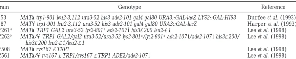
Strain Genotype Reference
Y153 MATa trp1-901 leu2-3,112 ura3-52 his3 ade2-101 gal4 gal80 URA3::GAL-lacZ LYS2::GAL-HIS3 Durfeeet al. (1993) Y187 MATatrp1-901 leu2-3,112 ura3-52 his3 ade2-101 gal4 gal80 URA3::GAL-lacZ Harperet al. (1993)
BY261a MATa TRP1 GAL2 ura3-52 lys2-801aade2-1078his3D200 leu2-D1 Leeet al. (1998)
BY262b MATa/aTRP1 GAL2/gal2 ura3-52/ura3-52 lys2-801a/lys2-801aade2-1078/ade2-1078his3D200/ Leeet al. (1998)
his3D200 leu2-D1/leu2-D1
BY508 MATa rvs167DTRP1 Leeet al. (1998)
BY561 MATa/arvs167DTRP1/rvs167DTRP1 ADE2/ade2-1078 Leeet al. (1998)
aThis strain and all following haploid strains are isogenic. bThis strain and all following diploid strains are isogenic.
cose and supplemented minimal medium containing 2% glu- TTGAATTCAGATCTAGCCGCGGCCGTAGCG. The follow-ing constructs were made to allow expression of RVS167 deriva-cose (SD) or 2% galactose (SG) were used (Kaiseret al. 1994).
To make SD plus NaCl, NaCl was added to SD to a final tives (see AD fusions for description of constructs) under control of the GAL1 promoter in vector p426 GAL1 (ATCC concentration of 6% w/v. For the sporulation assay, yeast cells
were initially grown in SD containing 10% glucose and then 87333;Ronickeet al. 1997): (1) pGAL-RVS167 (pBA1178); (2) pGAL-P473L (pBA1179); and (3) pGAL-GPASH3 (pBA1183). transferred to 1% potassium acetate supplemented with 0.253
amino acid mix minus methionine and histidine (Kaiser et Two-hybrid assay: Yeast strain Y153 was transformed with plasmids containing the Gal4 DNA-binding domain (pAS1) al. 1994). Standard methods for yeast transformations were
used (GuthrieandFink1991). For dot assays, cultures grown fusions. Yeast strain Y187 was transformed with plasmids con-taining Gal4 activation domain (pAD) fusions. Y153 and Y187 to midlog phase were serially diluted and spotted onto the
appropriate plates. containing these plasmids were then mated, and liquid
b-galactosidase assays were performed on the resulting
dip-Plasmids: Plasmids used in this study were generated by
PCR or restriction digest from source vectors and subcloned loids as described (AndrewsandMoore1992). The Las17p/ Bee1p two-hybrid screen was performed as described using into the vectors listed below. PCR products were sequenced
to confirm their integrity. Details of plasmid construction are yeast strain Y190 cotransformed with pAS1-LAS17/BEE1 and alACT yeast cDNA library (Durfeeet al. 1993;Harperet al. available upon request. The following constructs were made
as Gal4 activation domain fusions using vector pACT (Durfee 1993). A total of 150,000 independent transformants were selected on medium lacking Trp, Leu, and His but containing et al. 1993) or pACT II (Harperet al. 1993): (1) pAD-RVS167
(pBA1117, full length); (2) pAD-P473L (pBA1217, full-length 25 mmaminotriazole. Plasmids from positive clones were iso-lated and sequenced.
RVS167 with a mutation in codon 473). This construct was
created by in vitro overlap-extension PCR (LingandRobinson Western blotting:To prepare extracts for Western blotting, yeast cells were grown to log phase under inducing conditions 1997). The cytosine at the second position of codon 473 was
substituted for thymidine, changing proline 473 to leucine. (SG minus uracil for GAL vectors and SD minus methionine and histidine for MET vectors). Preparation of cell extracts The primers used for mutagenesis were universal and reverse
(from Stratagene, La Jolla, CA) and C2418T 59(GGTGTGT and Western blot analysis were performed as described (Lee et al. 1998).
TTCTTGGGAACTACG) and C2418T 39(CGTAGTTCCCAA
GAAACACACC). (Further details of mutagenesis are available Fluorescence microscopy:Lucifer yellow accumulation was performed as described except cells were grown in SD medium upon request.) (3) pAD-BARGPA (pBA1116, RVS167 codons
1–427 with a stop codon introduced at the end); (4) pAD- minus methionine and histidine and incubated with lucifer yellow for 1.5 hr (Leeet al. 1998). Calcofluor staining of bud GPASH3 (pBA1218, RVS167 codons 282–482); (5) pAD-BAR
(pBA1119, RVS167 codons 1–281 with a stop codon intro- scars was performed as described except the cells were grown in SD medium minus methionine and histidine and examined duced at the end); and (6) pAD-SH3 (pBA1259, RVS167
co-dons 423–482). at a magnification of31000 (Tennysonet al. 1998).
Sporulation:Cells were grown to midlog phase in supple-The following constructs were made as Gal4 DNA-binding
domain fusions using the vector pAS1 (Durfeeet al. 1993): mented medium (SD) plus 10% glucose minus methionine and histidine. Cells were washed once with distilled water and (1) pAS1-RVS167DSH3 (pBA1045, RVS167 codons 12–445);
(2) pAS1-BAR (pBA1219, RVS167 codons 1–281 with a stop resuspended in sporulation medium. The cells were shaken at 308for 4 days. The percentage of cells with two or four spores codon introduced at the end); (3) pAS1-RVS161 (pBA1121,
full length); (4) pAS1-PCL2 (pBA668; described inMeasday was determined by examining the cells at31000 magnification using a charge-coupled device camera mounted on a Leica et al. 1994); (5) pAS1-ABP1 PRO [pBA1122, ABP1 codons 444–
537; source of ABP1 PRO is pGABP1-2 (Lila and Drubin DM-LB microscope. 1997)]; and (6) pAS1-LAS17/BEE1 (pBA1101, full length).
The following constructs were made to allow expression of
RVS167 derivatives (see AD fusions for description of con- RESULTS structs) under control of the MET25 promoter in vector p413
MET25 (ATCC 87318;Ronickeet al. 1997): (1) pMET-RVS167 Interaction of Rvs167p with protein targets in the
two-(pBA1174); (2) pMET-P473L (pBA1175); (3) pMET-BARGPA hybrid assay: Evidence to date suggests that Rvs167p (pBA1176); (4) pMET-GPASH3 (pBA1177); (5) pMET-BAR interacts with several proteins and may be part of a large (pBA1213); and (6) SH3 (pBA1215). The
pMET-M-protein complex that regulates actin (Amberg et al. SH3 contains a single 59 Myc epitope tag generated during
1995;LilaandDrubin1997;Navarroet al. 1997;Wesp PCR amplificaton of the SH3 domain using primer Myc
TABLE 2
Interaction of Pcl2p with Rvs167p in the two-hybrid system
pAS1 pAS1-PCL2 Fold increase
Plasmid Miller unitsa Miller units in activation
pAD 0.0760.03 0.4760.07 6.7b
pAD-RVS167 0.2060.16 74.565.40 372
pAD-BARGPA 0.0460.03 28.065.61 700
pAD-GPASH3 0.0660.05 3.3960.09 56.5
pAD-BAR 0.1460.02 0.6460.10 4.6
ab-Galactosidase activity—seematerials and methodsand
Andrewsand Moore(1992); average of three independent cultures.
bFold increase is calculated by dividing the value of the
PAS1-PCL2 pAD-fusion interaction by the pAS1 pAD-infusion interaction value.
for interaction with Pcl2p. Because Pcl2p binds the BAR-GPA and the BAR-GPASH3 constructs, but not the BAR or SH3 domains individually, our data suggest that the GPA region of Rvs167p contains a binding site for Pcl2p. Yeast deleted for either RVS167 or RVS161 display
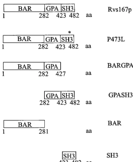
simi-Figure1.—Schematic diagram of the Rvs167p constructs.
lar phenotypes, and Rvs167p and Rvs161p interact in a
At the bottom of each construct is the amino acid position
two-hybrid assay (Baueret al. 1993;Navarroet al. 1997). that defines the boundaries between Rvs167p domains. The
asterisk on P473L indicates the position of the amino acid We used our Rvs167p domain fusions to assay
Rvs167p-substitution of proline 473 to leucine. Although the GPASH3 Rvs161p interactions in the two-hybrid system. As well, DNA construct begins at codon 282, the protein is predicted
we tested for Rvs167p-Rvs167p interactions using a Gal4
to start at methionine 286 in the MET25 and GAL1 vectors.
DB-Rvs167 fusion protein (pAS1-Rvs167DSH3) that
aa, amino acid.
lacks the SH3 domain of Rvs167p. We were unable to test full-length Rvs167p because it self-activated when fused to the Gal4 DNA-binding domain. In this assay, can be divided into three regions: the N-terminal BAR
both Rvs161p and Rvs167DSH3 interacted with full-domain, the GPA-rich region, and the C-terminal SH3
length Rvs167p, the BARGPA region, and the BAR do-domain (Figure 1). We sought to identify binding
part-main by itself (Table 3). A smaller construct expressing ners for individual regions of Rvs167p and to correlate
a Gal4 DB-BAR domain fusion of Rvs167p also bound the binding partners with the function of that region
full-length Rvs167p, the BARGPA region, and the BAR as determined by phenotypic complementation. To this
domain (Table 3). Therefore, it appears that Rvs167p end, we have used various constructs of Rvs167p in the
is capable of forming hetero- or homodimers through two-hybrid assay to test the binding of Rvs167p to
puta-its BAR domain. As noted above, other two-hybrid tive protein targets.
results have shown Rvs161p-Rvs167p heterodimers We have previously demonstrated that Rvs167p binds
but failed to show evidence of Rvs167p homodimers to Pcl2p, a cyclin component of the Pho85p kinase
(Navarroet al. 1997).
(Measdayet al. 1994;Leeet al. 1998). To better define
The SH3 domain of Rvs167p binds directly to a pro-the interaction between Rvs167p and Pcl2p, we
per-line-rich region of the actin-binding protein Abp1p in formed liquid b-galactosidase assays on transformants
a far-Western blot assay (Lilaand Drubin1997). We expressing Pcl2p fused to the Gal4 DNA-binding (DB)
used the two-hybrid assay to test the interaction of the domain (pAS1-Pcl2p) and various constructs expressing
SH3 domain of Rvs167p with the proline-rich region of fusions of Rvs167p to the Gal4 activation domain (pAD
Abp1p in vivo (pAS1-Abp1 PRO, Table 4). Abp1 PRO fusions, Table 2). Pcl2p interacted with full-length
interacted with full-length Rvs167p, the GPASH3 re-Rvs167p as well as the BARGPA and GPASH3 constructs.
gion, and the SH3 domain alone, confirming the in Pcl2p did not interact with either the BAR or SH3
do-vitro data (LilaandDrubin1997). We also constructed mains of Rvs167p alone (Tables 2 and 4). We were
a point mutation, P473L, within the SH3 domain of unable to detect the AD-GPA fusion or the GPA region
Rvs167p that is predicted to inactivate the SH3 domain alone in Western blotting of yeast extracts from
trans-(Clark et al. 1992; Musacchio et al. 1992;
Rozakis-formants expressing the GPA region from various
pro-Adcock et al. 1993). Abp1 PRO did not interact with
moters (data not shown). Therefore, we were unable
TABLE 3
Interaction of Rvs167p and Rvs161p with Rvs167p in the two-hybrid system
pAS1-RVS161 pAS1-RVS167DSH3 pAS1-BAR
pAS1
Plasmid Miller unitsa Miller units Fold increase Miller units Fold increase Miller units Fold increase
pAD 0.0560.02 0 0b 0.0960.04 1.8 0.1160.04 2.2
pAD-RVS167 0 26.0610.5 521 12.364.79c 247 2.4060.59 48.0
pAD-BARGPA 0 5.4360.69d 109 5.0860.42 102 2.3560.81 47.0
pAD-GPASH3 0.0360.04 0 0 0.0860.11c 1.6 0.0960.03 1.8
pAD-BAR 0.1060.06 8.5763.61 171 3.3660.96 67.2 1.3160.12 26.2
pAD-PCL2 0 0 0 NDe ND
ab-Galactosidase activity—see materials and methodsand Andrews and Moore (1992); average of three independent
cultures.
bFold increase is calculated by dividing the Miller units of the pAS1-fusion pAD-fusion interaction value by the interaction
value of pAS1-pAD (0.05).
cAverage of best three out of five values. dAverage of best three out of six values. eNot done.
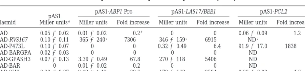
disrupts the function of the SH3 domain (Table 4). The the proline-rich region between amino acids 294 and 534 (data not shown). Thus, we propose that the SH3 P473L mutant remained competent to bind proteins
that interact with Rvs167p outside the SH3 domain (see domain of Rvs167p binds the proline-rich region of Las17p/Bee1p in vivo.
Pcl2p, Table 4).
Las17p/Bee1p, a proline-rich protein involved in ac- Expression analysis of Rvs167p constructs in vivo:
Our two-hybrid results suggest that Rvs167p can form tin regulation and endocytosis (Li 1997; Naqvi et al.
1998), may interact with SH3 domain-containing pro- several protein complexes and that the individual re-gions of Rvs167p are all likely to contribute to its func-teins involved in actin regulation. We performed a
two-hybrid screen with full-length Las17p/Bee1p and identi- tion. Yeast deleted for RVS167 displays several pheno-types including reduced viability upon starvation, salt fied Rvs167p as a Las17p/Bee1p-interacting protein
(data not shown). Seventeen clones encoding Rvs167p sensitivity, random budding in diploids, and endocytic defects (Baueret al. 1993;Munn et al. 1995;Sivadon
were isolated ranging from full length to one lacking
the first 212 amino acids. We tested the Gal4 DB- et al. 1995;Leeet al. 1998). To test the functional
impor-tance of the domains of Rvs167p, we expressed various Las17p/Bee1p against our AD-Rvs167p fusions and
found that the Las17p/Bee1p-Rvs167p interaction was RVS167 constructs (Figure 1) in vivo under control of
either the MET25 or GAL1 promoter. We anticipated dependent on the SH3 domain of Rvs167p (Table 4).
We also used the two-hybrid assay to demonstrate that that both the length of the Rvs167p constructs and their expression levels would affect the ability of the various the binding site for Rvs167p on Las17p/Bee1p is within
TABLE 4
Interaction of Abp1 PRO and Las17p with Rvs167p in the two-hybrid system
pAS1-ABP1 Pro pAS1-LAS17/BEE1 pAS1-PCL2
pAS1
Plasmid Miller unitsa Miller units Fold increase Miller units Fold increase Miller units Fold increase
pAD 0.0560.02 0.0160.02 0.2b 0 0 0.0660.09 1.2
pAD-RVS167 0.1060.11 3656240c 7306 3466159c 6915 NDd
pAD-P473L 0.1060.07 0 0 0.3260.49 6.4 91.9617.0 1838
pAD-BARGPA 0.0260.03 0 0 0 0 ND
pAD-GPASH3 0.0760.13 3.3960.49 67.8 2706118 5406 ND
pAD-BAR 0 0.0160.02 0.2 0 0 ND
pAD-SH3 0.3060.07 3.4361.43 68.6 1796163 3584 0.2360.09 4.6
ab-Galactosidase activity—see materials and methods and Andrews and Moore(1992); average of three independent
cultures.
bFold increase is calculated by dividing the Miller units of the pAS1-fusion pAD-fusion interaction value by the interaction
value of pAS1 pAD interaction (0.05).
Figure 2.—Expression of RVS167 con-structs in vivo. (A) Western blot analysis, using Rvs167p antiserum, of yeast extracts from transformants expressing various RVS167 constructs under control of the MET25 inducible promoter. Lane 1, wild-type diploid (BY262) transformed with vec-tor (pMET); lane 2, rvs167D diploid (BY561) transformed with vector (pMET). Lanes 3–7 show the Rvs167p in extracts from an rvs167D diploid (BY561) trans-formed with the RVS167 constructs indi-cated at the top of the figure. The arrow denotes the position of migration of the BAR protein derivative in lane 7. Equal amounts of total protein (30 mg) were loaded per lane. The position of migration of molecular weight markers is shown on the left. (B) Western blot analysis, using Rvs167p antiserum, of yeast extracts from transformants expressing various RVS167 constructs from the GAL1 promoter. Lane 1, rvs167Dhaploid (BY508) transformed with vector (pGAL); lane 2, wild-type haploid (BY261) transformed with vector (pGAL). Lanes 3–5 show the Rvs167p in extracts from a wild-type strain (BY261) transformed with the RVS167 constructs as noted at the top of the figure. The wild-type strain expresses endogenous Rvs167p so the signal for GAL-Rvs167p and GAL-P473L also contains endogenous Rvs167p. Because GAL-GPASH3 is smaller, the signals for endogenous Rvs167p and the GPASH3 region are separated. Equal amounts of total protein (20mg) were loaded per lane. The position of migration of molecular weight markers is indicated on the left. WT, wild type.
domains to rescue rvs167 loss-of-function phenotypes. described, but we only discuss results that showed rescue by the SH3 domain.
Because Rvs167p is not defined structurally, we used
other information to choose our domain boundaries. The same domains of Rvs167p were expressed from the GAL1 promoter in the wild-type strain BY261 (Figure Rvs161p contains only a BAR domain and is functional
in vivo (Crouzet et al. 1991). Our BAR domain con- 2B). Both full-length Rvs167p and full-length Rvs167p with proline 473 mutated to leucine (P473L; lanes 3 and struct of Rvs167p expresses a BAR region the same size
as Rvs161p and may define a functional domain in vivo. 4) are overexpressed compared to endogenous levels of Rvs167p (lane 2). The GPASH3 construct is over-We aligned the C terminus of Rvs167p with structurally
solved SH3 domains to choose the length of our SH3 expressed to some extent (compare the lower 25-kD GPASH3 band to the endogenous 57-kD band in lane domain construct (A. Davidson,personal
communica-tion). A recombinant version of this Rvs167-SH3 domain 5). We have included the GPASH3 construct in our overexpression assays as it represents the C terminus of folds in vitro and displays thermodynamic stability
ex-pected for an SH3 domain (A. Rath, K. Colwill, B. the protein, whereas the P473L with its nonfunctional SH3 domain (see above) represents the N terminus of
Andrews and A. Davidson, unpublished results).
Fi-nally, we defined the GPA region as the entire sequence the protein. The other constructs were not expressed to sufficient levels to be used in overexpression assays between the BAR and SH3 domains of Rvs167p.
Overexpression of Rvs167p in yeast inhibits growth at (data not shown).
From the Western analysis, we concluded that con-both 308and 378, indicating that the proper expression
levels of Rvs167p are important for normal cellular structs under control of the MET25 promoter were suit-able candidates for testing rescue of rvs167 loss-of-func-growth (Lee et al. 1998). Given the importance of
Rvs167p expression levels for our analysis, we expressed tion phenotypes, and a subset of these constructs under control of the GAL1 promoter were useful for assaying the RVS167 constructs from the MET25 promoter, which
allowed protein levels comparable to endogenous overexpression effects of Rvs167p.
Rescue of salt sensitivity: The first phenotype we
Rvs167p as demonstrated by Western blot analysis
[com-pare lane 1 (endogenous Rvs167p) to lanes 3–7 (plas- tested for rescue by our RVS167 constructs was the salt sensitivity of rvs167 mutants. Yeast deleted for rvs167 is mid-derived Rvs167p) in Figure 2A]. Both the BARGPA
region and the BAR domain were expressed at lower unable to grow on 6% NaCl (Baueret al. 1993;Leeet al.
1998). Log phase cultures of wild-type cells transformed levels than endogenous Rvs167p (lanes 5 and 7, Figure
2A). The GPA region and a myc-tagged SH3 domain with the MET vector alone or an rvs167 mutant trans-formed with the MET vector or the MET-RVS167 con-were not detected by Western blot analysis when
ex-pressed individually (data not shown). Because the small structs were serially diluted and spotted onto SD agar media that contained no NaCl or 6% NaCl (Figure 3). size of the SH3 domain made detection difficult, we
Figure3.—Effect of RVS167 constructs on the salt sensitivity of an rvs167 mutant (BY561, rvs167Ddiploid). The viability of yeast transformed with vari-ous constructs of RVS167 un-der control of the MET25 pro-moter in medium containing sodium chloride was tested. Se-rial dilutions of log phase cul-tures were spotted on an SD plate lacking NaCl (SD 2 NaCl), or on an SD plate con-taining NaCl (SD16% NaCl). The strain transformed by the various plasmids is indicated on the far left, followed by the plasmid transformed. WT, wild-type diploid BY262.
included in the medium. When 6% NaCl was added to the medium, the wild-type strain transformed with the MET vector alone was able to grow but the rvs167 mutant transformed with vector alone did not form colonies (Figure 3). The inability of the rvs167D yeast strain to grow on 6% NaCl was rescued by transformation with plasmids encoding full-length Rvs167p, the mutant P473L, the BARGPA region, or the BAR domain. The GPASH3 region was unable to rescue the salt sensitivity. The rvs167 mutant transformed with the BAR domain grew slightly slower than wild type, which may reflect the lower expression levels of the BAR domain (Figure 2A). We conclude that the BAR domain and not the GPASH3 region is required for growth in 6% NaCl. Our results are consistent with a previous report (Sivadon
et al. 1997).
Random budding assay: Diploid yeast cells bud in a
bipolar fashion with new buds forming at either the distal or proximal poles of the yeast cell (Chant and
Pringle1996). In contrast, diploids deleted for rvs167 tend to bud in a random fashion with the site of new bud selection not confined to the poles (Baueret al.
1993;Leeet al. 1998). We tested the ability of the MET-RVS167 constructs to rescue the random budding
phe-notype of an rvs167/rvs167 diploid strain (Figure 4). In our strain background, 95% of wild-type cells (BY262 transformed with pMET) showed a bipolar budding pat-tern. In contrast, only 38% of the rvs167 mutant cells
Figure4.—Effect of RVS167 constructs on the random
bud-(BY561 transformed with pMET) budded in a bipolar
ding pattern seen in rvs167 mutant diploids (BY561, rvs167D
pattern. The budding defect of the rvs167 mutant was diploid). The percentage of cells showing a bipolar budding rescued by transformation with plasmids encoding ei- pattern is graphed. Bud scars were visualized using Calcofluor, ther full-length Rvs167p or the P473L mutant. The BAR- and 150 yeast cells with three or more visible bud scars were counted for each sample. The standard deviations between
GPA region and the BAR domain also rescued the
phe-three independent cultures assayed in this experiment are
notype but not to the same extent. The incomplete
illustrated by the error bars. The percentage bipolar budding
rescue by the BARGPA region of the random budding is plotted on the y-axis and the strains transformed with various phenotype has been described before and was interpre- plasmids are indicated on the x-axis. BY262, wild-type diploid;
BY561, rvs167Ddiploid.
Figure 5.—Effect of RVS167 constructs on fluid-phase endocytotic uptake of luci-fer yellow. Yeast strains (Wild-type diploid, BY262; rvs167D diploid, BY561) trans-formed with various constructs of RVS167 (indicated on the far left) under control of the MET25 promoter were tested for lucifer yellow uptake. Lucifer yellow accumulation was visualized with fluorescein isothiocya-nate (FITC) fluorescence optics and photo-graphed at a magnification of3630 using an imaging system. Nomarski images are shown on the left and FITC images (LY) of the same field of cells are shown on the right.
(Sivadonet al. 1997). In our two-hybrid assay, neither to take up the fluorescent dye lucifer yellow by fluid-phase endocytosis. The endocytosis defect of the rvs167 the BARGPA construct nor the P473L mutant bound
SH3 targets, suggesting that the two are equivalent in mutant was rescued by expression of full-length RVS167 or the P473L mutant from the MET25 promoter (Figure lacking SH3 function (Table 4). Therefore, the lower
percentage of rescue by BARGPA as compared to the 5). The GPASH3 domain did not rescue the endocytic defect, indicating that the BAR domain is required for P473L mutant may be due to inadequate protein
expres-sion (Figure 2, lane 7) rather than a requirement for rescue (data not shown). Expression of the BARGPA construct also rescued the endocytosis defect but not the SH3 domain. However, the GPASH3 region allowed
a slight rescue of the random budding phenotype (506 to the same extent as wild-type Rvs167p or the P473L mutant (Figure 5). Similar to the budding assay, we 6 for the GPASH3 region vs. 3863 for vector alone),
suggesting that the SH3 domain may have a small role propose that the lower level of rescue is due to the underexpression of the BARGPA construct.
in bud placement. We conclude that the BAR domain
is required for budding and the SH3 domain may play Sporulation assay: Diploid yeast cells deleted for
RVS161 are sporulation defective (Desfarges et al.
a minor role.
Endocytosis assay: rvs167 mutants are defective in 1993). Given the similarity in phenotypes between
rvs167 and rvs161 mutants, we tested rvs167 mutants for
both receptor-mediated and fluid-phase endocytosis
cell morphology (Lee et al. 1998). We used RVS167
constructs under control of the GAL1 promoter to test the effect of overexpressing regions of Rvs167p. As men-tioned above, we analyzed only those constructs that allowed significant overexpression of the Rvs167p deriv-atives compared to endogenous Rvs167p: full-length Rvs167p, the mutant P473L, and the GPASH3 region. We tested the effect of expressing these constructs in a wild-type haploid strain (BY261). Log phase cultures of the various transformants were serially diluted and plated onto SD (noninducing) or SG (inducing) media. The wild-type strain transformed with vector alone was able to grow under all conditions tested (Figure 7). The same strain transformed with any of the three RVS167 constructs was able to grow on SD agar media at either 308or 378. Overexpressing the full-length RVS167 con-struct inhibited growth of wild-type cells at 308or 378. Previously, we reported that overexpression of Rvs167p inhibited growth at 378but not at 308(Leeet al. 1998). In
this assay, we used a different vector and the expression levels may be high enough that we saw a growth effect at a lower temperature. Overexpression of the GPASH3 construct did not inhibit growth at either temperature, demonstrating that the BAR domain is required for the overexpression phenotype. It is possible, however, that higher expression levels of the GPASH3 region could have affected growth. Expression of the P473L mutant
Figure6.—Effect of various RVS167 constructs on the
spor-caused poor growth at 308and prevented colony
forma-ulation defect of yeast strain BY561 (rvs167D diploid). The
percentage of cells forming spores is graphed. A total of 250 tion at 378. Therefore, both the BAR and the SH3
do-cells from each culture were scored for the percentage of mains are required for the full effect of the Rvs167p zero, two, or four spores formed. The percentage of two- or overexpression phenotype.
four-spore formation is graphed on the y-axis, and the strains
Our rescue and overexpression analyses have
demon-transformed with various plasmids are displayed on the
strated a function for both termini of Rvs167p. The
x-axis. The standard deviations between the three
indepen-dent cultures assayed in this experiment are represented by BAR domain appears essential for all Rvs167p functions
error bars. BY262, wild-type diploid; BY561, rvs167Ddiploid. except sporulation, whereas the SH3 domain has a
more subtle role as assessed in simple complementation assays. Combined with the two-hybrid results, our study sporulation media and incubated for 4 days to allow suggests that each domain of Rvs167p is involved in spore formation. After 4 days, the percentage of cells protein-protein interactions that are necessary for full that were able to form two or four spores was calculated. Rvs167p function.
In this assay, the wild-type strain (BY262 transformed with the MET25 vector) formed two spores 7% of the
DISCUSSION
time and four spores 21% of the time (Figure 6). The
rvs167 mutant strain (BY561 transformed with the Rvs167p, a proposed regulator of the actin
cytoskele-MET25 vector) formed two spores 4% of the time and ton, is a modular protein composed of three distinct four spores 8% of the time, indicating that the strain regions. In this article, we analyzed these regions using can sporulate, albeit at a lower efficiency than wild type. hybrid assays and phenotypic analyses. Our two-All expression constructs of RVS167 were able to rescue hybrid results indicate that each region of Rvs167p is the sporulation defect of the rvs167 mutant (Figure capable of protein-protein interactions. The BAR do-6). The lowest efficiency of rescue was conferred by main binds Rvs167p and Rvs161p; the GPA region binds expression of the SH3 domain. This result may be due Pcl2p; and the SH3 domain binds Abp1p and Las17p/ to low expression levels of the SH3 domain. Therefore, Bee1p. From our genetic studies, we conclude that the in contrast to the previous assays, either the BAR domain BAR domain is absolutely required for all Rvs167p func-or the SH3 domain was sufficient to rescue the spfunc-orula- tions tested except for sporulation. The SH3 domain, tion defect of the rvs167 mutant. on the other hand, can rescue the sporulation defect
Overexpression of Rvs167p in vivo:Overexpression of rvs167 mutants but not the other defects associated
Figure 7.—Effect of overexpression of various RVS167 constructs on cell growth. Wild-type yeast cells (strain BY261) transformed with various con-structs of RVS167 (shown to the left) un-der control of the GAL1 promoter were grown to midlog phase, and serial dilu-tions were spotted onto SD or SG plates. The ability of these yeast cells to form colonies was tested at both 30 and 378. The type of medium is indicated at the top and the incubation temperature is shown at the right.
random budding in diploids. We propose that the mod- form heterodimers in vivo, and heterodimerization may stimulate the function of Amphiphysin (Wigge et al.
ular nature of Rvs167p allows it to interact with
com-ponents of the actin cytoskeleton to signal structural 1997). In vitro assays demonstrate that an Amphiphysin heterodimer stimulates the GTPase activity of Dynamin change.
The BAR domain of Rvs167p:The BAR domain at the to a greater extent than either monomeric protein by
itself (Wiggeet al. 1997). We found that the BAR
do-N terminus of Rvs167p is conserved between Rvs167p,
Rvs161p, and the mammalian Amphiphysin proteins mains of two Rvs167p molecules interact in the two-hybrid system, suggesting that Rvs167p can form homo-(Baueret al. 1993;Davidet al. 1994; Sakamuroet al.
1996). The region is not defined structurally although dimers. Our result contrasts with a previous report that showed no interaction between two Rvs167 proteins in it is predicted to contain two coiled-coil motifs (
Nav-arroet al. 1997). BAR domains are not interchangeable; the two-hybrid system (Navarroet al. 1997). A
homodi-mer of Rvs167p would be more similar to an Amphiphy-in rescue assays, the BAR domaAmphiphy-in of Rvs161p and the
BAR domain of Rvs167p could not substitute for each sin heterodimer as both dimer pairs would have SH3 domains. More biochemical studies are required to de-other (Sivadonet al. 1997). Evidence suggests that the
BAR domain may contain more than one protein-pro- termine the in vivo relevance of homo- or heterodimers of Rvs167p.
tein interaction site. Rvs161p contains only the BAR
domain, but the endocytic and cell fusion functions The BAR domain is sufficient to rescue all rvs167 phenotypes tested, suggesting that the BAR domain of Rvs161p map to distinct regions of the protein (
Briz-zio et al. 1998). Correlating with these two distinct makes key nonredundant contacts that can compensate for lack of an SH3 domain. To rescue these phenotypes, functions of the BAR domain, only constructs of
Rvs161p functional for cell fusion bind Fus2p, a known the BAR domain is likely to be an effector domain as well as a potential dimerization motif. Several experiments cell fusion protein (Trueheartet al. 1987;Elionet al.
1995;Brizzioet al. 1998). suggest that the End4p/Sla2p protein may interact with Rvs167p in vivo: End4p/Sla2p is involved in endocytosis In this article, we showed that the BAR domain of
Rvs167p binds Rvs161p in the two-hybrid system. Al- and actin regulation (Holtzmanet al. 1993; Rathset al. 1993), END4/SLA2 deletion is synthetically lethal
though we have not defined this interaction in detail,
it is possible that the BAR domains dimerize through with rvs167 (LilaandDrubin1997), and End4p/Sla2p binds Rvs167p in the two-hybrid system through its the coiled-coil motifs. A heterodimer between Rvs167p
and Rvs161p has already been proposed based on bind- coiled-coil domain (Wespet al. 1997). Perhaps End4p/
Sla2p binds the coiled-coil motifs within the BAR do-ing assays and the similar phenotypes of yeast deleted
for RVS167 or RVS161 (Crouzet et al. 1991;Baueret main of Rvs167p.
The GPASH3 region of Rvs167: The GPA region of
al. 1993;Navarroet al. 1997). However, yeast deleted
for RVS161 but not RVS167 displays cell fusion defects, Rvs167p is poorly defined and not conserved among members of the Rvs/Amphiphysin family. A similar re-suggesting that the two proteins can perform separate
functions for which heterodimerization is not required gion enriched in glycine, proline, and alanine is found in Abp1p and Myosin I upstream of their SH3 domains, (Brizzioet al. 1998). The mammalian homologues of
domain function (Baueret al. 1993). SH3 domains are
common protein-protein interaction motifs that bind proline-rich ligands (PawsonandSchlessinger1993). Although rich in proline, the GPA region in Rvs167p does not contain any PXXP sequences that are targets for SH3 domains (Pawson and Schlessinger 1993). Thus, it is unlikely to bind either to its own SH3 domain or to the SH3 domain of another protein.
From our two-hybrid experiments, we propose that the GPA region is the site for Pcl2p binding. In this case, a probable function of Pcl2p is to deliver Rvs167p to Pho85p for phosphorylation. Substrate targeting by Pcl cyclins has already been demonstrated by the Pho80p and Pcl10p cyclins that target Pho85p to Pho4p and Gsy2p, respectively (Hirstet al. 1994;Huanget al.
1998). Pho85p does not interact detectably with Rvs167p even though the GPA region contains seven SP/TP sites that are potential Pcl–Pho85p phosphoryla-tion sites and the SH3 domain contains one such site (data not shown;Leeet al. 1998). Threonine 454 is the
putative Pho85p phosphorylation site within the SH3 domain (Leeet al. 1998). Threonine 454 aligns with the
Figure8.—Potential protein complexes involving Rvs167p.
n-src loop in other SH3 domains and likely contacts
(A) Model of a complex involving Rvs167p based on known
the SH3 ligand (Lee et al. 1998). Thus, regulation of
binding partners. The solid arrow indicates the potential for
the SH3 domain by Pcl-Pho85p may be mediated inter- an Rvs167p homodimer. The dotted arrows indicate putative nally as well as by modification of the upstream GPA binding partners suggested in this article as a means to link the BAR domain to the actin cytoskeleton. (B) An alternate
region.
model for a complex involving the SH3 domain of Rvs167p
The GPASH3 region was able to rescue only the
sporu-based on the two-hybrid results reported here of an
Rvs167p-lation defect in the rvs167 mutant. To promote
sporula-Las17p/Bee1p interaction. We suggest a link between the BAR
tion, Rvs167p may be part of a distinct protein complex domain of Rvs167p and Sla1p that can compensate for the in which the BAR or SH3 domain of Rvs167p is sufficient absence of the SH3 domain of Rvs167p.
for function. In this model, both the BAR and SH3 domains are redundant with other complex
compo-nents in the sporulation pathway. We propose that the leading to growth arrest, while excess Rvs167p may alter the timing of complex formation or may titrate out GPA region is a key linker between the BAR and SH3
domains that is required to position one or both ends key factors required in the complex. Evidence to date suggests that Rvs167p functions as part of a multipro-of Rvs167p for subsequent target binding. Thus, access
of Rvs167p to downstream targets may be controlled by tein complex (Lila andDrubin1997;Navarroet al.
1997;Wespet al. 1997). A potential complex containing
the GPA linker, which in turn is regulated by the
Pcl-Pho85p kinase (Leeet al. 1998). To test this model, we Rvs167p, Abp1p, Srv2p, and cyclase has already been suggested from genetic and biochemical studies (Lila
need to assay Rvs167p mutant derivatives in strains that
require both the BAR and SH3 domains for Rvs167p and Drubin 1997; Wendland et al. 1998; see Figure
8A). In this complex, the SH3 domain of Rvs167p con-function. Rvs167p is required for viability in strains
car-rying certain actin mutations or strains deleted for tacts the proline-rich region of Abp1p. Our results sug-gest that the BAR domain must make the key
nonredun-MYO1, SLA1, END4/SLA2, or SRV2 (LilaandDrubin
1997;BretonandAigle1998). In all of these mutant dant contacts within this protein complex. The BAR domain interacts with Rvs161p, and it may be through strains, actin is directly or indirectly compromised
(Botstein et al. 1997). Analysis of the function of the Rvs161p that contacts with actin are made. Alternately, End4p/Sla2p may bind the BAR domain of Rvs167p GPA and SH3 domains of Rvs167p may require
assess-ment of RVS167 mutants in strain backgrounds lacking (see above). In our model, the interaction of the BAR domain with End4p/Sla2p is either sufficient to relay other actin regulatory proteins such as SLA2/END4 or
SRV2. Rvs167p function or the BAR-Sla2p complex can
com-pensate for lack of the Rvs167 SH3 domain by
inter-Potential complexes of Rvs167:We propose that the
proper stoichiometry of Rvs167p is critical to cellular acting with proteins that the SH3 domain normally con-tacts (Figure 8A).
growth during stress conditions due to the participation
work was supported by grants from the National Cancer Institute of LAS17/BEE1 is the yeast homologue of the mammalian
Canada with funds from the Canadian Cancer Society to B.A. and J.F.
Wiscott-Aldrich Syndrome gene (Li 1997). Several
ob-and an Apotex, Inc./Medical Research Council of Canada University/
servations suggest a role for Las17p/Bee1p in actin regu- Industry award to B.A. B.A. is a Scientist of the Medical Research lation. First, a las17/bee1 mutant displays cytoskeletal Council of Canada.
defects and Las17p/Bee1p interacts with the actin regu-latory protein Sla1p (Li1997). Second, overexpression
of Las17p/Bee1p can suppress the endocytic defects in LITERATURE CITED
strains lacking another actin-regulatory protein, End5p/ Amberg, D. C., E. BasartandD. Botstein,1995 Defining protein
interactions with yeast actin in vivo. Nat. Struct. Biol. 2: 28–35.
verprolin (Naqviet al. 1998). Third, las17/bee1 mutants,
Anderson, B. L., I. Boldogh, M. Evangelista, C. Boone, L. A.
like end5/verprolin mutants, are defective in endocytosis
Greeneet al., 1998 The Src homology 3 domain (SH3) of a
and Las17p/Bee1p interacts with End5p/verprolin in yeast type I myosin, Myo5p, binds to verprolin and is required for
targeting to sites of actin polarization. J. Cell Biol. 141: 1357–1370.
a two-hybrid assay (Naqviet al. 1998). End5p/verprolin
Andrews, B.,andV. Measday,1998 The cyclin family of budding
also interacts with the type I myosin, Myo5p, in vivo
yeast: abundant use of a good idea. Trends Genet. 14: 66–72.
(Andersonet al. 1998). Together, these results suggest Andrews, B. J.,andL. Moore,1992 Interaction of the yeast Swi4 and Swi6 cell cycle regulatory proteins in vitro. Proc. Natl. Acad.
another protein complex involving Rvs167p, Las17p/
Sci. USA 89: 11852–11856.
Bee1p, End5p/verprolin, Myo5p, and actin (Figure 8B).
Ayscough, K. A.,andD. G. Drubin,1996 Actin: general principles
As noted above, Las17p/Bee1p interacts with the SH3 from studies with yeast. Annu. Rev. Cell Dev. Biol. 12: 129–160.
Bauer, F., M. Urdaci, M. AigleandM. Crouzet,1993 Alteration
domain-containing protein Sla1p (Li1997). Sla1p may
of a yeast SH3 protein leads to conditional viability and defects in
be genetically redundant with Rvs167p in a
Rvs167p-cytoskeletal and budding patterns. Mol. Cell. Biol. 13: 5070–5084.
Las17p/Bee1p-End5p/verprolin-type complex because Botstein, D., D. Amberg, J. Mulholland, T. Huffaker, A. Adams
et al., 1997 The yeast cytoskeleton, pp. 1–90 in The Molecular
both Sla1p and Rvs167p are predicted to bind the
pro-and Cellular Biology of the Yeast Saccharomyces, edited byJ. Pringle,
line-rich region of Las17p/Bee1p, and Sla1p is required
J. BroachandE. Jones.Cold Spring Harbor Laboratory Press,
for viability in an rvs167 mutant strain. Cold Spring Harbor, NY.
Breton, A. M.,andM. Aigle,1998 Genetic and functional
relation-Another candidate suggested to bind the SH3 domain
ship between Rvsp, myosin and actin in Saccharomyces cerevisiae.
of Rvs167p is Pan1p (WendlandandEmr1998). Pan1p
Curr. Genet. 34: 280–286.
is the yeast homologue of eps15 that is involved in endo- Brizzio, V., A. E. GammieandM. D. Rose,1998 Rvs161p interacts
with Fus2p to promote cell fusion in Saccharomyces cerevisiae.
cytosis in mammalian cells (TangandCai1996;Tang
J. Cell Biol. 141: 567–584. et al. 1997). Pan1p has been shown to bind yAP180p
Chant, J.,andJ. Pringle,1996 Patterns of bud-site selection in the
(homologue of mammalian AP180) and Sjl1p (homo- yeast Saccharomyces cerevisiae. J. Cell Biol. 129: 751–765.
Clark, S. G., M. J. SternandH. R. Horvitz,1992 C. elegans
cell-logue of mammalian synaptojanin;WendlandandEmr
signalling gene sem-5 encodes a protein with SH2 and SH3
do-1998). In mammalian cells, Amphiphysin I has been
mains. Nature 356: 340–344.
found in a complex with eps15, AP180, and synaptojanin Cross, F. R.,1995 Starting the cell cycle: what’s the point? Curr.
Opin. Cell Biol. 7: 790–797.
(Slepnevet al. 1998). Thus, an interaction of Rvs167p
Crouzet, M., M. Urdaci, L. DulauandM. Aigle,1991 Yeast
mu-with Pan1p would demonstrate conservation of binding
tant affected for viability upon nutrient starvation:
characteriza-partners, as well as function, through evolution. Interest- tion and cloning of the RVS161 gene. Yeast 7: 727–743.
David, C., M. SolimenaandP. DeCamilli,1994 Autoimmunity in
ingly, Pho85p is most similar to mammalian Cdk5 that
Stiff-Man Syndrome with breast cancer is targeted to the
C-termi-has been proposed to regulate cytoskeletal dynamics
nal region of human amphiphysin, a protein similar to the yeast
and neuronal differentiation (Leeet al. 1997). Similar to proteins, Rvs167 and Rvs161. FEBS Lett. 351: 73–79.
Desfarges, L., P. Durrens, H. Juguelin, C. Cassagne, M. Bonneu
Amphiphysin, Cdk5 co-localizes with actin in developing
et al., 1993 Yeast mutants affected in viability upon starvation
neurons and is involved in neural outgrowth (Nikolicet
have a modified phospholipid composition. Yeast 9: 267–277. al. 1996). Thus, following the Pho85-Rvs167p paradigm, Dorer, R., C. Boone, T. Kimbrough, J. KimandL. H. Hartwell,
1997 Genetic analysis of default mating behavior in
Saccharo-Amphiphysins are putative targets for Cdk5 in neuronal
myces cerevisiae. Genetics 46: 39–55.
cells.
Durfee, T., K. Becherer, P.-L. Chen, S.-H. Yeh, Y. Yanget al., 1993
In summary, we propose that Rvs167p is part of a The retinoblastoma protein associates with the protein
phospha-tase type 1 catalytic subunit. Genes Dev. 7: 555–569.
multivalent complex that is regulated by the Pcl-Pho85p
Elion, E. A., J. TrueheartandG. R. Fink,1995 Fus2 localizes near
kinase in response to environmental stress.
Phosphoryla-the site of cell fusion and is required for both cell fusion and
tion by the Pcl-Pho85p kinase changes the conformation nuclear alignment during zygote formation. J. Cell Biol. 130:
1283–1296.
of Rvs167p to allow access of the BAR and/or SH3
Espinoza, F. H., J. Ogas, I. HerskowitzandD. O. Morgan,1994
domains to their downstream targets.
Cell cycle control by a complex of the cyclin HCS26(PCL1) and the kinase PHO85. Science 266: 1388–1391.
We thank Thomas Lila and David Drubin for the gift of the Abp1
Floyd, S., M. Butler, O. Cremona, C. David, Z. Freyberget al.,
PRO construct and Charles Boone for the RVS161 plasmid and
com-1998 Expression of Amphiphysin I, an autoantigen of
para-ments on the manuscript. We are thankful to Arianna Rath and Alan
neoplastic neurological syndromes, in breast cancer. Mol. Med. Davidson for sharing unpublished data, for information on SH3
do-4:29–39. main alignments, and for comments on the manuscript. We are
grate-Guthrie, C.,andG. R. Fink,1991 Guide to Yeast Genetics and Molecular
ful to Jinhwa Lee for advice on phenotypic analyses of rvs167 mutants Biology. Academic Press, San Diego.
and Joanne Yu for initiating the Las17p/Bee1p two-hybrid screen. Harper, J. W., G. R. Adami, N. Wei, K. KeyomarsiandS. J. Elledge,
K.C. is a Research Fellow of the National Cancer Institute of Canada 1993 The p21 Cdk-interacting protein Cip1 is a potent inhibitor
Hirst, K., F. Fisher, P. C. McAndrewsandC. R. Goding,1994 The Nikolic, M., H. Dudek, Y. T. Kwon, Y. F. RamosandL. H. Tsai, 1996 The cdk5/p35 kinase is essential for neurite outgrowth transcription factor, the Cdk, its cyclin and their regulator:
direct-ing the transcriptional response to a nutritional signal. EMBO J. during neuronal differentiation. Genes Dev. 10: 816–825.
Pardee, A., 1989 G1 events and regulation of cell proliferation.
3:5410–5420.
Holtzman, D. A., S. YangandD. Drubin,1993 Synthetic-lethal Science 246: 603–608.
Pawson, T., andJ. Schlessinger, 1993 SH2 and SH3 domains. interactions identify two novel genes, SLA1 and SLA2, that control
membrane cytoskeleton assembly in Saccharomyces cerevisiae. J. Cell Curr. Biol. 3: 434–442.
Pawson, T.,andJ. D. Scott,1997 Signaling through scaffold, an-Biol. 122: 635–644.
Huang, D., J. Moffat, W. A. Wilson, L. Moore, P. J. Roachet al., choring, and adaptor proteins. Science 278: 2075–2080. Planas-Silva, M. D., andR. A. Weinberg, 1997 The restriction
1998 Cyclin partners determine Pho85 protein kinase substrate
specificity in vitro and in vivo: control of glycogen biosynthesis point and control of cell proliferation. Curr. Opin. Cell Biol. 9:
768–772. by Pcl8/Pcl10. Mol. Cell. Biol. 18: 3289–3299.
Kaiser, C., S. MichaelisandA. Mitchell,1994 Methods in Yeast Raths, S., J. Rohrer, F. CrausazandH. Riezman,1993 end3 and end4: two mutants defective in receptor-mediated and fluid-phase
Genetics. Cold Spring Harbor Laboratory Press, Plainview, NY.
Lee, K.-Y., Z. Qi, Y. P. YuandJ. H. Wang,1997 Neuronal cdc2-like endocytosis in Saccharomyces cerevisiae. J. Cell Biol. 120: 55–65. Ronicke, V., W. Graulich, D. Mumberg, R. MullerandM. Funk, kinases: neuron-specific forms of Cdk5. Int. J. Biochem. Cell Biol.
29:951–958. 1997 Use of conditional promoters for expression of
heterolo-gous proteins in Saccharomyces cerevisiae. Methods Enzymol. 283: Lee, J., K. Colwill, V. Aneliunas, C. Tennyson, L. Mooreet al.,
1998 Interaction of yeast Rvs167 and Pho85 cyclin-dependent 313–322.
Rozakis-Adcock, M., R. Fernley, J. Wade, T. PawsonandD. Bow-kinase complexes may link the cell cycle to the actin cytoskeleton.
Curr. Biol. 8: 1310–1321. tell,1993 The SH2 and SH3 domains of mammalian Grb2
couple the EGF-receptor to mSos1, an activator of Ras. Nature Lew, D. J.,andS. I. Reed,1993 Morphogenesis in the yeast cell cycle:
regulation by Cdc28 and cyclins. J. Cell Biol. 120: 1305–1320. 363:83–85.
Sakamuro, D., K. J. Elliott, R. Wechsler-ReyaandG. C. Prender-Lew, D. J.,andS. I. Reed,1995 Cell cycle control of morphogenesis
in budding yeast. Curr. Opin. Genet. Dev. 5: 17–23. gast,1996 BIN1 is a novel MYC-interacting protein with
fea-tures of a tumour suppressor. Nat. Genet. 14: 69–77. Lew, D. J., N. J. MariniandS. I. Reed,1992 Different G1 cyclins
control the timing of cell cycle commitment in mother and daugh- Sivadon, P., F. Bauer, M. AigleandM. Crouzet,1995 Actin
cy-toskeleton and budding patterns are altered in the yeast rvs161 ter cells of the budding yeast S. cerevisiae. Cell 69: 317–327.
Li, R.,1997 Bee1, a yeast protein with homology to Wiscott-Aldrich mutant: the Rvs161 protein shares common domains with the brain protein amphiphysin. Mol. Gen. Genet. 246: 485–495. Syndrome protein, is critical for the assembly of cortical actin
Sivadon, P., M. CrouzetandM. Aigle,1997 Functional assessment cytoskeleton. J. Cell Biol. 136: 649–658.
of the yeast Rvs161 and Rvs167 protein domains. FEBS Lett. 417: Lila, T.,andD. Drubin,1997 Evidence for physical and functional
21–27. interactions among two Saccharomyces cerevisiae SH3 domain
Slepnev, V. I., G.-C. Ochoa, M. H. Butler, D. Grabs andP. De proteins, an adenylyl cyclase-associated protein and the actin
Camilli, 1998 Role of phosphorylation in regulation of the cytoskeleton. Mol. Biol. Cell 8: 367–385.
assembly of endocytic coat complexes. Science 281: 821–824. Ling, M. M.,andB. H. Robinson,1997 Approaches to DNA
muta-Tang, P. K.,andM. Cai,1996 The EH-domain-containing protein genesis: an overview. Anal. Biochem. 254: 157–178.
Pan1 is required for normal organization of the actin cytoskeleton Measday, V., L. Moore, J. Ogas, M. TyersandB. Andrews,1994
in Saccharomyces cerevisiae. Mol. Cell. Biol. 16: 4897–4914. The PCL2 (ORFD)-PHO85 cyclin-dependent kinase complex: a
Tang, H. Y., A. MunnandM. Cai,1997 EH domain proteins Pan1p cell cycle regulator in yeast. Science 266: 1391–1395.
and End3p are components of a complex that plays a dual role Measday, V., L. Moore, R. Retnakaran, J. Lee, M. Donovielet al.,
in organization of the cortical actin cytoskeleton and endocytosis 1997 A family of like proteins that interact with the
cyclin-in Saccharomyces cerevisiae. Mol. Cell. Biol. 17: 4294–4304. dependent kinase, Pho85. Mol. Cell. Biol. 7: 1212–1223.
Tennyson, C., J. Lee andB. Andrews,1998 A role for the Pcl9-Mundigl, O., G. Ochoa, C. David, V. Slepnev, A. Kabanovet al.,
Pho85 cyclin-Cdk complex at the M/G1 boundary in Saccharomyces
1998 Amphiphysin I antisense oligonucleotides inhibit neurite
cerevisiae. Mol. Microbiol. 28: 69–79.
outgrowth in cultured hippocampal neurons. J. Neurosci. 18:
Trueheart, J., J. D. BoekeandG. R. Fink,1987 Two genes required 93–103.
for cell fusion during conjugation: evidence for a pheromone-Munn, A. L., B. J. Stevenson, M. I. GeliandH. Riezman,1995 end5,
induced surface protein. Mol. Cell. Biol. 7: 2316–2328.
end6 and end7: mutations that cause actin delocalization and
Wendland, B.,andS. D. Emr,1998 Pan1p, Yeast eps15, functions blot the internalization step of endocytosis in Saccharomyces
as a multivalent adaptor that coordinates protein-protein interac-cerevisiae. Mol. Biol. Cell 6: 1721–1742.
tions essential for endocytosis. J. Cell Biol. 141: 71–84. Musacchio, A., M. Noble, R. Pauptit, R. WierengaandM. Saraste,
Wendland, B., S. D. EmrandH. Reizman,1998 Protein traffic in
1992 Crystal structure of a Src-homology 3 (SH3) domain. Na- the yeast endocytic and vacuolar protein sorting pathways. Curr.
ture 359: 851–855. Opin. Cell Biol. 10: 513–522.
Naqvi, S. N., R. Zahn, D. A. Mitchell, B. J. StevensonandA. L. Wesp, A., L. Hicke, J. Palecek, R. Lombardi, T. Austet al., 1997 Munn,1998 The WASp homologue Las17p functions with the End4p/Sla2p interacts with actin-associated proteins for
endocy-WIP homologue End5p/verprolin and is essential for endocytosis tosis in Saccharomyces cerevisiae. Mol. Biol. Cell 8: 2291–2306.
in yeast. Curr. Biol. 8: 959–962. Wigge, P.,andH. T. McMahon,1998 The amphiphysin family of
Nasmyth, K.,1993 Control of the yeast cell cycle by the Cdc28 proteins and their role in endocytosis at the synapse. Trends
protein kinase. Curr. Opin. Cell Biol. 5: 166–179. Neurosci. 21: 339–344.
Navarro, P., P. DurrensandM. Aigle,1997 Protein-protein inter- Wigge, P., K. Kohler, Y. Vallis, C. A. Doyle, D. Owenet al., 1997
action between the RVS161 and RVS167 gene products of Sac- Amphiphysin heterodimers: potential role in clathrin-mediated
charomyces cerevisiae. Biochim. Biophys. Acta 1343: 187–192. endocytosis. Mol. Biol. Cell 8: 2003–2015.
Nigg, E. A.,1995 Cyclin-dependent protein kinases: key regulators