Copyright 0 1997 by the Genetics Society of America
Genetic Studies
of the Mouse Mutations mahogany
and
mahoganoid
K.
A.
Miller,* T.
M.
Gum,* M. M. CarrasquiUo,t M. L. Lamorem,: D. B. Galbraitht
and
G. S. Barsh*
*Departments of Pediatrics and Genetics, and the Howard Hughes Medical Institute, Stanford University School of Medicine, Stanford, California, 94305-5428, +Department of Biology, Trinity College, Hartford, Connecticut 06106 and
College of Veterinary Medicine, Texas A&M University, College Station, Texas 77843 Manuscript received March 11, 1997
Accepted for publication May 8, 1997
ABSTRACT
The mouse mutations mahogany ( m g ) and mahoganoid (md) are negative modifiers of the Agouti coat color gene, which encodes a paracrine signaling molecule that induces a switch in melanin synthesis from eumelanin to pheomelanin. Animals mutant for md or mgsynthesize very little or no pheomelanin depending on Agouti gene background. The Agouti protein is normally expressed in the skin and acts as an antagonist of the melanocyte receptor for a-MSH ( M c l r ) ; however, ectopic expression of Agouti
causes obesity, possibly by antagonizing melanocortin receptors expressed in the brain. To investigate where md and mg lie in a genetic pathway with regard to Agouti and Mclr signaling, we determined the effects of these mutations in animals that carried either a loss-of-function M c l r mutation (recessive yellow, Mc14) or a gain-of-function Agouti mutation (lethal yellow, Av). We found that the M c l f mutation
suppressed the effects of md and mg, but that md and mgsuppressed the effects of A? on both coat color and obesity. Plasma levels of a-MSH and of ACTH were unaffected by md or mg. These results suggest that md and mg interfere directly with Agouti signaling, possibly at the level of protein production or
receptor regulation.
P
REVIOUSLY existing mouse mutations collectedand characterized over the last several decades are a powerful set of tools for studying mammalian develop- ment and disease. In many cases, the ability to construct pathways using double mutant studies and/or trans- plantation experiments has helped to understand the site of gene action and the type of molecules likely involved (SILVERS and RUSSELL 1955; MCCULLOCH et al. 1965; COLEMAN 1973; KAPUR et al. 1993).
Among several well-characterized mouse mutations that produce obesity, the lethal yellow (Ay) and viable yellow (AT) alleles of the Agouti gene, are unusual be- cause they result from ectopic expression of a gene product whose normal function is to regulate fur color
(SIRACUSA 1994) (reviewed in YEN et al. 1994). Agouti
encodes a novel paracrine factor secreted by dermal papilla cells that causes hair follicle melanocytes to pro- duce yellow pigment, pheomelanin, instead of black or brown pigment, eumelanin (SILVERS 1979) (reviewed in JACKSON 1994). Agouti RNA is normally limited to hair follicles, where its transient expression during the midphase of the hair growth cycle gives rise to a subapi- cal yellow band or ticking present in individual hairs (VRIELING et al. 1994; MILLAR et al. 1995). However, in the AJ, ArT, and similar mutations, genomic rearrange- ments and/or insertions that cause ubiquitous expres- sion of Agouti protein have pleiotropic effects that in-
Correspondingauthor: Greg S. Barsh, Beckman Center B271A, School of Medicine, Stanford University, Stanford, CA 94305-5428. E-mail: [email protected]
clude a mostly yellow coat, obesity, and insulin resis- tance (DUHL et al. 1994a,b; MICHAUD et al. 1994b; MANNE et al. 1995).
The biochemical mechanism of Agouti protein ac- tion is controversial, In general, its effects are opposite to those observed by activating melanocortin receptors, a group of closely related seven transmembrane domain proteins that respond to alpha-melanocyte stimulating
hormone (a-MSH) or to adrenocorticotrophic hor-
mone (ACTH) (CHHAJLANI and WIKBERG 1992; MOUNT-
JOY et al. 1992; GANTZ et al. 1993a,b) (reviewed in CONE
et al. 1996). Indeed, the coat color of mice that carry Ay is very similar to that produced by a loss-of-function mutation in the melanocyte receptor for melanocortins
( M c l f ) , formerly known as recessive yellow (ROBBINS et al. 1993). Addition of purified Agouti protein to heter- ologous cells engineered to express the Mclr, McSr, or Mc4r inhibits the ability of radiolabeled melanocortins to bind these receptors, which indicates that Agouti protein is likely to act as a direct antagonist of a-MSH or ACTH (Lu et al. 1994; YANG et al. 1997). However, several groups have reported effects of Agouti protein in vitro that do not require addition of exogenous mela- nocortins (HUNT and THODY 1995; ZEMEI. et al. 1995; JONES et al. 1996) and there is some evidence to suggest that Agouti protein may alter intracellular calcium lev- els independently of a-MSH antagonism (reviewed in M A N N E et al. 1995).
To investigate further the mechanism of Agouti and melanocortin signaling, we have studied the previously
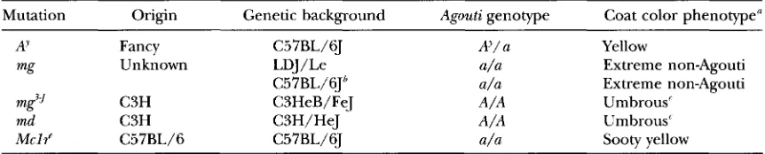
TABLE 1
Mutations and phenotypes studied
Mutation Origin Genetic background Agouti genotype Coat color phenotype"
AJ Fancy C57BL/6J A?/ a Yellow
mg Unknown LDJ/Le a/a Extreme nonhgouti
m$f C3H C3HeB/FeJ A/A Umbrous'
md C3H C3H/HeJ A/A Umbrous'
M c l 4 C57BL/6 C57BL/6J a/a Sooty yellow
C57BL/6Jb a/a Extreme nonhgouti
" See text and Figure 1 for explanation of phenotypes.
*
One of us (M.L.L.) has backcrossed the mg mutation to C57BL/6J for eight generations; these animals were used for the study depicted in Figure 3C.On an Agouti background, animals mutant for mg are darker than those mutant for md.
exisiting mouse coat color mutations mahogany (mg)
and mahoganoid ( m d ) , which lie on mouse chromosomes 2 and 16, respectively (GREEN 1989). Both mutations alter the balance between pigment types in favor of eumelanin synthesis, and therefore suppress the pheno- typic effects of Agouti protein in viuo (LANE 1960; LANE and GREEN 1960). To order these genes in a genetic pathway with respect to Agouti and the melanocortin 1 receptor, we generated animals doubly mutant for mg
or md and A' or the Mc14 allele. BEECHEY and SEARLE have previously reported that the nonagouti curly muta- tion, a probable allele of md ( m P ) is epistatic to the coat color effects of A? but not to those of the Mc14 allele (BEECHEY and SEARLE 1978, 1979). Our results indicate that mg and md are genetically downstream of
Agouti, but genetically upstream of the Mcl'. Surpris- ingly, mgand md suppress the effects of Agouti on obe- sity as well as on coat color.
MATERIALS AND METHODS
Mouse strains, mutations, and coat color phenotypes: All animals were obtained originally from the Jackson Labora- tory (Bar Harbor, ME). The strain of origin, source, genetic background, and relevant coat color genotype for each muta- tion is listed in Table 1. Most of the breeding studies de- scribed below involved a comparison of F2 animals whose Go
parents were from different strains. However, linkage be- tween AY and mg limited the number of doubly mutant ani- mals that could be recovered from an F2 cross. One of us (M.L.L.) has backcrossed the mgmutation to C57BL/6J for eight generations, and the studies shown in Figure 2C are based on comparison of non-littermate animals from the same strain backgrounds.
The effects of mg and md vary according to whether the Agouti genotype is A/A or a / a ; therefore interaction between the Me14 and the md mutations (whose original backgrounds are a / n and A/A, respectively) was carried out by first recov- ering a / a ; md/md F2 progeny from a cross to C57BL/6J-a/a mice. The coat color phenotypes described in Tables 2-5 as umbrous or dark brown with a yellow ventrum were very uni- form among different animals. However, the yellow and sooty yellow phenotypes were more of a continuum than discrete classes; in addition, nonagouti and extreme nonagouti pheno- types are classified together as "black." For measurement of
plasma melanocortin levels, isogenic C3HeB/FeJ-m$f/m$j animals were compared with C3H/HeJ animals (C3HeB/FeJ was derived from C3H/HeJ by embryo transfer).
Histology: Skin biopsies from the mid-dorsum, mid-ven- trum, and the tip of the pinna were obtained from male and female C3H/HeJ, C3HeB/FeJ-m$f/m$j, and C3H/HeJ-md/ md animals between 2 and 4 months of age. Biopsies were fixed in 10% formalin, rinsed with distilled water, and im- mersed in DOPA reagent (1
5%
D,Mihydroxyphenylalanine in 0.2 M phosphate buffer, pH 7.4) for 1 hr at 37". After 1 hr, the biopsies were transferred to fresh DOPA reagent for an additional 3-6 hr, then rinsed with distilled water and fixed in alcohol/formal/acetic acid. Biopsies were embedded in paraffin, sectioned (10 pm), and stained with alcoholic carmine.Genotyping: Genotype at mg or md was inferred from the allele sizes of closely linked simple sequence length polymor- phisms (SSLPs). D2Mit77 lies within 0.5 cM of mg (K A. MILLER and G. S. BARSH, unpublished results). The positions of md and D16Mit9 according to the International Mouse Genome Conference consensus map are 2 and 4 cM, respec- tively (MGD 1996). Linkage between md and SSLP markers has not been measured directly other than what is reported here (see below); therefore in the studies described in Table
2, allele sizes for additional markers [D16Mit182 and D16Mit88, which lie at 3.4 and 9.7 cM, respectively (MGD 1996)] were determined for all animals of the sooty yellow class (Mc14/Mc14). In addition, the results shown in Table 4
suggest that no recombination was observed between md and D16Mit9 in 42 meioses (22 Ay/A; +/md and 10 Ay/A; md/ md progeny). Oligonucleotide primer pairs for D2Mit77, D16Mit9, D16Mit88, and D16Mit182 were obtained from Re- search Genetics (Huntsville, A L ) ; allele sizes were determined using denaturing polyacrylamide gel electrophoresis to ana- lyze the PCR products from tail DNA of each animal. Allele sizes in animals carrying mg or md were identical to those observed in C3H/HeJ mice [174 nucleotides (nt) for D2Mit77; 126 nt for D16Mit91; allele sizes in animals carrying
AY or Me14 were identical to those observed in C57BL/6J mice (170 nt for D2Mit77; 146 nt for D16Mit9). Agouti and
M c l r genotypes were inferred from coat color phenotypes as
described in RESULTS; in the one equivocal situation, Ay/A; md/md us. A/A; md/md, Agouti genotype was determined by Southern hybridization using an exon 1A probe that detects the A'-associated deletion.
Interactions of mg, md, Mclr, and A 1409
were obtained between 9 AM and 10 AM after animals had remained undisturbed for at least 3 days. Animals were placed in a non-immobilizing restraining device, a small incision was made in the tail vein, and blood was collected by gravity over a 1-2-min period. In most cases, samples from two to three littermates of identical sex and genotype were pooled to reach a total of 0.3-0.5 ml of plasma required for the assay. Measure- ments of body weight were made on female animals housed together as littermates and allowed free access to water and standard laboratory mouse chow.
RESULTS
The coat colors of animals homozygous for md or mg are very similar. On an Agouti background ( A / A or A / a), md generally produces less darkening than mg, but the phenotype
of
both mg/mg or md/md animals is de- scribed as umbrous, black hair on the dorsum and dark Agouti hairs with greatly reduced yellow ticking on the flank and ventrum (Figure 1A). On a nonagouti back- ground ( u / u ) , small amounts of pheomelanin normally observed in perineal, mammary, and pinna hairs are replaced by eumelanin in mg/mg or md/md animals. In addition, the glabrous (nonhairy) skin of the ears and tail is much darker than normal (Figure IC). This so-called extreme nonagouti phenotype is nearly identical to that caused by an Agouti null allele, but can be diffi- cult to distinguish from nonagouti alone.
To investigate how md or mg caused darkening of glabrous areas of the skin, we examined split skin whole mount and paraffinembedded sections from
md/md,
mg/mg, m C J / m c J , and control C57BL/6J and C3H/ HeJ mice at ages of 2-4 months. Melanocyte counts were made separately for the dermis and epidermis of
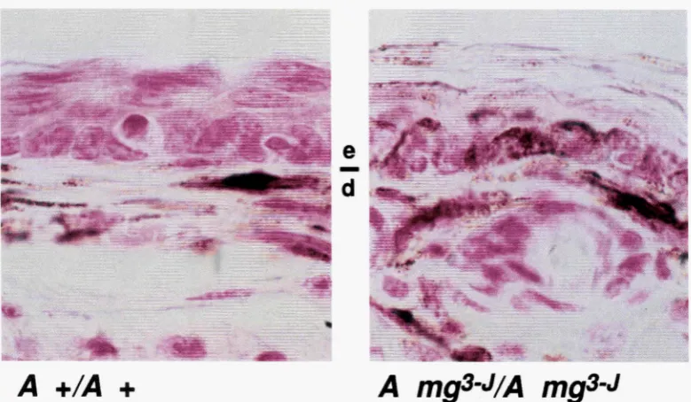
the pinna and tail, but we found no consistent differ- ences in the number of these cells that correlated with the darkened phenotype (data not shown). However, in both pinna and tail, we consistently observed increased transfer of melanosomes to epidermal keratinocytes (Figure
2).
We first considered whether the genes mutated in md and/or mg might encode structural or enzymatic components required for pheomelanin biosynthesis, and reasoned that, if so, the mutant phenotypes would be altered little or not at all by a defective Mclr (Figure 4). The Me14 mutation is a frameshift that produces a premature termination codon before the third trans- membrane domain and is thought to be a complete loss-of-function (ROBBINS et ul. 1993). A 50% gene dos- age reduction in M c l f / + animals has no phenotypic effect, but M c l f / M c l f animals are mostly yellow with some eumelanin in the dorsum and pinna hairs, a phenotype often described as sooty yellow. As pre- dicted, F1 progeny, + / M c l f ; + / m d ; u/u, or + / M c l f ; + / m g u/u, were nonagouti. Two classes of F2 progeny were obtained whose coat color phenotypes were sooty yellow or black, in a ratio of approximately 3:
1,
respec- tively (Tables 2 and 3). The sooty yellow F2 progenyF~GURE 1.-Coat color phenotypes of mutant animals. (A) On an Agouti background ( A / A or A / u ) , homozygosity for
md produces an umbrous phenotype, black hairs on the dor- sum and dark Agouti hairs on the flanks and ventrum. ( B ) AI/A; md/ md animals appear very similar to A / A ; md/ md ani- mals. ( C ) On a nonugouti background ( u / u ) , homozygosity for mg (or md, not shown) produces an extreme nonagouti phenotype, black hairs over the entire animal including the pinna, perineal, and mammary areas. Heterozygosity for mg
(or md, not shown) reveals a semidominant effect only when AY is present, AJ + / a mg animals are sooty yellow rather than completely yellow as in AJ + / a
+
animals. This phenotype also distinguishes the coat color effects of AJ from those of Mclf; C5’7BL/6J-McIf/Mclf animals are sooty yellow (not shown) whereas C57BL/6J-AJ/u animals are completely yel- low. The apparent differences between md and mg in A and C are due to the Agouti background.FIGURE 2.-Paraffinembedded section of DOPA-reacted pinna skin from control (C3H/HeJ-A + / A +) and mutant (C3I-.
FeT-A m2J/A m$g) animals. The pinna normally contains many melanocytes located centrallv in the dermis (d) whose dendr ites lie-padlel to &e long axis of c6nnective tissue cells of the dermis, in addition, there is a population of melanocytes that lies at the junction between the dermis and epidermis (e). In the tail and pinna of
md/md
and mg/mg animals (not shown) and inthe pinna of m8J/mg3'1 animals, basal keratinocytes and the overlying stratum corneum contain many more melanosomes compared to non-mutant animals. Magnification, 1 0 0 0 ~
To determine
if
md or mgwere genetically upstream of normal Agouti transcription, we made use of the AY allele, in which Agouti coding sequences are controlled by a heterologous promoter and, as a consequence, abnormally transcribed in a constitutive manner (DUHL et al. 1994a; M~CHAUD et al. 1994a). The coat colors of Ay/A; +/md, or Ay/a;+/mg
F1 progeny were similar but, surprisingly, not identical to their Ay/A;+/+
or Ay/a;+/+
parents, which are completely yellow. In-stead, the F1 animals were sooty yellow, which suggested that md and mg were genetically downstream of Agouti, and, further, that md or
mg
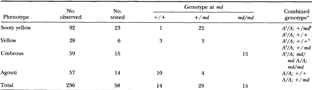
are semidominant rather than recessive. Both notions were confirmed in the F2 generation (Tables 4 and 5). For md, 59 of236
F2 prog- eny exhibited an umbrous phenotype (Table 4). Based on 10 animals tested for genotype at md and Agouti, Ay/ A; md/md animals could not be distinguished from A/ A;md/md
animals (Figure lB), demonstrating that mdTABLE 2
Fs progeny of + / M c l f ; +/md; a/a X + / M c l f ; +/md; a/a
No. No. Genotype at md
Phenotype observed tested
+/+
+/mdmd/md
Combined genotype"Sooty yellow 25 25 5 15 5 f / M c l M c l f ;
+
/+
M c l f / M c l f ; S/md M c l f / M c l 1"; md/md
+/+
or + / M c l f ;+/+
+/+
or + / M c l f ; +/md+/+
or + / M c l f ; md/md Total 117 112 34 54 24Black 92 87 29 39 19
Interactions of mg, md, Mclr, and A 141 1
TABLE 3
F2 progeny of + / M c l f ; +/mg; a/a X + / M c l f ; +/mg; a/a
No. No. Genotype at mg
Phenotype observed tested
+/+
+/mg mdmg Combined genotype"Sooty yellow 28 24 6 13 5 M c l f / M c l f ;
+/+
M c l f / M c l f ; + / m g Mcl f/Mc14; mg/mg
+/+
or +/Mc14; +/mg+/+
or + / M c l f ; mg/mgBlack 104 68 16 27 25
+/+
or +/Mc14;+/+
Total 132 92 22 40 30
Genotype for mgwas determined as described in MATERIALS AND METHODS. As for the case with md, sooty yellow animals were assumed to be homozygous for the Mc14 mutation, and this assumption was confirmed on one animal by direct sequence analysis of PCR-amplified genomic DNA. Similarly, among the black animals, no attempt was made to distinguish
+/+
from + / M c l 4 .is epistatic to Ay (Table 4). Of 23 sooty yellow F2 animals that were tested, 22 were md/+ and one was
+/+; of
six completely yellow animals that were tested, three were md/+
and three were+/+
(Table 4). These re- sults indicate that a weak semidominant effect of md is apparent in animals that carry Ay, but also suggest that strain differences between CSH/HeJ-md/md and C57BL/6J-AY/a can influence the extent of Ayinduced pheomelanogenesis. A similar phenomenon has been described for the DK/Lm-AY mouse strain (LAMOREUX and GALBRAITH 1986).For mgand Ay, three of 56 F2 progeny exhibited an unusual coat color phenotype, dark brown dorsum with a yellow ventrum, and were found to be Ay/a; mg/mg. (Because mg and A are linked, ratios of F2 progeny obtained are not those of independently seg- regating loci.) Similar to the situation with md, the sooty yellow phenotype was strongly but not exclu- sively associated with heterozygosity for mg (Figure
1C; Table 5). Thus, md and mg each can suppress Ay- induced pheomelanogenesis and therefore are un- likely to interfere with a process that normally regu- lates Agouti transcription.
The animals described above that were doubly mu- tant for Ay and md or mg were also used to determine the effects of md or mgon AY-induced obesity (Figure 3, A and B). Excess weight gain in mice that carry Ay first becomes apparent between 5 and 8 weeks of age, and can reach 30-60% above control levels de- pending on strain background (CASTLE 1941; CAR- PENTER and k h Y E R 1958). We found that heterozygos- ity or homozygosity for md suppressed Arinduced obesity in a dose-dependent manner (Figure 3A). The levels of weight gain in AY/a; md/md animals were similar to those observed in A/A; md/md animals, but -20% higher than in A/A animals, which suggests that md is not a general inhibitor of food intake or growth. For mg, only a small number of doubly mu-
TABLE 4
F2 progeny of W/A; md/+ X Ay/A; md/+
Genotype at md
No. No. Combined
Phenotype observed tested
+/+
+/mi
md/md genotype"Sooty yellow 92 23 1 22 Ay/A; +/md
Ay/A;
+/+
Yellow 28 6 3 3 Ay/A;
+
/+
A9/A; +/md
Umbrous 59 15 15 Ay/A; md/
md A/A; md/md
Agouti 57 14 10 4 A/A;
+
/+
A/A; +/md
Total 236 58 14 29 15
a Genotype for md was determined as described in MATERIALS AND METHODS. Genotype for Ay was determined in 10 of the umbrous animals by Southern hybridization with a probe that detects the AJ-associated deletion; seven were found to carry A?.
A. Miller et al.
TABLE 5
F2 progeny of Ay + / a mg X W + / a mg
No. No. Genotype at mg Combined
Phenotype observed tested
+/+
+/
mg m d m g genotype"Sooty yellow 25 18 18 Ay + / a mg
Yellow 7 2 1 1 Ay + / a
+'
Ay + / a mg
Black 21 14 2 12 a + / a mg
a mg/a mg
Dark brownb 3 3 3 Ay mg/a mg
Total 56 37 1 21 15
~ ~~ ~
Genotype for mg was determined as described in MATERIALS AND METHODS.
Dark brown dorsum and yellow ventrum; see Figure 1C.
The sooty yellow phenotype is strongly but not exclusively associated with heterozygosity for mg as described in the text.
~~
tant F2 animals were produced because of its linkage to AY, but in this sample, mginhibited AY-induced obe- sity (Figure 3B). As an alternative approach, we com- pared body weight gain among non-littermate Ay
+/
a+
,
Ay mg/ a mg, and a+ / a
+
animals from the same genetic background and found thatmg
suppressed the effects of Ay on obesity to a level similar to that observed in a + / a+
animals (Figure 3C).Administration of a-MSH or related compounds to mice that carry Ay affects coat color in a manner similar to that caused by md or mg (GESCHWIND 1966; GESCH- WIND et al. 1972), and therefore we considered whether one or both mutations might be explained by increased melanocortin production. Among C57BL/6J, C3H/
HeJ, CSH/HeJ-md/md, LDJ-mg/mg, and C3HeB/FeJ-
mg"J/m$j animals, we found that plasma levels of a- MSH or ACTH varied approximately three- or 1 .!%fold, respectively (Table 6). However, this variation did not correlate with mutant genotype and was therefore most likely caused by strain differences other than genotype at md or mg.
DISCUSSION
Gene interaction studies carried out with previously existing mouse mutations have played a key role in un- derstanding important biological processes including the cellular and physiologic action of endothelin 3, mast cell growth factor, leptin, and the Agouti protein
(SILVERS and RUSSELL 1955; MCCULLOCH et al. 1965; COLEMAN 1973; KAPuR et al. 1993). In the case ofAgouti, its effects on coat color though not on obesity require an intact Mclr, since animals that carry loss-of-function mutations for both genes ( a / a ; M c l f / M c l f ) are yellow and non-obese, whereas animals that carry Ay and the constitutively active Mclr mutation sombre (Mclr"") are black and obese (WOLFF et al. 1978; SILVERS 1979). By contrast, our results demonstrate that the md or mg mutations suppress the effects of AY on coat color and
obesity. Taken together with the interactions between md or mgand the M c l f mutation, these findings suggest a genetic pathway in which md and mgplay key roles in posttranscriptional production or reception of Agouti signaling (Figure 4).
The coat color effects of md and mg are similar to those caused by intraperitoneal or subcutaneous injec- tion of a-MSH (GESCHWIND 1966; GESCHWIND et al. 1972; SHIMIZU et al. 1989), but we found that plasma levels of a-MSH and ACTH were not correlated with either mutation. The source of melanocortins that nor- mally stimulates pigment cells is not clear (reviewed in SLOMINSKI et al. 1993; WINTZEN and GILCHREST 1996), and it is possible that the coat color effects of md or mg might still be explained by increased local production of melanocortins by keratinocytes. However, neither a local nor a systemic increase in melanocortin produc- tion would provide a simple explanation for the effect of md and mg on obesity, since injection of a-MSH or related compounds into AY/ a or a/ a animals stimulates rather than inhibits weight gain (SHIMIZU et al. 1989). Instead, md and mg are most likely to act by interfering directly with Agouti signaling, either in its processing or secretion, binding to a cell surface receptor, or post- receptor regulation. Each of these possibilities is dis- cussed below in more detail.
Agouti protein is normally secreted by dermal papilla cells; its sphere of action includes melanocytes in overly- ing but not adjacent hair follicles (POOLE 1975; MILLAR et al. 1995). Recombinant Agouti protein produced by insect cells is glycosylated and contains an amino-termi- nal basic domain that is not required for activity in
A
5 0 ,
n
4 0 ,
E
0,
.-
3 0 ,
20,
10
C
40
30
n
m
W
E
m
.-
s
2010
@ AVA: +/mcl(48)
d
0 AIA; +I? (27)
c?
1W A/A; mdmd(7)
ir
0 AVA; mcud(22)
+
0
L
1I&
P
P
,%,
. ..
.
3-5 12-1 4 26-28
.
.
38-40.
.
Age
(wks)
0 AYm#amg(15)
s x
s:
. . . .
4-6 9-1 0
. .
13-1 4
In r h - 0 , recombinant Agouti protein inhibits the
binding of radiolabeled melanocortins to cells that ex- press the
Mc
1 r, Mc2r. o r Mc4r(Lr.
PI 01. 1994; YASG P/nl. 199'i). Because the ability of Agouti protein to in- hibit CAMP accumulation in rdro requires the presence of a functional melanocortin receptor, these findings strongly suggest that the Mclr is the normal Agouti receptor. Although stnlctunl similarity behveen Agouti protein and omega;\conotoxins has led to speculation
H
&ro
F
i
*
.
3-5 12-14
.
.
.
.
26-28
Age (wks)
FIGC'RE J."Effects of mdor mgon A'-intlrlced wcight gain. FL' fcmale animals o f the indicated genotype were w e i ~ l w l e v e n 7-30 days as littermates. \';tlues tlepictcd ;Ire t h r nwan
2 SD 0 1 ) ; within an indicated internal, i . p . , 86-28 wrcks,
\dues for each group are artificially tlispl;~ced on thc ahwissa
to better indicate. thcdifferences bct\vcerl groups. ( A ) lntcmc- tion of !\' with mri. The group lahelerl ,,\/,.\: + / ? inclutlrs A / A;
+/+
and A / / \ ; +/md anim;ds. (R and C:) Interaction o f A' with mg. A'i s linked to mg. and among progeny ohtainetl from an F? intercross ( R ) , no 21 + / a+
;miln;ds were otminctl.Panel C shows a comp;Irison o f nonlittermate animals on a uniform genetic hackground.
that Agouti protein could act as a calcium channel an- tagonist (reviewed in MASSF. PI nl. l99.5), experiments in which Agouti protein was reported to affect calcium
flux may also be explained by impurities in the prepata- tion o r seconday effects (ZEMEI. P/ nl. 1995; JOSI<S P/
1414 li A. Miller d nl.
gesting that Agouti-induced obesity is caused bv Mc4r antagonism (HCS~AR and At. 1997). Thus. neither md nor mg are likely to encode a specific Agouti receptor that acts independently of melanocortin signaling. However, direct binding of Agouti to a cell surflce pro- tein has not vet been demonstrated, and it is possible that md or mg interfere with formation or assembly of a complex that includes a melanocortin receptor and additional proteins.
Finallv, md or mg might interfere with melanocortin receptor regulation. For example, an increased number of melanocortin receptors at the cell surface or a failure to undergo normal ligand-induced desensitimtion
would lead to increased melanocortin signaling, and therefore suppress the effects of increased antagonist production in mice doubly mutant for A" and md o r mg. Furthermore. increased melanocortin signaling would be apparent onlv in the presence of a functional r e c e p tor; therefore, md or mgwould have no effect in mice homozygous for the M r l f mutation.
Many mouse coat color mutations have pleiotropic effects because the molecular machinery required for pigment cell development and function is shared by other cells or tissues (reviewed in JACKSON 1994). In the case of Agouti, however, the pleiotropic effects of A' are apparent only because a signaling protein normallv restricted to the skin can elicit a response when ex- pressed abnormally in other parts of the body. O u r results indicate that md and mg have a wider spectrum of action than was previously apparent because both mutations suppress A'-induced obesity. Furthermore, multiple alleles with nearlv identical phenotypes have arisen for md and mg (GREEN 1989), which suggests that both mutations may represent a loss- rather than gain- of-function. We have suggested previously that variation in the human A p t i gene is unlikely to contribute to anv human phenotype other than skin or hair color (DVHI. PI nl. 1994b; M'ILSON PI nl. 199.5). The same may not he true, however, for human homologues of md and mg.
TABLE 6
Melanocortin levels in plasma
CY-MSH ACTH
Animal (pg/nII)" (pg/ml)"
C57RL/$J 63 t 17 358 2 47
C3H/He] 190 2 25 228 t 27
CSH/Hej-rnd/md 19.5 t 14 271 -t 69
LQJ-mg/mg 88 t 18 346 2 46
C3HeR/FqJ-mg7~/m$' 121 2 9 264 -t 41 " Plasma levels were determined using a radioimmunoassay as described in MATERIAIS AND MET1IOI)S. Initial results were obtained o n samples pooled together by sex and genotype. but no differences were apparent between males and females and the restdts shown here include both sexes.
0
m e ; mg+gizD7&
Agouti*.
obesity pheomelanlnf
(Redlyellow)f
d o p a q u i n o n e Mclr
@
(E) " ~\
~eumelanin (Brownlblack) ~ * ~ ~FI(;tIRF. 4.-Diagram summarizing the interactions k t w e e n md o r mg. and A' or the M r l t ' . The M r l t ' is epistatic to md and mg, which, in turn, are epistatic to the effects of A' on coat color and obesity. Recause mttltiple allrlrs o f m d antl m g have arisen with identical phenotypes, they are likclv to
represent loss-of-function mutations in a process o r processes required directly for elabontion or receipt o f Agouti signal- ing. An alternative hypothesis wherebv md or mg cottld inter- fere with melanocortin receptor regulation or drsensitization is discussed in the text.
This work \vas supported in part by grants from the National Insti-
tutes of Health to .M.1..1.. (EY-IO'L'L3) ant1 t o G.S.R. (DK-28506) who is an rlssistant Investigator o f the Howard Hughes Medical Institute.
LITERATURE CITED
REE(:IIEY.
c.
\'., and A. <;. SMR1.E. 1978 Epistasy of extension alleles.BEEc:lin.. C . \'., and A.
(;.
S~\\RI.E. 1979 Epistasv of nc over Ay.G\R\RPESrER. KJ.. and 1. ".4WR. 1958 Physiologic o h e m l i o n s o n yellow ohesih in the mouse. Am..J. Phyiol. 193 4IKLW4. GsII.E, H'. E.. I941 l n f l w n r e of certain coat color nlutations on
body sizr in mice, m s , and mhhits. Cknetics 26: l i i - l ! l l .
CIIIIAIIANI, V., and J. E. S. WIKRERG, IW! Molecular cloning and rxpression of the human melanocyte stimulating hormone re- ceptor cDNA. FERS Lxtt. 309 4 17-420.
< h t . E w \ s , D. I.., 1973 Effects 01' parabiosis of ohese with diabrtes and normal mice. Diahetolagia 9 29.1-298.
CX)SE. K. I).. 1). LL., S. K o r r t u , I). 1. VM;E, H. KI.~WUSI> d nl., 1996 The mrlanocortin rrcc-ptors: agonists. antagonists, and the hormonal control of pigmentation. Recent Prog. I - I o r m . Res.
Duttl.. D. .V.J., M. E. S'rnws, H. VRIEI.IN(;, P..j. SAXON. M. W .
MlI.I.ER rf nl.. 1 W 4 a Pleiotropic rffrcts o f the mouse lethal y d -
low (A(?)) mutation explained by drletion o f a maternally r x -
pressed genr antl the simulraneolls prtnlurtion o f agouti lilsion RNAs. Developmrnt 120: 1695- 17OX.
D ~ Y I I . . I). M.J.. H . \'RIEI.IX~;. I;. A. MII.I.ER,
<;.
I.. W01.n: and G . S.BARSII. t994h Nromorphic agouti mutations in ohese yellow mire. Nature <*net. 8: 59-65.
GA?.w., I., Y. KONIM, T. TASIIIRO, Y. SlllWOTO. H. MIWA rt nl., IW%a Molecular cloning of a novel melanocortin rcwptor. J. Riol. (:hem. 2 6 8 X24fi-8250.
G,~?.T/.. I., H. .MIWA, Y. K o s ~ ) , \ , Y. SIIIUOTO. T. TASIIIRO rf nl.. 1W3h
Molecular cloning, cxprrssion. antl gene localization of a 4th melanoconin reccpor. J. Riol. <:hem. 2 6 8 15174- 15179.
<;f-v:lt\r.lst). I. 1.. 196fi (:hange in hair color in mice intluced by injvrtion o f (I MSH. Endncrinolop 79: 1165-1 167.
Gv-v:ltwtsI>, 1. I., R. A. HUSEIW and R. SIS~IKWA. 1972 T h r effect o f melanocyte -stimulating hormone on coat color in the mouse. Recent Prog. Horm. Rrs. 28: 9 1 -130.
CRELS, M. C.. 1989 Catalog ol mutant genrs and polymorphic loci. pp. 12-403 in Cnrtir \ brinntc nnd Strnitu of thr I . d m n t n ~ M m w ,
etlitetl by M. F. 1,Yos and A. G . SFARI.F.. Oxford Cnivenitv Prcw. Oxford.
H r ~ r . G., and A. J. TIIODY, 1995 Agouti protein can art indeprn- dentlv of melanocyte-stimulating hormonr to inhibit mrlanogcn- esis.J. Entlocrinol. 147: RI -R4.
Monse Ncwr lrrr. 59: 1 9 .
MOIISC NCWY l r t t . 60: 45-47.
Interactions of mg, md, Mclr, and A 1415
HUSZAR, D., C. A. LYNCH, V. FAIRCHILDHUNTRESS, J. H. DUNMORE, Q. FANG et al., 1997 Targeted disruption of the melanocortin- 4 receptor results in obesity in mice. Cell 8 8 131-141. JACKSON, I. J., 1994 Molecular and developmental genetics of mouse
coat color. Annu. Rev. Genet. 28: 189-217.
JONES, B. H., J. H. KIM, M. B. ZEMEI., R. P. WOYCHIK, E. J. MICHAUD et al., 1996 Upregnlation of adipocyte metabolism by agouti protein: possible paracrine actions in yellow mouse obesity. Am.
J. Physiol. Endocrinol. Met. 33: E192-E196.
KAPUR, R. P., C. YOST and R. D. PALMITER, 1993 Aggregation chime- ras demonstrate that the primary defect responsible for aganglio- nic megacolon in lethal-spotted mice is not neuroblast autono- mous. Development 117: 993-999.
LAMOREUX, M. L., and D. B. GAI.BRAITH, 1986 DK/Lm: a strain of laboratory mouse with an unusual expression of the lethal yellow (Ay) phenotype. Genet. Res. 48: 35-40.
LANE, P. W., 1960 New mutants. Mouse News Lett. 22: 35.
LANE, P. W., and M. C. GREEN, 1960 Mahogany, a recessive color mutatioin in linkage group V of the mouse. J. Hered. 51: 228-
230.
Lu, D. S., D. WILLARD, I. R. PATEI., S. KADWELL, L. OVERTON et al., 1994 Agouti protein is an antagonist of the melanocyte-stimu- lating-hormone receptor. Nature 371: 799-802.
MANNE, J., A. C. ARGESON and L. D. SIRACUSA, 1995 Mechanisms for the pleiotropic effects of the agouti gene. Proc. Natl. Acad. Sci.
MCCUI.I.OCH, E. A,, I.. SIMINOVITCH, J. E. TILL, E. S. RUSSELL and S. E. BERNSTEIN, 1965 The cellular basis of the genetically deter- mined hemopoietic defect in anemic mice of genotype SI-Sld. Blood 26: 399-410.
MGD, 1996 Mouse Genome Database. Mouse h o m e InJmatics. The Jackson Laboratory, Bar Harbor, ME. World Wide Web (URL:
http://vmw.informatics.jax.org/).
MICHAL~D, E. J., S. J. BUI.TMAN, M. L. KLEBIG, V. M. VAN, L. J. STUBBS et al., 1994a A molecular model for the genetic and phenotypic characteristics of the mouse lethal yellow (Ay) mutation. Proc. Natl. Acad. Sci. USA 91: 2562-2566.
MICHAUD, E. J., M. J. VANVUGT, S. J. BULTMAN, H. 0. SWEET, M. T. DAWSSON et al., 199413 Differential expression of a new domi- nant agouti allele (A(iapy)) is correlated with methylation state and is influenced by parental lineage. Genes Dev. 8 1463-1472. MILLAR, S. E., M. W. MIILER, M. E. STEVENS and G. S. BARSH, 1995
Expression and transgenic studies of the mouse agouti gene pro- vide insight into the mechanisms by which mammalian coat color patterns are generated. Development 121: 3223-3232.
MOUNT~OY, K. G., L. S. ROBBINS, M. T. MORTRUD and R. D. CONE, 1992 The cloning of a family of genes that encode the melano- cortin receptors. Science 257: 1248-1251.
USA 92: 4721-4724.
POOLE, T. W., 1975 Dermal-epidermal interactions and the action of alleles at the agouti locus in the mouse. Dev. Biol. 42: 203-
210.
ROBBINS, L. S., J. H. NADEAU, K R. JOHNSON, M. A. KELLY, I,. ROSEI,
LIREHFUSS et al., 1993 Pigmentation phenotypes of variant ex- tension locus alleles result from point mutations that alter MSH receptor function. Cell 72: 827-834.
SHIMIZU, H., N. S. SHARGIIJ., G. A. BRAY, T. T. YEN and P. D. GE-
SEILCHEN, 1989 Effects of MSH on food intake, body weight and coat color of the yellow obese mouse. Life Sci. 45: 543-552. SILVERS, W. R, 1979 The Coat Colors ofMice: A Modelfor Mammalian
Gene Action and Interaction. Springer-Verlag, New York. SILVERS, W. R , and E. S. RUSSELL, 1955 An experimental approach
to action of genes at the agouti locus in the mouse. J. Exp. Zool. 130: 199-220.
SIRACUSA, L. D., 1994 The agouti gene: turned on to yellow. Trends Genet. 10: 423-428.
SI.OMINSKI, A,, R. PAUS and J. WORTSMAN, 1993 On the potential role of proopiomelanocortin in skin physiology and pathology. Mol. Cell. Endocrinol. 93: Cl-C6.
VRIELING, H., D. M. DUHI., S. E. MILIAR, K. A. MIILER and G. S. BARSH, 1994 Differences in dorsal and ventral pigmentation result from regional expression of the mouse agouti gene. Proc. Natl. Acad. Sci. USA 91: 5667-5671.
WILIARD, D. H., W. BODNAR, C. HARRIS, L. KIEFER, J. S. NICHOLS et al., 1995 Agouti structure and function: characterization of a potent alpha-melanocyte stimulating hormone receptor antago- nist. Biochemistry 34: 12341-12346.
WIISON, B. D., M. M. OLLMANN, L. KANG, M. STOFFEI., G. I. BELL. et al., 1995 Structure and function of ASP, the human homolog of the mouse agouti gene. Hum. Mol. Genet. 4: 223-230. WINTZEN, M., and B. A. CILCHREST, 1996 Proopiomelanocortin, its
derived peptides, and the skin. J. Invest. Dermatol. 106: 3-10. WOI.FF, G. L., D. B. GALBRAITH, 0. E. DOMON and J. M. Row, 1978
Phaeomelanin synthesis and obesity in mice. Interaction of the viable yellow ( A T ) and sombre (Eso) mutations. J. Hered. 69: YANG, Y. R, M. M. OILMANN, B. D. WILSON, C. DICKINSON, T. YAMAUA
et al., 1997 Effects of recombinant agouti-signaling protein on melanocortin action. Mol. Endocrinol. 11: 274-280.
YEN, T. T., A. M. GILL, L. G. FRIGERI, G. S. BARSH and G. L. WOLFF, 1994 Obesity, diabetes, and neoplasia in yellow A(vy)/- mice: ectopic expression of the agouti gene. FASEB J. 8: 479-488. ZEMEL, M. B., J. H. KIM, R. P. WOYCHIK, E. J. MICHAUD, S. H. KADWELI.
et al., 1995 Agouti regulation of intracellular calcium: role in the insulin resistance of viable yellow mice. Proc. Natl. Acad. Sci. 295-298.
USA 92: 4733-4737.