Review
Papillomaviruses in dogs and cats
John S Munday a,*, Neroli A Thomson a, Jennifer A Luff b
a Pathobiology, Institute of Veterinary, Animal and Biomedical Sciences, Massey University, Palmerston North, New Zealand.
b Department of Population Health and Pathobiology, College of Veterinary Medicine, North Carolina State University, Raleigh, NC, USA.
* Corresponding author. Tel.: +64 356 9099.
E-mail address: j.munday@massey.ac.nz (J.S. Munday).
3
6
9
12
Abstract
Papillomaviruses (PVs) are recognised to cause disease in both dogs and cats.
Currently in dogs, PVs are thought to cause oral papillomatosis, cutaneous papillomas, and
canine viral pigmented plaques. Papillomaviruses have been rarely associated with the
development of oral and cutaneous squamous cell carcinomas in dogs. In cats, PVs are
currently thought to cause oral papillomas, feline viral plaques, Bowenoid in situ carcinomas,
and feline sarcoids. However, there is increasing evidence that PVs may also be an important
cause of cutaneous squamous cell carcinomas and basal cell carcinomas in cats. These
diseases are discussed in this review. Additionally, there is a brief overview of PV biology
including how these viruses cause disease. Diagnostic techniques and possible methods to
prevent PV infection are also discussed.
Keywords: Papillomavirus, dogs, cats, papillomas, oncogenesis, pigmented plaques, warts.
18
21
24
27
Introduction
Warts have been recorded in folk-lore for centuries in people. A viral cause of warts
was confirmed in 1907 and the first human papillomavirus (PV) was fully sequenced in 1982
(Danos et al., 1982). The first evidence that PVs can cause cancer was reported in from
studies in rabbits in 1935 (Rous and Beard, 1935). This was followed in 1981 by the
breakthrough demonstration that PVs cause the majority of human cervical cancers (zur
Hausen et al., 1981). Subsequent research has expanded the range of human cancers that can
be caused by PV infection and it is currently estimated that around 5% of all human cancers
are due to PV infection (Parkin, 2006).
In dogs transmissible warts were first noted in 1898 with a viral aetiology confirmed
in 1959 (Nicholls and Stanley, 1999). By using light and electron microscopy, evidence of a
PV aetiology was suggested in 1969 and the first canine PV was fully sequenced in 1994
(Delius et al., 1994). In contrast, as PVs only rarely cause papillomas in cats, the first
evidence of PV-induced disease in this species was not reported until 1990 (Carney et al.,
1990) with the first PV from domestic cats fully sequenced in 2002 (Tachezy et al., 2002;
Terai and Burk, 2002). In the years since these initial studies of PVs, additional PV types
have been identified and PVs have been associated with an expanded range of canine and
feline diseases. In addition, it is now accepted that host factors play a significant role in
determining whether or not a PV will cause clinically-relevant disease. This review will
provide a general overview of the biology of PVs and the mechanisms by which they cause
disease followed by a detailed discussion of the diseases of dogs and cats that are currently
associated with PV infection. The different methods that can be used to diagnose PV-induced
disease will be outlined, and for each distinct disease, the clinical presentation, histology,
prognosis, and any treatments are described. Lastly will be a brief discussion of the possible
ways that could be used to prevent diseases due to PVs in dogs and cats.
33
36
39
42
45
48
51
Papillomavirus biology
Papillomaviruses are small, non-enveloped, icosahedral viruses that infect the
stratified squamous epithelium of many mammalian, as well as some avian and reptilian,
species. Their circular double-stranded DNA genome is around 8000 base pairs long and
includes five or six early (E) and two late (L) open reading frames (ORF) (Munday and
Pasavento, 2017). Papillomaviruses are classified using the L1 ORF sequence.
Papillomaviruses within the same genus have greater than 60% L1 ORF similarity and
typically demonstrate similar host, location, and behavioural characteristics. Different
papillomavirus types have less than 90% similarity in their L1 ORF (Bernard et al., 2010).
Currently 18 Canis familiaris papillomavirus (CPV) types have been fully sequenced and
classified as Lambapapillomaviruses, Taupapillomaviruses and Chipapillomaviruses (Delius
et al., 1994; Tobler et al., 2006; Yuan et al., 2007; Tobler et al., 2008; Lange et al., 2009a;
Lange et al., 2012a; Lange et al., 2012b; Luff et al., 2012a; Yuan et al., 2012; Lange et al.,
2013; Zhou et al., 2014; Luff et al., 2015; Zhou et al., 2015; Munday et al., 2016b; Tisza et
al., 2016; Table 1). Four Felis catus (FcaPV) types have been fully sequenced including one
that has been classified as a Lambdapapillomaviruses one as a Dyothetapapillomaviruses and
two PV types remaining unclassified (Tachezy et al., 2002; Terai and Burk, 2002; Lange et
al., 2009b; Munday et al., 2013a; Dunowska et al., 2014). However, short sequences of
additional PV types have been amplified from dogs and cats suggesting that new PV types
are likely to be recognised from both species in the future. While the bovine
Deltapapillomaviruses are an important exception, PVs are typically highly species specific
(Sundberg et al., 2000).
57
60
63
66
69
72
75
Papillomaviruses are primarily spread by direct contact, although indirect spread is
also possible due to their ability to survive in the environment (Roden et al., 1997). Once the
PV comes into contact with a mucocutaneous epithelium, the presence of microabrasions
allows infection of basal cells resulting in the production of small numbers of circular PV
DNA copies (episomes) within the cell (Schiller et al., 2010). These episomes are maintained
in the basal cells as they replicate, providing a reservoir of infection. However, the viral
life-cycle is only completed when an infected cell undergoes terminal differentiation (Doorbar,
2005). Keratinocyte differentiation results in the expression of PV E6 and E7 proteinsthat
promote replication of the normally post-mitotic suprabasal cell and allow large-scale
amplification of the viral genome (Doorbar et al., 2012). Expression of the PV capsid
proteins (L1 and L2) and viral assembly occurs as the infected cell reaches the upper
epithelium. Papillomavirus-laden mature keratinocytes are sloughed from the epithelial
surface with the subsequent rupture of these cells releasing the infectious virions (Doorbar et
al., 2012). The bovine Deltapapillomaviruses are unique because they can also infect
mesenchymal cells, although these cells probably do not permit viral replication (Jelinek and
Tachezy, 2005).
The clinical presentation of a PV infection is largely determined by the degree of cell
proliferation induced by the PV. Most PV types only mildly increase cell proliferation and
PV replication occurs slowly in the absence of any visible lesions (Doorbar et al., 2012).
Alternatively, a minority of PV types markedly increase cell replication resulting in rapid
production of large numbers of viral particles. Such infections cause marked epithelial
hyperplasia that is visible clinically as a papilloma (wart) (Munday, 2014a).
As the majority of viral replication occurs in the external epithelial layers in the
absence of cell lysis, PV infections often only illicit a weak host immune response (Doorbar,
2006).However, when an immune reaction occurs, the response can be subdivided into
81
84
87
90
93
96
99
humoral and cell-mediated immunity. The production of circulating IgG antibodies blocks
entry of the PV into the basal cells preventing further infections by this PV type, although
antibodies do not influence resolution of an established PV infection (Nicholls et al., 1999;
Ghim et al., 2000). The development of a cell-mediated response results in the resolution of
established infections (Egawa and Doorbar, 2017). As there is significant intra-individual
variation in the time taken by the body to mount a cell-mediated response, there is also
variation in the time taken to resolve a clinically visible papilloma. As discussed later,
resolution may also be delayed in immunosuppressed animals.
Most dogs and cats are asymptomatically infected by PVs (Munday and Witham,
2010; Lange et al., 2011; Thomson et al., 2015). These infections usually do not result in
clinically-visible epithelial hyperplasia because the immune system is able to prevent the PV
from markedly changing normal epithelial cell regulation (Egawa and Doorbar, 2017).
However, if changes in the host allow greater PV protein expression, the resultant epithelial
hyperplasia can become visible as a lesion. Currently, the host factors that determine whether
or not a PV infection will cause a visible hyperplastic lesion are poorly-understood. However,
immunosuppression appears to predispose to the development of some PV-induced lesions
(Callan et al., 2005) and the increased frequency of pigmented plaques in certain dog breeds
(Narama et al., 2005; Luff et al., 2016) suggests genetic factors may also influence whether a
PV infection remains asymptomatic or results in clinical disease.
In humans, the major significance of PVs is the ability of the high risk
Alphapapillomaviruses to cause cervical, anogenital and oral cancer (zur Hausen, 2009). An
important process in PV-induced cancer is the accidental integration of the PV E6 and E7
genes into the host DNA resulting in rapid uncontrolled cell growth, inhibition of cell
apoptosis, telomerase loss, and disruption of processes that ensure accurate assembly of
replicated host DNA (Doorbar et al., 2012). These rapidly-dividing genetically-unstable cells
105
108
111
114
117
120
123
quickly accumulate additional mutations resulting in malignant progression (Pett et al., 2004).
While there is accumulating evidence that PVs may cause cancer in dogs and cats (Munday
and Kiupel, 2010; Munday et al., 2011d; Munday, 2014b; Altamura et al., 2016b; Luff et al.,
2016; Thomson et al., 2016), the precise functions of the canine and feline PV E6 and E7
proteins have not been fully determined. Furthermore, some neoplasms have been shown to
contain productive infections, suggesting that the PV DNA may not be integrated in the host
DNA (Munday et al., 2015d; Thomson et al., 2016). Therefore, additional research is
required to define the precise role that PVs have in the development of cancer in dogs and
cats.
Diagnosis of papillomaviral disease
Oral and cutaneous warts are the most frequent manifestation of PV disease in dogs
and the majority of these cases can be diagnosed clinically. While feline oral papillomas
likewise have a typical clinical presentation, they may not be recognised due to their rarity.
Due to variation in the clinical presentation of the remainder of the PV-induced lesions,
histopathology is recommended to allow definitive diagnosis.
As PVs complete their life cycle by promoting proliferation of epithelial cells,
histology of a PV-induced lesion will reveal thickening of the epithelium. If the PV induces
marked proliferation of epithelial cells, this can cause folding of the thickened epithelium and
a papilloma (Fig. 1). Papillomaviruses that only mildly promote epithelial replication contain
more moderate epidermal thickening that typically appears as a raised plaque. The presence
of viral replication within a lesion may be visible histologically as PV-induced cell changes
that could include: enlarged cells with a shrunken nucleus surrounded by a clear cytoplasmic
halo (koilocytes), cells with increased quantities of grey or blue fibrillar cytoplasm, cells with
intracytoplasmic inclusions, or cells with enlarged vesicular nuclei (Fig. 2). Intranuclear
129
132
135
138
141
144
147
inclusions can be visible, although in the authors’ experience these can be transient and
difficult to differentiate from nucleoli. Clumping of keratohyalin granules in the granular
layer is also often present within PV-induced lesions. Generally lesions with more marked
epithelial proliferation (such as papillomas) support greater viral replication and are more
likely to contain PV-induced cell changes. In contrast, lesions with more modest epithelial
proliferation (such as a viral plaque) contain less viral replication and only variably contain
PV-induced cell changes. The presence of PV-induced cell changes within a lesion does not
necessarily prove that the lesion was caused by PV infection. However, PV-induced changes
do confirm that the lesion contains viral replication and therefore suggest that the normal
behaviour of cells has been influenced by the PV. The histological features of each
PV-induced disease are described in more detail below.
Immunohistochemistry to detect L1 protein production can be used to investigate a
PV aetiology of a lesion. However, as the L1 protein is only produced late in the PV life
cycle immunostaining is restricted to lesions that contain active viral replication (Longworth
and Laimins, 2004). In addition, as specific antibodies against canine and feline PVs are not
available, some PV types may not be detectable. In humans, immunohistochemistry to detect
p16CDKN2A protein (p16) is used as a marker for a PV aetiology in some lesions (Smeets et al.,
2007). This protein is increased because most PVs promote cell replication by degrading
retinoblastoma protein (pRb), a change which subsequently increases p16 (Parry et al., 1995).
Unlike PV immunostaining, p16 immunostaining therefore detects an effect of PV infection
rather than the presence of PV L1 protein in the lesion. Additionally, p16 immunostaining
will be increased even if no viral replication is present. Furthermore, human anti-p16
antibodies have been validated for use in dogs and cats. While p16 immunostaining has been
associated with PV infection in both dogs and cats and FcaPV-2 has been shown to degrade
pRb (Munday et al., 2011a; Munday and Aberdein, 2012; Munday et al., 2015d; Altamura et
153
156
159
162
165
168
171
174
al., 2016b), it is currently uncertain whether all canine and feline PVs degrade pRb.
Additionally, as other causes of pRb dysfunction have been shown to increase cell p16 in
human cancers, it is possible that increased cell p16 can be present in some non-PV-induced
lesions in dogs and cats.
By using PCR it is possible to detect very small quantities of PV DNA within a
lesion, even in the absence of PV replication. The use of consensus primers even allows the
amplification of DNA from PV types that have not previously been reported. In addition to
conventional PCR, reverse transcriptase PCR can be used to detect PV gene expression.
Detecting PV RNA suggests that the PV was producing proteins and was therefore able to
influence cell growth and differentiation. In situ hybridisation techniques can also be used to
localise either PV DNA or RNA within a lesion. While molecular techniques are useful to
detect PV DNA within a sample, PVs are a common commensal of the skin and oral cavity of
dogs and cats so that the detection of PV DNA within a lesion does not prove causality.
Therefore, PCR-based molecular techniques are better suited for research rather than for
routine diagnostic testing.
Papillomaviral diseases of dogs
Oral papillomatosis
Oral papillomas caused by CPV-1 (formerly referred to as canine oral PV) are
common in young dogs (Lange and Favrot, 2011). The epidemiology of infection by CPV-1
is poorly understood. There are anecdotal reports of outbreaks of canine oral papillomatosis,
suggesting that the disease can be acquired through contact with affected dogs. However, as
dogs are commonly asymptomatically infected by CPV-1, avoiding dogs with papillomas
may not be sufficient to prevent disease (Lange et al., 2011). The development of oral
papillomatosis results in the development of antibodies that prevent new infections by
CPV-180
183
186
189
192
195
198
1. However, it is probable that some dogs remain asymptomatically infected by CPV-1
following resolution of the initial papillomas (Doorbar et al., 2012) and reports of
papillomatosis in older immunosuppressed dogs is consistent with reactivation of an
asymptomatic infection (Sundberg et al., 1994; Radowicz and Power, 2005). Oral
papillomatosis presents as multiple exophytic vegetative warts involving the lips and mouth
(Lange and Favrot, 2011; Fig. 3). Most dogs do not show any systemic signs of disease
although there are rare reports of extensive disease that interferes with eating or respiration
(Nicholls et al., 1999). Histology reveals an exophytic mass comprised of thickened folded
epithelium, often with numerous PV-induced cell changes.
The overwhelming majority of canine oral papillomas spontaneously regress and
surgical excision is rarely necessary. Regression is due to the development of a cell-mediated
immune response. In experimentally-induced papillomas resolution was typically reported
within 8 weeks. However, resolution of natural papillomas appears to be more variable with
resolution taking up to 12 months in some dogs (Sancak et al., 2015). Numerous treatments to
hasten the resolution of oral papillomas have been proposed. Most have not been assessed in
appropriate studies. However, in a prospective, randomised, double-blinded
placebo-controlled study of 17 dogs with papillomas, lesion regression was observed within 50 days
in all 10 dogs that were treated with azithromycin, but only 1 of 7 untreated dogs (Yagci et
al., 2008). Stimulating antibody production is not expected to influence lesion regression and
papilloma regression was not observed in a dog that received both viral capsid and
autologous vaccines despite the dog developing raised antibody titres (Nicholls et al., 1999).
There are rare reports both of persistent and of repeatedly recurrent oral papillomas
and these are thought to develop due to an ineffective cell-mediated immune response against
PV-infected cells (Nicholls et al., 1999). Most dogs do not have any other detectable signs of
immunosuppression, suggesting an immune deficiency that is specific to PVs. These dogs can
204
207
210
213
216
219
222
develop extensive papillomatosis and may be predisposed to oral squamous cell carcinoma
(SCC) (Regalado, 2016). It is suggested that a guarded prognosis be given to dogs that have
papillomas for longer than 18 months.
Cutaneous papillomas
Canine cutaneous papillomas have been associated with CPV-1, -2, -6, and -7
(Sundberg et al., 1994; Yuan et al., 2007; Lange et al., 2009a). Most develop in young dogs,
presumably at the time of first infection by the causative PV. Cutaneous papillomas can be
single or multiple. They can occur anywhere on the body, but are most common on the face,
ears, and extremities (Gross et al., 2005; Fig. 4). Both the presence of a skin abrasion and
exposure to the relevant PV type currently appear to be important for papilloma development
(Debey et al., 2001; Munday et al., 2010a).
Histologically, papillomas can be subdivided into exophytic papillomas in which the
folded epidermis protrudes above the surface of the skin and inverted papillomas in which the
folded epithelium is contained within a depressed cup-shaped structure (Campbell et al.,
1988; Munday and Pasavento, 2017). Papillomaviral-induced cell changes are typically
frequent in both subtypes.
Spontaneous resolution is expected although persistent papillomas have been rarely
reported and progression of CPV-2-induced inverted papillomas to SCCs was reported in
multiple dogs in a research colony of immunocompromised dogs (Goldschmidt et al., 2006).
Cutaneous viral pigmented plaques
Canine pigmented plaques are associated with a number of closely-related
Chipapillomavirus types (Tobler et al., 2006; Tobler et al., 2008; Lange et al., 2009a; Lange
et al., 2012b; Luff et al., 2012a; Luff et al., 2012b; Yuan et al., 2012; Zhou et al., 2014; Luff
228
231
234
237
240
243
246
et al., 2015). It is likely that dogs are often asymptomatically infected by these PV types, but
only develop plaques when host factors allow increased PV replication. The role of the host
in disease development is illustrated by the predisposition to plaques observed in dogs
receiving immunosuppressive therapy and in pug dogs (Narama et al., 2005). However, as
pigmented plaques also occur in dogs of other breeds without any identifiable
immunosuppressive disease, the precise host factors that allow plaque formation are
unknown.
Plaques are typically dark, multiple, and 1-10mm in diameter. They are most common
on the ventrum and medial aspects of the limbs (Gross et al., 2005; Munday et al., 2011d;
Fig. 5). Histology reveals moderate epidermal acanthosis, hyperkeratosis, a typical scalloped
appearance of the skin surface, and prominent epidermal and dermal melanin pigmentation.
Pigmented plaques can contain viral replication (Lange et al., 2013) and PV-induced cell
changes and PV immunostaining are variably present.
Pigmented plaques can spontaneously regress, persist, or progress to involve
extensive areas of the skin. Generally pigmented plaques are considered cosmetically
undesirable, but do not have a negative impact on health. However, pigmented plaques have
rarely been reported to undergo malignant transformation to an invasive SCC (Munday et al.,
2011d; Luff et al., 2015; Luff et al., 2016). Although malignant transformation has only been
reported in plaques that were associated with CPV-9, -12 and -16, the small number of
published cases suggests that malignant potential cannot be excluded for plaques caused by
other PV types.
Dogs with pigmented plaques should be assessed to exclude any underlying
immunosuppressive disease. While treatment is often not required, surgical excision of small
numbers of plaques is possible. A dog with numerous viral plaques was reported to be
252
255
258
261
264
267
270
successfully treated using laser therapy (Knight et al., 2016). Plaques should be observed
carefully for any evidence of malignant transformation.
Squamous cell carcinomas
While PV DNA has been detected in canine cutaneous SCCs (Zaugg et al., 2005),
there is currently no evidence that PVs are a frequent cause of these cancers (Waropastrakul
et al., 2012; Munday et al., 2013c; Sabattini et al., 2016).
Canine papillomavirus -17 was detected in multiple oral papillomas that progressed to
SCCs in a dog (Munday et al., 2016b). However, most canine oral SCCs do not contain
detectable PV DNA suggesting that the majority are not caused by PV infection (Porcellato et
al., 2014; Munday et al., 2015b).
Papillomaviral diseases of cats
Oral papillomas
Feline oral papillomas are caused by FcaPV-1 (Munday et al., 2015a). While oral
papillomas have been rarely reported in cats, the true incidence is unknown as the majority of
these lesions probably spontaneously resolve without causing clinical signs of disease.
Papillomas present as a cluster of small exophytic masses on the ventral surface of the tongue
(Sundberg et al., 2000). Histologically they are typical exophytic papillomas that contain
prominent PV-induced cell changes including characteristic eosinophilic intracytoplasmic
inclusions. Although feline oral SCCs can also develop on the ventral surface of tongue, there
is no evidence that oral papillomas progress to SCCs in cats (Munday and French, 2015).
276
279
282
285
288
291
Viral plaques and Bowenoid in situ carcinomas (BISCs)
Viral plaques and BISCs are most often caused by FcaPV-2 (Munday et al., 2007;
Lange et al., 2009b; Munday and Peters-Kennedy, 2010). Most cats are infected with this PV
and infection is thought to occur shortly after birth due to shedding of the virus from the
queen (Thomson et al., 2015). As infection is common, but clinical disease is rare, it appears
host factors are important in disease development due to FcaPV-2. Whether
immunosuppressed cats are predisposed is uncertain as viral plaques and BISCs often
develop in cats without any immunosuppressive disease. Currently, the changes in the host
that allow disease development are poorly understood.
Feline viral plaques and BISCs have been traditionally classified as separate disease
entities. However, as both are typically caused by FcaPV-2 and as transitional lesions
between viral plaques and BISCs have reported (Wilhelm et al., 2006), they probably
represent different severities of the same disease process.
Lesions are often multiple and most frequently develop on the head and neck of cats.
Viral plaques are typically mildly raised, hairless, and less than 1 cm diameter while BISCs
tend to be larger and can be ulcerated or covered by thick scaling (Wilhelm et al., 2006;
Munday et al., 2016a; Figs. 6 and 7). Both lesions are often pigmented. Histology of a feline
viral plaque reveals a well-demarcated focus of mild epidermal hyperplasia that often
contains prominent PV-induced cell changes. Viral plaques may rarely contain sebaceous
gland hyperplasia (Munday et al., 2017c). In comparison, a BISC contains more marked
epidermal hyperplasia that often extends into follicular infundibula and can result in a
nodular mass that bulges into the underlying dermis (Gross et al., 2005). Cell atypia and
crowding is present with groups of basal cells having nuclei that are dorso-ventrally
elongated resulting in a ‘windblown’ appearance. Papillomavirus-induced cell changes may
297
300
303
306
309
312
315
be visible; however, the changes become less common as dysplasia increases (Wilhelm et al.,
2006).
Viral plaques and BISCs can spontaneously resolve, be present persistently without
progressing, or slowly increase in size and number. In addition, BISCs are pre-neoplastic and
all BISCs should be carefully monitored for progression to a SCC. Bowenoid in situ
carcinomas in Devon Rex and Sphinx cats appear predisposed to rapid progression with the
resultant invasive SCCs also demonstrating high metastatic potential. Interestingly, high
FcaPV-2 copy numbers and p16 protein immunostaining is detectable within the metastatic
lesions suggesting that the PV infection could have contributed to the malignant progression
(Ravens et al., 2013; Munday et al., 2016a).
Surgical excision of a viral plaque or BISC is expected to be curative, although
additional lesions may subsequently develop at different locations. In cases in which the large
size and number of lesions makes surgical excision impractical, imiquimod cream has been
suggested as a topical therapy. Topical imiquimod may promote cell-mediated immunity by
locally increasing alpha interferon and tumour necrosis factor-α (Miller et al., 1999) and has
been used to treat genital papillomas in humans. In an uncontrolled study of 12 cats with
BISCs, imiquimod resulted in partial resolution of a lesion in all cats and complete remission
of at least one BISC was observed in five cats (Gill et al., 2008). However, significant side
effects were reported including local erythema in five cats and systemic toxicity in two cats.
Additional controlled studies are required to determine the efficacy and safety of this
treatment for BISCs in cats.
Squamous cell carcinomas
Papillomaviruses were first associated with feline cutaneous SCCs in 2008 when
FcaPV-2 DNA was amplified significantly more frequently from SCCs than from
non-321
324
327
330
333
336
339
neoplastic skin samples (Munday et al., 2008). Subsequent evidence that FcaPV-2 may be
causally associated with SCCs includes the presence of increased p16 and decreased pRb in
SCCs that contain FcaPV-2 DNA and evidence that SCCs that contain PV DNA have a
different biological behavior to those that do not contain PV DNA (Munday et al., 2011b;
Munday et al., 2013b). Furthermore, FcaPV-2 RNA can be detected within a proportion of
SCCs with the transforming properties of the corresponding proteins demonstrated in cell
culture, confirming that the PV has the potential to influence cell behavior and contribute to
cancer development (Altamura et al., 2016a; Altamura et al., 2016b; Thomson et al., 2016).
An association with PV infection is observed most frequently in SCCs that develop in
areas of the body protected from UV light, such as haired or pigmented skin (Munday et al.,
2011b). However, PV DNA and p16 immunostaining is also detectable in a proportion of
SCCs from sun-exposed skin suggesting that PVs could also act as co-factor with UV light
(Munday and Kiupel, 2010; Altamura et al., 2016b). A PV aetiology may also be more likely
for SCCs that have an exophytic component (Munday et al., 2017b). While the precise role of
the PV is currently uncertain, evidence suggests that FcaPV-2 may influence the development
of 33 - 45% of feline cutaneous SCCs (Munday et al., 2011b; Thomson et al., 2016).
Cutaneous SCCs do not contain histological evidence of PV infection. They are
typically highly infiltrative neoplasms that can be difficult to surgically excise and, while
SCCs are generally slow to metastasize, they can cause significant disease due to the local
effects of the neoplasm.
While PV DNA is detectable in a small proportion of feline oral SCCs, there is no
evidence that PVs are a significant cause of these neoplasms (Munday et al., 2009; Munday
et al., 2011c; O'Neill et al., 2011; Munday and French, 2015; Altamura et al., 2016b).
Basal cell carcinomas
345
348
351
354
357
360
363
An association between cutaneous basal cell carcinomas (BCCs) and PVs was first
proposed after BISC-like changes were observed in the epidermis overlying some BCCs
(Gross et al., 2005). Subsequently, BCCs containing PV-induced cell changes and PV DNA
have been reported suggesting that some BCCs may be caused by infection by non-FcaPV-2
types (Munday et al., 2017a).
Feline sarcoid
Feline sarcoids are most likely to be caused by a dead-end cross-species infection by
bovine papillomavirus (BPV)-14 (Munday et al., 2015c). Evidence of a role of this PV
includes the consistent detection of the PV in feline sarcoids, but not any non-sarcoid feline
sample (Munday et al., 2010b). BPV-14 is a Deltapapillomavirus that is closely related to the
BPVs that cause equine sarcoids. Unsurprisingly feline sarcoids are restricted to cats that
have contact with cattle and affected cats are typically younger male cats that live on farms
(Schulman et al., 2001).
Feline sarcoids most frequently develop on the nasal philtrum or lips, although they
can develop anywhere on the body (Schulman et al., 2001; Teifke et al., 2003). While the
mechanism of transmission from cow to cat is currently unknown, the lesion distribution
suggests biting flies or cat-fight wounds may be important for BPV-14 infection. Feline
sarcoids are typically non-ulcerated exophytic firm masses. Histology reveals a proliferation
of mesenchymal and epithelial cells with well-differentiated dermal fibroblast-like cells
underlying a thickened epidermis that has characteristic broad interlacing rete pegs.
Papillomaviral DNA can be detected by in situ hybridisation within the mesenchymal cells
(Teifke et al., 2003). However, as the infection does not result in viral replication, neither
PV-induced cell changes nor anti-PV immunostaining are visible.
369
372
375
378
381
384
387
Sarcoids tend to be infiltrative and local recurrence is common after surgical excision.
Treatment of a feline sarcoid with imiquimod did not appear to slow disease progression
(Munday et al., 2015c).
Prevention of papillomaviral diseases
Papillomas in dogs are thought to develop when an uninfected dog is first infected by
a specific PV type. The development of a papilloma coincides with the shedding of large
numbers of infectious virions (Sancak et al., 2015). Therefore, preventing contact between an
affected dog and a dog that has never had papillomas is advisable. However, as many dogs
are asymptomatically infected by PVs and these viruses are resistant within the environment,
infection may be possible even without contact with an affected dog (Roden et al., 1997;
Debey et al., 2001).
As host factors appear to be important in the development of pigmented plaques in
dogs and viral plaques and BISCs in cats, minimising immunosuppressive drugs and treating
immunosuppressive diseases should reduce the likelihood of disease. However, other host
factors also appear to influence disease development and PV-induced disease can occur in
animals without any identifiable immunosuppressive disease.
In humans, virus-like particle PV vaccines are used to prevent PV infection and
subsequent PV-induced disease. While such vaccines also prevent PV infection in animals
(Suzich et al., 1995), there are three important limitations to the use of vaccines to prevent
PV-induced disease in dogs and cats. Firstly, each virus-like particle type can only protect
against a single PV type. In humans, small numbers of high-risk HPV types cause most
PV-induced cancers so producing a vaccine against these types is feasible (Pitisuttithum et al.,
393
396
399
402
405
408
2015). Similarly in cats, the majority of PV-induced diseases appear to be due to FcaPV-2.
However, many different PV types cause disease in dogs suggesting that a canine PV vaccine
would need numerous components. Secondly, for a PV vaccine to prevent infection it has to
be given prior to first exposure to the PV (Pitisuttithum et al., 2015). This is possible in
humans as the high-risk HPVs are sexually transmitted. However, as FcaPV-2 appears to be
acquired from the queen soon after birth (Thomson et al., 2015), novel vaccination strategies
would have to be developed to protect cats against FcaPV-2 induced cancer. Thirdly, a
vaccine has to be economically viable and it is uncertain whether a vaccine to prevent a
common, but typically self-resolving disease such as oral papillomatosis or a rare, but
life-threatening disease such as feline BISCs would be commercially viable.
Conclusions
Papillomaviruses are becoming increasingly recognised as a cause of oral and skin
disease in dogs and cats. The vast majority of PV-induced papillomas are self-resolving.
However, there is evidence that PVs also cause pre-neoplastic and neoplastic diseases of dogs
and cats. While further research is required to determine the epidemiology of infection and
the pathogenesis of disease, the role of PVs in some disease may provide novel strategies for
prevention or treatment.
Conflict of interest statement
None of the authors of this paper has a financial or personal relationship with other
people or organisations that could inappropriately influence or bias the content of the paper.
References
414
417
420
423
426
429
432
Altamura, G., Corteggio, A., Borzacchiello, G., 2016a. Felis catus papillomavirus type 2 E6 oncogene enhances mitogen-activated protein kinases and Akt activation but not EGFR expression in an in vitro feline model of viral pathogenesis. Veterinary Microbiology 195, 96-100.
Altamura, G., Corteggio, A., Pacini, L., Conte, A., Pierantoni, G.M., Tommasino, M., Accardi, R., Borzacchiello, G., 2016b. Transforming properties of Felis catus
papillomavirus type 2 E6 and E7 putative oncogenes in vitro and their transcriptional activity in feline squamous cell carcinoma in vivo. Virology 496, 1-8.
Bernard, H.U., Burk, R.D., Chen, Z., van Doorslaer, K., Hausen, H., de Villiers, E.M., 2010. Classification of papillomaviruses (PVs) based on 189 PV types and proposal of taxonomic amendments. Virology 401, 70-79.
Callan, M.B., Preziosi, D., Mauldin, E., 2005. Multiple papillomavirus-associated epidermal hamartomas and squamous cell carcinomas in situ in a dog following chronic
treatment with prednisone and cyclosporine. Veterinary Dermatology 16, 338-345.
Campbell, K.L., Sundberg, J.P., Goldschmidt, M.H., Knupp, C., Reichmann, M.E., 1988. Cutaneous inverted papillomas in dogs. Veterinary Pathology 25, 67-71.
Carney, H.C., England, J.J., Hodgin, E.C., Whiteley, H.E., Adkison, D.L., Sundberg, J.P., 1990. Papillomavirus infection of aged Persian cats. Journal of Veterinary Diagnostic Investigation 2, 294-299.
Danos, O., Katinka, M., Yaniv, M., 1982. Human papillomavirus 1a complete DNA
sequence: a novel type of genome organization among papovaviridae. EMBO Journal 1, 231-236.
Debey, B.M., Bagladi-Swanson, M., Kapil, S., Oehme, F.W., 2001. Digital papillomatosis in a confined Beagle. Journal of Veterinary Diagnostic Investigation 13, 346-348.
Delius, H., Van Ranst, M.A., Jenson, A.B., zur Hausen, H., Sundberg, J.P., 1994. Canine oral papillomavirus genomic sequence: a unique 1.5-kb intervening sequence between the E2 and L2 open reading frames. Virology 204, 447-452.
Doorbar, J., 2005. The papillomavirus life cycle. Journal of Clinical Virology. 32, S7-15.
Doorbar, J., 2006. Molecular biology of human papillomavirus infection and cervical cancer. Clinical Science 110, 525-541.
Doorbar, J., Quint, W., Banks, L., Bravo, I.G., Stoler, M., Broker, T.R., Stanley, M.A., 2012. The biology and life-cycle of human papillomaviruses. Vaccine 30, F55-70.
Dunowska, M., Munday, J.S., Laurie, R.E., Hills, S.F., 2014. Genomic characterisation of Felis catus papillomavirus 4, a novel papillomavirus detected in the oral cavity of a domestic cat. Virus Genes 48, 111-119.
Egawa, N., Doorbar, J., 2017. The low-risk papillomaviruses. Virus Research 231, 119-127
438
441
444
447
450
453
456
459
462
465
468
471
474
477
480
483
Ghim, S., Newsome, J., Bell, J., Sundberg, J.P., Schlegel, R., Jenson, A.B., 2000.
Spontaneously regressing oral papillomas induce systemic antibodies that neutralize canine oral papillomavirus. Experimental and Molecular Pathology 68, 147-151.
Gill, V.L., Bergman, P.J., Baer, K.E., Craft, D., Leung, C., 2008. Use of imiquimod 5% cream (Aldara) in cats with multicentric squamous cell carcinoma in situ: 12 cases (2002-2005). Veterinary and Compative Oncology 6, 55-64.
Goldschmidt, M.H., Kennedy, J.S., Kennedy, D.R., Yuan, H., Holt, D.E., Casal, M.L., Traas, A.M., Mauldin, E.A., Moore, P.F., Henthorn, P.S.and others, 2006. Severe
papillomavirus infection progressing to metastatic squamous cell carcinoma in bone marrow-transplanted X-linked SCID dogs. Journal of Virology 80, 6621-6628.
Gross, T.L., Ihrke, P.J., Walder, E.J., Affolter, V.K., 2005. Skin Diseases of the Dog and Cat: Clinical and Histopathologic Diagnosis, 2nd Edition. Blackwell Science, Oxford, United Kingdom. pp. 567-589
Jelinek, F., Tachezy, R., 2005. Cutaneous papillomatosis in cattle. Journal of Comparative Pathology 132, 70-81.
Knight, E.C., Munday, J.S., Stone, B.M., Shipstone, M.A., 2016. Carbon dioxide laser treatment of extensive pigmented viral plaque lesions in a golden retriever dog. Veterinary Dermatology 27, 442-e117.
Lange, C.E., Ackermann, M., Favrot, C., Tobler, K., 2012a. Entire genomic sequence of novel canine papillomavirus type 13. Journal of Virology 86, 10226-10227.
Lange, C.E., Favrot, C., 2011. Canine papillomaviruses. The Veterinary Clinics of North America. Small Animal Practice 41, 1183-1195.
Lange, C.E., Tobler, K., Ackermann, M., Panakova, L., Thoday, K.L., Favrot, C., 2009a. Three novel canine papillomaviruses support taxonomic clade formation. Journal of General Virology 90, 2615-2621.
Lange, C.E., Tobler, K., Lehner, A., Vetsch, E., Favrot, C., 2012b. A case of a canine pigmented plaque associated with the presence of a Chi-papillomavirus. Veterinary Dermatology 23, 76-80, e18-79.
Lange, C.E., Tobler, K., Markau, T., Alhaidari, Z., Bornand, V., Stockli, R., Trussel, M., Ackermann, M., Favrot, C., 2009b. Sequence and classification of FdPV2, a papillomavirus isolated from feline Bowenoid in situ carcinomas. Veterinary Microbiology 137, 60-65.
Lange, C.E., Tobler, K., Schraner, E.M., Vetsch, E., Fischer, N.M., Ackermann, M., Favrot, C., 2013. Complete canine papillomavirus life cycle in pigmented lesions. Veterinary Microbiology 162, 388-395.
489
492
495
498
501
504
507
510
513
516
519
522
525
528
531
Lange, C.E., Zollinger, S., Tobler, K., Ackermann, M., Favrot, C., 2011. Clinically healthy skin of dogs is a potential reservoir for canine papillomaviruses. Journal of Clinical Microbiology 49, 707-709.
Longworth, M.S., Laimins, L.A., 2004. Pathogenesis of human papillomaviruses in
differentiating epithelia. Microbiology and Molecular Biology Reviews 68, 362-372.
Luff, J., Mader, M., Britton, M., Fass, J., Rowland, P., Orr, C., Schlegel, R., Yuan, H., 2015. Complete genome sequence of canine papillomavirus type 16. Genome
Announcements 3.
Luff, J., Moore, P., Zhou, D., Wang, J., Usuda, Y., Affolter, V., Schlegel, R., Yuan, H., 2012a. Complete genome sequence of canine papillomavirus type 10. Journal of Virology 86, 11407.
Luff, J., Rowland, P., Mader, M., Orr, C., Yuan, H., 2016. Two canine papillomaviruses associated with metastatic squamous cell carcinoma in two related Basenji dogs. Veterinary Pathology 53, 1160-1163.
Luff, J.A., Affolter, V.K., Yeargan, B., Moore, P.F., 2012b. Detection of six novel
papillomavirus sequences within canine pigmented plaques. Journal of Veterinary Diagnostic Investigation 24, 576-580.
Miller, R.L., Gerster, J.F., Owens, M.L., Slade, H.B., Tomai, M.A., 1999. Imiquimod applied topically: a novel immune response modifier and new class of drug. International Journal of Immunopharmacology 21, 1-14.
Munday, J.S., 2014a. Bovine and human papillomaviruses: a comparative review. Veterinary Pathology 51, 1063-1075.
Munday, J.S., 2014b. Papillomaviruses in felids. Veterinary Journal 199, 340-347.
Munday, J.S., Aberdein, D., 2012. Loss of retinoblastoma protein, but not p53, is associated with the presence of papillomaviral DNA in feline viral plaques, Bowenoid in situ carcinomas, and squamous cell carcinomas. Veterinary Pathology 49, 538-545.
Munday, J.S., Benfell, M.W., French, A., Orbell, G.M., Thomson, N., 2016a. Bowenoid in situ carcinomas in two Devon Rex cats: evidence of unusually aggressive neoplasm behaviour in this breed and detection of papillomaviral gene expression in primary and metastatic lesions. Veterinary Dermatology 27, 215-e255.
Munday, J.S., Dunowska, M., Hills, S.F., Laurie, R.E., 2013a. Genomic characterization of Felis catus papillomavirus-3: a novel papillomavirus detected in a feline Bowenoid in situ carcinoma. Veterinary Microbiology 165, 319-325.
Munday, J.S., Dunowska, M., Laurie, R.E., Hills, S., 2016b. Genomic characterisation of canine papillomavirus type 17, a possible rare cause of canine oral squamous cell carcinoma. Veterinary Microbiology 182, 135-140.
537
540
543
546
549
552
555
558
561
564
567
570
573
576
579
Munday, J.S., Fairley, R.A., Mills, H., Kiupel, M., Vaatstra, B.L., 2015a. Oral papillomas associated with Felis catus papillomavirus type 1 in 2 domestic Cats. Veterinary Pathology 52, 1187-1190.
Munday, J.S., French, A., Harvey, C.J., 2015b. Molecular and immunohistochemical studies do not support a role for papillomaviruses in canine oral squamous cell carcinoma development. Veterinary Journal 204, 223-225.
Munday, J.S., French, A., Thomson, N., 2017a. Detection of DNA sequences from a novel papillomavirus in a feline basal cell carcinoma. Veterinary Dermatology, 28, 236-e60.
Munday, J.S., French, A.F., 2015. Felis catus papillomavirus types 1 and 4 are rarely present in neoplastic and inflammatory oral lesions of cats. Research in Veterinary Science 100, 220-222.
Munday, J.S., French, A.F., Gibson, I.R., Knight, C.G., 2013b. The presence of p16 CDKN2A protein immunostaining within feline nasal planum squamous cell carcinomas is associated with an increased survival time and the presence of papillomaviral DNA. Veterinary Pathology 50, 269-273.
Munday, J.S., French, A.F., MacNamara, A.R., 2010a. The development of multiple
cutaneous inverted papilloma following ovariohysterectomy in a dog. New Zealand Veterinary Journal 58, 168-171.
Munday, J.S., French, A.F., Peters-Kennedy, J., Orbell, G.M., Gwynne, K., 2011a. Increased p16CDKN2A protein within feline cutaneous viral plaques, bowenoid in situ
carcinomas, and a subset of invasive squamous cell carcinomas. Veterinary Pathology 48, 460-465.
Munday, J.S., Gibson, I., French, A.F., 2011b. Papillomaviral DNA and increased
p16CDKN2A protein are frequently present within feline cutaneous squamous cell carcinomas in ultraviolet-protected skin. Veterinary Dermatology 22, 360-366.
Munday, J.S., Gwyther, S., Thomson, N.A., Malik, R., 2017b. Bilateral pre-auricular papillary squamous cell carcinomas associated with papillomavirus infection in a domestic cat. Veterinary Dermatology28, 232-e58.
Munday, J.S., Howe, L., French, A., Squires, R.A., Sugiarto, H., 2009. Detection of
papillomaviral DNA sequences in a feline oral squamous cell carcinoma. Research in Veterinary Science 86, 359-361.
Munday, J.S., Kiupel, M., 2010. Papillomavirus-associated cutaneous neoplasia in mammals. Veterinary Pathology 47, 254-264.
Munday, J.S., Kiupel, M., French, A.F., Howe, L., 2008. Amplification of papillomaviral DNA sequences from a high proportion of feline cutaneous in situ and invasive squamous cell carcinomas using a nested polymerase chain reaction. Veterinary Dermatology 19, 259-263.
Munday, J.S., Kiupel, M., French, A.F., Howe, L., Squires, R.A., 2007. Detection of papillomaviral sequences in feline Bowenoid in situ carcinoma using consensus primers. Veterinary Dermatology 18, 241-245.
Munday, J.S., Knight, C.G., French, A.F., 2011c. Evaluation of feline oral squamous cell carcinomas for p16CDKN2A protein immunoreactivity and the presence of papillomaviral DNA. Research in Veterinary Science 90, 280-283.
Munday, J.S., Knight, C.G., Howe, L., 2010b. The same papillomavirus is present in feline sarcoids from North America and New Zealand but not in any non-sarcoid feline samples. Journal of Veterinary Diagnostic Investigation 22, 97-100.
Munday, J.S., Marshall, S., Thomson, N.A., Kiupel, M., Heathcott, R.W., French, A., 2017c. Multiple viral plaques with sebaceous differentiation associated with an unclassified papillomavirus type in a cat. New Zealand Veterinary Journal, doi:
10.1080/00480169.2017.1313146.
Munday, J.S., O'Connor, K.I., Smits, B., 2011d. Development of multiple pigmented viral plaques and squamous cell carcinomas in a dog infected by a novel papillomavirus. Veterinary Dermatology 22, 104-110.
Munday, J.S., Pasavento, P., 2017. Papillomaviridae and Polyomaviridae, In: MacLachlan N.J., Dubovi, E.J. (Eds). Fenner's Veterinary Virology, 5th Edition. Academic Press, London, United Kingdom, pp. 229-243.
Munday, J.S., Peters-Kennedy, J., 2010. Consistent detection of Felis domesticus
papillomavirus 2 DNA sequences within feline viral plaques. Journal of Veterinary Diagnostic Investigation 22, 946-949.
Munday, J.S., Thomson, N., Dunowska, M., Knight, C.G., Laurie, R.E., Hills, S., 2015c. Genomic characterisation of the feline sarcoid-associated papillomavirus and proposed classification as Bos taurus papillomavirus type 14. Veterinary Microbiology 177, 289-295.
Munday, J.S., Tucker, R.S., Kiupel, M., Harvey, C.J., 2015d. Multiple oral carcinomas associated with a novel papillomavirus in a dog. Journal of Veterinary Diagnostic Investigation 27, 221-225.
Munday, J.S., Waropastrakul, S., Gibson, I., French, A.F., 2013c. Papillomaviral DNA sequences are not amplifiable from canine subungual squamous cell carcinomas. New Zealand Veterinary Journal 61, 234-236.
Munday, J.S., Witham, A.I., 2010. Frequent detection of papillomavirus DNA in clinically normal skin of cats infected and noninfected with feline immunodeficiency virus. Veterinary Dermatology 21, 307-310.
Narama, I., Kobayashi, Y., Yamagami, T., Ozaki, K., Ueda, Y., 2005. Pigmented cutaneous papillomatosis (pigmented epidermal nevus) in three pug dogs; histopathology, electron microscopy and analysis of viral DNA by the polymerase chain reaction. Journal of Comparative Pathology 132, 132-138.
Nicholls, P.K., Klaunberg, B.A., Moore, R.A., Santos, E.B., Parry, N.R., Gough, G.W., Stanley, M.A., 1999. Naturally occurring, nonregressing canine oral papillomavirus infection: host immunity, virus characterization, and experimental infection.
Virology 265, 365-374.
Nicholls, P.K., Stanley, M.A., 1999. Canine Papillomavirus -A Centenary Review. Journal of Comparative Pathology 120, 219-233.
O'Neill, S.H., Newkirk, K.M., Anis, E.A., Brahmbhatt, R., Frank, L.A., Kania, S.A., 2011. Detection of human papillomavirus DNA in feline premalignant and invasive squamous cell carcinoma. Veterinary Dermatology 22, 68-74.
Parry, D., Bates, S., Mann, D.J., Peters, G., 1995. Lack of cyclin D-Cdk complexes in Rb-negative cells correlates with high levels of p16INK4/MTS1 tumour suppressor gene product. EMBO Journal 14, 503-511.
Parkin, D.M., 2006. The global health burden of infection-associated cancers in the year 2002. International Journal of Cancer. 118, 3030-3044.
Pett, M.R., Alazawi, W.O., Roberts, I., Dowen, S., Smith, D.I., Stanley, M.A., Coleman, N., 2004. Acquisition of high-level chromosomal instability is associated with
integration of human papillomavirus type 16 in cervical keratinocytes. Cancer Research 64, 1359-1368.
Pitisuttithum, P., Velicer, C., Luxembourg, A., 2015. 9-Valent HPV vaccine for cancers, pre-cancers and genital warts related to HPV. Expert Review of Vaccines 14, 1405-1419.
Porcellato, I., Brachelente, C., Guelfi, G., Reginato, A., Sforna, M., Bongiovanni, L.,
Mechelli, L., 2014. A retrospective investigation on canine papillomavirus 1 (CPV1) in oral oncogenesis reveals dogs are not a suitable animal model for high-risk HPV-induced oral cancer. PLoS One 9, e112833.
Radowicz, S.N., Power, H.T., 2005. Long-term use of cyclosporine in the treatment of canine atopic dermatitis. Veterinary Dermatology 16, 81-86.
Ravens, P.A., Vogelnest, L.J., Tong, L.J., Demos, L.E., Bennett, M.D., 2013. Papillomavirus-associated multicentric squamous cell carcinoma in situ in a cat: an unusually
extensive and progressive case with subsequent metastasis. Veterinary Dermatology 24, 642-645, e161-642.
Regalado, A., 2016. Malignant transformation of a canine viral papilloma to oral squamous cell carcinoma, In: Veterinary Dental Forum Minneapolis, Minnesota.
Roden, R.B., Lowy, D.R., Schiller, J.T., 1997. Papillomavirus is resistant to desiccation. Journal of Infectious Disease 176, 1076-1079.
Rous, P., Beard, J.W., 1935. The progression to carcinoma of virus-induced rabbit papillomas (Shope). Journal of Experimaental Medicine 62, 523-548.
687
690
693
696
699
702
705
708
711
714
717
720
723
726
729
Sabattini, S., Savini, F., Gallina, L., Scagliarini, A., Bassi, P., Bettini, G., 2016. p16 Immunostaining of canine squamous cell carcinomas is not associated with papillomaviral DNA. PLoS One 11, e0159687.
Sancak, A., Favrot, C., Geisseler, M.D., Muller, M., Lange, C.E., 2015. Antibody titres against canine papillomavirus 1 peak around clinical regression in naturally occurring oral papillomatosis. Veterinary Dermatology 26, 57-59, e19-20.
Schiller, J.T., Day, P.M., Kines, R.C., 2010. Current understanding of the mechanism of HPV infection. Gynecologic Oncology 118, S12-17.
Schulman, F.Y., Krafft, A.E., Janczewski, T., 2001. Feline cutaneous fibropapillomas: clinicopathologic findings and association with papillomavirus infection. Veterinary Pathology 38, 291-296.
Smeets, S.J., Hesselink, A.T., Speel, E.J., Haesevoets, A., Snijders, P.J., Pawlita, M., Meijer, C.J., Braakhuis, B.J., Leemans, C.R., Brakenhoff, R.H., 2007. A novel algorithm for reliable detection of human papillomavirus in paraffin embedded head and neck cancer specimen. International Journal of Cancer 121, 2465-2472.
Sundberg, J.P., Smith, E.K., Herron, A.J., Jenson, A.B., Burk, R.D., Van Ranst, M., 1994. Involvement of canine oral papillomavirus in generalized oral and cutaneous verrucosis in a Chinese Shar Pei dog. Veterinary Pathology 31, 183-187.
Sundberg, J.P., Van Ranst, M., Montali, R., Homer, B.L., Miller, W.H., Rowland, P.H., Scott, D.W., England, J.J., Dunstan, R.W., Mikaelian, I.and others, 2000. Feline papillomas and papillomaviruses. Veterinary Pathology 37, 1-10.
Suzich, J.A., Ghim, S.J., Palmer-Hill, F.J., White, W.I., Tamura, J.K., Bell, J.A., Newsome, J.A., Jenson, A.B., Schlegel, R., 1995. Systemic immunization with papillomavirus L1 protein completely prevents the development of viral mucosal papillomas. Proceedings of the National Academy of Sciences of the United States of America 92, 11553-11557.
Tachezy, R., Duson, G., Rector, A., Jenson, A.B., Sundberg, J.P., Van Ranst, M., 2002. Cloning and genomic characterization of Felis domesticus papillomavirus type 1. Virology 301, 313-321.
Teifke, J.P., Kidney, B.A., Lohr, C.V., Yager, J.A., 2003. Detection of papillomavirus-DNA in mesenchymal tumour cells and not in the hyperplastic epithelium of feline
sarcoids. Veterinary Dermatology 14, 47-56.
Terai, M., Burk, R.D., 2002. Felis domesticus papillomavirus, isolated from a skin lesion, is related to canine oral papillomavirus and contains a 1.3 kb non-coding region between the E2 and L2 open reading frames. Journal of General Virology 83, 2303-2307.
Thomson, N.A., Dunowska, M., Munday, J.S., 2015. The use of quantitative PCR to detect Felis catus papillomavirus type 2 DNA from a high proportion of queens and their kittens. Veterinary Microbiology 175, 211-217.
Thomson, N.A., Munday, J.S., Dittmer, K.E., 2016. Frequent detection of transcriptionally active Felis catus papillomavirus 2 in feline cutaneous squamous cell carcinomas. Journal of General Virology 97, 1189-1197.
Tisza, M.J., Yuan, H., Schlegel, R., Buck, C.B., 2016. Genomic sequence of canine papillomavirus 19. Genome Announcements 4, e01380-01316.
Tobler, K., Favrot, C., Nespeca, G., Ackermann, M., 2006. Detection of the prototype of a potential novel genus in the family Papillomaviridae in association with canine epidermodysplasia verruciformis. Journal of General Virology 87, 3551-3557.
Tobler, K., Lange, C., Carlotti, D.N., Ackermann, M., Favrot, C., 2008. Detection of a novel papillomavirus in pigmented plaques of four pugs. Veterinary Dermatology 19, 21-25.
Waropastrakul, S., Munday, J.S., French, A.F., 2012. Infrequent detection of papillomaviral DNA within canine cutaneous squamous cell carcinomas, haemangiosarcomas and healthy skin on the ventrum of dogs. Veterinary Dermatology 23, 197-201.
Wilhelm, S., Degorce-Rubiales, F., Godson, D., Favrot, C., 2006. Clinical, histological and immunohistochemical study of feline viral plaques and bowenoid in situ carcinomas. Veterinary Dermatology 17, 424-431.
Yagci, B.B., Ural, K., Ocal, N., Haydardedeoglu, A.E., 2008. Azithromycin therapy of papillomatosis in dogs: a prospective, randomized, double-blinded, placebo-controlled clinical trial. Veterinary Dermatology 19, 194-198.
Yuan, H., Ghim, S., Newsome, J., Apolinario, T., Olcese, V., Martin, M., Delius, H., Felsburg, P., Jenson, B., Schlegel, R., 2007. An epidermotropic canine
papillomavirus with malignant potential contains an E5 gene and establishes a unique genus. Virology 359, 28-36.
Yuan, H., Luff, J., Zhou, D., Wang, J., Affolter, V., Moore, P., Schlegel, R., 2012. Complete genome sequence of canine papillomavirus type 9. Journal of Virology 86, 5966.
Zaugg, N., Nespeca, G., Hauser, B., Ackermann, M., Favrot, C., 2005. Detection of novel papillomaviruses in canine mucosal, cutaneous and in situ squamous cell carcinomas. Veterinary Dermatology 16, 290-298.
Zhou, D., Luff, J., Paul, S., Alkhilaiwi, F., Usuda, Y., Wang, N., Affolter, V., Moore, P., Schlegel, R., Yuan, H., 2015. Complete genome sequence of canine papillomavirus type 12. Genome Announcements 3.
Zhou, D., Luff, J., Usuda, Y., Affolter, V., Moore, P., Schlegel, R., Yuan, H., 2014. Complete genome sequence of canine papillomavirus type 11. Genome Announcements 2.
zur Hausen, H., 2009. Papillomaviruses in the causation of human cancers - a brief historical account. Virology 384, 260-265.
786
789
792
795
798
801
804
807
810
813
816
819
822
825
828
831
zur Hausen, H., de Villiers, E.M., Gissmann, L., 1981. Papillomavirus infections and human genital cancer. Gynecologic Oncology 12, S124-128.
Table Legend
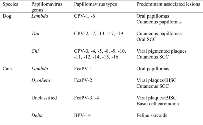
Table 1.Summary of papillomaviruses that have been detected in dogs and cats and their
predominant associated lesions. It should be noted that not all papillomavirus types within a
genus will cause all the associated lesions listed. For example, Canis familiaris
papillomavirus (CPV)-17 is the only canine Taupapillomavirus type associated with oral
SCCs and only CPV-2 and -7 have been associated with cutaneous papillomas. SCC,
squamous cell carcinoma; BISC, Bowenoid in situ carcinoma; FcaPV, Felis catus
papillomavirus; BPV, bovine papillomavirus.
Species Papillomavirus
genus Papillomavirus types Predominant associated lesions
Dog Lambda CPV-1, -6 Oral papillomas
Cutaneous papillomas
Tau CPV-2, -7, -13, -17, -19 Cutaneous papillomas Oral SCC
Chi CPV-3, -4, -5, -8, -9, -10, -11, -12, -14, -15, -16
Viral pigmented plaques Cutaneous SCC
Cats Lambda FcaPV-1 Oral papillomas
Dyotheta FcaPV-2 Viral plaques/BISC
Cutaneous SCC
Unclassified FcaPV-3, -4 Viral plaques/BISC Basal cell carcinoma
Delta BPV-14 Feline sarcoids
837
840
843
Figure Legends
Figure 1. Inverted papilloma, dog. As papillomaviruses promote their replication by
increasing proliferation of epithelial cells, some papillomaviral infections can result in
marked epithelial hyperplasia. If this hyperplasia causes folding of the epidermis, an
exophytic or endophytic papilloma can develop. Scale bar = 100µm. Haematoxylin and eosin.
Figure 2. Inverted papilloma, dog. Papillomaviral replication in a lesion can be detected
histologically by the presence of papillomavirus-induced cell changes. Visible in this
papilloma are numerous enlarged keratinocytes are visible that contain increased quantities of
blue fibrillar material. Additionally, rare cells with shrunken nuclei surrounded by a clear
cytoplasmic halo (koilocytes) are visible (arrows). Scale bar = 20µm. Haematoxylin and
eosin.
Figure 3. Oral papillomatosis, dog. This disease is characterised by the presence of numerous
exophytic vegetative growths involving the lips and mouth. This dog also has cutaneous
papillomas involving the skin surrounding the mouth (Photo courtesy of Dr Stephen White,
UC Davis).
Figure 4. Cutaneous papillomas, dog. Papillomas frequently develop around the ears,
possibly secondary to self-trauma caused by scratching in this area (Photo courtesy of Dr
Stephen White, UC Davis).
Figure 5. Pigmented plaques, dog. Numerous plaques are visible on the ventrum and legs of
this dog. The plaques are dark and are covered by keratin scale (Photo courtesy of Dr Mark
Turnwald, Belmont Veterinary Clinic, North Shore City, New Zealand).
849
852
855
858
861
864
867
870
Figure 6. Feline viral plaque. Plaques most frequently appear as single focal slightly raised
lesions that often develop around the face of cats. Feline viral plaques and Bowenoid in situ
carcinomas appear to be different severities of the same disease process with viral plaques the
more mild manifestation of the disease (Photo courtesy of Dr Sharon Marshall, Veterinary
Associates, Hastings, New Zealand).
Figure 7. Bowenoid in situ carcinoma, cat. This Devon Rex cat developed multiple
pigmented, raised ulcerated lesions that were covered by a layer of keratin. Feline Bowenoid
in situ carcinomas can develop anywhere on the cat, but are often are more common dorsally
on the head and neck (Photo courtesy of Dr Linda Vogelnest, Small Animal Specialist
Hospital, New South Wales, Australia).
876
879
882