Can Genetic Factors Compromise the Success of Dental Implants? A Systematic Review and Meta-Analysis
Joel Ferreira Santiago Junior1*, Claudia Cristina Biguetti2, Mariza Akemi Matsumoto2 Guilherme Abu Halawa Kudo1, Raquel Barroso Parra da Silva2, Patrícia Pinto Saraiva1, Walid D. Fakhouri3,4*
1Pró–Reitoria de Pesquisa e Pós-graduação (PRPPG), Universidade do Sagrado Coração, USC, 10–50 Irmã Arminda, Jardim Brasil, Bauru, 17011–160, SP, Brazil. 2Department of Basic Sciences, São Paulo State University (Unesp), School of Dentistry, Araçatuba, SP. 3Center for Craniofacial Research, Department of Diagnostic and Biomedical Sciences, School of Dentistry, University of Texas Health Science Center at Houston, Houston, TX 77054. 4Department of Pediatrics, McGovern Medical School, University of Texas Health Science Center, Houston, TX 77030.
Abstract
Dental implants provide a predictable treatment option for partial and complete edentulism via the placement of fixed permanent artificial root to support prosthetic dental crowns. Despite the high survival rates, long-term implant failures are still reported leading to implant removal and additional financial and health burdens. While extrinsic factors that improve survival rate of implants have been well explored, the impact of genetic factors on this matter is poorly understood. A systematic review and meta-analysis study was conducted to
determine whether genetic factors contribute to increase the risk of dental implant failure. A comprehensive search for peer-reviewed articles on dental implants and genetics was performed using various literature database libraries. The study design was conducted according to PRISMA guidelines, and the obtained records were registered in PROSPERO database. According to the exclusion/inclusion criteria, 11 studies were eligible for this study out of 808 articles. The meta-analysis of the combined association studies of DNA variations and dental implants did not indicate an increase risk for implant failure due to DNA
variations in IL-1B, IL-10 and TNF-α. This study emphasizes the need for larger randomized controlled clinical trails to inform clinicians and patients about the role of genetic factors on dental implant survival and success rate in healthy and compromised patients.
1. Introduction
Association studies between common DNA variations and human diseases have been proven very useful to identify genetic factors that increase the risk or provide protection to human complex diseases [1, 2]. DNA variations can modify gene expression and function which can increase susceptibility for a disease and affect an individuals phenotype. Each individual carries on average 3 million single nucleotide polymorphisms (SNPs) that can be defined as an alteration of a single nucleotide base that occurs in at least 1% of the human population [1-4]. Therefore, applying this approach should be considered to determine the contribution of genetic factors when it comes to dental implant failure and bone loss [1, 3, 4, 6, 7]. Previous studies reported that various systemic diseases can lead to implant failure, including the influence of immune system regulation on bone metabolism and bone density [6, 8, 9]. It has been emphasized that the host-implant interaction and the response to foreign object could be a major cause for severe marginal loss and consequent implant failure rather than a secondary biofilm-mediated infection [9]. Furthermore, recent reports indicated that poor bone quality can reduce dental implant survival [10-12]. In this context, knowledge of the genetic factors that influence osseointegration and possible longevity of dental implants is pertinent to be investigated in the field of implantology, in order to identify intrinsic risk factors [3]. Notably, the research involving the host response regarding the marginal bone loss in the osseointegrated interface remains unexplored.
Failure of osseointegration occurs due to multifactorial conditions, including individual susceptibility or risk factors. Failure can occur even under proper conditions of bone tissue due to possible host immune responses [6, 13, 14]. Considering this possibility, it is important to investigate the intrinsic characteristics of individuals who experience dental implant failures to identify genetic factors that influence osseointegration [4]. The immune system is important in regulating the balance of cytokines and chemokines during
inflammatory conditions. Depending on the local condition, the presence of pathogens in the oral cavity can alter the components of the immune response such as the cytokines and growth factors involved in the regulation of healing process [14, 15]. In the case of dental implants, the surgical procedures stimulate an initial inflammatory response to the implanted artificial root by the production of several types of cytokines and other mediators, such as interleukins [16]. Different types of interleukins play an important role in bone remodeling by inducing bone resorption (e.g. IL-6), or stimulating bone formation (e.g. IL-10) [1, 4, 7, 17]. While some cytokines act as an anti-inflammatory profile, such as IL-10, other
molecules and cytokines such as IL-2 and IL-6 are involved with pro-inflammatory activity and bone loss [18].
Polymorphisms in the promoters of IL-2 and IL-6 genes were associated with an increase in the expression of both cytokines and with the development of chronic diseases such as periodontitis [1]. High levels of inflammatory mediators have been detected in patients with local infections, which would indicate that local infections could aggravate marginal bone loss and threaten dental implants success [19]. Moreover, inflammatory conditions can affect the balance of other molecules involved in bone matrix homeostasis, such as Matrix Metalloproteinases (MMPs) and their tissue inhibitors (TIMPs) [3].
been conducted to define the contribution of multiple DNA variations and genetic factors to dental implant failure. Therefore, it is important to understand the underlying molecular mechanism that leads to dental implant failure to improve the clinical outcome by preventing or developing targeted therapy. Thus, the aim of the present study is to analyze the relation between genetics and implant failures by means of a systematic review and meta-analysis. The null hypothesis is that genetic factors do not influence dental implant survival rate, while the alternative hypothesis is that certain genetic factors increase the risk for dental implants failure.
2. Methodology
2.1. Standardized criteria and type of study
This systematic review and meta-analysis study was designed according to the established criteria by Cochrane collaboration for the design of the systematic review and meta-analysis [21]. The authors followed the PRISMA criteria [22], as well as the recently published models of systematic review to ensure the standardization of the data
inclusion/exclusion criteria and analysis [12, 23-27].
2.2. Registry protocol
The data of this study was registered by the PROSPERO database under the number CRD42018088458. This data is publicly available for download or analysis on
https://www.crd.york.ac.uk/prospero/display_record.php?RecordID=88458.
2.3. Eligibility criteria
The analysis was designed based on the PICO index as follows; (1) Population: Patients who received oral rehabilitation, (2) Intervention/Exposure: Effects of genetic factors on dental implant failure, (3) Comparison: Group that lost dental implants vs. group that did not lose installed implants within 6 months of treatment, (4) Outcomes: Potential association between DNA variations and dental implant failure with other characteristic phenotypes.
2.3. Inclusion/Exclusion criteria and cohort size 2.3.1. Inclusion criteria
A literature search until February of 2018 was performed to select studies that
contained the following criteria: 1) published in English; 2) minimum clinical follow up of 6 months of retrospective and prospective studies, controlled and randomized clinical trials; 3) Adult patients (≥18 years) that received dental implants were considered.
2.3.2. Exclusion criteria
In vitro, animal studies, clinical reports, reviews, non-controlled or incomplete data were not considered and consequently excluded. Clinical studies with a greater focus on smoking, periodontal disease, or systemic diseases were not included in the systematic review. Clinical studies with less than 10 patients were excluded.
2.4. Search strategy
A search for articles published until February, 2018 were made by PubMed, Cochrane, and Web of Science databases. Boolean operators based on MeSH and PubMed included the following: ``Dental Implants`` and ``bone genes``, ``dental implants`` and `` genetic risk factors``; ``dental implants failure`` and ``genetic risk factors``. Related search by PubMed was: ("dental implants"[MeSH Terms] OR ("dental"[All Fields] AND
Terms] OR ("bone"[All Fields] AND "bones"[All Fields]) OR "bone and bones"[All Fields] OR "bone"[All Fields]) AND ("genes"[MeSH Terms] OR "genes"[All Fields]); ("genetic therapy"[MeSH Terms] OR ("genetic"[All Fields] AND "therapy"[All Fields]) OR "genetic therapy"[All Fields] OR "genetic"[All Fields]) AND ("risk factors"[MeSH Terms] OR ("risk"[All Fields] AND "factors"[All Fields]) OR "risk factors"[All Fields]) AND ("dental implants"[MeSH Terms] OR ("dental"[All Fields] AND "implants"[All Fields]) OR "dental implants"[All Fields]) AND failure[All Fields]; ("dental implants"[MeSH Terms] OR ("dental"[All Fields] AND "implants"[All Fields]) OR "dental implants"[All Fields]) AND failure[All Fields] AND ("genetic therapy"[MeSH Terms] OR ("genetic"[All Fields] AND "therapy"[All Fields]) OR "genetic therapy"[All Fields] OR "genetic"[All Fields]) AND ("risk factors"[MeSH Terms] OR ("risk"[All Fields] AND "factors"[All Fields]) OR "risk factors"[All Fields]).
A manual search from September 2017 until February 2018 was conducted for the following journals: Implantology, Clinical Implant Dentistry and Related Research, Clinical Oral Implants Research, European Journal of Oral Implantology, Implant Dentistry,
International Journal of Oral and Maxillofacial Implants, International Journal of Oral and Maxillofacial Surgery, International Journal of Periodontics and Restorative Dentistry, International Journal of Prosthodontics, Journal of Clinical Periodontology, Journal of Dental Research, Journal of Oral Implantology, Journal of Oral and Maxillofacial Surgery, Journal of Oral Rehabilitation, Journal of Periodontal Research, Journal of Periodontology, and Journal of Prosthetic Dentistry. Additionally, a manual search was conducted in the references of included articles.
2.5. Data collection process
Three previously calibrated reviewers (RBPS, MAM and JFSJ) selected the articles and performed the data collection. Discrepancies in the analysis were solved in a consensus meeting for analysis of the selected titles and abstracts, with an agreement test value for the selected articles in the 3 databases. In order to decrease bias in the selection of the articles, authors GAHK and CB participated in the selection of the sample, data collection and examination of the databases as well. Consensus meetings for the selection of each article within the selected sample pool were held on weekly bases (November, December, 2017 and February, 2018). The group and other researchers worked together to consolidate the analysis of the topics (PPS and WDF).
2.6. Items of extracted data
Data extracted from studies that passed the inclusion criteria were analyzed and the main standardized information were obtained as follows: 1) author; 2) year of publication; 3) country and origin of the study; 4) number of patients; 5) analyzed group of patients; 6) mean age of the patients; 7) number and sites of implants; 8) trade mark of the implants; 9)
implants failure; 10) main data of the implants; 11) data of periodontal evaluation; 12)
studied variables; 13) methodologies; 14) rate of peri-implant bone loss; 15) follow-up period of each study. Data were collected using a standardized file built in Excel software.
2.7. Evaluation of the study quality and risk of bias
The selected clinical studies were evaluated based on their consideration of their methodology approach on a bias scale by the National Health and Medical Research Council (NHMRC - Australian Government). The bias scale allows for evidence levels of different categorical studies (intervention, diagnostic accuracy, prognosis, etiology, screening
controlled study; III-1 pseudorandomised controlled study; III-2 clinical study with a group control; III-3 A comparative study without control group; IV case series [28, 29].
2.8. Measurements and statistical analysis
The quantitative data collected from the articles was tabulated for the analysis of relative risk (RR) with a correspondent 95% confidence interval (CI). For all analysis, significant values were considered as p<0.05. Reviewer Manager 5.3 (Cochrane Group) was used for the meta-analysis and graphic elaboration.
2.9. Anticipated Outcome 2.9.1. Primary outcome
The primary outcome was to analyze if there is a significant association between genetic factors and the failure of dental implants based on previously published studies that passed the inclusion criteria.
2.9.2. Risk of bias of quantitative data
A fixed-effects model was applied in case no significant differences were observed in the data. Alternatively, a random effects model was applied in case significant differences (high heterogeneity among the tests) were observed. Heterogeneity was considered
significant at p<0.1 and was evaluated using the Q (x2) test and I2 value. Statistical I2 value was used in the analysis of heterogeneity variations, and values above 75 (0-100) were considered to indicate significant heterogeneity [23, 30-32].
2.9.3. Additional analysis
Sensitivity tests for the analysis of patient subgroups and the allele frequency of different types of alleles for the genes ILs and TNFα were made in order to avoid potential heterogeneity due to different groups of patients (failure vs. control group) [12, 33].
3. Results
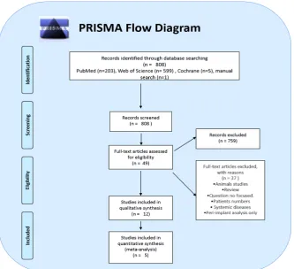
A total of 808 articles were identified from the three online databases searches. After applying the inclusion/exclusion criteria, 49 articles were eligible for full-text assessment. After a complete assessment, 12 articles fulfilled the inclusion criteria for qualitative analysis and five studies for meta-analysis, as shown in Figure 1.
Figure 1. A flowchart representing the literature search, screening, eligibility and selection of this study.
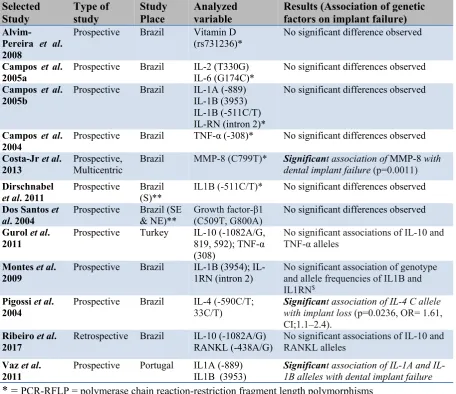
For statistical analysis different kinds of cellular mediators were evaluated based on PICO criteria, with a focus on selecting studies that provided data on genetic factors that can lead to implant failure,such as vitamin D receptor polymorphism D [6], interleukins; IL-1B [4], IL1A and IL1B [17, 34], IL1B and IL1RN [20], IL-2 [1], IL-4 [16] IL-10 [14], and IL-10 [5], Tumor necrosis factor-α [19], metalloproteinase-8 [3], growth factor β1 [7], RANKL [5]. The selected studies presented similar related methodologies to process their samples,
however, there were several distinct differences. Eight of the studies used epithelial cells extracted from the oral mucosa, amplified by polymerase chain reaction (PCR) and then analyzed by restriction fragment length polymorphism (RFLP) [1, 4, 6, 7, 16, 19, 20, 34]. One study used PCR and restriction endonuclease digestion [3], while two other studies used amplification refractory mutation system coupled with PCR [5, 14]. One study used the variable number of tandem repeat (VNTR) [16], and another study used amplification for PCR and reversal hybridization [17].
Table 1. A summarized data of the articles selected in this study and the association between genetic factors and dental implant failure.
Selected Study Type of study Study Place Analyzed variable
Results (Association of genetic factors on implant failure)
Alvim-Pereira et al. 2008
Prospective Brazil Vitamin D
(rs731236)* No significant difference observed
Campos et al.
2005a Prospective Brazil IL-2 (T330G) IL-6 (G174C)* No significant differences observed Campos et al.
2005b Prospective Brazil IL-1A (-889) IL-1B (3953) IL-1B (-511C/T) IL-RN (intron 2)*
No significant differences observed
Campos et al.
2004 Prospective Brazil TNF-α (-308)* No significant differences observed Costa-Jr et al.
2013 Prospective, Multicentric Brazil MMP-8 (C799T)* Significandental implant failuret association of (p=0.0011)MMP-8 with Dirschnabel
et al. 2011 Prospective Brazil (S)** IL1B (-511C/T)* No significant differences observed Dos Santos et
al. 2004 Prospective Brazil (SE & NE)** Growth factor-β1 (C509T, G800A) No significant differences observed
Gurol et al.
2011 Prospective Turkey IL-10 (-1082A/G, 819, 592); TNF-α (308)
No significant associations of IL-10 and TNF-α alleles
Montes et al. 2009
Prospective Brazil 1B (3954); IL-1RN (intron 2)
No significant association of genotype and allele frequencies of IL1B and IL1RN$
Pigossi et al.
2004 Prospective Brazil IL-4 33C/T)(-590C/T; Significanwith implant losst association of IL-4 (p=0.0236, OR= 1.61, C allele CI;1.1–2.4).
Ribeiro et al.
2017 Retrospective Brazil IL-10 (-1082A/G) RANKL (-438A/G) No significant associations of IL-10 and RANKL alleles
Vaz et al.
2011 Prospective Portugal IL1A (-889) IL1B (3953) Significan1B alleles with dental implant failure t association of 1A and IL-* = PCR-RFLP = polymerase chain reaction-restriction fragment length polymorphisms
** = S (South), SE (Southeast), NE (Northeast)
$ = The number of teeth present was observed to influence implant loss, p=0,027.
3.1. Clinical Parameters
A total of 1,853 patients combined from twelve studies were included for this
systematic review and meta-analysis. Among them, only one study considered a single group of patients without including a control group [5]. The other studies used the comparison between two groups, one with successful implants (control group: 1106 patients), and the other with patients who presented with at least one implant failure (test or study group: 647 patients) [1, 3, 4, 6, 7, 14, 16, 17, 19, 20, 34]. In relation to the follow-up time, some studies limited the monitoring period of the control and study groups to one year [4, 16, 19]. One study examined the survival rate 9 months post-operation [3], and another study chose a 6 months follow-up [7].
number of failures. Alvim-Pereira et al., (2008) emphasized that 50% of the failures occurred before 20 weeks (range: 0-237 weeks) from the implant date. Out of all the studies, only five studies revealed the trademark of the dental implants [5-7, 16, 19]. Some studies highlighted clinical conditions which could reflect on the survival of dental implants. Alvim-Pereira et al. (2008) reported the effects of the site of installation (maxillae and mandible, p=0.003), posterior/anterior (p=0.037), mean length of the implant (p=0.001), primary stability
(p=0.001), surgical technique (p=0.016), the quality of bone tissue (p=0.049), and edentulism (p=0.009) on dental implant survival. Dirschnabel et al. (2011) also related the loss of
implants as due to the edentulism (p=0.019), site of implant installation (p=0.001) and
medical/systemic conditions of the patient (p=0.04). Similarly, Pigossi et al. (2004) identified edentulism (p=0.031), maxillae/mandible (p=0.003), anterior/posterior regions (p=0.037), primary stability (p=0.0010, and implant load (p=0.001) as factors relating to implant survival. Montes et al. (2009) highlighted that genotype 2/2 of IL1RN (intron 2) was associated with failure of implants in individuals with multiple dental implant losses in addition to factors such as edentulism and the number of teeth present. In addition, some studies analyzed the periodontal condition of the remaining teeth, detecting significant differences in probing depth index in comparison to the study and control groups, (p=0,002 [16], p=0,011 [6], p=0,005 [4], p=0,011 [20]). Other clinical periodontal variables did not reveal any significant differences, as shown in Table 2.
3.2. Meta-analysis outcome
Studies that analyzed the same allele of the same gene of interest were grouped together to increase the power of statistical analysis and the level of significance by comparing the control groups with no dental implant failure (control group) and the group with implant failure (study group). Increase of power analysis can be achieved by having a larger sample size of experimental and control groups with similar environmental conditions. Therefore, this study sought to combine all related studies that analyzed the association between DNA variations of genetic factors and dental implant failure. For each DNA allele, two studies were combined for the frequency distribution of genetic alleles. A high heterogeneity in the frequencies of the alleles and Odds Ratio (OR) across the individual studies (I2 > 50%) was
observed for IL1B (study group), TNF-α (study group). On the other hand, acceptable measures (I2) were identified for study group of IL10 (Failure group), IL1B (Control G vs.
Table 2. The size of the cohortsand the dental implants clinical data of the articles selected in this study. Selected
Studies
No. Patient
Groups Ave.Age (years)
Implants Trade Mark
Periodontal evaluation (partially edentulous patients)
Alvim-Pereira et al. 2008
217 CG: 137
SG: 80
51.7±11.3 CG:1232
SG: 135
Neodent Gingival Index: 0,64±0,38 (CG), 0,65± 0,55 (SG).
Plaque Index: 0,14±0,26 (CG), 0,25± 0,42 (SG). Calculus Index: 0,08±0,13 (CG), 0,14± 0,25 (SG).
Probing attachment (mm): 2,68±0,41 (CG), 2,52±0,47 (SG). Clinical attachment (mm): 3,61±0,76 (CG), 3,66±1,10 (SG). Mobility (absence/presence): 98/13 (CG); 60/15 (SG)
Campos et al.
2005a 74 CG: 40 SG: 34 43.8 49.3 NI NI NI
Campos et al. 2005b
72 CG: 34
SG: 28
43.3 52.7
NI 97
3i/
Conexão
NI
Campos et al.
2004 66 CG:38* SG: 28 NC NI 3i
/
Conexão NI
Costa-Jr et al. 2013
180 CG:100**
SG: 80
>18 NI NI NI
Dirschnabel
et al. 2011 277 CG:185* SG: 92 53.6±11.1 NI NI Gingival Index: 0,64±0,37 (CG), 0,65± 0,53 (SG) Plaque Index: 0,12±0,23 (CG), 0,23± 0,41 (SG) Calculus Index: 0,07±0,12 (CG), 0,13± 0,24 (SG)
Probing attachment (mm): 2,72±0,46 (CG), 2,54±0,47 (SG) Clinical attachment (mm): 3,62 ±0,85 (CG), 3,66±1,07 (SG) Mobility (absence/presence): 132/19 (CG), 70/15 (SG)
Dos Santos et al. 2004
68 CG:40$
SG: 28
>18 NI 3i/
Conexão
NI
Gurol et al.
2011 108 CG: 70 SG: 38 25-48 NI 16 NI NI
Montes et al. 2009
266 SG: 90
CG: 176
51.5±11.5 1232 135
Neodent Gingival Index: 0.63±0.38 (CG) and 0.65± 0.53 (SG)
Plaque Index: 0.12±0.24 (CG) and 0.24± 0.42 (SG) Calculus Index: 0.07±0.12 (CG) and 0.13± 0.24 (SG). Probing attachment (mm): 2.72±0.46 (CG) and 2.55±0.47 (SG).
Clinical attachment (mm): 3.61 ±0.85 (CG) and 3.67±1.07 (SG). Dental Mobility 18 (142) (CG); 16 (83) (SG)
doi:10.20944/preprints201807.0443.v1
Peer-reviewed version available at
Pigossi et al.
2014 280 CG: 186* SG: 94 56.1 ±11.3 1232 135 Neodent
Gingival Index: 0.63±0.38 (CG) and 0.64± 0.28 (SG)
Plaque Index: 0.12±0.23 (CG) and 0.23± 0.41 (SG) Calculus Index: 0.07±0.12 (CG) and 0.13± 0.24 (SG).
Probing attachment level (mm): 2.72±0.46 (CG) and 2.55±0.47 (SG).
Clinical attachment level (mm): 3.61 ±0.85 (CG) and 3.67±1.07 (SG).
Dental Mobility 19 (12.5) (CG); 16 (18.6) (SG)
Ribeiro et al.
2017 90 1 Group 54.5 245 Straumann
NI
Vaz et al.
2011 155 CG:100 SG: 55 NI NI NI NI
NI = Not Informed; Neodent = Curitiba, PR, Brazil; 3i: 3i = Palm Beach Gardens, Florida, USA; Straummann = Bern, Switzerland; SG = Study group; CG = Control
group without loss of implant; SG = Study group with loss of at least 1 implant; *CG = patients without implant loss for 1 year of follow-up; **CG = implants installed with at least 9 months of follow-up; $CG = implants installed with at least 6 months of follow-up.
doi:10.20944/preprints201807.0443.v1
Peer-reviewed version available at
Genes
2018
,
9
, 444;
The genetic data of two studies on the role of IL1B on dental implant failure were combined because they used the same SNP (rs16944) or allele (IL1B-C511) of IL1B. The genetic data was divided into four subgroups including the prevalence of IL1B-C511 allele in study (failure) group and control group, and the prevalence of IL1B-T511 allele in failure group and control group. The subgroups for each allele were combined from the two individual studies and analyzed for an association between study groups from both studies and dental implant failure [4, 34]. Analysis of the two subgroups IL1B-C511 in study group (failure G) and control group (control G) shows a pooled odd ratio of 0.85, 95% CI of 0.62-1.18, and p-value 0.33, indicating no significant association in carrying this allele and increase risk for implant failure (Figure 2). Similarly, no significant association between T511 allele and implant failure was identified compared to control group with a pooled OR of 1.17 (95% CI: 0.85-1.61), p = 0.33 (Figure 3). The confidence intervals of both pooled ORs crossed the line of no effect indicating that both alleles do not increase the risk for dental implant failure under these circumstances of both studies.
Figure 2. Odd ratio and forest plot for the prevalence of IL1B-C511 allele in the failure group (at least one implant failure) vs. control group (without implant failure). The odd ratio of the combined data crossed the line of no difference.
Figure 3. Odd ratio and forest plot for the prevalence of IL1B-T511 allele in the failure group (at least one implant failure) vs. control group (without implant failure). The odd ratio of the combined data crossed the line of no difference.
Other two studies showed a comparison between the failure and control groups for IL-10 [5, 14] in the analysis of the comparison between groups the pooled OR was 0.72 (95% CI: 0.43-1.22), p = 0.22, indicating no significant odds for the presence of the G allele in any of the groups (Figure 4). Furthermore, no significant difference in A allele expression was identified for both groups, the pooled OR was 1.38 (95% CI: 0.82-2.33), p = 0.22 (Figure 5).
Figure 5. Odd ratio and forest plot for the prevalence of IL10-A1082 allele in the failure group (at least one implant failure) vs. control group (without implant failure). The odd ratio of the combined data crossed the line of no difference.
Finally, two studies showed a comparison between the failure and control groups for TNF-α [14, 19], in the analysis of the comparison between groups the pooled OR was 1.04 (95% CI: 0.48-2.24), p = 0.92, indicating no significant odds for the presence of the A allele in any of the groups (TNF-α: failure vs. control group), (Figure 6). As well as, no significant difference in G allele expression was identified for both groups, the pooled OR was 0.96 (95% CI: 0.45-2.08), p = 0.92 (Figure 7).
Figure 6. Odd ratio and forest plot for the prevalence of TNF-α-A308 allele in the failure group (at least one implant failure) vs. control group (without implant failure). The odd ratio of the combined data crossed the line of no difference.
Figure 7. Odd ratio and forest plot for the prevalence of TNF-α-G308 allele in the failure group (at least one implant failure) vs. control group (without implant failure). The odd ratio of the combined data crossed the line of no difference.
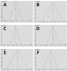
Figure 8. A funnel graph for the homogeneity representation of the meta-analysis. The data shows that the plotted homogeneity is acceptable for the DNA alleles used in the meta-analysis, including the IL1B-C allele study group (A), IL1B-T allele (B), IL10-G allele (C), IL10-A allele (D), TNFa-A allele (E), and TNFa-G allele (F).
4. Discussion
Dental implants provide an excellent treatment option for patients with missing teeth via the replacement of tooth roots with a fixed permanent artificial root to match the natural ones and support prosthetic dental crowns. Despite the high success rate, implant failures are still common. In this context, long-term implant failure is generally a result of a severe bone marginal loss and bone resorption related to secondary infection [9]. Additionally, despite of some clinical similarities with tooth lost post periodontitis, recent clinical perspective studies in dentistry have shown that implant failure caused by severe bone resorption is not
implants, mainly in cases of low bone density [12, 38, 39]. Most of the implant failures occurred before a functional loading (around 81.3%), suggesting an important role of the host recipient site during the osseointegration process [16].
Due to the limitation of the included studies in this systematic review, it is important to note that none of these studies had indicated a sample randomization process, which might reflect a lower score on the bias scale of funnel graph analysis. Some of the selected studies presented initial sample randomization, however a smaller number of the patients from those samples were chosen for their study and control groups [5, 6, 16, 34]. Vaz et al. (2017) performed sample calculations for the constituents of their test and control samples. In order to reduce possible biases in sample selection, including a large number of patients with randomized control trails that take into consideration a match for age, sex, ethnicity and gender among control and patients, some studies performed an equalization in order to homogenize the groups with a better division between smoking patients, ethnic groups, gender and age [4, 6, 16].
Bone peri-implant marginal loss can be aggravated by chronic or systemic diseases such as cardiovascular diseases, osteoporosis, diabetes, hepatitis, severe periodontal disease, chemo or radiotherapy, HIV positive infection, pregnancy or lactation, or even large bone reconstructions that compromise bone regeneration and integration. The included studies used in this systematic review selected patients who were healthy and with no systemic diseases [1, 3, 5-7, 14, 16, 17, 19]. However, one of the studies did not reveal the exclusion criteria of the patients [4]. Several clinical studies have focused on the role of interleukins in dental implant failure, because the clinical procedure of implant exhibits a higher level of interleukins after the first day of the implant installation due to normal local inflammation [1]. A higher cytokine activity in bone metabolism could enhance peri-implant bone loss leading to failure of the implant [1, 40-43]. Unfortunately, this biological effect was not fully proven in this study because the meta-analysis of combined studies did not show increase in the risk of dental implant failure in presence of a specific type of allele in two IL genes. These results need to be analyzed with caution, since only two studies were considered in the sample: IL1B (C511T, rs16944) [34], IL1B (C511T) [4]. Therefore, additional well-designed studies should be performed in order to analyze the influence of IL1B on failure of dental implants.
A study by Campos et al. (2005) analyzed DNA polymorphism in only one promoter of each IL gene, and reported that genes can present a number of polymorphic sites that act together. In this context, another publication suggests that genetic polymorphisms probably interfere with the process of osseointegration though a cumulative effect of multiple
polymorphisms [3]. Another important point to consider is ethnicity because the majority of the selected studies are concentrated in Brazil, a country where its heterogeneity is composed mainly of Portuguese, Spanish and Italians [1, 19]. The clinical study developed by
Dirschanebel et al. (2011) included 96.4% Caucasians individuals, likely from southeast Brazil, in their sample. It is possible that samples of other populations or ethnic groups can provide more insights and information [1].
Two clinical studies justified the lack of significant evidence due to sample size [17, 19]. In fact, literature guidelines have stated that the clinical studies should be delineated, including a sample calculation [3, 14], since it is difficult to compile a group of patients with implant loss and with specific exclusions such as smokers [3]. Furthermore, more studies should evaluate other cytokine genes that could influence an association between periodontal disease and the failure of dental implants [14]. One of the studies reported that the key for the success of osseointegration was not related to the level of cytokines production, but an
the meta-analysis and on the majority of the included studies since they did not identify significant associations of the analyzed genetic factors with the failure of dental implants [1, 4-7, 14, 19]. However, a recent study reported that while only the +33C allele of the IL-4 gene was associated with susceptibility of implant loss, when a SNP was included in the analysis of haplotype IL-4, a statistical difference was not identified [16]. It emphasizes the importance of a balanced analysis of the patients’ profiles in control and study groups, reinforcing the positive impact of randomized control studies.
Multiple published studies highlighted the importance of considering the levels of marginal bone loss around dental implants for future clinical trails [4]. Furthermore, a recent systematic review study showed that there is a moderate association between peri-implantitis and higher expression of inflammatory cytokines, indicating that the evidences are still limited in this research area [44]. Finally, it is important that further controlled and
randomized studies be developed to establish a more precise answer to the question of this systematic review. The planning of the surgical technique, the study of anatomy, pre-existence risk factors, primary stability, quality and quantity of bone tissue have all been indicated as important factors involved in dental implant survival.
In conclusion, the meta-analysis of the combined genetic studies did not show
increase risk or protection in dental implant failure due to DNA variations in IL1B, IL10 and TNFα in study groups compared to control groups. Hence, there is no strong evidence that genetic factors can lead to the failure of oral rehabilitation with dental implants. It is
important that additional randomized controlled studies with large sample size be conducted in order to conclusively determine whether there is possible effect of genetic risk factors on implant failure and on marginal bone loss. These findings will help to identify those
individuals with higher risk for dental implant loss facilitating the preparation of prevention strategies and individualized therapies in order to increase survival rates of oral rehabilitation with dental implants [3-5].
Author contributions: Conceptualization, MAM, JFSJR, CCB, WDF; original draft preparations, MAM, JFSJR, CCB, RBPS, GAHK, PPS; Review and editing: MAM, JFSJR,CCB, WDF; supervision, PPS,WDF; Funding acquisition, JFSJR, WDF.
Acknowledgements: We are grateful to Ms. Jamie Jiang and Ms. Neha Sharma for reviewing the manuscript.
Funding: This study was funded by the Research Foundation of the State of São Paulo, (FAPESP) grant #2017/13276-5 (expansion of the research group with international
participation), and grant #2015/20827-2 (support for the software acquisition and subsidy for development of systematic review). We also thank the support from NIH grant
R15GM122030-01 to WDF.
References
1. Campos, M. I.; Godoy dos Santos, M. C.; Trevilatto, P. C.; Scarel-Caminaga, R. M.; Bezerra, F. J.; Line, S. R., Interleukin-2 and interleukin-6 gene promoter
polymorphisms, and early failure of dental implants. Implant Dent 2005a, 14, 391-6. 2. Thompson, M.; McInnes, R.; Willard, H., Genes in population. In Genetics in Medicine,
6th ed.; Company, W. S., Ed. Philadelphia, 2001; p 540.
3. Costa-Junior, F. R.; Alvim-Pereira, C. C.; Alvim-Pereira, F.; Trevilatto, P. C.; de Souza, A. P.; Santos, M. C., Influence of MMP-8 promoter polymorphism in early osseointegrated implant failure. Clin Oral Investig 2013, 17, 311-6.
4. Dirschnabel, A. J.; Alvim-Pereira, F.; Alvim-Pereira, C. C.; Bernardino, J. F.; Rosa, E. A.; Trevilatto, P. C., Analysis of the association of IL1B(C-511T) polymorphism with dental implant loss and the clusterization phenomenon. Clin Oral Implants Res 2011, 22 , 1235-41.
5. Ribeiro, R.; Melo, R.; Tortamano Neto, P.; Vajgel, A.; Souza, P. R.; Cimoes, R., Polymorphisms of Il-10 (-1082) and RANKL (-438) Genes and the Failure of Dental Implants. Int J Dent 2017, 2017, 3901368.
6. Alvim-Pereira, F.; Montes, C. C.; Thome, G.; Olandoski, M.; Trevilatto, P. C., Analysis of association of clinical aspects and vitamin D receptor gene polymorphism with dental implant loss. Clin Oral Implants Res 2008, 19, 786-95.
7. Dos Santos, M. C.; Campos, M. I.; Souza, A. P.; Scarel-Caminaga, R. M.;
Mazzonetto, R.; Line, S. R., Analysis of the transforming growth factor-beta 1 gene promoter polymorphisms in early osseointegrated implant failure. Implant Dent 2004, 13, 262-9.
8. Mombelli, A.; Cionca, N., Systemic diseases affecting osseointegration therapy. In Clin Oral Implants Res, Denmark, 2006; Vol. 17 Suppl 2, pp 97-103.
9. Albrektsson, T.; Dahlin, C.; Jemt, T.; Sennerby, L.; Turri, A.; Wennerberg, A., Is marginal bone loss around oral implants the result of a provoked foreign body reaction?
Clin Implant Dent Relat Res 2014, 16, 155-65.
10. Gaetti-Jardim, E. C.; Santiago-Junior, J. F.; Goiato, M. C.; Pellizer, E. P.; Magro-Filho, O.; Jardim, E. G., Dental implants in patients with osteoporosis: A clinical reality?
Journal of Craniofacial Surgery 2011, 22, 1111-1113.
11. Goiato, M. C.; dos Santos, D. M.; Santiago, J. F. J.; Moreno, A.; Pellizzer, E. P., Longevity of dental implants in type IV bone: a systematic review. International Journal
of Oral and Maxillofacial Surgery 2014, 43, 1108-1116.
12. de Medeiros, F.; Kudo, G. A. H.; Leme, B. G.; Saraiva, P. P.; Verri, F. R.; Honorio, H. M.; Pellizzer, E. P.; Santiago Junior, J. F., Dental implants in patients with
osteoporosis: a systematic review with meta-analysis. Int J Oral Maxillofac Surg 2017. 13. Esposito, M.; Hirsch, J. M.; Lekholm, U.; Thomsen, P., Biological factors contributing
to failures of osseointegrated oral implants. (I). Success criteria and epidemiology. Eur J
Oral Sci 1998, 106, 527-51.
14. Gurol, C.; Kazazoglu, E.; Dabakoglu, B.; Korachi, M., A comparative study of the role of cytokine polymorphisms interleukin-10 and tumor necrosis factor alpha in
susceptibility to implant failure and chronic periodontitis. Int J Oral Maxillofac Implants 2011, 26, 955-60.
15. Takashiba, S.; Naruishi, K., Gene polymorphisms in periodontal health and disease.
Periodontol 2000 2006, 40, 94-106.
17. Vaz, P.; Gallas, M. M.; Braga, A. C.; Sampaio-Fernandes, J. C.; Felino, A.; Tavares, P., IL1 gene polymorphisms and unsuccessful dental implants. Clin Oral Implants Res 2012, 23, 1404-13.
18. Cavalla, F.; Araujo-Pires, A. C.; Biguetti, C. C.; Garlet, G. P. Cytokine Networks regulating inflammantion and immune defense in the oral cavity Curr Oral Health Rep [Online], 2014, p. 104-113.
19. Campos, M. I.; dos Santos, M. C.; Trevilatto, P. C.; Scarel-Caminaga, R. M.; Bezerra, F. J.; Line, S. R., Early failure of dental implants and TNF-alpha (G-308A) gene
polymorphism. Implant Dent 2004, 13, 95-101.
20. Montes, C. C.; Alvim-Pereira, F.; de Castilhos, B. B.; Sakurai, M. L.; Olandoski, M.; Trevilatto, P. C., Analysis of the association of IL1B (C+3954T) and IL1RN (intron 2) polymorphisms with dental implant loss in a Brazilian population. Clin Oral Implants Res 2009, 20, 208-17.
21. Higgins, J.; Green, S., Cochrane Handbook for Systematic Reviews of Interventions Version 5.1.0. Collaboration, T. C., Ed. The Cochrane Collaboration: 2011.
www.cochrane-handbook.org.
22. Moher, D.; Liberati, A.; Tetzlaff, J.; Altman, D. G., Preferred reporting items for systematic reviews and meta-analyses: the PRISMA statement. PLoS Med 2009, 6, e1000097.
23. Santiago Junior, J. F.; de Souza Batista, V. E.; Verri, F. R.; Honorio, H. M.; de Mello, C. C.; Almeida, D. A. d.; Pellizzer, E. P., Platform-switching implants and bone
preservation: a systematic review and meta-analysis. International Journal of Oral and
Maxillofacial Surgery 2016, 45, 332-345.
24. Lopes, L. F. d. P.; da Silva, V. F.; Santiago, J. F., Jr.; Panzarini, S. R.; Pellizzer, E. P., Placement of dental implants in the maxillary tuberosity: a systematic review.
International Journal of Oral and Maxillofacial Surgery 2015, 44, 229-238.
25. Araujo Lemos, C. A.; Verri, F. R.; de Souza Batista, V. E.; Santiago Junior, J. F.; Mello, C. C.; Pellizzer, E. P., Complete overdentures retained by mini implants: A systematic review. Journal of Dentistry 2017, 57, 4-13.
26. Lemos, C. A.; de Souza Batista, V. E.; Almeida, D. A.; Santiago Junior, J. F.; Verri, F. R.; Pellizzer, E. P., Evaluation of cement-retained versus screw-retained implant-supported restorations for marginal bone loss: A systematic review and meta-analysis. J
Prosthet Dent 2015.
27. Lemos, C. A. A.; Verri, F. R.; Bonfante, E. A.; Santiago Junior, J. F.; Pellizzer, E. P., Comparison of external and internal implant-abutment connections for implant
supported prostheses. A systematic review and meta-analysis. J Dent 2017.
28. Lopes, L. F.; da Silva, V. F.; Santiago, J. F., Jr.; Panzarini, S. R.; Pellizzer, E. P., Placement of dental implants in the maxillary tuberosity: a systematic review. Int J Oral
Maxillofac Surg 2015, 44, 229-38.
29. NHMRC. National Health and Medical Research Council. How to use the evidence: assessment and application of scientific evidence. (accessed 02/17/2018).
30. Higgins, J. P.; Thompson, S. G., Quantifying heterogeneity in a meta-analysis. Stat Med 2002, 21, 1539-58.
31. Atieh, M. A.; Ibrahim, H. M.; Atieh, A. H., Platform switching for marginal bone preservation around dental implants: a systematic review and meta-analysis. J
Periodontol 2010, 81, 1350-66.
33. Santiago, J. F. J.; de Souza Batista, V. E.; Verri, F. R.; Honorio, H. M.; de Mello, C. C.; Almeida, D. A.; Pellizzer, E. P., Platform-switching implants and bone preservation: a systematic review and meta-analysis. Int J Oral Maxillofac Surg 2016, 45, 332-345. 34. Campos, M. I.; Santos, M. C.; Trevilatto, P. C.; Scarel-Caminaga, R. M.; Bezerra, F.
J.; Line, S. R., Evaluation of the relationship between interleukin-1 gene cluster polymorphisms and early implant failure in non-smoking patients. Clin Oral Implants Res 2005b, 16, 194-201.
35. de Souza Batista, V. E.; Santiago Junior, J. F.; de Faria Almeida, D. A.; de Toledo Piza Lopes, L. F.; Verri, F. R.; Pellizzer, E. P., The Effect of Offset Implant Configuration on Bone Stress Distribution: A Systematic Review. Journal of Prosthodontics-Implant
Esthetic and Reconstructive Dentistry 2015, 24, 93-99.
36. Lemos, C. A.; Verri, F. R.; Batista, V. E.; Junior, J. F.; Mello, C. C.; Pellizzer, E. P., Complete overdentures retained by mini implants: A systematic review. J Dent 2016. 37. Goiato, M. C.; Pellizzer, E. P.; Moreno, A.; Gennari-Filho, H.; dos Santos, D. M.;
Santiago, J. F., Jr.; dos Santos, E. G., Implants in the zygomatic bone for maxillary prosthetic rehabilitation: a systematic review. Int J Oral Maxillofac Surg 2014, 43, 748-57.
38. Goiato, M. C.; dos Santos, D. M.; Santiago, J. F., Jr.; Moreno, A.; Pellizzer, E. P., Longevity of dental implants in type IV bone: a systematic review. Int J Oral Maxillofac
Surg 2014, 43, 1108-16.
39. Santiago Junior, J. F.; Verri, F. R.; de Faria Almeida, D. A.; de Souza Batista, V. E.; Araujo Lemos, C. A.; Pellizzer, E. P., Finite element analysis on influence of implant surface treatments, connection and bone types. Materials Science & Engineering
C-Materials For Biological Applications 2016, 63, 292-300.
40. Pietruski, J. K.; Pietruska, M. D.; Stokowska, W.; Pattarelli, G. M., Serum levels of interleukin-1 (IL-1), interleukin-6 (IL-6) and interleukin-8 (IL-8) in patients treated with dental implants. Rocz Akad Med Bialymst 2001, 46, 28-37.
41. Kao, R. T.; Curtis, D. A.; Richards, D. W.; Preble, J., Increased interleukin-1 beta in the crevicular fluid of diseased implants. Int J Oral Maxillofac Implants 1995, 10, 696-701.
42. Panagakos, F. S.; Aboyoussef, H.; Dondero, R.; Jandinski, J. J., Detection and
measurement of inflammatory cytokines in implant crevicular fluid: a pilot study. Int J
Oral Maxillofac Implants 1996, 11, 794-9.
43. Salcetti, J. M.; Moriarty, J. D.; Cooper, L. F.; Smith, F. W.; Collins, J. G.; Socransky, S. S.; Offenbacher, S., The clinical, microbial, and host response characteristics of the failing implant. Int J Oral Maxillofac Implants 1997, 12, 32-42.