International Journal of Nanomedicine
A role of cellular glutathione in the differential
effects of iron oxide nanoparticles on
antigen-specific T cell cytokine expression
Chien-Chang Shen1 Hong-Jen Liang2 Chia-Chi Wang3 Mei-Hsiu Liao4 Tong-Rong Jan1
1Department and Graduate Institute
of Veterinary Medicine, School of Veterinary Medicine, National Taiwan University, Taipei, 2Innovation
and Incubation Center, Yuanpei University, Hsinchu, 3School of
Pharmacy, Kaohsiung Medical University, Kaohsiung, 4Division of
Isotope Application, Institute of Energy Research, Taoyuan, Taiwan
Correspondence: Tong-Rong Jan Department and Graduate Institute of Veterinary Medicine, School of Veterinary Medicine, National Taiwan University, No 1, Sec 4, Roosevelt Road, Taipei 10617, Taiwan Tel +886 2 3366 1287 Fax +886 2 2366 1475 Email tonyjan@ntu.edu.tw
Background: Accumulating evidence indicates that iron oxide nanoparticles modulate immune responses, and induce oxidative stress in macrophages. It was recently reported that iron oxide nanoparticles attenuated antigen-specific immunity in vivo, though the underlying mechanism remains elusive. The present study investigates the direct effect of iron oxide nanoparticles on antigen-specific cytokine expression by T cells, and potential underlying mechanisms.
Methods: Ovalbumin-primed splenocytes were exposed to iron oxide nanoparticles, followed by restimulation with ovalbumin. Cell viability, cytokine production, and cellular levels of glutathione and reactive oxygen species were measured.
Results: The splenocyte viability and the production of interleukin-2 and interleukin-4 were unaffected, whereas interferon-γ production was markedly attenuated by iron oxide nanopar-ticles (10–100 µg iron/mL) in a concentration-dependent manner. Iron oxide nanoparticles also transiently diminished the intracellular level of glutathione, with a peak response at 6 hours posttreatment. The effects of iron oxide nanoparticles on interferon-γ and glutathione were attenuated by the presence of N-acetyl-L-cysteine, a precursor of glutathione. However, iron oxide nanoparticles did not influence the generation of reactive oxygen species.
Conclusion: Iron oxide nanoparticles induced a differential effect on antigen-specific cytokine expression by T cells, in which the T helper 1 cytokine IFN-γ was sensitive, whereas the T helper 2 cytokine interleukin-4 was refractory. In addition, the suppressive effect of iron oxide nanoparticles on interferon-γ was closely associated with the diminishment of glutathione.
Keywords: iron oxide nanoparticle, T cell, antigen-specific, glutathione, cytokine
Introduction
Nanomaterials have been increasingly applied in numerous fields, including nanomedicine. Among various biomedical nanoparticles, superparamagnetic iron oxide nanoparticles have been used in clinical settings as a contrasting agent to
enhance magnetic resonance imaging.1,2 In addition, iron oxide particles have shown
promising potential for cell labeling, cancer therapy, and drug delivery.2–4 It has been
well-documented that iron oxide nanoparticles administered systemically are rapidly engulfed by the reticuloendothelial system, with the liver and spleen being the main
distribution sites for the particles.5–7 Because phagocytes are one of the major cell
groups exposed to iron oxide nanoparticles, the potential effect of nanoparticles on the functionality of immune cells and host immune competency is a concern, and is under intensive investigation.
Accumulating evidence indicates that exposure to iron oxide nanoparticles causes
apoptosis and alters the functionality of macrophages.8–12 Primary macrophages exposed
Dove
press
O R I G I N A L R E S E A R C H open access to scientific and medical research
Open Access Full Text Article
International Journal of Nanomedicine downloaded from https://www.dovepress.com/ by 118.70.13.36 on 23-Aug-2020
For personal use only.
Number of times this article has been viewed
This article was published in the following Dove Press journal: International Journal of Nanomedicine
to iron oxide nanoparticles in culture showed a marked increase of apoptosis, accompanied by elevated generation of
reactive oxygen species (ROS).10 Similarly, exposure of the
murine macrophage cell line J774 to iron oxide nanoparticles resulted in an increased production of intracellular ROS,
with subsequent cell injury and apoptosis.13 Murine studies
further showed that a single intratracheal instillation of iron oxide nanoparticles had proinflammatory and prooxidative effects, as evidenced by a marked infiltration of inflammatory cells into the lungs and a diminished level of intracellular
glutathione in bronchoalveolar lavage cells.12 Collectively,
these results have demonstrated the immunomodulatory and cytotoxic effects of iron oxide nanoparticles on macrophages, possibly through oxidative stress-related mechanisms.
In addition to macrophages, other immune cells are also sensitive to iron oxide nanoparticles. For example, the function of dendritic cells to process antigens and stimulate
T cells were suppressed by iron oxide nanoparticles.14 Several
animal studies have reported that T cells are another target in the immune system sensitive to iron oxide nanoparticles. Both oral and intravenous administrations of iron oxide nanoparticles to normal nonsensitized mice altered T cell cellularity.15,16 The serum levels of interleukin (IL)-2, IL-10,
and interferon (IFN)-γ were elevated in mice intravenously
treated with iron oxide nanoparticles.16 Furthermore, it was
recently reported that a single systemic administration of iron oxide nanoparticles attenuated the serum production of
antigen-specific immunoglobulin (IgG)1 and IgG2a and the
expression of IL-4 and IFN-γ by splenocytes, in ovalbumin
(OVA)-sensitized mice.17 These results demonstrate that
exposure to iron oxide nanoparticles influences T cell func-tionality in both nonsensitized and antigen-sensitized mice. However, the underlying mechanisms remain elusive, and whether iron oxide nanoparticles produce a direct effect on T cells remains unclear.
The present study investigated the direct effect of iron oxide nanoparticles on antigen-specific T cell reactivity by using OVA-primed splenocytes in culture. Furthermore, in light of the available evidence showing the cytotoxic and prooxidative effects of iron oxide nanoparticles on macrophages, the potential role of oxidative stress and cellular glutathione in iron oxide nanoparticle-mediated effects on T cells was also addressed.
Materials and methods
Reagents and chemicals
All reagents were obtained from Sigma-Aldrich Corporation (St Louis, MO), unless otherwise stated. Reagents and
antibodies for enzyme-linked immunosorbent assay were purchased from BD Biosciences – Pharmingen (Becton, Dickinson and Company, San Diego, CA). Fetal bovine serum and cell culture supplies were purchased from Thermo Fisher Scientific (Logan, UT). A commercial preparation of carboxydextran-coated iron oxide nanoparticles containing
28 mg iron (Fe)/mL, namely ferucarbotran (Resovist®;
Schering AG, Berlin-Wedding, Germany), was used in the present study. According to its package insert, the hydrody-namic diameters of the coated particles range from 45 nm to 60 nm. Results from confirmatory experiments revealed that Resovist exhibited a monodisperse population of particles with an average diameter to be 58.7 nm, using a particle size analyzer (Zetasizer Nano S, Malvern Instruments Ltd, Malvern, Worcestershire, United Kingdom). The crystal-line core of ferucarbotran is composed of magnetite and
maghemite.5
Animals
Male BALB/c mice (5–6 weeks old) were purchased from BioLasco Taiwan Co, Ltd (Taipei, Taiwan). On arrival, the mice were randomly transferred to plastic cages containing aspen bedding, with five mice per cage, and quarantined for at least 1 week. The mice were housed in an environment in
which temperature (22°C ± 2°C), humidity (50% ± 20%),
and light (12-hour light/dark cycle) were controlled. Food and water were supplied ad libitum. All animal experiments were approved by the Institutional Animal Care and Use Committee of the National Taiwan University.
OVA-primed splenocytes
BALB/c mice were sensitized twice with OVA by intraperi-toneal injection on days 0 and 14. On these days, each mouse
was injected with 250 µL sensitization solution, containing
20 µg OVA and 2 mg alum (as adjuvant) in saline. On day
15, the mice were euthanized and their spleens were harvested aseptically and made into single cell suspensions as described
previously.17 The obtained OVA-primed splenocytes were
cultured in Roswell Park Memorial Institute 1640 medium
supplemented with 100 U/mL penicillin, 100 µg/mL
strepto-mycin, and 5% heat-inactivated fetal bovine serum. In all cases,
splenocytes were cultured at 37°C in 5% carbon dioxide.
Measurement of splenocyte viability
The viability of splenocytes was determined by the 3-(4,5-dimethylthiazol-2-yl)-2,5-diphenyl-tetrazolium bromide (MTT) assay described previously.18 In brief, splenocytes
(5 × 10⁶ cells/mL) were seeded into 96-well plates (100 µL/well) Dovepress
Shen et al
International Journal of Nanomedicine downloaded from https://www.dovepress.com/ by 118.70.13.36 on 23-Aug-2020
and treated with iron oxide nanoparticles (1–100 µg Fe/mL) and/or vehicle (VH; Roswell Park Memorial Institute medium). Each iron oxide treatment was administered in quadruplicate. The iron oxide concentration range used corresponded with
0.11–11 times the estimated plasma concentration (9 µg/
mL) at a dose of 447 µg Fe/kg.19 For the next 44 hours, the
cells were either left unstimulated or were stimulated with
OVA (100 µg/mL). An MTT stock solution (5 mg/mL in
phosphate buffered saline) was then added to each well (10 µL/
well) and incubated for 4 hours. The formed formazan was dissolved with 0.1 N acid- isopropanol (100 µL/well), and optical density was measured at 570 nm, and at 630 nm as a background reference, using a microplate reader (SpectraMax M5, Molecular Devices, Sunnyvale, CA).
Cytokine measurement
Splenocytes (5 × 10⁶ cells/mL) were cultured in 48-well
plates (250 µL/well) and treated with iron oxide nanoparticles
(1–100 µg Fe/mL) and/or VH, followed by stimulation with
OVA (100 µg/mL) for 48 hours. Each iron oxide treatment
was administered in triplicate. The supernatants were
har-vested and quantified for IL-2, IL-4, and IFN-γ by
enzyme-linked immunosorbent assay. The level of cytokines in the VH group was designated as 100%, and the percentage of inhibition induced by treatment of iron oxide nanoparticles was calculated according to the following formula:
% of inhibition
= ([Cytokine levelVH - Cytokine levelIron oxide nanoparticles]
/Cytokine levelVH) × 100%
Flow cytometric analysis of intracellular
glutathione
Splenocytes (5 × 10⁶ cells/mL) were seeded into 48-well
plates (250 µL/well) and treated with iron oxide
nanopar-ticles (1–100 µg Fe/mL) and/or VH, followed by stimulation
with OVA (100 µg/mL) for 6 hours. Each iron oxide
treat-ment was administered in triplicate. The cells were incubated
with monochlorobimane (20 µM) for 20 minutes. The single
cell fluorescence for each sample was measured at emission of 525 nm and excitation of 355 nm using a flow cytometer (BD LSRFortessa cell analyzer; BD Biosciences – Immu-nocytometry Systems, Becton, Dickinson and Company, San Jose, CA). The data were analyzed using the software Flowjo 5.7 (Tree Star, Inc, Ashland, OR).
ROS measurement
Splenocytes (5 × 10⁶ cells/mL) were preincubated with 20 µM
dichlorofluorescin diacetate for 30 minutes at 37°C. The
splenocytes preloaded with dichlorofluorescin diacetate were
cultured in a 96-well opaque plate (100 µL/well) and treated
with iron oxide nanoparticles (1–100 µg Fe/mL) and/or VH,
followed by stimulation with OVA (100 µg/mL) for 6 hours.
Each iron oxide treatment was administered in triplicate. Each iron oxide treatment was administered in triplicate. To mea-sure the levels of intracellular ROS, the cells were lysed with
dimethyl sulfoxide (10 µL/well) and measured at excitation of
488 nm and emission of 525 nm using a microplate reader.
Statistical analysis
The mean ± standard error was determined for each
treat-ment group in the individual experitreat-ments. Normality and homoscedasticity of data were tested by the Shapiro–Wilk test. Homogeneous data were then evaluated by a parametric analysis of variance, and Dunnett’s two-tailed t-test was used to compare the results for the treatment groups with those of the control group. For experiments with N-acetyl-L-cysteine pretreatment (NAC), the data were evaluated by two-way analysis of variance and Duncan’s multiple range test, using the Statistical Analysis System (v 9.1; SAS Institute Inc, Cary, NC). Statistical significance was defined as a P value of less than 0.05.
Results
No effect of iron oxide nanoparticles
on splenocyte viability
Using an MTT assay, it was examined whether iron oxide nanoparticles caused cytotoxicity. OVA-primed splenocytes
were exposed to iron oxide nanoparticles (1–100 µg Fe/mL)
and/or VH, followed by stimulation with OVA (100 µg/mL).
For nonstimulated controls, the splenocytes were exposed to iron oxide nanoparticles but did not receive OVA stimulation. The results showed that regardless of whether cells were or were not stimulated by OVA, the iron oxide nanoparticles did not influence the viability of splenocytes (Figure 1).
Differential effects of iron oxide
nanoparticles on the expression
of antigen-specific cytokines
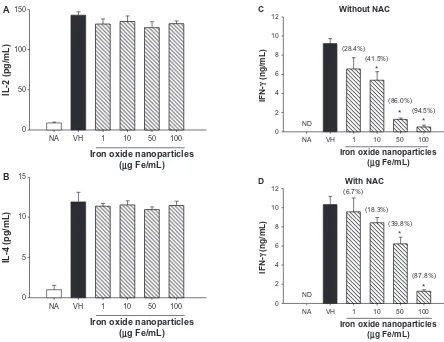
The effects of iron oxide nanoparticles on the expression of three cytokines predominantly expressed by T cells, namely
IL-2, IFN-γ, and IL-4, were examined. The production of
cytokines by unstimulated splenocytes was very low, whereas OVA stimulation strongly induced the expression of the three cytokines (Figure 2A–C, naïve versus VH). The
pres-ence of iron oxide nanoparticles (10–100 µg Fe/mL) did not
influence the production of IL-2 and IL-4, whereas IFN-γ was
Dovepress Iron nanoparticles modulate T cell cytokines
International Journal of Nanomedicine downloaded from https://www.dovepress.com/ by 118.70.13.36 on 23-Aug-2020
0.0
VH 1 10
Iron oxide nanoparticles (µg Fe/mL)
50 100 0.1
OD at 570 nm
0.2 0.3 0.4
No stimulation OVA (100 µg/mL) 0.5
Figure 1 No effect of iron oxide nanoparticles on viability of splenocytes. Splenocytes (5 × 10⁶ cells/mL) were treated with iron oxide nanoparticles (1–100 µg iron [Fe]/mL) and/or vehicle (VH; Roswell Park Memorial Institute medium) and then either left unstimulated or stimulated with ovalbumin (OVA; 100 µg/mL) for 44 hours. The viability of splenocytes was determined by 3-(4,5-dimethylthiazol- 2-yl)-2,5-diphenyl-tetrazolium bromide assay.
Notes: Data are expressed as the mean ± standard error of quadruplicate cultures. Results are representative of three independent experiments.
Abbreviation: OD, optical density.
markedly suppressed in a concentration-dependent manner (Figure 2A–C).
Previous reports have indicated that iron oxide
nanoparticles cause oxidative stress in macrophages.10,13
Therefore the potential role of oxidative stress as a possible
mechanism for the effect of iron oxide nanoparticles on IFN-γ
was investigated. For this purpose we used NAC, a thiol antioxidant as well as a precursor of glutathione. The presence of NAC (1 mM) markedly attenuated iron oxide
nanoparticle-mediated inhibition of IFN-γ production (Figure 2D). In the
absence of NAC, the magnitude of inhibition induced by 10,
50, and 100 µg Fe/mL of iron oxide nanoparticles on IFN-γ
was 45.1%, 86.1%, and 95.4%, respectively (Figure 2C). In the presence of NAC, these percentages were attenuated to 18.3%, 39.8%, and 87.8%, respectively (Figure 2D).
Attenuation of iron oxide
nanoparticle-mediated inhibition of IFN-
γ
by thiol,
but not nonthiol, antioxidants
In addition to NAC, several thiol and nonthiol antioxidants were employed to further address the involvement of oxida-tive stress. Both NAC and exogenous glutathione (1–4 mM of each) were found to significantly attenuate iron oxide
nanoparticle (50 µg Fe/mL)-mediated suppression of IFN-γ in
a concentration-dependent manner (Table 1). In contrast, the nonthiol antioxidants pyruvate (1–4 mM), dimethylthiourea
(4 mM), and tiron (100 µM) did not reverse the effects of
iron oxide nanoparticles (Table 1).
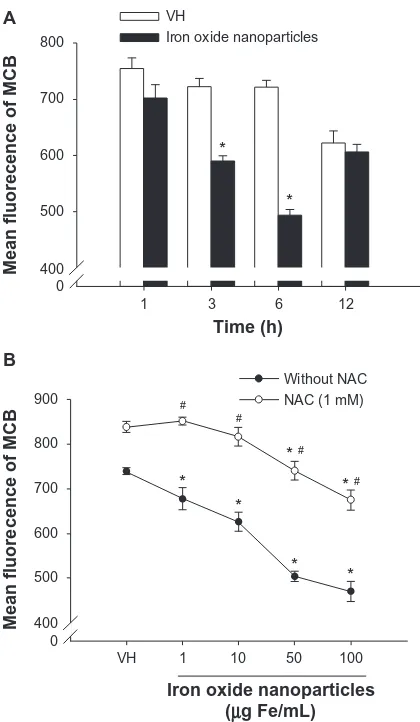
Diminishment of intracellular glutathione
by iron oxide nanoparticles
Based on the results showing the effectiveness of thiol antioxidants, the influence of iron oxide nanoparticles on intracellular levels of glutathione in splenocytes was examined. Exposure of splenocytes to iron oxide
nanopar-ticles (50 µg Fe/mL) markedly decreased the
monochlo-robimane fluorescence with a peak response at 6 hours postexposure, indicating a diminished level of intracellular glutathione (Figure 3A). The effect of iron oxide
nanopar-ticles (1–100 µg Fe/mL) on glutathione diminishment was
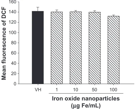
concentration-dependent, and was significantly reversed by the presence of NAC (1 mM) (Figure 3B). In addition to glutathione, the level of intracellular ROS was also measured in splenocytes exposed to iron oxide nanoparticles. At the time point (6 hours) showing peak glutathione diminish-ment, no significant changes in ROS levels were detected in splenocytes exposed to iron oxide nanoparticles up to
100 µg Fe/mL (Figure 4).
Discussion
Although iron oxide nanoparticles have been shown to
affect the functionality and apoptosis of macrophages,8–12
evidence pertaining to their effects on other immune cells is limited. It was previously reported that systemic expo-sure of OVA-sensitized mice to iron oxide nanoparticles attenuated the production of antigen-specific antibodies
and T cell cytokines.17 In the present study, it was further
investigated whether iron oxide nanoparticles induced a direct effect on T cells in culture. The data demonstrated that direct exposure of OVA-primed splenocytes to iron oxide nanoparticles in culture resulted in a marked suppression
of IFN-γ expression and a decrease in intracellular
glutathi-one levels. These results provide evidence that iron oxide nanoparticles produce a direct effect on antigen-specific T cell responses.
In contrast to the suppressive effect on IFN-γ, iron oxide
nanoparticles did not influence OVA-induced production of
IL-2 and IL-4. Previous research has established that IFN-γ
and IL-4 are signature cytokines expressed by T helper (Th)1
and Th2 cells, respectively.20 The current findings suggested
a differential sensitivity between Th1 and Th2 cells, with Th1 cells being a more sensitive target to the nanoparticles. This notion is in line with a recent report showing that
antigen-specific IgG2a and IFN-γ were slightly more
sensi-tive than IgG1 and IL-4 regarding suppression by iron oxide
nanoparticles in vivo.17 On the basis of these results, it was
speculated that exposure to iron oxide nanoparticles may
Dovepress
Shen et al
International Journal of Nanomedicine downloaded from https://www.dovepress.com/ by 118.70.13.36 on 23-Aug-2020
switch the Th1/Th2 immunobalance toward Th2-dominant immunity in response to antigen stimulation. The potential effect of iron oxide nanoparticles on immune responses medi-ated by Th1 and Th2 cells warrants further investigations using appropriate models.
These data on antigen-specific T cell responses contradict the findings of a previous study, which reported that a single intravenous administration of iron oxide nanoparticles to normal nonsensitized mice elevated the serum level of
IFN-γ.15 Although the mechanism responsible for these
contradictory results remains to be elucidated, apparently a crucial factor dictating the effect of iron oxide nanoparticles on
IFN-γ production may be the absence or presence of antigen
sensitization. In the authors’ studies, IFN-γ production is antigen-specific. In contrast, the elevated IFN-γ in nonsensitized mice has been interpreted as a possible proinflammatory status
induced by iron oxide nanoparticles.15
Iron oxide nanoparticles reportedly cause cytotoxicity in macrophages.13 In the present study, the cell viability
of splenocytes was monitored using an MTT assay, which showed comparable viability between nanoparticle-treated and VH-treated groups. Hence, the effect of iron oxide nanoparticles on cytokine expression evidently should not be attributed to a general cytotoxic mechanism.
The authors’ mechanistic studies revealed that both the
IFN-γ suppression and the glutathione diminishment induced
by iron oxide nanoparticles were significantly reversed by NAC, a glutathione precursor. Glutathione plays a critical role in maintaining the cellular homeostasis of redox balance.
Both NAC and glutathione can function as ROS scavengers.21
Notably, iron oxide nanoparticles have been reported to cause oxidative stress in several cells, including those of immune
origin.10,13 It was therefore also evaluated whether ROS were
induced in the present study’s experimental conditions.
0
VH
NA 1 10
Iron oxide nanoparticles (µg Fe/mL) A
50 100
IL-2 (pg/mL) 50
100 150
0
VH
NA 1 10
Iron oxide nanoparticles (µg Fe/mL) B
50 100
IL-4 (pg/mL) 5
10 15
Figure 2 Differential effects of iron oxide nanoparticles on the production of antigen-specific interleukin (IL)-2, IL-4, and interferon (IFN)-γ by splenocytes. (A−C) Splenocytes (5 × 10⁶ cells/mL) were either left untreated (naïve; NA) or treated with iron oxide nanoparticles (1-100 µg iron [Fe]/mL) and/or vehicle (VH) followed by stimulation with ovalbumin (100 µg/mL) for 48 hours. (D) Splenocytes were pretreated with N-acetyl-L-cysteine (NAC; 1 mM) prior to the treatment of iron oxide nanoparticles and ovalbumin stimulation described above. The levels of (A) IL-2, (B) IL-4, and (C and D) IFN-γ in the supernatants were measured by enzyme-linked immunosorbent assay. Notes: Data are expressed as the mean ± standard error of triplicate cultures. The level of cytokines in the VH group was designated as 100%, and the percentage of inhibition induced by iron oxide nanoparticles (10-100 µg Fe/mL) was calculated against this standard (as indicated in parentheses). *P, 0.05, comparison with VH group. Results are representative of three independent experiments.
Abbreviation: ND, no data.
0
VH NA ND
10 1
Iron oxide nanoparticles
(µg Fe/mL)
C
50 100 (41.5%)
(28.4%)
(86.0%)
* *
(94.5%)
*
IFN-γ
(ng/mL
)
8
6
4
2 10 12
0
VH NA ND
10 1
Iron oxide nanoparticles
(µg Fe/mL)
D
Without NAC
With NAC
50 100 (18.3%)
(6.7%)
(39.8%)
*
(87.8%)
*
IFN-γ
(ng/mL)
8
6
4
2 10 12
Dovepress Iron nanoparticles modulate T cell cytokines
International Journal of Nanomedicine downloaded from https://www.dovepress.com/ by 118.70.13.36 on 23-Aug-2020
However, all three nonthiol antioxidants which were used, namely pyruvate, dimethylthiourea, and tiron, failed to coun-teract the effects of iron oxide nanoparticles. Moreover, iron oxide nanoparticles did not alter the intracellular level of ROS in splenocytes restimulated with OVA, as measured by dichlorofluorescin fluorescence. Overall, these results provided evidence to differentiate the respective contributions of gluta-thione and ROS in iron oxide nanoparticle-mediated effects, with glutathione rather than ROS playing a central role.
Similar findings were reported from a previous study on human cardiac endothelium, in which ROS was not found to play a positive mediating role. In that study, iron oxide nanoparticles were found not to induce cytotoxicity and ROS
production in human cardiac endothelial cells.22 Another
recent report showed that titanium dioxide nanoparticles induced ROS production in both Escherichia coli MG1655 and Cupriavididus metallidurans CH34, but caused
cytotox-icity only in E. coli MG1655.23 Overall, these results suggest
that the induction of ROS production and the contribution of ROS in metal oxide nanoparticle-mediated cytotoxic effects vary across different cells.
By contrast, previous research has documented that glutathione plays a pivotal role in various T cell functions,
such as the regulation of cytokine expression.24,25 Glutathione
is considered the hallmark redox buffer in living cellular systems. Depletion of cellular glutathione in mice fed with a liquid control diet containing 30% ethanol-derived calories was found to downregulate the production of
antigen-specific IFN-γ when also upregulating IL-4 production,
but did not affect the level of IL-2.24 In addition, depletion
of intracellular glutathione by buthionine sulfoximine differentially influenced the functionality of Th1/Th2 cell clones. Specifically, IL-2-induced DNA synthesis was
attenuated in the IFN-γ producing Th cell clone 29, whereas
DNA synthesis in the IL-4 producing Th cell clone D10.
G4.1.HD was not affected.26 The current findings were
0
1 3 6 12
Time (h) 400
Mean fluorecence of MCB
500 600 700
VH
Iron oxide nanoparticles 800
Iron oxide nanoparticles (µg Fe/mL) 0
VH 1 10 50 100
400
Mean fluorecence of MCB
500 600 700
# #
* # * #
* * *
* *
*
800
Without NAC NAC (1 mM) 900
A
B
Figure 3 Diminishment of intracellular glutathione in the presence of iron oxide nanoparticles in splenocytes. (A) Splenocytes (5 × 10⁶ cells/mL) were treated with iron oxide nanoparticles (50 µg iron [Fe]/mL) and/or vehicle (VH) followed by stimulation with ovalbumin (100 µg/mL) for 1–12 hours. (B) Splenocytes were treated with iron oxide nanoparticles (1–100 µg Fe/mL) and/or VH, followed by stimulation with ovalbumin (100 µg/mL) for 6 hours in the absence or presence of N-acetyl-L-cysteine (NAC; 1 mM). The levels of intracellular glutathione were measured as monochlorobimane (MCB) fluorescence by flow cytometry. Notes: Data are expressed as the mean ± standard error of triplicate cultures. *P, 0.05, comparison with matched control group without nanoparticle treatment. #P, 0.05, comparison with matched nonNAC group. Results are representative of three independent experiments.
Table 1 Effects of thiol and nonthiol antioxidants on iron oxide nanoparticle-mediated suppression of interferon-γ production by splenocytes
Antioxidants Iron oxide nanoparticle-mediated inhibition of interferon-γ
production (%)a
Control 85.8 ± 2.6
N-acetyl-L-cysteine (mM)
1 39.3 ± 8.9*
2 35.6 ± 6.4*
4 27.9 ± 4.8*
Glutathione (mM)
1 41.5 ± 6.2*
2 36.1 ± 8.5*
4 29.7 ± 4.1*
Pyruvate (mM)
1 82.3 ± 6.9
4 84.3 ± 3.1
Dimethylthiourea (mM)
4 89.7 ± 1.0
Tiron (mM)
0.1 86.6 ± 1.2
Notes: aSplenocytes (5 × 10⁶ cells/mL) were pretreated with iron oxide nanoparticles (50 µg iron/mL) in the absence (control) or presence of N-acetyl-L-cysteine, glutathione, pyruvate, dimethylthiourea or tiron, followed by stimulation with ovalbumin (100 µg/mL) for 48 hours. The levels of interferon-γ in the supernatants were measured by enzyme-linked immunosorbent assay. The percentage of inhibition induced by iron oxide nanoparticles was calculated as described in Materials and methods. Data are expressed as the mean ± standard error of six samples pooled from two experiments; *P, 0.001 compared to the control.
Dovepress
Shen et al
International Journal of Nanomedicine downloaded from https://www.dovepress.com/ by 118.70.13.36 on 23-Aug-2020
congruent with these reports and showed that iron oxide nanoparticles diminished the intracellular glutathione and
suppressed the production of IFN-γ, whereas IL-2 and IL-4
were unaffected.
Conclusion
The present study demonstrated that direct exposure of antigen-primed splenocytes to iron oxide nanoparticles resulted in a marked suppression of the expression of
antigen-specific IFN-γ, accompanied by a decrease in
intracellular glutathione. Both the IFN-γ suppression
and glutathione diminishment were reversed by the thiol antioxidant NAC, but not by nonthiol antioxidants. These findings constitute the first reported evidence of the critical role played by glutathione in the immunosuppressive effect of iron oxide nanoparticles on T cells. Accumulating evi-dence suggests that iron oxide nanoparticles may drive macrophages toward proinflammatory responses and affect T cell reactivity.8–10,15–17 Thus, it would be prudent
to further investigate the immunopharmacology and immunotoxicology of these nanoparticles, and to consider their possible biomedical application in vivo.
Acknowledgments
This work was supported by grants NSC98-2320-B-002-036-MY3 from the National Science Council, Executive Yuan, Taiwan. The authors thank the Consulting Center of Statistics and Bioinformatics in the College of BioResources
and Agriculture, National Taiwan University, for help in statistical analysis.
Disclosure
The authors report no conflicts of interest in this work.
References
1. Chouly C, Pouliquen D, Lucet I, Jeune JJ, Jallet P. Development of superparamagnetic nanoparticles for MRI: effect of particle size, charge and surface nature on biodistribution. J Microencapsul. 1996;13(3): 245–255.
2. Xie J, Huang J, Li X, Sun S, Chen X. Iron oxide nanoparticle platform for biomedical applications. Curr Med Chem. 2009;16(10):1278–1294. 3. Liong M, Lu J, Kovochich M, et al. Multifunctional inorganic
nanopar-ticles for imaging, targeting, and drug delivery. ACS Nano. 2008;2(5): 889–896.
4. Yu MK, Jeong YY, Park J, et al. Drug-loaded superparamagnetic iron oxide nanoparticles for combined cancer imaging and therapy in vivo.
Angew Chem Int Ed Engl. 2008;47(29):5362–5365.
5. Hamm B, Staks T, Taupitz M, et al. Contrast-enhanced MR imaging of liver and spleen: first experience in humans with a new superpara-magnetic iron oxide. J Magn Reson Imaging. 1994;4(5):659–668. 6. Reimer P, Muller M, Marx C, et al. T1 effects of a bolus-injectable
super-paramagnetic iron oxide, SH U 555 A: dependence on field strength and plasma concentration – preliminary clinical experience with dynamic T1-weighted MR imaging. Radiology. 1998;209(3):831–836. 7. Wang J, Chen Y, Chen B, et al. Pharmacokinetic parameters and
tis-sue distribution of magnetic Fe(3)O(4) nanoparticles in mice. Int J
Nanomedicine. 2010;5:861–866.
8. Hsiao JK, Chu HH, Wang YH, et al. Macrophage physiological function after superparamagnetic iron oxide labeling. NMR Biomed. 2008;21(8): 820–829.
9. Cho WS, Cho M, Kim SR, et al. Pulmonary toxicity and kinetic study of Cy5.5-conjugated superparamagnetic iron oxide nanoparticles by optical imaging. Toxicol Appl Pharmacol. 2009;239(1):106–115. 10. Lunov O, Syrovets T, Buchele B, et al. The effect of carboxydextran-coated
superparamagnetic iron oxide nanoparticles on c-Jun N-terminal kinase-mediated apoptosis in human macrophages. Biomaterials. 2010;31(19): 5063–5071.
11. Yeh CH, Hsiao JK, Wang JL, Sheu F. Immunological impact of mag-netic nanoparticles (Ferucarbotran) on murine peritoneal macrophages.
J Nanopart Res. 2010;12(1):151–160.
12. Park EJ, Kim H, Kim Y, Yi J, Choi K, Park K. Inflammatory responses may be induced by a single intratracheal instillation of iron nanoparticles in mice. Toxicology. 2010;275(1–3):65–71.
13. Naqvi S, Samim M, Abdin M, et al. Concentration-dependent toxicity of iron oxide nanoparticles mediated by increased oxidative stress. Int
J Nanomedicine. 2010;5:983–989.
14. Blank F, Gerber P, Rothen-Rutishauser B, et al. Biomedical nano-particles modulate specific CD4(+) T cell stimulation by inhibition of antigen processing in dendritic cells. Nanotoxicology. January 13; 2011. [Epub ahead of print.]
15. Chen BA, Jin N, Wang J, et al. The effect of magnetic nanoparticles of Fe(3)O(4) on immune function in normal ICR mice. Int J Nanomedicine. 2010;5:593–599.
16. Wang J, Chen B, Jin N, et al. The changes of T lymphocytes and cytokines in ICR mice fed with Fe3O4 magnetic nanoparticles. Int J
Nanomedicine. 2011;6:605–610.
17. Shen CC, Wang CC, Liao MH, Jan TR. A single exposure to iron oxide nanoparticles attenuates antigen-specific antibody production and T-cell reactivity in ovalbumin-sensitized BALB/c mice. Int J Nanomedicine. 2011;6:1229–1235.
18. Mosmann T. Rapid colorimetric assay for cellular growth and survival: application to proliferation and cytotoxicity assays. J Immunol Methods. 1983;65(1–2):55–63.
0
VH 1 10
Iron oxide nanoparticles (µg Fe/mL)
50 100 20
40
Mean fluorescence of DC
F
60 80 100 120 140 160
Figure 4 No effect of iron oxide nanoparticles on the intracellular levels of reactive oxygen species. Splenocytes (5 × 10⁶ cells/mL) preloaded with dichlorofluorescin (DCF) diacetate (20 µM) were treated with iron oxide nanoparticles (1–100 µg iron [Fe]/mL) and/or vehicle (VH), followed by stimulation with ovalbumin (100 µg/mL) for 6 hours. The levels of intracellular reactive oxygen species were measured as DCF fluorescence using a microplate reader.
Notes: Data are expressed as the mean ± standard error of triplicate cultures. Results are representative of three independent experiments.
Dovepress Iron nanoparticles modulate T cell cytokines
International Journal of Nanomedicine downloaded from https://www.dovepress.com/ by 118.70.13.36 on 23-Aug-2020
International Journal of Nanomedicine
Publish your work in this journal
Submit your manuscript here: http://www.dovepress.com/international-journal-of-nanomedicine-journal The International Journal of Nanomedicine is an international,
peer-reviewed journal focusing on the application of nanotechnology in diagnostics, therapeutics, and drug delivery systems throughout the biomedical field. This journal is indexed on PubMed Central, MedLine, CAS, SciSearch®, Current Contents®/Clinical Medicine,
Journal Citation Reports/Science Edition, EMBase, Scopus and the Elsevier Bibliographic databases. The manuscript management system is completely online and includes a very quick and fair peer-review system, which is all easy to use. Visit http://www.dovepress.com/ testimonials.php to read real quotes from published authors. 19. Metz S, Bonaterra G, Rudelius M, Settles M, Rummeny EJ,
Daldrup-Link HE. Capacity of human monocytes to phagocytose approved iron oxide MR contrast agents in vitro. Eur Radiol. 2004;14(10): 1851–1858.
20. Finkelman FD, Holmes J, Katona IM, et al. Lymphokine control of in vivo immunoglobulin isotype selection. Annu Rev Immunol. 1990;8: 303–333.
21. Meister A, Anderson ME. Glutathione. Annu Rev Biochem. 1983;52: 711–760.
22. Sun J, Wang S, Zhao D, Hun FH, Weng L, Liu H. Cytotoxicity, permeability, and inflammation of metal oxide nanoparticles in human cardiac microvascular endothelial cells: cytotoxicity, permeability, and inflammation of metal oxide nanoparticles. Cell Biol Toxicol. 2011;27(5): 333–342.
23. Simon-Deckers A, Loo S, Mayne-L’hermite M, et al. Size-, composition- and shape-dependent toxicological impact of metal oxide nanopar-ticles and carbon nanotubes toward bacteria. Environ Sci Technol. 2009;43(21):8423–8429.
24. Peterson JD, Herzenberg LA, Vasquez K, Waltenbaugh C. Glutathione levels in antigen-presenting cells modulate Th1 versus Th2 response patterns. Proc Natl Acad Sci U S A. 1998;95(6):3071–3076.
25. Gmunder H, Roth S, Eck HP, Gallas H, Mihm S, Droge W. Interleukin-2 mRNA expression, lymphokine production and DNA synthesis in glutathione-depleted T cells. Cell Immunol. 1990;130(2): 520–528.
26. Gmunder H, Droge W. Differential effects of glutathione depletion on T cell subsets. Cell Immunol. 1991;138(1):229–237.
Dovepress
Dove
press
Shen et al
International Journal of Nanomedicine downloaded from https://www.dovepress.com/ by 118.70.13.36 on 23-Aug-2020
![Figure 1 No effect of iron oxide nanoparticles on viability of splenocytes. Splenocytes (5 × 10⁶ cells/mL) were treated with iron oxide nanoparticles (1–100 µg iron [Fe]/mL) and/or vehicle (VH; Roswell Park Memorial Institute medium) and then either left u](https://thumb-us.123doks.com/thumbv2/123dok_us/8346485.1382045/4.612.68.276.50.221/nanoparticles-viability-splenocytes-splenocytes-nanoparticles-roswell-memorial-institute.webp)