1556-6811/09/$08.00⫹0 doi:10.1128/CVI.00116-09
Copyright © 2009, American Society for Microbiology. All Rights Reserved.
Highly Efficient Antiviral CD8
⫹
T-Cell Induction by Peptides Coupled
to the Surfaces of Liposomes
䌤
Akira Takagi,
1Masanori Matsui,
1Satoshi Ohno,
1Hongying Duan,
1Osamu Moriya,
1Nobuharu Kobayashi,
1Hiroshi Oda,
2Masahito Mori,
2Akiharu Kobayashi,
2Maiko Taneichi,
3Tetsuya Uchida,
3and Toshitaka Akatsuka
1*
Department of Microbiology, Faculty of Medicine, Saitama Medical University, Moroyama-Cho, Iruma-Gun, Saitama 350-0495, Japan1; Drug Delivery System Development Division, Nippon Oil and Fat Corporation, 4-20-3 Ebisu, Shibuya-Ku, Tokyo 150-6019,
Japan2; and Department of Safety Research on Blood and Biological Products, National Institute of
Infectious Diseases, 4-7-1 Gakuen, Musashimurayama, Tokyo 208-0011, Japan3
Received 15 March 2009/Returned for modification 28 May 2009/Accepted 4 August 2009
In previous studies, we have demonstrated that liposomes with differential lipid components display differ-ential adjuvant effects when antigens (Ags) are chemically coupled to their surfaces. When ovalbumin was coupled to liposomes made by using unsaturated fatty acids, it was found to be presented not only to CD4ⴙT cells but also to CD8ⴙT cells and induced cytotoxic T lymphocytes (CTLs) which effectively eradicated the tumor from mice. In this study, we coupled liposomes to immunodominant CTL epitope peptides derived from lymphocytic choriomeningitis virus (LCMV) and evaluated its potency as an antiviral vaccine. The intramus-cular immunization of mice with the peptide-liposome conjugates along with CpG resulted in the efficient induction of antiviral CD8ⴙT-cell responses which conferred complete protection against not only LCMV Armstrong but also a highly virulent mutant strain, clone 13, that establishes persistent infections in immu-nocompetent mice. The intranasal vaccination induced mucosal immunity effective enough to protect mice from the virus challenge via the same route. Complete protection was achieved in mice even when the Ag dose was reduced to as low as 280 ng of liposomal peptide. This form of vaccination with a single CTL epitope induced Ag-specific memory CD8ⴙT cells in the absence of CD4ⴙT-cell help, which could be shown by the complete protection of CD4-knockout mice in 10 weeks as well as by the analysis of recall responses. Thus, surface-linked liposomal peptide might have a potential advantage for the induction of antiviral immunity.
The development of practical vaccines has been greatly po-tentiated by the availability of synthetic antigens (Ags), but progress has been hampered by the poor immunogenicity of Ags. Liposomes have successfully been used as drug carriers (35) and have also been proposed to be carriers of Ags and adjuvants to induce immune responses (33). Most of the lipo-somal vaccines proposed have been prepared by Ag encapsu-lation within the aqueous lumen of liposomes. However, it is known that the immune responses induced by encapsulated liposomal Ags are different from those induced by surface-linked liposomal Ags.
We have demonstrated that Ags chemically coupled to the surfaces of liposomes induce Ag-specific immunoglobulin G (IgG) but not IgE antibody production (28). The inducibility of Ag-specific IgG production was found to vary among liposome preparations: the greater that the membrane mobility in the liposomes is, the greater that the antibody production induced by Ag-liposome conjugates is (29). In our previous study, we have reported that ovalbumin (OVA) coupled to liposomes made with unsaturated fatty acids was presented to both CD4⫹ and CD8⫹T cells, whereas OVA coupled to liposomes made with saturated fatty acids was presented only to CD4⫹T cells. Furthermore, the cross-presentation of OVA coupled to
lipo-somes consisting of unsaturated fatty acids was further con-firmed by the in vivo induction of cytotoxic T lymphocytes (CTLs) which conferred tumor eradication (42) and protection against influenza virus (27) in mice. However, the advantages of the surface-linked liposomal peptides for other forms of vaccines, especially regarding the efficiency of effector CD8⫹T cells and the inducibility of long-term memory CD8⫹T cells, have not been demonstrated.
In the present study, we evaluated the potency of surface-coupled liposomal peptides as an antiviral vaccine using the infection of mice with lymphocytic choriomeningitis virus (LCMV) as a model. Use of that model enabled us to compare the effectiveness of this system with that of various vaccine formulas prepared with the same epitope peptide as the Ag reported elsewhere. We also elucidate the induction and main-tenance of memory CD8⫹T cells by a minimum CTL epitope peptide which does not seem to stimulate the help of CD4 cells.
MATERIALS AND METHODS
Mice and viruses.Female C57BL/6 mice (age, 6 weeks) were purchased from Clea Japan, Inc. (Tokyo, Japan), and Tokyo Laboratory Animal Science Co. Ltd. (Tokyo, Japan). CD4-knockout (KO) mice of a C57BL/6 background were pur-chased from the Jackson Laboratory (Bar Harbor, ME). After they were bred, 6-week-old male CD4-KO mice were used for the experiments. The mice were housed in appropriate animal care facilities at Saitama Medical University (Saitama, Japan) and were handled according to international guidelines. The experimental protocols were approved by the Animal Research Committee of
Saitama Medical School (approval number 634). The mice received 2⫻105PFU
of LCMV Armstrong (Arm) intraperitoneally (i.p.) or intranasally (i.n.) to
ini-* Corresponding author. Mailing address: Department of Microbi-ology, Faculty of Medicine, Saitama Medical University, Moroyama-cho, Iruma-gun, Saitama 350-0495, Japan. Phone: 81-49-276-1165. Fax: 81-49-295-9107. E-mail: [email protected].
䌤Published ahead of print on 12 August 2009.
1383
on August 17, 2020 by guest
http://cvi.asm.org/
phosphatidylcholine, dioleoyl phosphatidylethanolamine, dioleoyl phosphatidyl-glycerol acid, and cholesterol in a 4:3:2:7 molar ratio (29). Each peptide was coupled to the surfaces of the liposomes by the use of disuccinimidyl suberate, as described previously (42). The final preparations contained 0.7 mg of the peptide and 10 mg of the liposome per ml. Liposomes conjugated with the GP33-41 peptide and NP396 (Lip-GP33/NP396), Lip-GP33/GP61, and Lip-GP33/NP309 were made by mixing peptide GP33-41 with peptide NP396-404, peptide GP61-80, and peptide NP309-328, respectively, in equal amounts before conjugation.
Immunization.The mice received the liposomal peptides at the indicated
amounts together with 5g of CpG in 100l of phosphate-buffered saline (PBS)
by one of the three different routes: subcutaneously (s.c.), intramuscularly (i.m.),
or i.p. The i.n. immunization was performed by introducing 20l of a liposome
solution (14g of peptide) and 5g of CpG (5l) into the nasal cavity. Mice
that had recovered from an acute infection of LCMV Arm served as positive controls.
51Cr-release assay.Spleen cells were prepared 7 days after immunization to
obtain effector cells. The cytotoxic activities of the GP33-specific CTLs were
measured by standard 4-h51Cr-release assays. For peptide-pulsed target cells,
1⫻106
MC57G cells were pulsed with 10M of peptide GP33 for 2 h and then
labeled with 100Ci of Na251CrO4 for 1 h. For virus-infected targets, the
MC57G cells were infected with VVGP33 or wild-type vaccinia virus at a mul-tiplicity of infection of 5. After 1 h of infection, the cells were washed, resus-pended in medium, and incubated overnight. The infected targets were then
labeled with51
Cr, as described above. The labeled target cells were plated in
wells of a round-bottom 96-well plate at 2⫻103cells/well with effector cells at
various effector-cell-to-target-cell (E/T) ratios. Blocking by antibodies was
per-formed by adding anti-CD4 or anti-CD8 (5g/ml; eBioscience, San Diego, CA)
to the mixture. Percent lysis was calculated as [(cpmsample⫺cpmspontaneous)/
(cpmmaximum⫺cpmspontaneous)]⫻100. The maximum amount of51Cr released
was determined with the supernatants of cells that were lysed by addition of 2%
Nonidet P-40. The spontaneous release of51Cr from target cells incubated
without effector cells was measured in the supernatants. Percent specific lysis was calculated by subtracting the percent lysis with unpulsed targets from that with peptide-pulsed targets.
In vivo CTL assay.The in vivo CTL assay was carried out as reported else-where (42). Briefly, spleen cells from naive C57BL/6 mice were split into two equal populations. One population was pulsed with a peptide at a final
concen-tration of 10M for 1 h at 37°C and then labeled with a high concentration (2.5
M) of carboxyfluorescein diacetate succinimidyl ester (CFSE; Molecular
Probes, Eugene, OR) for 10 min at 37°C. The other was unpulsed and labeled
with a lower concentration (0.25M) of CFSE. An equal number of cells (1⫻
107) from each population was mixed and transferred into immunized mice
intravenously. Twelve hours later, spleen cells were prepared and analyzed by
flow cytometry. Percent specific lysis was calculated as 1⫺[(number of CFSElow
cells in healthy mice/number of CFSEhigh
cells in healthy mice)/(number of CFSElow
cells in immunized mice/number of CFSEhighcells in immunized mice)]⫻100,
where CFSElow
and CFSEhigh
represent low and high CFSE concentrations, respectively.
IFN-␥ELISPOT assay.The detection of gamma interferon (IFN-␥)-secreting
cells was performed by using a mouse IFN-␥ enzyme-linked immunospot
(ELISPOT) assay set (BD Biosciences–Pharmingen, San Diego, CA). Each well of a sterile 96-well ImmunoSpot ELISPOT assay plate (BD
Biosciences–Pharm-ingen) was precoated with 0.5g of unlabeled anti-IFN-␥capture antibody
(clone R4-6A2; BD Biosciences–Pharmingen) per well at 4°C overnight. The plates were washed and then blocked with RPMI 1640 containing 10% FCS for 2 h at room temperature. Spleen cells from individual immunized mice were
96-well round-bottom plate, 2⫻106spleen cells were incubated with 1⫻105
cells of syngeneic spleen cells which had been pulsed with an appropriate peptide
for 3 h in the presence of 0.2l/well brefeldin A (GolgiPlug; BD Biosciences) for
5 h at 37°C. The cells were then washed once and incubated for 10 min at 4°C with a rat anti-mouse CD16/CD32 monoclonal antibody (MAb; Fc Block; BD
Biosciences) at a concentration of 1g/well. Following incubation, the cells were
stained with a fluorescein isothiocyanate (FITC)-conjugated rat anti-mouse
CD8␣MAb (clone 53-6.7; BD Biosciences) at a concentration of 0.5g/well for
30 min at 4°C. After the cells were washed twice, they were fixed and permeab-ilized by using a Cytofix/Cytoperm kit (BD Biosciences) and stained with a
phycoerythrin (PE)-conjugated rat anti-mouse IFN-␥MAb (clone XMG1.2; BD
Biosciences). After the cells were washed, flow cytometric analyses were per-formed with a FACScan flow cytometer (Becton Dickinson, Franklin Lakes, NJ), and the data were analyzed with CellQuest software (Becton Dickinson). The experiment was repeated three times.
Tetramer and CD62L staining.Spleen cells were prepared and treated with an
anti-mouse CD16/CD32 MAb as described above for intracellular IFN-␥staining
and were then stained with a PE-conjugated GP33H-2Db
tetramer (Medical and Biological Laboratories, Nagoya, Japan), peridinin chlorophyll protein-conju-gated anti-CD8a (BD Bioscience), and FITC-conjuprotein-conju-gated anti-CD62L (BD Bio-science) for 30 min at 4°C. After the cells were washed twice, they were fixed with PBS containing 1% formaldehyde and 2% FCS and analyzed by flow cytometry.
Statistical analysis.Statistical analyses were performed by Student’sttest. A
Pvalue of⬍0.05 was considered statistically significant.
RESULTS
Induction of LCMV-specific CD8ⴙT-cell response in mice immunized with liposome-coupled GP33 peptide.Ag chemi-cally coupled to the surfaces of liposomes composed of unsat-urated fatty acids has been shown to be presented to CD8⫹T cells (42) and to induce protective immunity against influenza virus (27). For further evaluation of the use of surface-linked liposomal peptide as an antiviral vaccine, we chose an immu-nodominant CTL epitope, GP33-41, of LCMV, which is pre-sented by theH-2Db molecule (10, 16, 36).H-2Kd-restricted
epitope GP283-292 (10) is not presented in C57BL/6 mice (44) and served as the negative control in our experiments. We conjugated liposomes with GP33-41 peptide (Lip-GP33) or GP283-292 (Lip-GP283) and immunized C57BL/6 mice by one of three different routes (s.c., i.m., or i.p.) together with CpG. The immune responses were evaluated by51Cr-release assays
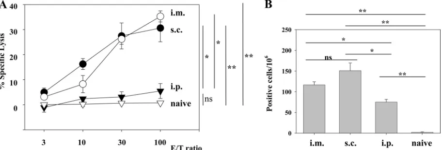
and ELISPOT assays. As shown in Fig. 1A and B, single i.m. and s.c. immunizations induced substantial levels of CTL ac-tivity and IFN-␥-producing cells, whereas i.p. immunizations induced much lower responses, and the unconjugated peptide induced no detectable response in either assay (see Fig. 6 for data only for the ELISPOT assay). In the absence of CpG, the responses were very low (data not shown), in accordance with the findings presented in a previous report (27). Therefore, in
on August 17, 2020 by guest
the following experiments, the immunizations with CpG were done mainly by the i.m. route. We confirmed the specificities of the reactions observed in the experiments described above as follows. Intracellular IFN-␥staining experiments revealed the peptide-specific IFN-␥production by CD8⫹T cells (Fig. 2A).
51Cr-release assays with target cells expressing the GP33
epitope by infection with recombinant vaccinia virus VVGP33 demonstrated that the effector CTLs induced by the GP33-liposome conjugates recognized the epitope, which was
endo-genously processed and presented by major histocompatibility complex class I molecules (Fig. 2B). Blocking experiments with anti-CD4 and anti-CD8 antibodies demonstrated that the CTL activities were exerted by CD8⫹T cells (Fig. 2B). The CTL activities induced by the liposome conjugate were also con-firmed by in vivo CTL assays (see Fig. 4B).
Experimental challenge of immunized mice with LCMV.To examine the protective efficacy of Lip-GP33, the immunized mice were challenged with LCMV Arm. Four days after the FIG. 1. Comparison of three different routes of immunization with Lip-GP33. Each C57BL/6 mouse received 20l of Lip-GP33 diluted in 100 l of PBS containing 5g of CpG. Spleen cells were prepared 7 days after immunization for the51Cr-release assays (A) and ELISPOT assays (B).
(A)51Cr-release assays were performed to detect GP33-specific CTL activity at various E/T ratios by using MC57G cells pulsed with peptide GP33
as targets. (B) IFN-␥-producing cells responding to peptide GP33 were detected by ELISPOT assays, as described in Materials and Methods. Spleen cells from nonimmunized (naive) mice were used as a negative control. The data are representative of those from two independent and reproducible experiments. The results are shown as the means of four mice per group⫾standard errors of the means.*,P⬍0.05;**,P⬍0.01; ns, not statistically significant.
FIG. 2. Confirmation of GP33-specific CD8⫹T-cell responses of immunized mice. C57BL/6 mice were injected i.m. with 20l of Lip-GP33 or Lip-GP283 diluted in 100l of PBS containing 5g of CpG, and spleen cells were prepared 7 days later for analysis. (A) The numbers of IFN-␥-producing CD8⫹T cells specific to GP33 were determined by intracellular IFN-␥staining. Spleen cells from mice immunized with Lip-GP33 or Lip-GP283 were stimulated with either GP33-pulsed or unpulsed syngeneic spleen cells for 5 h and were then stained to detect their surface expression of CD8 (x axis) with FITC-conjugated MAb and their intracellular expression of IFN-␥ (y axis) with PE-conjugated MAb. All lymphocytes were gated and analyzed on a FACScan flow cytometer by the use of CellQuest software (BD Biosciences). The values shown in the upper right quadrants indicate the percentage of CD8⫹cells that are positive for intracellular IFN-␥after stimulation with GP33-pulsed spleen cells. The results of stimulation with unpulsed spleen cells were almost zero (data not shown). Each experiment used four mice per group, and the spleen cells of the mice in each group were pooled. The data shown are representative of those from three independent and reproducible experiments. (B)51Cr-release assays with MC57G cells infected with VVGP33 as targets were performed to detect CTL activity against the
endogenously expressed GP33 epitope (gray bars). As control targets, MC57G cells were infected with wild-type vaccinia virus. The same effector cells were used as GP33 peptide-pulsed and unpulsed targets as well for comparison (black bar). Lysis was also tested in the presence of an anti-CD4 or an anti-CD8 MAb (5g/ml) in the CTL assay. Specific lysis is demonstrated at an E/T ratio of 50. Each experiment used three mice, and the spleen cells of the three mice were pooled. The data are representative of those from two independent and reproducible experiments.
on August 17, 2020 by guest
http://cvi.asm.org/
infection, the virus titers in the spleens of the immunized mice were determined. As LCMV Arm is cleared from all tissues during the first week of infection and infection with this virus results in the formation of functional and protective memory CD8⫹T cells (2), the mice which recovered from the LCMV Arm infection were used as positive controls. As shown in Fig. 3A, immunization with Lip-GP33 as well as LCMV Arm in-fection conferred complete protection to the mice, and the titers in the spleen were below the detection limit (2.0⫻101
PFU/g), whereas the mice immunized with the negative control peptide (Lip-GP283) showed titers of (1.69 ⫾ 0.40) ⫻ 107
PFU/g (Fig. 3A). Again, in the absence of CpG, the immunized mice were not protected at all (data not shown). The protective efficacy of Lip-G33 with CpG was further tested by challenging the immunized mice with LCMV Cl.13, a mutant strain of LCMV Arm which is known to induce viremia for⬃3 months, with the virus persisting in some tissues for life (1). Eight days after infection, the mice immunized with Lip-GP33 or LCMV Arm completely cleared the Cl.13 virus, whereas those which received the negative control peptide (Lip-GP283) had a high titer of the virus in the spleen (Fig. 3B), and the persistence of the virus was confirmed 4 weeks later (data not shown).
Liposome-coupled peptide NP396 induces low CTL re-sponses but complete protection. The bulk of the CTL re-sponse to LCMV inH-2bmice is directed against three
dom-inant epitopes, GP33-41, GP276-286, and NP396-404, which are presented by major histocompatibility complex class I
H-2Dbmolecules (10, 16, 36). We investigated whether
pep-tides other than GP33 were able to induce antiviral T-cell responses when they were coupled to the surfaces of lipo-somes. Peptide NP396-404 was coupled to liposomes by itself (Lip-NP396) or with peptide GP33 (Lip-GP33/NP396). Mice were immunized with either Lip-GP33, Lip-NP396, Lip-GP33/ NP396, or a mixture of Lip-GP33 and Lip-NP396 (Lip-GP33– Lip-NP396), and the responses were compared by ex vivo CTL assays (Fig. 4A), in vivo CTL assays (Fig. 4B), ELISPOT assays
(Fig. 4C), and challenge experiments with LCMV Arm (Fig. 4D).
The level of induction of CTL activities by Lip-NP396 was very low in both the ex vivo and the in vivo assays, and Lip-GP33/NP396 and Lip-GP33–Lip-NP396 induced intermediate levels of CTL activity compared to the levels induced by Lip-GP33 and Lip-NP396 (Fig. 4A and B). On the other hand, Lip-NP396 induced substantial levels of IFN-␥-producing cells, and Lip-GP33/NP396 and Lip-GP33–Lip-NP396 induced even higher, although not significantly higher, numbers of IFN-␥ -producing cells compared with the numbers induced by Lip-GP33 and Lip-NP396 (Fig. 4C). When these immunized mice were challenged with LCMV Arm, all the liposomal conjugates except Lip-GP283 were found to have the potency necessary to confer complete protection to mice (Fig. 4D).
i.n. immunization of mice with liposome-coupled peptide.
LCMV is transmitted through contact with secretions from infected animals or by inhalation of dried particles from them. Therefore, LCMV infection is also suitable for the study of the capacity of liposomal peptides to induce mucosal immunity and protect mice from infection by viruses through the mode of transmission described above. We introduced Lip-GP33 and CpG by the i.n. or i.m. route, and 1 week later, the numbers of IFN-␥-producing CD8⫹ T cells in draining cervical lymph nodes and spleens were measured (Fig. 5A). The i.n. immuni-zations resulted in the induction of IFN-␥-producing CD8⫹T cells at high levels in the cervical lymph nodes but at only low levels in the spleens, indicating that effector CD8⫹T cells were induced locally near the nasal cavity. On the other hand, the i.m. immunizations induced high numbers of IFN-␥-producing CD8⫹ T cells in the spleens but only small numbers in the cervical lymph nodes (Fig. 5A). When the immunized mice were challenged with LCMV Arm via the i.n. route, both i.m. and i.n. immunizations with Lip-GP33 were found to provide complete protection to the mice (Fig. 5B).
FIG. 3. Challenge experiments with vaccinated and control mice. C57BL/6 mice were immunized by injecting Lip-GP33 or Lip-GP283, as described in the legends to Fig. 1 and 2, or by i.p. inoculation of 2⫻105PFU of LCMV Arm. After 2 weeks, the mice received 2⫻105PFU of
LCMV Arm i.p. to initiate acute infection (A) or 2⫻106PFU of Cl.13 intravenously to initiate chronic infection (B). The virus titers in the spleens
were quantitated by plaque assay on Vero cells at day 4 (A) or day 8 (B) postchallenge. Virus titers are indicated for each animal in the study. A dotted line represents the lower limit of detection (2⫻101PFU/g [spleen]). The data are representative of those from three independent and
reproducible experiments.
on August 17, 2020 by guest
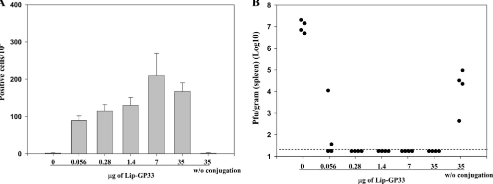
Dose-response study of liposome-coupled peptides.To eval-uate further the efficacies of the liposomal peptides, we made serial fivefold dilutions of Lip-GP33 and injected them into mice in the presence of CpG. To our surprise, as little as 0.08
l of Lip-GP33, which contained only 56 ng of the peptide, induced a significant level of IFN-␥-producing CD8⫹T cells (Fig. 6A); and half of the immunized mice were completely protected against LCMV challenge (Fig. 6B) in two indepen-dent experiments. Immunizations with 0.4l or more of Lip-GP33 (ⱖ280 ng peptide) conferred complete protection to all the mice, but the peptide (35g) not conjugated to liposomes did not induce either IFN-␥-producing CD8⫹T cells or pro-tection against the virus challenge (Fig. 6), which suggests the essential role of the liposome on the induction of the antiviral responses. The results were confirmed in three independent and reproducible experiments.
Effector and memory CD8ⴙ T-cell induction by Lip-GP33 without the help of CD4.Several studies have demonstrated that CD4⫹ T cells play critical roles in generating primary CD8⫹T cells or maintaining memory CD8⫹T cells (5, 17, 19, 37, 39). When mice were immunized with minimal CTL epitope peptides, no functional CD8⫹ T cells could be de-tected past day 20 in an IFN-␥ELISPOT assay (8). Therefore, we wished to know how long the antiviral T-cell response persists and whether memory CD8⫹ T cells are established after immunization with Lip-GP33. We also wished to rule out the possibility that CD4⫹T cells are involved in the induction of an antiviral T-cell response by Lip-GP33. We immunized both C57BL/6 and CD4-KO mice with a single dose of Lip-GP33 and compared the number of Ag-specific IFN-␥ -produc-ing T cells in their spleens by ELISPOT assay (Fig. 7A). When the primary CD8⫹T-cell responses to GP33 were compared at FIG. 4. Comparison of GP33 and NP396 peptides as vaccine components. C57BL/6 mice were immunized i.m. with either Lip-GP33, Lip-NP396, Lip-GP283, or Lip-GP33/NP396 (20l each) or with Lip-GP33–Lip-NP396 (10l each) in 100l of PBS containing 5g of CpG. Seven days later,51Cr-release assays (A), in vivo CTL assays (B), ELISPOT assays (C), and challenge experiments with LCMV Arm (D) were
performed. The51Cr-release assays (A) and ELISPOT assays (C) were done as described in the legend to Fig. 1. The in vivo CTL assays (B) were
performed only for the mice immunized with either Lip-GP33, Lip-NP396, or Lip-GP283. One week after immunization, the mice received an equal number of a relevant peptide (GP33 or NP396)-pulsed CFSEhightargets and unpulsed CFSElowtargets. The numbers show the percentages
of specific lysis. The experiment was repeated twice. The challenge experiments with LCMV Arm (D) were performed by the methods described in the legend to Fig. 3A.*,P⬍0.05;**,P⬍0.01. No statistically significant difference was observed between the four groups by the ELISPOT assays (C).
on August 17, 2020 by guest
http://cvi.asm.org/
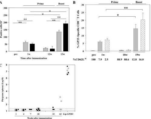
1 week postimmunization, there was no significant difference between the two groups. Twelve weeks after priming, when the Ag-specific IFN-␥-producing T cells in the spleen diminished, the mice were boosted in the same manner used to prime them. One week postboosting, clear recall responses of 6- and
10-fold expansions of Ag-specific IFN-␥-producing T cells could be seen in the two groups, respectively, and the differ-ence was not significant between the two groups. The recall responses of the C57BL/6 mice were also assessed 18 weeks after priming by tetramer staining and compared with those of FIG. 5. i.n. immunizations with Lip-GP33. C57BL/6 mice were immunized by the i.n. or i.m. route with 20l of Lip-GP33 or Lip-GP283 and 5g of CpG. One week later, lymphocytes from cervical lymph nodes and spleens were prepared and subjected to ELISPOT (IFN-␥) assays (A). The immunized mice were also challenged with 2⫻105PFU of LCMV Arm via the i.n. route, and the virus titers in the spleens were quantitated
(B). (A) ELISPOT assays of IFN-␥-producing CD8⫹T cells from cervical lymph nodes (left) and spleens (right) of the mice immunized via the i.n. or i.m. route with Lip-GP33 or Lip-GP283. The data are representative of those from two independent and reproducible experiments. The results for one pool of cells from the lymph nodes and the means for spleen cells from four mice per group⫾standard errors of the means are shown. *, P ⬍0.05 compared with the results obtained with Lip-GP283 immunization; ns, not statistically significant. (B) One week after immunization, the mice received 2⫻105PFU of LCMV Arm i.n., and the virus titers in the spleens were quantitated by plaque assay on Vero
cells at day 4 postchallenge. The virus titers are indicated for each animal in the study. A dotted line represents the lower limit of detection (2⫻ 101PFU/g [spleen]). The data are representative of those from three independent and reproducible experiments.
FIG. 6. Dose-response experiments. Serial fivefold dilutions of Lip-GP33 starting at 50l were prepared in 100l of PBS containing 5g of CpG and injected i.m. to each C57BL/6 mouse. Seven days later, ELISPOT (IFN-␥) assays (A) and challenge experiments with LCMV Arm (B) were performed. (A) ELISPOT assays were performed with spleen cells from immunized mice. One group of mice received peptide GP33 (35 g) without (w/o) conjugation, and one group received GP33 at the same dose conjugated with 5g of CpG as one of the Lip-GP33 immunizations. The data are representative of those from three independent and reproducible experiments. The results are shown as the means for four mice per group⫾standard errors of the means. (B) The immunized mice received 2⫻105PFU of LCMV Arm i.p., and the virus titers in the spleens were
quantitated by plaque assay on Vero cells at day 4 postchallenge. Virus titers are indicated for each animal in the study. The dotted line represents the lower limit of detection (2⫻101PFU/g [spleen]). The data are representative of those from two independent and reproducible experiments.
on August 17, 2020 by guest
the mice which acquired sterile immunity by LCMV Arm in-fection (Fig. 7B). One week after the boost, 22- and 33-fold expansions of GP33-specific CD8⫹T cells were observed in the two groups, respectively. We also analyzed the expression of CD62L, a surface molecule associated with T-cell memory function, of GP33-specific CD8⫹T cells in both Lip-GP33- and LCMV Arm-immunized mice. As shown in Fig. 7B, CD62L was downregulated after the mice were primed. Eighteen weeks after the mice were primed, more than 88% of the GP33-specific CD8⫹T cells recovered their ability to express CD62L and were again downregulated after the boost. To confirm the establishment of memory CD8⫹T cells, the
im-munized mice were challenged with LCMV Arm at different times after vaccination. As shown in Fig. 7C, complete protec-tion was seen even in the absence of CD4⫹T cells at 10 weeks after priming. In the 62nd week, half of the C57BL/6 mice showed perfect protection, and on average, about 4-log-titer reductions were observed.
We then examined if the presence of CD4⫹ T-cell help would augment the induction of effector and memory CD8⫹ cells by Lip-GP33. As helper T-cell epitopes, I-Ab-restricted
LCMV GP61-80 and NP309-328 were chosen and conjugated to liposomes by themselves (Lip-GP61 and Lip-NP309) or with peptide GP33 (Lip-GP33/GP61 and Lip-GP33/NP309). Mice FIG. 7. Analyses of effector and memory CD8⫹T-cell induction in the absence of CD4 help. Each of C57BL/6 and CD4-KO mice received 20 l of Lip-GP33 or Lip-GP283 diluted in 100l of PBS containing 5g of CpG; and ELISPOT assays (A), tetramer and CD62L staining (B), and challenge experiments with LCMV Arm (C) were performed at different times. (A) At 1 and 12 weeks postimmunization, IFN-␥-producing cells responding to peptide GP33 were detected by ELISPOT assays of spleen cells from C57BL/6 (gray bars) and CD4-KO (black bars) mice. At week (w) 12, the mice were boosted in the same manner in which they received the primary immunizations, and ELISPOT assays were performed 1 week later. (B) GP33H-2Dbtetramer staining was performed 1 and 18 weeks postimmunization for C57BL/6 mice (gray bars). For comparison, mice immunized by inoculating 2⫻105PFU of LCMV Arm i.p. were included in these experiments (shaded bars). At week 18, both groups of mice
were boosted with Lip-GP33 and were analyzed for their recall responses 1 week later. The expression of CD62L on gated GP33H-2Db tetramer-positive CD8⫹T cells was also determined by flow cytometry. (C) Challenge experiments were performed by the i.p. injection of 2⫻105
PFU of LCMV Arm at the indicated times. The virus titers are shown for each of the C57BL/6 (open circles) and the CD4-KO (closed circles) mice. The data are shown as the means for at least three and five mice per group⫾standard errors of the means for the ELISPOT and tetramer assays, respectively.*,P⬍0.05;**,P⬍0.01.
on August 17, 2020 by guest
http://cvi.asm.org/
In the study described in this report, we demonstrate that antigenic peptides coupled to the surfaces of liposomes serve as efficient vaccine vehicles for the induction of antiviral im-munity mediated by CD8⫹T cells. We used LCMV infection of mice as a model system because LCMV has been extensively used to study CD8⫹T-cell-mediated antiviral immunity (21, 25, 43). In addition, LCMV causes respiratory tract as well as percutaneous infections; and some LCMV variants, such as Cl. 13, WE, and Docile, induce persistent infections in immuno-competent mice. These features of LCMV enabled us to eval-uate the efficacy of our vaccine preparation by comparing the findings obtained with this preparation with those obtained in many other studies in which the same epitope was presented as the peptide (3, 7, 18, 31) or in which liposome-encapsulated peptide (23), plasmid DNA (4, 15, 24, 34), recombinant vac-cinia virus (14, 20, 30, 44), recombinant lentivirus (46), and recombinantListeria(38) vaccines were used.
Lip-GP33 efficiently induced CTLs, IFN-␥-producing CD8⫹ T cells, and complete protection against LCMV challenge. Although the numbers of positive cells in the ELISPOT assays were low compared to the results of the intracellular cytokine staining (Fig. 2A), that result seems to be simply due to tech-nical factors because spleen cells from the same immunized animal showed such differences, and these were also observed in mice immunized by LCMV Arm infection.
The minimum amount of GP33 peptide (0.28g) required to yield perfect protection in this study was extremely low. Peptide vaccines with adjuvants, cytokines, CpGs, etc., usually require multiple injections in amounts ranging from 10 to 500
g (3, 7, 18, 31). When peptide GP33 was encapsulated within liposomes and injected with CpG, two immunizations with 80
g were required to obtain perfect protection, while a smaller dose gave only partial protection (23). In distinction, we ex-pressed Ags on the surfaces of liposomes and speculate that they might be more efficiently recognized by Ag-presenting cells, which resulted in an enhanced presentation to T cells. In direct comparisons, we have found that surface-linked liposo-mal Ags induced a significantly higher level of Ag-specific IgG production than that induced by liposome-encapsulated Ags in mice (our unpublished observation). In addition, a significant difference was observed between liposomes with different lipid components; more Ags coupled to the unsaturated liposomes were engulfed by macrophages in vitro, and a higher level of Ag-specific antibody production was induced in vivo when un-saturated liposomes were used than when un-saturated liposomes were used (29). These lines of evidence indicate that the
ad-the application to immunoad-therapy of a persistent infection (6, 13). However, the induction of the shock-like signs by peptide vaccination is dose dependent; less than 4 g of peptide showed no evident effects (22). Therefore, the high level of efficiency of surface-linked liposomal peptides may circumvent these issues.
Of note, Lip-NP396 also conferred perfect protection, al-though only low levels of cytolytic activity were detected in both the ex vivo and the in vivo CTL assays (Fig. 4). This is in contrast to the immunization with GP33 peptide encapsulated in liposomes, which induced very strong cytolytic activity by CD8⫹T cells but very low protective immunity (23). This may be explained by the difference between GP33- and NP396-specific CTLs. NP396-spepcific CTLs have been found to be
⬃1,000 times more sensitive to Ag and to have more protective capacity than GP33-specific CTLs (9). Moreover, LCMV and hepatitis B virus have been found to be susceptible to noncy-topathic antiviral control mechanisms that depend on local IFN-␥and tumor necrosis factor alpha induction (11, 12, 32, 41). This noncytopathic mechanism may underlie the protec-tive immunity exerted by Lip-NP396. It is unclear why the two immunodominant epitopes, GP33 and NP396, exhibited differ-ent CD8⫹ T-cell responses when they were coupled on the surfaces of liposomes. In our further study of liposomal vac-cines with hepatitis C virus epitopes, some immunodominant CTL epitopes also showed such differences when they were used as conventional peptide vaccines and surface-linked lipo-somal peptide vaccines (unpublished data). Further elucida-tion of the mechanisms underlying the differences between the induction of CTL activity and cytokine production by surface-linked liposomal peptides may lead to the development of more beneficial vaccines.
Several studies have demonstrated that while the primary expansion of antiviral CD8⫹T cells can occur independently of CD4⫹ T-cell help, CD4 help is required for the long-term (⬎2-month) survival of memory CD8⫹ T cells (17, 39, 40). Unhelped memory populations have been reported to be de-fective in their ability to generate a recall response following secondary challenge (39). LCMV peptide GP33 has been re-ported to induce effector CD8⫹T cells by being emulsified in incomplete Freund adjuvant (3), mixed with CpG (18, 31), or encapsulated in liposomes (23); however, the induction and maintenance of memory CD8⫹T cells by those immunizations have not been elucidated. In the present study, it was demon-strated that IFN-␥production and protective immunity were induced and maintained for more than 1 year after a single
on August 17, 2020 by guest
immunization with Lip-GP33 (Fig. 7). Significant recall re-sponses were observed in ELISPOT assays and staining of the tetramer after the booster injection with Lip-GP33. These find-ings as well as the analysis of CD62L expression indicate that the immunization with Lip-GP33 and CpG induced the matu-ration and maintenance of Ag-specific memory CD8⫹T cells. To our knowledge, this is the first time that long-lasting pro-tective immunity against LCMV infection has been induced by the use of minimum CTL epitope peptides. These findings may be useful in the development of vaccines against human patho-gens.
In summary, we report that the peptides coupled on the surfaces of liposomes consisting of unsaturated fatty acids very efficiently induce protective antiviral immunity in mice. The mode of action is still unclear, but it seems to be very unique on the basis of the differences in the responses depending on the epitope peptides used and the induction/maturation of memory CD8⫹T cells without the help of CD4 cells.
ACKNOWLEDGMENTS
This work was supported by a grant from the Ministry of Health, Labor, and Welfare of Japan and a grant from the Ministry of Edu-cation, Culture, Sports, Science, and Technology of Japan.
We thank Hiroe Akatsuka for technical assistance. We are also grateful to J. Lindsay Whitton (The Scripps Research Institute) for providing us VVGP33 and technical advice, Rafi Ahmed (Emory Uni-versity School of Medicine) for providing LCMV Arm and Cl.13, and Stephen M. Feinstone (CBER, FDA) for helpful discussions and pre-paring the manuscript.
REFERENCES
1.Ahmed, R., and M. B. Oldstone. 1988. Organ-specific selection of viral
variants during chronic infection. J. Exp. Med.167:1719–1724. (Erratum,
168:457.)
2.Ahmed, R., A. Salmi, L. D. Butler, J. M. Chiller, and M. B. Oldstone.1984. Selection of genetic variants of lymphocytic choriomeningitis virus in spleens of persistently infected mice. Role in suppression of cytotoxic T lymphocyte
response and viral persistence. J. Exp. Med.160:521–540.
3.Aichele, P., K. Brduscha-Riem, R. M. Zinkernagel, H. Hengartner, and H. Pircher.1995. T cell priming versus T cell tolerance induced by synthetic
peptides. J. Exp. Med.182:261–266.
4.Bartholdy, C., A. Stryhn, J. P. Christensen, and A. R. Thomsen.2004. Single-epitope DNA vaccination prevents exhaustion and facilitates a broad
antiviral CD8⫹T cell response during chronic viral infection. J. Immunol.
173:6284–6293.
5.Bourgeois, C., B. Rocha, and C. Tanchot.2002. A role for CD40 expression
on CD8⫹T cells in the generation of CD8⫹T cell memory. Science297:
2060–2063.
6.Brooks, D. G., A. M. Lee, H. Elsaesser, D. B. McGavern, and M. B. A. Oldstone.2008. IL-10 blockade facilitates DNA vaccine-induced T cell re-sponses and enhances clearance of persistent virus infection. J. Exp. Med.
205:533–541.
7.Ciupitu, A. M., M. Petersson, C. L. O’Donnell, K. Williams, S. Jindal, R. Kiessling, and R. M. Welsh.1998. Immunization with a lymphocytic cho-riomeningitis virus peptide mixed with heat shock protein 70 results in protective antiviral immunity and specific cytotoxic T lymphocytes. J. Exp.
Med.187:685–691.
8.Fernando, G. J. P., V. Khammanivong, G. R. Leggatt, W. J. Liu, and I. H. Frazer.2002. The number of long-lasting functional memory CD8⫹T cells generated depends on the nature of the initial nonspecific stimulation. Eur.
J. Immunol.32:1541–1549.
9.Gallimore, A., T. Dumrese, H. Hengartner, R. M. Zinkernagel, and H. G. Rammensee.1998. Protective immunity does not correlate with the hierarchy of virus-specific cytotoxic T cell responses to naturally processed peptides. J.
Exp. Med.187:1647–1657.
10.Gallimore, A., H. Hengartner, and R. Zinkernagel. 1998. Hierarchies of
antigen-specific cytotoxic T-cell responses. Immunol. Rev.164:29–32.
11.Guidotti, L. G., P. Borrow, A. Brown, H. McClary, R. Koch, and F. V. Chisari.1999. Noncytopathic clearance of lymphocytic choriomeningitis
vi-rus from the hepatocyte. J. Exp. Med.189:1555–1564.
12.Guidotti, L. G., T. Ishikawa, M. V. Hobbs, B. Matzke, R. Schreiber, and F. V. Chisari.1996. Intracellular inactivation of the hepatitis B virus by cytotoxic
T lymphocytes. Immunity4:25–36.
13.Ha, S. J., S. N. Mueller, E. J. Wherry, D. L. Barber, R. D. Aubert, A. H. Sharpe, G. J. Freeman, and R. Ahmed.2008. Enhancing therapeutic vacci-nation by blocking PD-1-mediated inhibitory signals during chronic
infec-tion. J. Exp. Med.205:543–555.
14.Harrington, L. E., R. van der Most, J. L. Whitton, and R. Ahmed.2002. Recombinant vaccinia virus-induced T-cell immunity: quantitation of the
response to the virus vector and the foreign epitope. J. Virol.76:3329–3337.
15.Hassett, D. E., J. Zhang, and J. L. Whitton.1999. Plasmid DNA vaccines are
effective in the absence of IFN-␥. Virology263:175–183.
16.Hudrisier, D., H. Mazarguil, F. Laval, M. B. A. Oldstone, and J. E. Gairin.
1996. Binding of viral antigens to major histocompatibility complex class I
H-2Dbmolecules is controlled by dominant negative elements at peptide
non-anchor residues. J. Biol. Chem.271:17829–17836.
17.Janssen, E. M., E. E. Lemmens, T. Wolfe, U. Christen, M. G. von Herrath, and S. P. Schoenberger.2003. CD4⫹T cells are required for secondary
expansion and memory in CD8⫹T lymphocytes. Nature421:852–856.
18.Johansen, P., T. Storni, L. Rettig, Z. Qiu, A. Der-Sarkissian, K. A. Smith, V. Manolova, K. S. Lang, G. Senti, B. Mullhaupt, T. Gerlach, R. F. Speck, A. Bot, and T. M. Kundig.2008. Antigen kinetics determines immune reactivity.
Proc. Natl. Acad. Sci. USA105:5189–5194.
19.Kaech, S. M., and R. Ahmed.2003. CD8 T cells remember with a little help.
Science300:263–265.
20.Klavinskis, L. S., J. L. Whitton, and M. B. A. Oldstone.1989. Molecularly engineered vaccine which expresses an immunodominant T-cell epitope duces cytotoxic T lymphocytes that confer protection from lethal virus
in-fection. J. Virol.63:4311–4316.
21.Lau, L. L., B. D. Jamieson, T. Somasundaram, and R. Ahmed.1994.
Cyto-toxic T-cell memory without antigen. Nature369:648–652.
22.Liu, F., R. Feuer, D. E. Hassett, and J. L. Whitton.2006. Peptide vaccination
of mice immune to LCMV or vaccinia virus causes serious CD8⫹T
cell-mediated, TNF-dependent immunopathology. J. Clin. Investig.116:465–475.
23.Ludewig, B., F. Barchiesi, M. Pericin, R. M. Zinkernagel, H. Hengartner, and R. A. Schwendener.2000. In vivo antigen loading and activation of dendritic cells via a liposomal peptide vaccine mediates protective antiviral
and anti-tumour immunity. Vaccine19:23–32.
24.Martins, L. P., L. L. Lau, M. S. Asano, and R. Ahmed.1995. DNA
vaccina-tion against persistent viral infecvaccina-tion. J. Virol.69:2574–2582.
25.Matloubian, M., S. R. Kolhekar, T. Somasundaram, and R. Ahmed.1993. Molecular determinants of macrophage tropism and viral persistence: im-portance of single amino acid changes in the polymerase and glycoprotein of
lymphocytic choriomeningitis virus. J. Virol.67:7340–7349.
26.Matsui, M., O. Moriya, T. Yoshimoto, and T. Akatsuka.2005. T-bet is
required for protection against vaccinia virus infection. J. Virol.79:12798–
12806.
27.Nagata, T., T. Toyota, H. Ishigaki, T. Ichihashi, K. Kajino, Y. Kashima, Y. Itoh, M. Mori, H. Oda, H. Yamamura, M. Taneichi, T. Uchida, and K. Ogasawara.2007. Peptides coupled to the surface of a kind of liposome
protect infection of influenza viruses. Vaccine25:4914–4921.
28.Nakano, Y., M. Mori, S. Nishinohara, Y. Takita, S. Naito, A. Horino, H. Kato, M. Taneichi, Y. Ami, Y. Suzaki, K. Komuro, and T. Uchida.1999. Antigen-specific, IgE-selective unresponsiveness induced by antigen-lipo-some conjugates. Comparison of four different conjugation methods for the
coupling of antigen to liposome. Int. Arch. Allergy Immunol.120:199–208.
29.Nakano, Y., M. Mori, S. Nishinohara, Y. Takita, S. Naito, H. Kato, M. Taneichi, K. Komuro, and T. Uchida.2001. Surface-linked liposomal antigen induces IgE-selective unresponsiveness regardless of the lipid components of
liposomes. Bioconjug. Chem.12:391–395.
30.Oldstone, M. B., A. Tishon, M. Eddleston, J. C. de la Torre, T. McKee, and J. L. Whitton.1993. Vaccination to prevent persistent viral infection. J.
Vi-rol.67:4372–4378.
31.Oxenius, A., M. M. Martinic, H. Hengartner, and P. Klenerman.1999. CpG-containing oligonucleotides are efficient adjuvants for induction of protective antiviral immune responses with T-cell peptide vaccines. J. Virol.
73:4120–4126.
32.Pasquetto, V., S. F. Wieland, S. L. Uprichard, M. Tripodi, and F. V. Chisari.
2002. Cytokine-sensitive replication of hepatitis B virus in immortalized
mouse hepatocyte cultures. J. Virol.76:5646–5653.
33.Peek, L. J., C. R. Middaugh, and C. Berkland.2008. Nanotechnology in
vaccine delivery. Adv. Drug Deliv. Rev.60:915–928.
34.Rodriguez, F., S. Harkins, J. M. Redwine, J. M. de Pereda, and J. L. Whitton.
2001. CD4⫹T cells induced by a DNA vaccine: immunological consequences
of epitope-specific lysosomal targeting. J. Virol.75:10421–10430.
35.Samad, A., Y. Sultana, and M. Aqil.2007. Liposomal drug delivery systems:
an update review. Curr. Drug Deliv.4:297–305.
36.Selin, L. K., and R. M. Welsh. 1997. Cytolytically active memory CTL present in lymphocytic choriomeningitis virus-immune mice after clearance
of virus infection. J. Immunol.158:5366–5373.
37.Shedlock, D. J., and H. Shen.2003. Requirement for CD4 T cell help in
generating functional CD8 T cell memory. Science300:337–339.
38.Shen, H., M. K. Slifka, M. Matloubian, E. R. Jensen, R. Ahmed, and J. F. Miller.1995. Recombinant Listeria monocytogenes as a live vaccine vehicle