Electronic Thesis and Dissertation Repository
11-15-2016 12:00 AM
Evaluating Factors that Affect Hand Dexterity after Distal Radius
Evaluating Factors that Affect Hand Dexterity after Distal Radius
Fracture
Fracture
Pavlos Bobos
The University of Western Ontario
Supervisor
Dr. Joy C MacDermid
The University of Western Ontario
Graduate Program in Health and Rehabilitation Sciences
A thesis submitted in partial fulfillment of the requirements for the degree in Master of Science © Pavlos Bobos 2016
Follow this and additional works at: https://ir.lib.uwo.ca/etd
Part of the Physical Therapy Commons
Recommended Citation Recommended Citation
Bobos, Pavlos, "Evaluating Factors that Affect Hand Dexterity after Distal Radius Fracture" (2016). Electronic Thesis and Dissertation Repository. 4258.
https://ir.lib.uwo.ca/etd/4258
This Dissertation/Thesis is brought to you for free and open access by Scholarship@Western. It has been accepted for inclusion in Electronic Thesis and Dissertation Repository by an authorized administrator of
Objectives: The primary objective of this study was to determine the effect of age and gender
on hand dexterity after distal radius fracture (DRF). The second aim of this study was to
evaluate the recovery of hand dexterity in 1-year follow-up of DRF. The third purpose of this
study was to determine the extent to which loss of range of motion (ROM) and grip strength
predicts hand dexterity 6-months after injury
Methods: A prospective cohort study of 242 patients with DRF examined the recovery of hand
dexterity across 3 time-points (3, 6 and 12 months). Dexterity testing was performed using the
small, medium and large objects subtests of the NK Dexterity testing; in both hands. The mean
of two trials was computed. A generalized lineal model (GLM) multivariate analysis was
performed to determine the effect of age and gender on hand dexterity. Repeated measures
(GLM) was performed to test recovery over time controlling for age and gender. A second
prospective study of 391 patients examined if physical impairments predict hand dexterity at
3 months and 6 months after the DRF. A stepwise multiple regression was performed. Scatter
plots were analyzed and the probability level was set at α=0.05, CI 95%
Results: Age was a statistically significant predictor for hand dexterity for all size of objects
R2=0.227, p<0.001 (n=242) with older adults have slower times. Gender was associated with
less dexterity to manipulate both large and medium objects, (R2=0.038, p=0.003, R2=0.044,
p=0.01) but no significant effects were found on small objects (R2=0.000, p=0.860). Males had
better hand dexterity scores on large and medium objects in the 3 to 6-month period. From
6-months to 12-6-months showed that males on medium objects were worsened while females had
a slightly worst dexterity scores on that period. The manipulation of small objects indicated
that females were performing much better in all three evaluation time points. Age, sex and
radial-ulnar deviation arc of motion were significant predictors of large hand dexterity
explaining the 23.2% of the variation in scores while, age and flexion-extension were
significant predictors for the manipulation of small objects explaining the 10.9% of the variable
at 3-months after fracture (n=391). At 6-months post injury, grip strength, ROM
flexion-extension and age were found to be significant predictors explaining 34% of the variation in
large hand dexterity. For the small objects, age, grip strength, sex and radial-ulnar deviation
ii
and slowly worsened until 1-year following DRF; and it does not recover to the state of the
uninjured hand even by 1 year. This would support the need for greater attention to hand
dexterity during rehabilitation. Also, this study confirms that demographics and wrist
impairments determine dexterity following DRF. At the 3-month follow-up, hand dexterity is
determined primarily by ROM radio-ulnar deviation and flexion-extension. At the 6-month
follow-up hand dexterity is determined primarily by grip strength and flexion-extension ROM.
Identifying predictors of hand dexterity following a DRF can assist clinicians understand the
relationship between hand dexterity and physical impairments to improve hand function
iii
Thesis design and the research questions were formulated by Pavlos Bobos under the
supervisory and assistance of Dr. Joy C. MacDermid. My advisory committee members Dr.
Emily A. Lalone and Dr. Ruby Grewal reviewed the whole project and provide valuable
feedback and suggestions. Data collection was performed by a research assistant in Roth
McFarlane Hand and Upper Limb clinic at St. Joseph’s Hospital in London, Ontario. Data
analysis, interpretation of results and the manuscripts preparation for publication were
formulated by Pavlos Bobos.
CHAPTER 1: INTRODUCTION
Pavlos Bobos- sole author
CHAPTER 2: DETERMINE THE EFFECT OF AGE AND GENDER ON HAND
DEXTERITY AND EVALUATE THE RECOVERY OF HAND DEXTERITY AFTER
DISTAL RADIUS FRACTURE IN 1-YEAR FOLLOW-UP
Pavlos Bobos – primary author, study design, data analysis, interpretation of the results,
manuscript preparation
Dr. Joy C. MacDermid- study design, statistical consultation, suggestions for revision of the
manuscript
Dr. Emily A. Lalone- suggestions for revisions of the manuscript
iv
RADIUS FRACTURE
Pavlos Bobos – primary author, study design, data analysis, interpretation of the results,
manuscript preparation
Dr. Joy C. MacDermid- study design, statistical consultation, suggestions for revision of the
manuscript
Dr. Emily A. Lalone- suggestions for revisions of the manuscript
Dr. Ruby Grewal- suggestions for revisions of the manuscript
CHAPTER 4: GENERAL DISCUSSION AND FUTURE DIRECTION
v
Initially, I would like to say a big ‘’thank you’’ for my supervisor and my mentor Dr. Joy C.
MacDermid, which she assisted me throughout this period to develop technical research skills
and the ability to present details of my research work to various audiences. Also, she has
prepared me for communication in a high-standard scientific and academic environment and
was the key contributor to draw of this work. Also, I thank her sincerely for willing to mentor
me when I was thinking to do my first steps in research in the start of my undergraduate studies.
Furthermore, I would like to express my gratitude to my advisory committee members Dr.
Emily A. Lalone and Dr. Ruby Grewal for their guidance and their support. Their professional
approach and their smile were a motivation for me to remain determined on my work. The
feedback and the suggestions that they provided was very valuable to complete this project.
Moreover, special thanks to the research assistants in my clinical lab Dr. Joshua Vincent and
Ms. Katrina Munro for their assistance. Also, I would like to thank my lab colleagues for their
willing to provide feedback on my work, as well as Margaret Lomotan for her precious
assistance. Additionally, I would like to express many thanks to the staff of my department
Health and Rehabilitation Sciences in Western University for their support (Janet Holmes,
Nancy A. Inchley, Barbara Nikolakakos and the chair of my department Dr. Andrew Johnson).
Also, I would like to thank my family, my father Ioannis, my mother Danae, my brother George
and my grandmother Helen for their support, for the endless love and for their complete trust
for these years. I miss you all.
Last, but not least, I would like to dedicate the rest of acknowledgements for my spouse, my
role model and my true love Georgia. After so many years together I feel the most fortunate
husband in the world. Her stubborn integrity, her knowledge and her stickler of perfection were
a true inspiration for me to succeed in my postgraduate studies. The ladder of success that I
have climbed so far is because I have you always in my side. Thank you for making me so
vi
Abstract ... Error! Bookmark not defined.
Co-Authorship Statement ... iii
Acknowledgments ... v
List of Tables ... ix
List of Figures ... x
Chapter 1Introduction... 1
1.1.1 Types and Classification of Distal Radius Fracture ... 1
1.2 Epidemiology of Distal Radius Fracture... 2
1.2.1 Risk Factors of Distal Radius Fracture ... 3
1.3 Management of Distal Radius Fractures ... 4
1.4 Hand Dexterity ... 4
1.4.1 Measurement of Hand Dexterity as Functional Test ... 5
1.5 Description of the Gap in the Existing Literature ... 5
1.6 Objectives of this Thesis ... 6
Reference ... 7
Chapter 2: Determine the effect of age and gender on hand dexterity and evaluate the recovery of dexterity after distal radius fracture (DRF). A 1-year prospective cohort study ... 18
Abstract ... 18
2.1 Introduction ... 19
2.2 Materials and Methods ... 20
2.2.1 Study design ... 20
2.2.2 Recruitment and Participants ... 20
2.2.3 Outcome Variable ... 20
vii
2.5 Discussion... 22
2.6 Conclusions ... 23
Reference ... 24
Chapter 3 Do impairments predict hand dexterity after distal radius fracture. A 6-month prospective cohort study ... 43
Abstract ... 43
3.1 Introduction ... 44
3.2 Materials and Methods ... 45
3.2.1 Study design ... 45
3.2.2 Recruitment and Participants ... 45
3.2.3 Outcome variable ... 45
3.2.4 NK Hand dexterity board device ... 46
3.2.5 JTech medical grip strength device ... 46
3.3 Statistical analysis ... 46
3.4 Results ... 47
3.5 Discussion... 48
3.6 Conclusions ... 50
Reference ... 50
Chapter 4 Overall Discussion ... 71
4.1 Thesis Overview ... 71
4.2 Future Applications, Clinical and Research Implications ... 73
4.3 Limitations ... 74
Reference ... 74
Appendix A: Ethics Approval ... 86
viii
ix
Table 1. Demographic features of 242 DRF patients ... 35
Table 2. Results of GLM multivariate analysis with variable predictors age and gender ... 36
Table 3. Mean comparisons and significance levels on hand dexterity recovery ... 37
Table 4. Descriptive Analysis of DRF Patients ... 62
Table 5. Predictors of Large Hand Dexterity in 3-months period ... 63
Table 6. Predictors of Small Hand Dexterity in 3-months period ... 65
Table 7. Predictors of Large Hand Dexterity in 6-months period ... 65
x
Figure 1. NK Hand dexterity device ... 38
Figure 2. DRF Patients Flow diagram ... 39
Figure 3. Hand dexterity recovery plots on large objects by hand and by gender ... 40
Figure 4. Hand dexterity recovery plots on medium objects by hand and gender ... 41
Figure 5. Hand dexterity recovery plots on small objects by hand and gender ... 42
Figure 6. Large hand dexterity Scatter plot in 3-month period ... 68
Figure 7. Small hand dexterity scatter plot in 3-month period ... 68
Figure 8. Large hand dexterity scatter plot in 6-month period ... 69
xi
Appendix A: Ethics Approval ... 86
Appendix: B Patient information and Consent Form ... 87
Appendix C: Boxplots for hand dexterity variables ... 95
Chapter 1Introduction
Distal radius fracture (DRF) is one of the most common fractures and can affect all ages
(MacIntyre & Dewan, 2016). One of the characteristics of DRF is that they commonly occur due to
low energy falls. A DRF occurs around 2 cm above the distal articular surface at the point where the
cortical bone is thinner and is supported by the trabecular bone network (Resnik, 2000). The most
commonly described mechanism of DRF is a fall on the outstretched hand typically from a standing
position or low height. The older patients tend to sustain an extra-articular DRF in the metaphyseal
area; whereas, younger individuals tend to sustain an intra-articular fracture (Goldfarb, Yin, Gilula,
Fisher, & Boyer, 2001). Loss of range of motion, swelling and potential deformities can follow a DRF
(Ilyas & Jupiter, 2010). Effective management of the DRF must take into consideration the injured
muscles and the soft tissue around the hand area as well as, to restore successfully the alignment of
the involved bones. There are common features to DRF rehabilitation despite the uniqueness of each
injury (Cherubino, Bini, & Marcolli, 2010).
1.1.1 Types and Classification of Distal Radius Fracture
Colles fracture was named from Dr. Abraham Colles in 1814 (Ellis, 2012). The current
definition of Colles fracture can be described as a fracture of the radius within 2 cm from the distal
radius with dorsal displacement of the dorsal segment (Altizer, 2008). The mechanism of injury is
usually reported as a fall on outstretched hand. The evaluation of each fracture is assessed individually
since the degree of the injury varies from person to person. One of the main objectives for the
management of this type of fracture is to restore the anatomical position of the distal radius with no
pain in the wrist (Altizer, 2008). Another type of fracture is the Smith’s fracture which sometimes
maybe defined as “reversed Colles”. Initially, this type of fracture was described by Robert William
Smith in 1847 (Paterson, 1966). Smith’s fracture is a type of fracture of the distal radius within 2 cm
of the articular surface, associated with subluxation of the distal fragment and the carpus (Richard &
Terry, 1984). This type of fracture has 3 different categories of classification depending on the
obliquity and location of the fracture. Type I fractures are transverse fractures extending from the
dorsal cortex to the volar cortex, whereas type II injuries are extending from the dorsal lip of the distal
radial articular surface to the volar metaphyseal region. Type III are extending from within the articular
injury on Smith’s fracture is commonly described by falling on back of the hand. Moreover, the Barton’s fracture should be confined to the anterior fracture dislocation in which a wedge-shaped
fragment of radius of varying dimension is sheared of the anterior margin of the radius and displaced
with the carpus forward and proximal (de Oliveira, 1973).
1.2 Epidemiology of Distal Radius Fracture
Every year almost 90.000 individuals in USA suffer from DRF commonly by fall from
standing height and most often during outdoor activities (yen, Rohde, Hochberg, Johnsen, &
Haugeberg, 2010; Chung, Shauver, & Birkmeyer, 2009; Chung, Shauver, Yin, & Birkmeyer, 2010).
DRF injuries cause annual expenses in health care exceeding 230 million dollars in the USA (Shauver,
Yin, Banerjee, & Chung, 2011). Moreover, these fractures can lead to functional limitations and
particularly to the elderly population which can result in loss of independence (Diaz-Garcia, Oda,
Shauver, & Chung, 2011; Rozental, Branas, Bozentka, & Beredjiklian, 2002). The highest incidence
of DRF has been recorded during the winter months on the grounds that, the falls might be related to
outdoor activities and slippery walkways (Jacobsen, Sargent, Atkinson, O’Fallon, & Melton, 1999;
Thompson, Taylor, & Dawson, 2004). Μore recent studies found that an increasing incidence of DRF
and reported that lifestyle might play a significant role (Porrino et al., 2014). In the pediatric
population, DRF incidence is most common during puberty where the level of the bone mineralization
is considered relatively low. DRF injuries have been recorded to be more common to young boys than
girls (yen, Rohde, Hochberg, Johnsen, & Haugeberg, 2010; Chung, Shauver, & Birkmeyer, 2009;
Chung, Shauver, Yin, & Birkmeyer, 2010). It has been estimated that the cost in USA for the pediatric
population extents to 2 billion US dollars annually (yen, Rohde, Hochberg, Johnsen, & Haugeberg,
2010; Chung, Shauver, & Birkmeyer, 2009; Chung, Shauver, Yin, & Birkmeyer, 2010). Since bone
remodelling in the pediatric population is faster, better outcomes can usually be achieved (Nellans,
Kowalski, & Chung, 2012). Overall, children are less likely to incur a DRF compared to the adult
population (Jupiter, 2012). It has been recorded that between the ages 19 to 49, the DRF incidence
ratio is higher in men than women. Above the age of 50, the incidence is higher to women than men,
partially because of the effect of osteoporosis (Koo, Tan, & Chong, 2013). The common mechanism
of injury for the young adult individuals involves outdoor activities and motor vehicle accidents
(Nellans et al., 2012). In the elderly population, 85.000 individuals are suffering from DRF annually
common mechanism of injury for this type of population (Nellans et al., 2012). The elderly population
has several functional limitations after the incidence and potential medical complications after the
injury (Shauver et al., 2011). The impact of a DRF on older adults can affect their ability to be
independent in tasks of daily living (Shauver et al., 2011).
1.2.1 Risk Factors of Distal Radius Fracture
Age and gender have been considered as risk factors for DRF; although the gender risk varies
age. (MacIntyre & Dewan, 2016). According to recent epidemiological studies (Chung et al., 2010;
Giladi et al., 2014; MacIntyre & Dewan, 2016), DRF incidence seems to have two periods of
increasing rate during the lifespan. Individuals above the age of 50 and children 18 years old or lower
experience higher rates of DRF (MacIntyre & Dewan, 2016). The distribution of gender and age has
shown that boys 18 years old or younger they have relatively higher risk compared to girls
(Court-Brown & Caesar, 2006). This male predominance it seems to be maintained until the age of 49. After
this age, the incidence rate of DRF is much higher for women; who overall have a 15% lifetime risk
(Cummings, Black, & Rubin, 1989; Haentjens et al., 2003). The DRF risk levels of men above the age
of 50 remain low until the age of 80 (Cummings et al., 1989; Haentjens et al., 2003).
Other risk factors that has been reported in the literature are lifestyle and seasonal factors
(MacIntyre & Dewan, 2016). Sport and outdoor activities as well as motor vehicle accidents are among
the most common causes of DRF in the pediatric and young adult population respectively (Tsai et al.,
2011). Interestingly, a low energy fracture (fall from standing height) seems to be the most common
way of injury in the older adult population (Earnshaw, Cawte, Worley, & Hosking, 1998; Tsai et al.,
2011; Vogt et al., 2002). Individuals who live in rural areas they have higher chance to sustain high
energy fracture even after the adjustment of other risk factors such as age and gender (Diamantopoulos
et al., 2012). This study (Diamantopoulos et al., 2012) reported that DRF incidence was higher in
winter months. This finding highlights the higher risk of people who living in colder places with snow
and ice. In contrast, a study from Tsai et al. (2011) (Tsai et al., 2011) found that the higher chance of
DRF incidence in Taiwan is during the summer months because of the typhoons and rains. The
epidemiologic literature suggests that environmental factors, lifestyle and the density of the population
1.3 Management of Distal Radius Fractures
The management of the DRF is based on many factors such as the type of fracture, the quality
of the bone, the patient and the personal experience of the clinicians (Cherubino et al., 2010). The
diagnostic criteria, indications or type of fracture that indicate the surgical treatment is needed varies.
There is a variety of surgical techniques for DRF and there is no clear indication for a gold standard
treatment approach (Cherubino et al., 2010).
The rehabilitation management of DRF aims to manage pain and restore range of motion, grip
strength and hand function (Diaz-Garcia & Chung, 2012),(Weinstock, 1999). During the
immobilization phase therapy goals are to control the degree of oedema and range of motion exercises
for the digits (Weinstock, 1999),(D. W. Smith & Henry, 2002). After the immobilization phase
(mobilization phase) therapy goals include oedema and pain control, restore hand, wrist and forearm
range of motion and improve grip strength and hand function (Weinstock, 1999). The last
rehabilitation phase involves exercises and functional patterns to restore the hand function in their
normal activity levels (Michlovitz, LaStayo, Alzner, & Watson, 2001).
1.4 Hand Dexterity
Speed, force, endurance and dexterity are among the basic physiological mechanisms in order
a normal subject to manipulate objects (Wiesendanger & Serrien, 2001). Force directed to task
manipulation depends on the ability of the muscles to produce force and speed. Endurance requires
the previous two elements to be sustained over time. Dexterity is the complex integration of higher
brain functions (Wiesendanger & Serrien, 2001). A study from Lemon et al. (2011)(Lemon, Mantel,
& Muir, 2011) in conscious monkeys showed that the motor cortex through the pyramid tract and the
cortico-motoneuronal connections are involved during the reaching to grasp movement, to the
formation of precise grip and to independent finger actions. Also, the sensorimotor cortical areas in
grasp actions haven been very well established through transcranial magnetic stimulation in human
subjects (RN, RS, Westling, Lemon, & Johansson, 1995). The hand can be described as the most active
and interactive tool in the upper extremity in daily life. Manual hand dexterity is defined as a term that
can address the combination of different abilities during task movements (Martin, Ramsay, Hughes,
Peters, & Edwards, 2015). Martin et al. (2015) (Martin et al., 2015) address those task movements by
finger tapping speed, aim, stability of arm and wrist range of motion speed were the terms that were
used to describe the combination of hand abilities (Martin et al., 2015).
1.4.1 Measurement of Hand Dexterity as Functional Test
Hand dexterity performance is pivotal to execute daily tasks and can be affected after each
injury (Yancosek & Howell, 2009). With respect to the available evidence hand dexterity can be
classified as static which does not require any manipulative skills or dynamic which includes power
grip and precision together (Aaron & Jansen, 1992). Also, dexterity can be subcategorized as manual
dexterity which describes the ability to move hands skillfully or as fine finger dexterity which is the
ability to manipulate objects by fingers (Weintraub, Gilmour-Grill, & Weiss, 2010; Yancosek &
Howell, 2009). Many tests have been developed for the measurement of hand dexterity however the
most widely used are the Jebsen-Taylor Hand Functional Test (JTHFT)(Jebsen, Taylor, Trieschmann,
Trotter, & Howard, 1969), the Box and Block Test (BBT)(V. Mathiowetz, Volland, Kashman, &
Weber, 1985), the Purdue Pegboard Test (PPT)(Tiffin & Asher, 1948), the Nine-hole Peg Test
(NHPT)(Virgil Mathiowetz, Weber, Kashman, & Volland, 1985), the Functional Dexterity Test
(FDT)(Aaron & Jansen, 1992), the NK Hand Dexterity Board (NKHDT)(Turgeon, MacDermid, &
Roth, 1999) and the ReSense Test(Kalron, Greenberg-Avrahami, & Achiron, 2014). All these
instruments have been developed to assess the hand dexterity after neurological and musculoskeletal
disorders. A brief description of these instruments is the different task execution against time by using
your hand and your fingers to manipulate objects. It is important to understand that such assessments
are very pivotal to understand a person’s hand function after an injury. The evaluation of hand
dexterity can be very challenging and complex especially when the dexterity tests have been shown
poor clinimetric properties. A systematic review and a meta-analysis from Lucelle et al. (2009) (van
de Ven-Stevens, Munneke, Terwee, Spauwen, & van der Linde, 2009) identified 5 different hand
dexterity tests from the 23 that their clinimetric properties have been adequately described. In
conclusion, none of the instruments was found with a positive rating of their clinimetric properties.
This highlights the lack of gold standard for measuring hand dexterity.
1.5 Description of the Gap in the Existing Literature
Although there are many studies evaluating hand dexterity for healthy subjects, the existing
hand dexterity as a functional outcome evaluation test to identify impairments. Two hand dexterity
tests have been found to stratify upper limb and hand function after stroke (Thompson-Butel, Lin,
Shiner, & McNulty, 2014). Hand dexterity test has also been used to evaluate hand function in
Parkinson’s disease(Mak, Lau, Tam, Woo, & Yuen, 2015). Physiotherapists and Occupational
Therapists evaluate hand function to have a baseline for their treatment plan. Dexterity is a
performance-based assessment of hand function, that can provide information useful for treatment
planning. Previous clinical studies such as therapy practice patterns (Michlovitz et al., 2001) and
randomized controlled trials (Kay, McMahon, & Stiller, 2008; Maciel, Taylor, & McIlveen, 2005)
focused on improving activity after DRF but, none of them provided hand dexterity exercises or used
any hand dexterity test to evaluate the hand function.
1.6 Objectives of this Thesis
The purpose of this thesis is to determine factors that affect hand dexterity and investigate hand
dexterity recovery after DRF injury. There were specific 3 research questions addressed with respect
to dexterity following DRF:
1) Do age and gender affect hand dexterity scores?
2) Do hand dexterity scores change across 3 intervals 3, 6 and 12-months after DRF?
3) Do physical impairments in range of motion (ROM) and grip strength predict hand dexterity
Reference
yen, J., Rohde, G. E., Hochberg, M., Johnsen, V., & Haugeberg, G. (2010). Low-energy distal radius
fractures in middle-aged and elderly women - Seasonal variations, prevalence of osteoporosis,
and associates with fractures. Osteoporosis International, 21(7), 1247–1255.
Aaron, D. H., & Jansen, C. W. S. (1992). Development of the Functional Dexterity Test (FDT):
construction, validity, reliability, and normative data. Journal of Hand Therapy : Official
Journal of the American Society of Hand Therapists, 16(1), 12–21.
Altizer, L. L. (2008). Colles’ fracture. Orthopedic Nursing, 27(2), 140–5; quiz 146–7.
Amadio, P. C., Silverstein, M. D., Ilstrup, D. M., Schleck, C. D., & Jensen, L. M. (1996). Outcome
after colles fracture: The relative responsiveness of three questionnaires and physical
examination measures. Journal of Hand Surgery, 21(5), 781–787.
http://doi.org/10.1016/S0363-5023(96)80192-4
Amrhein, P. C., Stelmach, G. E., & Goggin, N. L. (1991). Age differences in the maintenance and
restructuring of movement preparation. Psychol Aging, 6(3), 451–66.
Armstrong, a D., MacDermid, J. C., Chinchalkar, S., Stevens, R. S., & King, G. J. (1998).
Reliability of range-of-motion measurement in the elbow and forearm. Journal of Shoulder and
Elbow Surgery / American Shoulder and Elbow Surgeons ... [et Al.], 7(6), 573–580.
http://doi.org/10.1016/S1058-2746(98)90003-9
Bashardoust Tajali, S., MacDermid, J. C., Grewal, R., & Young, C. (2016). Reliability and Validity
of Electro-Goniometric Range of Motion Measurements in Patients with Hand and Wrist
Limitations. The Open Orthopaedics Journal, 10(1), 190–205.
http://doi.org/10.2174/1874325001610010190
Bowden, J. L., Lin, G. G., & McNulty, P. A. (2014). The prevalence and magnitude of impaired
cutaneous sensation across the hand in the chronic period post-stroke. PLoS ONE, 9(8), 1–8.
http://doi.org/10.1371/journal.pone.0104153
Friedland, R. B. (2016). Lack of Agreement on Distal Radius Fracture Treatment. Journal of
the American Board of Family Medicine : JABFM, 29(2), 218–225.
http://doi.org/10.3122/jabfm.2016.02.150233
Bruder, A. M., Taylor, N. F., Dodd, K. J., & Shields, N. (2013). Physiotherapy intervention practice
patterns used in rehabilitation after distal radial fracture. Physiotherapy (United Kingdom),
99(3). http://doi.org/10.1016/j.physio.2012.09.003
Cederlund, R. (1996). The use of dexterity tests in hand rehabilitation. Journal of Hand Surgery:
European Volume, 21(1 SUPPL.), 41. http://doi.org/10.1016/S0266-7681(96)80325-1
Cerella, J. (1985). Information processing rates in the elderly. Psychological Bulletin, 98(1), 67–83.
Cherubino, P., Bini, A., & Marcolli, D. (2010). Management of distal radius fractures: Treatment
protocol and functional results. Injury.
Chung, K. C., Shauver, M. J., & Birkmeyer, J. D. (2009). Trends in the United States in the
treatment of distal radial fractures in the elderly. The Journal of Bone and Joint Surgery.
American Volume, 91(8), 1868–73. http://doi.org/10.2106/JBJS.H.01297
Chung, K. C., Shauver, M. J., Yin, H., & Birkmeyer, J. D. (2010). The Epidemiology of Distal
Radius Fracture in the United States Medicare Population. The Journal of Hand Surgery,
35(10), 24–25. http://doi.org/10.1016/S0363-5023(10)60102-5
Clerke, A. M., Clerke, J. P., & Adams, R. D. (2005). Effects of hand shape on maximal isometric
grip strength and its reliability in teenagers. Journal of Hand Therapy, 18(1), 19–29.
http://doi.org/10.1197/j.jht.2004.10.007
Court-Brown, C. M., & Caesar, B. (2006). Epidemiology of adult fractures: A review. Injury.
Cummings, S. R., Black, D. M., & Rubin, S. M. (1989). Lifetime risks of hip, Colles’, or vertebral
fracture and coronary heart disease among white postmenopausal women. Archives of Internal
Medicine, 149, 2445–2448.
Dayanidhi, S., & Valero-Cuevas, F. J. (2014). Dexterous manipulation is poorer at older ages and is
Sciences and Medical Sciences, 69(9), 1139–1145.
de Oliveira, J. C. (1973). Barton’s fractures. The Journal of Bone and Joint Surgery. American
Volume, 55(3), 586–94.
Dekkers, M., & Søballe, K. (2004). Activities and impairments in the early stage of rehabilitation
after Colles’ fracture. Disability and Rehabilitation, 26(11), 662–8.
Diamantopoulos, A. P., Rohde, G., Johnsrud, I., Skoie, I. M., Hochberg, M., & Haugeberg, G.
(2012). The epidemiology of low- and high-energy distal radius fracture in middle-aged and
elderly men and women in Southern Norway. PLoS ONE, 7(8).
Dias, J., Wray, C., Jones, J., & Gregg, P. (1987). The value of early mobilisation in the treatment of
Colles’ fractures. J Bone Joint Surg BR, 69(3), 463–7.
Diaz-Garcia, R. J., & Chung, K. C. (2012). The Evolution of Distal Radius Fracture Management: A
Historical Treatise. Hand Clinics.
Diaz-Garcia, R. J., Oda, T., Shauver, M. J., & Chung, K. C. (2011). A systematic review of
outcomes and complications of treating unstable distal radius fractures in the elderly. The
Journal of Hand Surgery, 36(5), 824–35.e2. http://doi.org/10.1016/j.jhsa.2011.02.005
Dorfman, L. J., & Bosley, T. M. (1979). Age-related changes in peripheral and central nerve
conduction in man. Neurology, 29(1), 38–44.
Earnshaw, S. A., Cawte, S. A., Worley, A., & Hosking, D. J. (1998). Colles’ {Fracture} of the
{Wrist} as an {Indicator} of {Underlying} {Osteoporosis} in {Postmenopausal} {Women}:
{A} {Prospective} {Study} of {Bone} {Mineral} {Density} and {Bone} {Turnover} {Rate}.
Osteoporos Int, 8(1), 53–60. http://doi.org/10.1007/s001980050048
Einhorn, T. a, & Gerstenfeld, L. C. (2015). Fracture healing: mechanisms and interventions. Nature
Reviews. Rheumatology, 11(1), 45–54.
Ellis, H. (2012). Abraham Colles: Colles’ fracture. Journal of Perioperative Practice, 22(8), 270–
Ghandi Dezfuli, M., Akbarfahimi, M., Nabavi, S. M., Hassani Mehraban, A., & Jafarzadehpur, E.
(2015). Can hand dexterity predict the disability status of patients with multiple sclerosis?
Medical Journal of the Islamic Republic of Iran, 29(1), 760–767.
Giladi, A., Shauver, M., Ho, A., Zhong, L., Kim, H., & Chung, K. (2014). Variation in the Incidence
of Distal Radius Fractures in the U.S. Elderly as Related to Slippery Weather Conditions. Plast
Reconstr Surg, 133(2), 321–332. http://doi.org/10.1097/01.prs.0000436796.74305.38.Variation
Goldfarb, C. a, Yin, Y., Gilula, L. a, Fisher, a J., & Boyer, M. I. (2001). Wrist fractures: what the
clinician wants to know. Radiology, 219(1), 11–28.
Grewal, R., MacDermid, J. C., Pope, J., & Chesworth, B. M. (2007). Baseline predictors of pain and
disability one year following extra-articular distal radius fractures. Hand, 2(3), 104–111.
Gutow, A. P. (2005). Avoidance and treatment of complications of distal radius fractures. Hand
Clinics.
Haentjens, P., Autier, P., Collins, J., Velkeniers, B., Vanderschueren, D., & Boonen, S. (2003).
Colles fracture, spine fracture, and subsequent risk of hip fracture in men and women. A
meta-analysis. The Journal of Bone and Joint Surgery. American Volume, 85-A(10), 1936–43.
Haward, B. M., & Griffin, M. J. (2002). Repeatability of grip strength and dexterity tests and the
effects of age and gender. International Archives of Occupational and Environmental Health,
75(1-2), 111–119.
Holmberg, A. H., Johnell, O., Nilsson, P. M., Nilsson, J., Berglund, G., & Åkesson, K. (2006). Risk
factors for fragility fracture in middle age. A prospective population-based study of 33,000 men
and women. Osteoporosis International, 17(7), 1065–1077.
http://doi.org/10.1007/s00198-006-0137-7
Ilyas, A. M., & Jupiter, J. B. (2010). Distal Radius Fractures-Classification of Treatment and
Indications for Surgery. Hand Clinics.
Jacobsen, S. J., Sargent, D. J., Atkinson, E. J., O’Fallon, W. M., & Melton, L. J. (1999).
in Rochester, Minnesota. Osteoporosis International, 9(3), 254–259.
Jakobson, L. S., & Goodale, M. A. (1991). Factors affecting higher-order movement planning: a
kinematic analysis of human prehension. Experimental Brain Research, 86(1), 199–208.
Jebsen, R. H., Taylor, N., Trieschmann, R. B., Trotter, M. J., & Howard, L. A. (1969). An objective
and standardized test of hand function. Archives of Physical Medicine and Rehabilitation,
50(6), 311–319.
Jupiter, J. (2012). Future Treatment and Research Directions in Distal Radius Fracture. Hand
Clinics.
Kalron, A., Greenberg-Abrahami, M., Gelav, S., & Achiron, A. (2013). Effects of a new sensory
re-education training tool on hand sensibility and manual dexterity in people with multiple
sclerosis. NeuroRehabilitation, 32(4), 943–948.
Kalron, A., Greenberg-Avrahami, M., & Achiron, A. (2014). Validity and test-retest reliability of a
measure of hand sensibility and manual dexterity in people with multiple sclerosis: the ReSense
test. Disability and Rehabilitation, 8288(August), 1–7.
http://doi.org/10.3109/09638288.2014.948128
Kay, S., McMahon, M., & Stiller, K. (2008). An advice and exercise program has some benefits over
natural recovery after distal radius fracture: A randomised trial. Australian Journal of
Physiotherapy, 54(4), 253–259.
Kellor, M., Frost, J., Silberberg, N., Iversen, I., & Cummings, R. (1971). Hand strength and
dexterity. The American Journal of Occupational Therapy. : Official Publication of the
American Occupational Therapy Association, 25(2), 77–83.
Kong, K. H., Chua, K. S. G., & Lee, J. (2011). Recovery of upper limb dexterity in patients more
than 1 year after stroke: Frequency, clinical correlates and predictors. NeuroRehabilitation,
28(2), 105–111.
Koo, K. O. T., Tan, D. M. K., & Chong, A. K. S. (2013). Distal radius fractures: an epidemiological
Krampe, R. T. (2002). Aging, expertise and fine motor movement. Neuroscience and Biobehavioral
Reviews.
Krampe, R. T., & Ericsson, K. A. (1996). Maintaining excellence: deliberate practice and elite
performance in young and older pianists. Journal of Experimental Psychology. General, 125(4),
331–359.
Kurokawa, K., Mimori, Y., Tanaka, E., Kohriyama, T., & Nakamura, S. (1999). Age-related change
in peripheral nerve conduction: Compound muscle action potential duration and dispersion.
Gerontology, 45(3), 168–173.
Lee, M. S., Lyoo, C. H., Lee, M. J., Sim, J., Cho, H., & Choi, Y. H. (2010). Impaired finger dexterity
in patients with parkinson’s disease correlates with discriminative cutaneous sensory
dysfunction. Movement Disorders, 25(15), 2531–2535.
Lemon, R. N., Mantel, G. W. H., & Muir, R. B. (2011). Corticospinal facilitation of hand muscles
during voluntary movement in the conscious monkey. Journal of Physiology, 381, 497–527.
Lexell, J. (1997). Evidence for nervous system degeneration with advancing age. The Journal of
Nutrition, 127(5), 1011S–1013S.
Lichtman, D. M., Bindra, R. R., Boyer, M. I., Putnam, M. D., Ring, D., Slutsky, D. J., … Raymond,
L. (2010). Treatment of distal radius fractures. The Journal of the American Academy of
Orthopaedic Surgeons, 18(3), 180–9.
Liu, Y., & Rouiller, E. M. (1999). Mechanisms of recovery of dexterity following unilateral lesion of
the sensorimotor cortex in adult monkeys. In Experimental Brain Research (Vol. 128, pp. 149–
159). Springer Verlag.
MacDermid, J., Alyafi, T., Richards, R., & Roth, J. (2001). Test-retest reliability of isometric
strength and endurance grip tests performed on th Jamar and NK devices. Physiotherapy
Canada, 53(1), 48–54. Retrieved from
http://ovidsp.ovid.com/ovidweb.cgi?T=JS&CSC=Y&NEWS=N&PAGE=fulltext&D=amed&A
N=0028181\nhttp://mlsfx.lib.uea.ac.uk:8888/sfx_local?sid=OVID:ameddb&id=pmid:&id=doi:
&issn=0300-0508&isbn=&volume=53&issue=1&spage=48&pages=48-54&date=2001&title=Physiotherapy+Cana
Macdermid, J. C. (2001). The Effect of Physical Factors on Grip Strength and Dexterity, 112–118.
MacDermid, J. C., Donner, A., Richards, R. S., & Roth, J. H. (2002). Patient versus injury factors as
predictors of pain and disability six months after a distal radius fracture. Journal of Clinical
Epidemiology, 55(9), 849–854. Retrieved from http://www.ncbi.nlm.nih.gov/pubmed/12393071
MacDermid, J. C., & Mulè, M. (2001). Concurrent validity of the NK hand dexterity test.
Physiotherapy Research International : The Journal for Researchers and Clinicians in Physical
Therapy, 6(2), 83–93.
MacDermid, J. C., Roth, J. H., & McMurtry, R. (2007). Predictors of time lost from work following
a distal radius fracture. Journal of Occupational Rehabilitation, 17(1), 47–62.
http://doi.org/10.1007/s10926-007-9069-0
Maciel, J. S., Taylor, N. F., & McIlveen, C. (2005). A randomised clinical trial of activity-focussed
physiotherapy on patients with distal radius fractures. Archives of Orthopaedic and Trauma
Surgery, 125(8), 515–520. http://doi.org/10.1007/s00402-005-0037-x
MacIntyre, N. J., & Dewan, N. (2016). Epidemiology of distal radius fractures and factors predicting
risk and prognosis. Journal of Hand Therapy, 29(2). http://doi.org/10.1016/j.jht.2016.03.003
Mackenzie, R. A., & Phillips, L. H. (1981). Changes in peripheral and central nerve conduction with
aging. Clinical and Experimental Neurology, 18, 109–16.
Mak, M. K. Y., Lau, E. T. L., Tam, V. W. K., Woo, C. W. Y., & Yuen, S. K. Y. (2015). Use of
Jebsen Taylor Hand Function Test in evaluating the hand dexterity in people with Parkinson’s
disease. Journal of Hand Therapy, 28(4), 389–395. http://doi.org/10.1016/j.jht.2015.05.002
Martin, J. A., Ramsay, J., Hughes, C., Peters, D. M., & Edwards, M. G. (2015). Age and grip
strength predict hand dexterity in adults. PLoS ONE, 10(2), 1–19.
http://doi.org/10.1371/journal.pone.0117598
Mathiowetz, V., Volland, G., Kashman, N., & Weber, K. (1985). Adult norms for the Box and Block
Publication of the American Occupational Therapy Association, 39(6), 386–391.
Mathiowetz, V., Weber, K., Kashman, N., & Volland, G. (1985). Adult norms for the Nine Hole Peg
Test of finger dexterity. The Occupational Therapy Journal of Research, 5(1), 24–38.
McAuliffe, T. B., Hilliar, K. M., Coates, C. J., & Grange, W. J. (1987). Early mobilisation of Colles’
fractures. A prospective trial. Journal of Bone and Joint Surgery. British Volume.
Mehta, S. P., MacDermid, J. C., Richardson, J., MacIntyre, N. J., & Grewal, R. (2015). A systematic
review of the measurement properties of the patient-rated wrist evaluation. Journal of
Orthopaedic & Sports Physical Therapy, 45(4), 289–298.
http://doi.org/10.2519/jospt.2015.5236
Mellstrand-Navarro, C., Pettersson, H. J., Tornqvist, H., & Ponzer, S. (2014). The operative
treatment of fractures of the distal radius is increasing: Results from a nationwide Swedish
study. The Bone & Joint Journal, 96-B(7), 963–9.
http://doi.org/10.1302/0301-620X.96B7.33149
Metcalf, C. D., Irvine, T. A., Sims, J. L., Wang, Y. L., Su, A. W. Y., & Norris, D. O. (2014).
Complex hand dexterity: A review of biomechanical methods for measuring musical
performance. Frontiers in Psychology. Frontiers Research Foundation.
Michlovitz, S. L., LaStayo, P. C., Alzner, S., & Watson, E. (2001). Distal radius fractures: therapy
practice patterns. Journal of Hand Therapy : Official Journal of the American Society of Hand
Therapists, 14(4), 249–257. http://doi.org/10.1016/S0894-1130(01)80002-8
Millet, P. J., & Rushton, N. (1995). Early mobilization in the treatment of Colles’ fracture: a 3 year
prospective study. Injury, 26(10), 671–675.
Murata, Y., Higo, N., Oishi, T., Yamashita, A., Matsuda, K., Hayashi, M., & Yamane, S. (2008).
Effects of motor training on the recovery of manual dexterity after primary motor cortex lesion
in macaque monkeys. Journal of Neurophysiology, 99(2), 773–786.
Nellans, K. W., Kowalski, E., & Chung, K. C. (2012). The Epidemiology of Distal Radius Fractures.
Paterson, D. C. (1966). SMITH’S FRACTURE: A REVIEW. Australian and New Zealand Journal
of Surgery, 36(2), 145–152.
Payne, A. M., & Delbono, O. (2004). Neurogenesis of Excitation-Contraction Uncoupling in Aging
Skeletal Muscle. Exercise and Sport Sciences Reviews, 32(1), 36–40.
Porrino, J. A., Maloney, E., Scherer, K., Mulcahy, H., Ha, A. S., & Allan, C. (2014). Fracture of the
distal radius: Epidemiology and premanagement radiographic characterization. American
Journal of Roentgenology, 203(3), 551–559. http://doi.org/10.2214/AJR.13.12140
Resnik, C. S. (2000). Wrist and hand injuries. Seminars in Musculoskeletal Radiology.
Ribeiro, F., & Oliveira, J. (2007). Aging effects on joint proprioception: The role of physical activity
in proprioception preservation. European Review of Aging and Physical Activity, 4(2), 71–76.
Richard, S., & Terry, R. (1984). Smith ’ s Fracture , Type III, (May).
RN, L., RS, J., Westling, G., Lemon, R. N., & Johansson, R. S. (1995). Corticospinal control during
reach, grasp, and precision lift in man. The Journal of Neuroscience : The Official Journal of
the Society for Neuroscience, 15(9), 6145–6156.
Rozental, T. D., Branas, C. C., Bozentka, D. J., & Beredjiklian, P. K. (2002). Survival among elderly
patients after fractures of the distal radius. The Journal of Hand Surgery, 27(6), 948–952.
http://doi.org/10.1053/jhsu.2002.36995
Shauver, M. J., Yin, H., Banerjee, M., & Chung, K. C. (2011). Current and Future National Costs to
Medicare for the Treatment of Distal Radius Fracture in the Elderly. The Journal of Hand
Surgery, 36, 1282–1287. http://doi.org/10.1016/j.jhsa.2011.05.017
Shinohara, M., Latash, M. L., & Zatsiorsky, V. M. (2003). Age effects on force produced by
intrinsic and extrinsic hand muscles and finger interaction during MVC tasks. Journal of
Applied Physiology (Bethesda, Md. : 1985), 95, 1361–1369.
Smith, C. D., Umberger, G. H., Manning, E. L., Slevin, J. T., Wekstein, D. R., Schmitt, F. a, …
Gash, D. M. (1999). Critical decline in fine motor hand movements in human aging. Neurology,
Smith, D. W., & Henry, M. H. (2002). Comprehensive management of soft-tissue injuries associated
with distal radius fractures. Journal of the American Society for Surgery of the Hand, 2(3), 153–
164.
Swart, E., Nellans, K., & Rosenwasser, M. (2012). The effects of pain, supination, and grip strength
on patient-rated disability after operatively treated distal radius fractures. Journal of Hand
Surgery, 37(5), 957–962. http://doi.org/10.1016/j.jhsa.2012.01.028
Thompson, P. W., Taylor, J., & Dawson, A. (2004). The annual incidence and seasonal variation of
fractures of the distal radius in men and women over 25 years in Dorset, UK. Injury, 35(5),
462–466.
Thompson-Butel, a. G., Lin, G. G., Shiner, C. T., & McNulty, P. a. (2014). Two Common Tests of
Dexterity Can Stratify Upper Limb Motor Function After Stroke. Neurorehabilitation and
Neural Repair, 28(8), 788–796. http://doi.org/10.1177/1545968314523678
Tiffin, J., & Asher, E. J. (1948). The Purdue pegboard; norms and studies of reliability and validity.
The Journal of Applied Psychology, 32(3), 234–247.
Tsai, C. H., Muo, C. H., Fong, Y. C., Lo, W. Y., Chen, Y. J., Hsu, H. C., & Sung, F. C. (2011). A
population-based study on trend in incidence of distal radial fractures in adults in Taiwan in
2000-2007. Osteoporosis International, 22(11), 2809–2815.
Turgeon, T. R., MacDermid, J. C., & Roth, J. H. (1999). Reliability of the NK dexterity board.
Journal of Hand Therapy : Official Journal of the American Society of Hand Therapists, 12(1),
7–15. Retrieved from http://dx.doi.org/10.1016/S0894-1130(99)80028-3
Valtola, A., Honkanen, R., Kröger, H., Tuppurainen, M., Saarikoski, S., & Alhava, E. (2002).
Lifestyle and other factors predict ankle fractures in perimenopausal women: A
population-based prospective cohort study. Bone, 30(1), 238–242.
van de Ven-Stevens, L. A., Munneke, M., Terwee, C. B., Spauwen, P. H., & van der Linde, H.
(2009). Clinimetric Properties of Instruments to Assess Activities in Patients With Hand Injury:
A Systematic Review of the Literature. Archives of Physical Medicine and Rehabilitation,
Vogt, M. T., Cauley, J. A., Tomaino, M. M., Stone, K., Williams, J. R., & Herndon, J. H. (2002).
Distal radius fractures in older women: A 10-year follow-up study of descriptive characteristics
and risk factors. The study of osteoporotic fractures. Journal of the American Geriatrics
Society, 50(1), 97–103.
Walenkamp, M. M. J., Aydin, S., Mulders, M. a. M., Goslings, J. C., & Schep, N. W. L. (2015).
Predictors of unstable distal radius fractures: a systematic review and meta-analysis. Journal of
Hand Surgery (European Volume). http://doi.org/10.1177/1753193415604795
Warwick, D., Field, J., Prothero, D., Gibson, a, & Bannister, G. C. (1993). Function ten years after
Colles’ fracture. Clinical Orthopaedics and Related Research, (295), 270–274.
Weinstock, T. B. (1999). Management of fractures of the distal radius: therapist’s commentary.
Journal of Hand Therapy : Official Journal of the American Society of Hand Therapists, 12(2),
99–102. http://doi.org/10.1016/S0894-1130(99)80008-8
Weintraub, N., Gilmour-Grill, N., & Weiss, P. L. (2010). Relationship between handwriting and
keyboarding performance among fast and slow adult keyboarders. American Journal of
Occupational Therapy, 64(1), 123–132.
Wiesendanger, M., & Serrien, D. J. (2001). Neurological problems affecting hand dexterity. Brain
Research Reviews, 36(2-3), 161–168. http://doi.org/10.1016/S0165-0173(01)00091-1
Wishart, L. R., Lee, T. D., Murdoch, J. E., & Hodges, N. J. (2000). Effects of aging on automatic
and effortful processes in bimanual coordination. The Journals of Gerontology. Series B,
Psychological Sciences and Social Sciences, 55(2), P85–P94.
World Health Organization. (2001). The World Health Report 2001. Social Psychiatry and
Psychiatric Epidemiology, 36(10), 473–474. http://doi.org/10.1007/s001270170010
Yancosek, K. E., & Howell, D. (2009). A Narrative Review of Dexterity Assessments. Journal of
Chapter 2: Determine the effect of age and gender on hand dexterity and evaluate the recovery of dexterity after distal radius fracture (DRF). A 1-year prospective cohort study
Abstract
Background
Factors that predict hand dexterity as an advanced functional outcome following a distal radius fracture
(DRF) have not yet been examined. The first objective of this study was to determine the effect of age
and gender on hand dexterity. The second objective was to evaluate the recovery of hand dexterity
after DRF during 1-year period
Methods
Hand dexterity was examined bilaterally for the manipulation of 3 different objects (small, medium
and large). The measurements took place at 3-months, 6-months and 12-months after DRF was
occurred. Generalized linear model (GLM) multivariate analysis and GLM repeated measures was
performed with variable predictors age and gender.
Results
Overall, 242 DRF patients participated in this study. Age was a statistically significant predictor for
hand dexterity for all size of objects R2=0.227, p<0.001 (n=242) with older adults have slower times.
Gender was associated with less dexterity to manipulate both large and medium objects, (R2=0.038,
p=0.003, R2=0.044, p=0.01) but no significant effects were found on small objects (R2=0.000,
p=0.860). Males had better hand dexterity scores on large and medium objects in the 3 to 6-month
period. From 6-months to 12-months showed that males on medium objects were worsened while
females had a slightly worst dexterity scores on that period. The manipulation of small objects
indicated that females were performing much better in all three evaluation time points.
Conclusions
Our study found that age and gender mediate hand dexterity scores after DRF on 12-months period.
Therapists should compare recovery of dexterity to the age-and gender matched comparisons.
tracking dexterity may guide hand therapists in designing and monitoring recovery of had function
following DRF.
Key words: hand dexterity, hand fracture, distal radius fracture
Level of evidence: Prognosis, 2a
2.1 Introduction
Distal radius fracture (DRF) has become a very common injury among all age groups (Porrino et al.,
2014). A common complication after distal radius fracture is impaired mobility and function of
adjacent upper limb joints, usually worsened in the presence of post traumatic oedema (Dekkers &
Søballe, 2004; Gutow, 2005; Warwick, Field, Prothero, Gibson, & Bannister, 1993). Rigid forearm
and wrist motion is one of the most pervasive implications affecting the individuals’ daily activities
and hand performance (Dekkers & Søballe, 2004; Gutow, 2005; Warwick et al., 1993). Also, it has
been reported that individuals with high self-reported disability following a DRF not only risk absence
from work (MacDermid, Roth, & McMurtry, 2007) but also, long term-disability (Holmberg et al.,
2006; Valtola et al., 2002). The assessment of hand dexterity provides a distinct method of evaluating
the neuromotor function of the whole hand on the basis that sensation, hand movement and strength
are combined to execute detailed dextrous tasks (Yancosek & Howell, 2009). Hand skill assessment
is very critical during rehabilitation recovery periods because dexterity is one of the pivotal elements
for hand functionality and a skill dexterity test provides an indication of functional ability to hand
therapists (Cederlund, 1996). However, it seems that dexterity is an undervalued functional outcome
since most DRF studies focus on grip strength, wrist range of motion, wrist stability and to a lesser
extent swelling (Dias, Wray, Jones, & Gregg, 1987; McAuliffe, Hilliar, Coates, & Grange, 1987;
Millet & Rushton, 1995). The main goal of a hand therapy program is to optimize hand activity
(Cherubino et al., 2010). There is no clear guidelines for managing hand dexterity (Kay et al., 2008;
Maciel et al., 2005; Michlovitz et al., 2001). Initial steps to defining interventions that optimize hand
dexterity are understanding what deficits occur, how they change over time and the factors that predict
dexterity. The primary aim of this study was to investigate the predictors that may contribute to hand
dexterity after DRF. The second aim of this study is to evaluate the recovery of hand dexterity followed
2.2 Materials and Methods
2.2.1 Study design
This study was a prospective cohort study design that was conducted at the Roth McFarlane Hand and
Upper Limb Centre at St. Joseph’s Health Care in London Ontario. Institutional research ethics was
obtained. The study design complied with the STROBE reporting guidelines (Vandenbroucke JP, von
Elm E, Altman DG, et al., 2007)
2.2.2 Recruitment and Participants
Individuals were eligible to participate in the study if they had a DRF. Participants were excluded if
they could not speak English or if they had neurological disorders or any other pre-fracture
comorbidities limiting their ability to easily manipulate objects. A consent form was signed by all
participants. Patients were recruited between 18 to 65 years old. Demographic features were collected
by a research assistant such as injured hand, the mechanism of fracture, dominant hand, treatment,
age, gender and medical history. Assessments took place from September 2011 until August 2015 and
the dexterity data were recorded at 3 time points, 3 months, 6 months and 12 months after the DRF by
a research assistant who was responsible for the measurements.
2.2.3 Outcome Variable
The dependent variable was hand dexterity. Hand dexterity was measured bilaterally 3 months after
DRF (affected and unaffected hand) using 3 sizes of objects (small, medium and large). Dexterity was
measured with NK dexterity board (NKHDT) which the reliability and validity have been tested in
previous studies (MacDermid & Mulè, 2001; Turgeon et al., 1999) ranging from fair to excellent
reliability and validity from moderate to strong. Previous studies have been found that the NKHDT
is a responsive test to evaluate the dexterity recovery from DRF (Amadio, Silverstein, Ilstrup, Schleck,
& Jensen, 1996). The NKHDT (FIGURE 1) is a computerized timing evaluation tool which comprises
of three different levels of hand dexterity tests (small, medium large). The NK hand dexterity board
measures the ability of the individual to manipulate objects against time (seconds). The testing
procedure and the testing protocol was adopted by Turgeon et al. (1999)(Turgeon et al., 1999)
The independent variables were the demographic data. Demographic data were collected by an initial
dominant hand (left or right), injured hand (left or right) and mechanism of fracture (1=Fall on ice or
snow, 2=other fall, 3=motor vehicle accident, 4=industrial accidents, 5=during sports, 6=other).
2.3 Statistical Analysis
Descriptive analysis was used to identify the demographic and clinical features of our sample.
Hypothesis was tested with multivariate analysis generalized linear model (GLM) to evaluate
predictors (age and gender) that affect hand dexterity and with repeated measures to analyze the hand
dexterity recovery between 3 and 12-months period (3 times points). A post hoc statistical power
analysis for multiple regression was performed to identify the power of the sample. Plots were used
for both available predictors to examine the data relationship and the hand dexterity recovery between
3-months and 12-months period. An SPSS 22.0 software was used and significance level was set at
α=0.05
2.4 Results
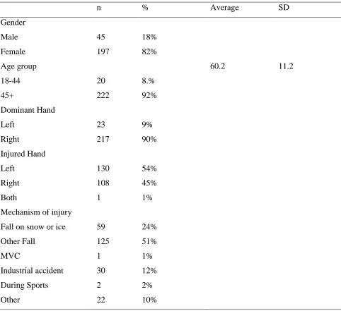
Overall, 242 patients with mean age 60.2 years old, SD ±11.26 with DRF were found eligible and
agreed to participate in the study. Our sample included 45 males and 197 females with 217 patients
having right hand dominance and 23 the left (2 were not reported). The right hand was injured from
44% of the included sample and the left hand from 54% (Table 1). The highest incidence of mechanism
of fracture was by “fall from standing height” at a total of 76.1% (24.4% “Fall on ice or snow” and 51.7% “other fall”) (Table 1). A post hoc statistical power analysis for multiple regression was
performed. The number of predictors were 2 (age and gender), the number of the average observed
R2=0.127 for large hand dexterity, R2=0.173 for medium dexterity and R2=0.056 for small hand
dexterity. The probability level was set at α=0.05 and the sample size at 238 DRF patients. Therefore,
the observed power was 0.99 for large and medium objects hand dexterity and 0.92 for the small
objects.
None of the individuals were excluded from the study since all recruited patients met our eligibility
criteria. 240 patients completed the 3-month evaluation time point, 237 at the 6-month and 240 at the
12-month period (Figure 2 Flow diagram). Multivariate analysis showed that age was a statistically
significant predictor for hand dexterity for all size of objects R2=0.227, p<0.001 (n=242) with older
adults have slower times (Table 2). Gender as variable was associated with less dexterity to manipulate
found on small objects (R2=0.000, p=0.860) (Table 2). Significant improvements in large object
dexterity were found between 3-months to 6-months and from 3-months to 12-months period
(p=0.002) but no statistical difference was observed from 6-months to 12-months (p=0.977) indicating
that the majority of improvement occurred by 6 months (Figure 3-5), (Table 3). Medium object
dexterity improved from 3-months to 6-months period (p=0.002) and from 6-months to 12-months
(p=0.016), but no statistically significant difference was observed from the 3-months to 12-months
period (p=0.773) (Figure 3-5), (Table 3). Hand dexterity recovery analysis for small objects showed a
statistically significant difference from 3-months to 6-months period (p=0.013) and from 6-months to
months period (p=0.024) but no statistically significant difference was found from 3-months to
12-months (p=0.152) (Figure 3-5), (Table 3). Moreover, the injured performed significantly poorer than
the injured hand compared at all three evaluation time points during the 12-month period.
2.5 Discussion
This study indicates that dexterity improves more in between 3 and 6 months, and slowly worsened
until 1-year following DRF; and it does not recover to the state of the uninjured hand even by 1 year.
This would support the need for greater attention to hand dexterity during rehabilitation. This study
also determined that age and gender are predictors of hand dexterity, so that scores should only
compared between people or groups when these factors have been controlled for. Age-gender based
norms are important for comparison.
The proportion of females compared to males was large (197 females and 45 males) which is reflective
in part to the higher predominance of females having DRF; but may also indicate some volunteer bias.
Since we did record all eligible fractures we cannot determine this. However, our drop out in the three
evaluation time point was negligible.
This study indicated 2 factors (age and gender) influence on hand dexterity scores. The association
between poor hand dexterity and increasing age has been reported in previous studies (Amrhein,
Stelmach, & Goggin, 1991; Cerella, 1985; Jakobson & Goodale, 1991; Martin et al., 2015; C. D. Smith
et al., 1999; Wishart, Lee, Murdoch, & Hodges, 2000). Furthermore, several studies have shown that
the reduction of mass muscles with increasing age also has an effect on reduced neuromuscular
junctions (Dorfman & Bosley, 1979; Kurokawa, Mimori, Tanaka, Kohriyama, & Nakamura, 1999;
the central nervous system (Lexell, 1997; Payne & Delbono, 2004) (motor cortex) which may be
linked to reduced hand dexterity performance. Although general exercise used to improve ROM and
strength may also have a positive effect on dexterity exercise specificity would suggest that dexterity
exercises might have a role in changing the hand dexterity and therefore improving the hand function.
Gender was a significant predictor. With respect to the available evidence from the literature our
preliminary literature review identified two studies (Haward & Griffin, 2002; Macdermid, 2001) that
examined the relationship of gender and hand dexterity scores on healthy subjects. More specifically,
the two studies showed conflicting results about hand dexterity scores and gender. Haward and
colleagues (2002) (Haward & Griffin, 2002) found no difference on 72 healthy subjects while the
other study from MacDermid and colleagues (2001)(Macdermid, 2001) found that gender was a
significant predictor for dexterity scores on 50 healthy individuals. The latter study was conducted
with the NK dexterity test so the results are more comparable to the present. Although few studies of
dexterity have specifically tested age and gender as predictors, many have assumed that these are
predictors when presenting norms as these are often used to subcategorize the data. Males had better
(faster) hand dexterity scores on large and medium objects in the 3 and 6-month period. Our visual
plot (Figure 4) for medium objects from 6-months to 12-months showed that males were worsened
while females had a slightly worst dexterity scores on that period. The data analysis for the
manipulation of small objects indicated that females were performing much better in all three
evaluation time points. This interesting observation may be explained by the fact that females may be
more capable on the manipulation of small objects because these tasks require fine coordination,
repeated precise performance of more selective accurate movements and are not based on grip
strength. However, it is a limitation of our study that male population with DRF was underrepresented
in our sample and there is a lack of gold standard for hand dexterity tests, this may indicate some
potential biases to our results.
2.6 Conclusions
Our study found that dexterity improves between 3 and 6 months and slightly worsened between 6
and 12 months after fracture; without reaching the unaffected side by 12-months following fracture.
Age and gender influence both dexterity scores and recovery patterns during the year following DRF.
Greater attention to measuring and treating dexterity may be investigated which means to provide
Reference
yen, J., Rohde, G. E., Hochberg, M., Johnsen, V., & Haugeberg, G. (2010). Low-energy distal radius
fractures in middle-aged and elderly women - Seasonal variations, prevalence of osteoporosis,
and associates with fractures. Osteoporosis International, 21(7), 1247–1255.
Aaron, D. H., & Jansen, C. W. S. (1992). Development of the Functional Dexterity Test (FDT):
construction, validity, reliability, and normative data. Journal of Hand Therapy : Official
Journal of the American Society of Hand Therapists, 16(1), 12–21.
Altizer, L. L. (2008). Colles’ fracture. Orthopedic Nursing, 27(2), 140–5; quiz 146–7.
Amadio, P. C., Silverstein, M. D., Ilstrup, D. M., Schleck, C. D., & Jensen, L. M. (1996). Outcome
after colles fracture: The relative responsiveness of three questionnaires and physical
examination measures. Journal of Hand Surgery, 21(5), 781–787.
http://doi.org/10.1016/S0363-5023(96)80192-4
Amrhein, P. C., Stelmach, G. E., & Goggin, N. L. (1991). Age differences in the maintenance and
restructuring of movement preparation. Psychol Aging, 6(3), 451–66.
Armstrong, a D., MacDermid, J. C., Chinchalkar, S., Stevens, R. S., & King, G. J. (1998).
Reliability of range-of-motion measurement in the elbow and forearm. Journal of Shoulder and
Elbow Surgery / American Shoulder and Elbow Surgeons ... [et Al.], 7(6), 573–580.
http://doi.org/10.1016/S1058-2746(98)90003-9
Bashardoust Tajali, S., MacDermid, J. C., Grewal, R., & Young, C. (2016). Reliability and Validity
of Electro-Goniometric Range of Motion Measurements in Patients with Hand and Wrist
Limitations. The Open Orthopaedics Journal, 10(1), 190–205.
http://doi.org/10.2174/1874325001610010190
Bowden, J. L., Lin, G. G., & McNulty, P. A. (2014). The prevalence and magnitude of impaired
cutaneous sensation across the hand in the chronic period post-stroke. PLoS ONE, 9(8), 1–8.