Scholarship@Western
Scholarship@Western
Electronic Thesis and Dissertation Repository
1-6-2017 12:00 AM
Effect of L-Ascorbic Acid and All-trans Retinoic Acid on Smooth
Effect of L-Ascorbic Acid and All-trans Retinoic Acid on Smooth
Muscle Cells Cultured on PCL Scaffolds
Muscle Cells Cultured on PCL Scaffolds
Brandon Chaffay
The University of Western Ontario
Supervisor
Dr. Kibret Mequanint
The University of Western Ontario
Graduate Program in Chemical and Biochemical Engineering
A thesis submitted in partial fulfillment of the requirements for the degree in Master of Engineering Science
© Brandon Chaffay 2017
Follow this and additional works at: https://ir.lib.uwo.ca/etd
Part of the Biological Engineering Commons, and the Biomaterials Commons
Recommended Citation Recommended Citation
Chaffay, Brandon, "Effect of L-Ascorbic Acid and All-trans Retinoic Acid on Smooth Muscle Cells Cultured on PCL Scaffolds" (2017). Electronic Thesis and Dissertation Repository. 4369.
https://ir.lib.uwo.ca/etd/4369
This Dissertation/Thesis is brought to you for free and open access by Scholarship@Western. It has been accepted for inclusion in Electronic Thesis and Dissertation Repository by an authorized administrator of
Abstract
The aim of vascular tissue engineering (VTE) is to fabricate tissues that are both
mechanically and biologically competent similar to the native vessel they are intended to
replace. To this end, the incorporation of sufficient extracellular matrix elastin and collagen
is important. The objective of this thesis work was to evaluate the effect of two
biochemical factors, L-ascorbic acid (AA) and all-trans retinoic acid (atRA), on elastin
synthesis when coronary artery smooth muscle cells were cultured on 3D polycaprolactone
(PCL) scaffolds. First, porous PCL scaffolds were fabricated using a solvent casting and
particulate leaching approach. The effect of different solvents (ethyl acetate, chloroform
and tetrahydrofuran) and PCL concentration on the morphology and porosity of the
resulting scaffolds were studied. The best scaffolds (based on SEM and micro-CT analyses)
were fabricated from 30% w/w PCL in ethyl acetate. Second, smooth muscle cells were
cultured on these scaffolds to evaluate elastin synthesis. It was found that concurrent
addition of AA and atRA in both 2-D and 3-D cultures suppressed elastin protein
expression compared with atRA alone. To overcome this effect, sequential biochemical
factors addition was tested. The results demonstrated that sequential but not concurrent
addition of biochemical agents promoted tropoelastin synthesis. This study suggested the
importance of biochemical factor addition strategy to engineer a viable vascular tissue.
Keywords: vascular tissue engineering, vascular smooth muscle cells, elastin, all-trans
Acknowledgements
I am sincerely grateful to my supervisor, Dr. Kibret Mequanint, who has guided me these
past years. While the learning curve was steep, Dr. Mequanint was always there to ensure
that I stayed on the right track and focused on the big picture. His continued mentorship
made me to be a well-rounded and independent student. I would like to thank Dr. Shigang
Lin and Dr. Kalin Penev, both of whom provided invaluable advice and were always
available for a quick discussion about the field and ways to constantly improve. I am very
appreciative of Dibakar Mondal for his assistance with the micro-CT data reconstruction.
Most importantly, I am thankful for having an understanding family that has been by my
Table of Contents
Abstract
... ii
Acknowledgements
... iii
List of Tables
... vii
List of Figures
... viii
List of Abbreviations
... xi
Chapter 1 - Introduction ... 1
1.1 Overview ... 1
1.2 Thesis Outline ... 2
Chapter 2 – Literature Review ... 4
2.1 Vasculature Organization and Function ... 4
2.1.1 Circulation Anatomy ... 4
2.1.2 Arterial Circulation: Structural and Histological Detail ... 5
2.1.3 Arterial Circulation: Extracellular Matrix (ECM) Proteins and Associated Mechanical Properties ... 6
2.2 Coronary Arterial Circulation ... 11
2.3 Coronary Artery Disease (CAD) ... 12
2.4 Therapeutic Interventions for CAD... 14
2.5 Vascular Tissue Engineering (VTE) ... 16
2.5.1 Overview ... 16
2.5.2 Cell Source ... 17
2.5.3 Scaffolds ... 19
2.5.4 Bioreactors ... 22
2.6 Small-Diameter VTE: Challenges and Potential Solutions... 22
2.7 Vascular Smooth Muscle Cell (vSMC) Culture ... 24
2.7.1 vSMC Phenotype ... 24
2.7.2 vSMC Response to Microenvironments: 2-D vs. 3-D Culture ... 25
2.8 Retinoids... 27
2.8.1 Overview ... 27
2.8.2 atRA Impact on vSMC Phenotype ... 29
2.8.4 atRA in Combination with Ascorbic Acid: Impact on Elastin ... 31
2.9 Study Rationale and Objectives ... 31
Chapter 3 – Materials and Methods ...33
3.1 Materials ... 33
3.2 Methods ... 34
3.2.1 Scaffold Fabrication via Solvent Casting and Particulate Leaching (SCPL) ... 34
3.2.2 Scaffold Characterization ... 36
3.2.3 Cell Culture Conditions ... 36
3.2.4 Scaffold Preparation for Cell Culture ... 37
3.2.5 2-D and 3-D Culture ... 37
3.2.6 Cell Viability and Proliferation Assays ... 38
3.2.7 Immunofluorescence Staining and Confocal Imaging in 3-D Culture ... 39
3.2.8 RNA Isolation and Gene Expression Studies Using Real-Time PCR (qPCR) Analysis ... 39
3.2.9 Protein Analysis Using Western Blotting ... 40
3.2.10 Statistical Analysis ... 41
Chapter 4 – Results and Discussion ...42
4.1 Scaffold Characterization ... 42
4.1.1 General Observations during Scaffold Fabrication ... 42
4.1.2 Scanning Electron Microscopy (SEM) ... 44
4.1.3 Micro-CT analysis of 3-D Scaffolds ... 48
4.2 Assessing Cell Viability and Proliferation ... 50
4.3 The Effect of atRA on Cultured Smooth Muscle Cells ... 54
4.3.1 Cell Morphological Response to atRA Treatment ... 54
4.3.2 The effect of atRA Concentration on Elastin Gene Expression ... 58
4.4 The Effect of AA and atRA Combination on Tropoelastin Synthesis ... 59
4.5 Comparative Study of Elastin Synthesis in 2D Plates and 3D PCL Scaffolds ... 62
4.5.1 Spatial Effects on Elastin Synthesis ... 62
4.5.2 Combinational Approach to Rescue Elastin Expression in 3-D Culture ... 64
Chapter 5 – Conclusions and Future Directions ...69
5.1 Conclusions ... 69
5.2 Future Directions ... 71
6. References ...72
Appendix: Copyright Permission ... 88
List of Tables
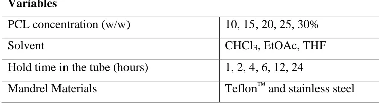
Table 4.1: Variables considered for solvent casting particulate leaching (SCPL) process to fabricate PCL Scaffolds. ... 43
List of Figures
Figure 2.1: A. Longitudinal section of an artery indicating the exposed vessel wall layers. B. Histological cross-section indicating extensive lamellar elastin distribution within the medial layer.[11] Used with permission from the Publisher. ... 6
Figure 2.2: Nonlinear mechanical behavior of an artery. A: average circumferential stress versus stretch ratio. B: circumferential incremental elastic modulus (Einc) versus stretch ratio. Einc was calculated by determining the local slope of the stress-stretch ratio relationship in Fig. A. Data shown is for adult mouse aorta.[5] Used with permission from the Publisher... 8
Figure 2.3: A. Assembly process of elastic fibers beginning at tropoelastin secretion and association with the cellular membrane where cross-linking occurs by lysyl oxidase. B. Silver stain (van Gieson) indicating that elastin is most evident within the medial layer of the vessel wall.[25] Used with permission from the Publisher. ... 10
Figure 2.4: Process description for vascular tissue engineering (VTE). The overall approach is to harvest cells from patients, expand them in culture and seed them to a 3-D scaffold for maturation and remodeling in a bioreactor. ... 17
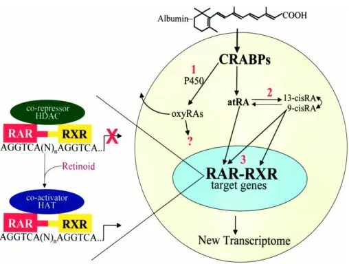
Figure 2.5: Intracellular effects of atRA. After being transported through the plasma by a protein carrier, albumin, atRA translocates across the membrane and associates with the CRABPs that allow nuclear translocation and subsequent transcriptional effects.[113] Used with permission from the Publisher ... 28
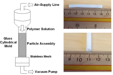
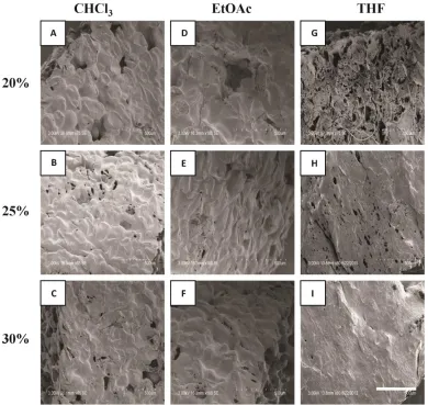
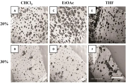
Figure 3.1: Solvent casting and particulate leaching apparatus. The digital images shown are the tubular scaffolds fabricated using the apparatus. ... 35 Figure 4.1: Ablumenal SEM images of 20-30% w/w PCL scaffolds fabricated using SCPL. PCL was dissolved in CHCl3 (A-C), EtOAc (D-F) and THF (G-I). Scale bar represents 500 μm. ... 45
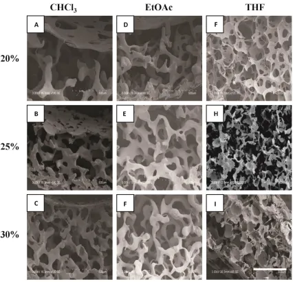
Figure 4.2: Lumenal SEM images of 20-30% w/w PCL scaffolds fabricated using SCPL. PCL dissolved in CHCl3 (A, B), EtOAc (C, D) and THF (E, F). Scale bar represents 1000 μm. ... 46
Figure 4.4: Micro-CT images of PCL scaffolds fabricated by dissolving in either CHCl3 or EtOAc at varying w/w concentrations. A-C. PCL dissolved in CHCl3. D-F. PCL dissolved in EtOAc. The images are specific volume elements representing the cross-section within the scaffolds. The white within the image represents PCL and the grey regions represent the pores. ... 49
Figure 4.5: MTT assays of NIH-3T3 fibroblasts or hcSMCs on 20%, 25%, or 30% w/w PCL scaffolds dissolved in EtOAc. A: Viability of NIH-3T3 fibroblasts assessed at day 4 and 7. B: Viability of hcSMCs assessed at day 4 and 7. Significance: p<0.05 (*). ... 52
Figure 4.6: DNA quantification of hcSMCs seeded onto 30% w/w PCL scaffolds at days 4 and 7. Significance: p<0.05 (*), p<0.01 (**), p<0.001 (***). ... 54
Figure 4.7: Confocal images of hcSMCs seeded onto porous 3-D polyurethane scaffolds and exposed to 100 μM of atRA. Confocal images were taken after 4 and 7 days of culture. Scale bar represents 200 μm. Staining: F-actin (green) and DAPI (red). ... 56
Figure 4.8: Confocal images of hcSMCs seeded on a porous 3-D polyurethane scaffold with or without fibronectin pre-treatment and 150 μM atRA. Cells were cultured for 14 days before fixation and confocal imaging. Scale bar represents 200 μm. Staining: F-actin (green) and DAPI (red). ... 57
Figure 4.9: The effect of atRA concentration on tropoelastin gene expression in hcSMCs cultured on 2D plates for 4 days. 10 μM atRA produced the highest tropoelastin fold increase and was utilized for subsequent experiments. Significance: p<0.05 (*) was observed for 10 μM of atRA compared to the untreated control (C) and 0.1 and 1 μM of atRA. ... 59
Figure 4.10: Time-course evaluation tropoelastin synthesis (as determined by Western blot) isolated from whole-cell lysates of hcSMCs in response to various biochemical factor treatments. Data are normalized to GAPDH and a control without biochemical factor treatment. Significance: p<0.05 (*) was observed as indicated in the graph. ... 61
Figure 4.12: Western blotting of whole-cell lysates of hcSMCs assessing for tropoelastin expression in response to biochemical factor treatment in 2D culture plates (A) and 3D PCL scaffolds (B). Data was normalized to GAPDH and a non-treated control.
Significance: p<0.05 (*). ... 63
Figure 4.13: Elastin expression assessed by Western blotting of whole-cell lysates of hcSMCs after sequential non-overlapping factor exposure for different times. The schematic of the experimental design is shown in A where cells were cultured either without treatment (control), 6 days of AA treatment, 5 days AA followed by 1 day atRA treatment or 3 days AA followed by 3 days atRA treatment. Cells were harvested on the 7th day for protein extraction and Western blotting. Significance: p<0.05 (*) was observed for 3 days of atRA rescue compared to AA alone. ... 65
List of Abbreviations
2-D Two dimensional
3-D Three-dimensional
α-SMA Smooth muscle alpha actin
atRA all-trans Retinoic Acid
BSA Bovine serum albumin
bFGF Basic fibroblast growth factor
CAD Coronary artery disease
CVD Cardiovascular disease
DMEM Dulbecco’s modified eagle’s medium
EC Endothelial cell
ECM Extracellular matrix
EtOAc Ethyl Acetate
FBS Fetal bovine serum
GAPDH Glyceraldehyde 3-phosphate dehydrogenase
hcSMC Human coronary artery smooth muscle cell
LAD Left anterior descending
L-AA L-Ascorbic Acid
LCA Left coronary artery
LDL Low-density lipoprotein
LOX Lysyl oxidase
NO Nitric oxide
qPCR Real-time PCR
PCL ε-Polycaprolactone
PBS Phosphate buffered saline
PAGE Polyacrylamide gel electrophoresis
PCR Polymerase chain reaction
RCA Right coronary artery
SCPL Solvent casting and particulate leaching
SDS Sodium dodecyl sulfate
SEM Scanning electron microscopy
SMC Smooth muscle cell
THF Tetrahydrofuran
TEBV Tissue-engineered blood vessel
vSMC Vascular smooth muscle cell
Chapter 1 - Introduction
1.1 Overview
Despite significant progress made in intervention outcomes, cardiovascular disease (CVD)
represents the leading cause of death worldwide. Diseases impacting the small diameter
vasculature, such as the vessels servicing the coronary circulation, are becoming the
leading cause of mortality for all of CVD [1]. Alarmingly, these trends are predicted to
increase in the future, likely attributed to an aging population prone to making poor health
decisions. Prophylactic measures to reduce the incidence of CVD have not been effective
due to compliance issues.[2] For replacement of the diseased coronary arteries, autologous
vessels such as the great saphenous vein or internal thoracic artery are the most viable
options yet the likely presence of co-morbidities or removal from previous harvests
indicate that other options are required. While it is possible to utilize allografts or
xenografts, there are significant immune-mediated issues that arise that decrease the
patency of these grafts. As such, there is need for the development of a therapeutically
viable and highly patent small-diameter graft replacement. This can be accomplished
through vascular tissue engineering (VTE) that aims to mimic a native vessel through
synthetic fabrication methods. However, the success of such designs relies on matching the
mechanical properties to that of the native vessels.[3] As collagen and elastin are the two
main proteins that endow the native vessels with their mechanical properties, it is important
While collagen can be increased in vitro through L-Ascorbic Acid (AA) addition, a main
challenge of VTE is an inability to understand and promote elastogenesis. Elastin provides
resiliency to the arteries and allows for the accommodation of cyclic loading during the
cardiac cycle. It is thought that elastogenic factors, that aid in stimulating the process,
represent viable avenues to explore. One such factor is all-trans retinoic acid (atRA) that is
a derivative of vitamin A and essential during embryonic development. atRA has been
shown to promote the expression of elastin in 2-D cultures. Additionally, atRA is suggested
to have significant phenotypic control over vascular smooth muscle cells (vSMCs)
whereby a contractile state is produced that is non-proliferative. Thus, atRA appears to be
an ideal candidate for investigation as it can increase elastin while concurrently mitigating
the risk of restenosis usually seen with surgical interventions.[4] That said, few studies have
investigated the combination of AA and atRA on 3-D scaffolds and those that did utilize
long end-points and failed to assess elastogenesis acutely compared to either factor alone.
1.2 Thesis Outline
The thesis contains five chapters that are based on investigations pertaining to the
interaction of hcSMCs with 2-D and 3-D substrates and the fabrication process of 3-D
scaffolds. Chapter 2 introduces the relevant literature of the vasculature anatomy, vascular
disease progression, therapeutic options to treat the pathologies, followed by the need for
vascular tissue engineering and options to augment elastogenesis. The motivation and
objectives of this study are discussed at the end of the chapter. Chapter 3 introduces the
findings and results from the work. Chapter 5 comments on the future directions of the
Chapter 2 – Literature Review
2.1 Vasculature Organization and Function 2.1.1 Circulation Anatomy
Blood vessels are hollow tubes that act as conduits for the cardiac output of the heart,
allowing for systemic tissue perfusion with an oxygenated blood flow while facilitating the
return of deoxygenated blood to the heart. The contraction of the left ventricle of the heart
allows for the distribution of oxygenated blood systemically via the aorta, the initial
segment of the arterial circulation. Distal to the aorta, numerous branches and bifurcations
occur. Each newly created continuous segment is associated with an alternative lumenal
diameter and deviation in the underlying structural detail of the vessel wall which imparts
specific functionality.[5] The aorta transitions to smaller elastic then more muscular arteries
which give rise to numerous arterioles and ultimately the capillaries.[5] The aorta acts as a
Windkessel vessel, attempting to limit pulsatile blood flow in the periphery by dampening
pulse pressure as it possesses relatively higher compliance than other derivatives in the
arterial system.[6] The smaller arteries and the arterioles are more resistive in nature and
thus have a larger impact on the maintenance of mean arterial pressure and control of distal
flow. As the transition to arterioles occurs, there is a slight increase in the mean surface
area of the vessels that reaches a maximum at the capillaries, which have the slowest flow
velocity. Capillaries have the ideal hemodynamic characteristics for gas exchange and
filtration or absorption due to their small wall thickness.[7] The capillaries converge after
gas-exchange occurs at their proximal ends and become venules more distally, the first
that drain into the inferior or superior vena cava and back into the right atrium of the heart.
Abnormalities in any section of this closed system will compromise downstream tissue
perfusion and thus organ function.
2.1.2 Arterial Circulation: Structural and Histological Detail
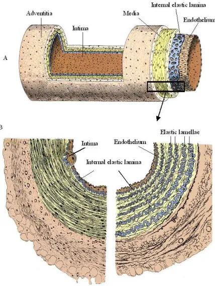
The arterial wall is a tri-layered laminar structure consisting of the tunica intima, the tunica
media, and the tunica adventitia.[5] Along the arterial tree, the composition of the arteries
varies and similar, yet variant, stress-strain curves are observed.[8] Numerous studies have
shown that the stress-strain curves will range from elastic to viscoelastic more distally with
a continuum between these points.[5, 8, 9] The proceeding text describes the general features
of an elastic or a more proximal muscular artery to which Figure 2.1 provides an overview
of the arterial wall layers and underlying histological detail in cross-section.
The tunica intima is the innermost layer of the vessel wall that is exposed to the blood flow.
It is composed of a mono-layer lining of endothelial cells (ECs) upon a basement
membrane with a sub-endothelial matrix consisting of loose connective tissue, myointimal
cells, fibroblasts, and extra-cellular constituents. Deep to the tunica intima lies the tunica
media, a muscular layer consisting of circumferentially orientated smooth muscle cells
(SMCs) and interspersed extra-cellular matrix fibers in a lamellar organization. The
junction between the tunica media and overlying tunica intima is delineated by an internal
elastic lamina, which assists in volume accommodation during the cardiac cycle and may
have a role in SMC migration from the medial layer.[10] An external elastic lamina lies
adventitia. The latter layer is more connective tissue in nature with fibroblasts in addition
to collagenous fibers orientated longitudinally.
Figure 2.1: A. Longitudinal section of an artery indicating the exposed vessel wall layers. B. Histological cross-section indicating extensive lamellar elastin distribution within the medial layer.[11] Used with permission from the Publisher.
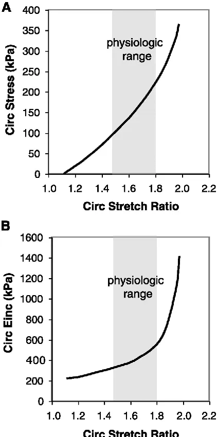
2.1.3 Arterial Circulation: Extracellular Matrix (ECM) Proteins and Associated Mechanical Properties
Compared with the highly compliant venous circulation, arteries have significantly higher
elasticity, reversible recoil, and resilience at low strains.[5] These properties allow the
arterial circulation to act as a pressure reservoir, to ensure that blood flows distally
especially during diastole. However, the mechanical properties vary during alternative
loading conditions and relate to the composite nature of arteries facilitating anisotropic
mechanical responses. The classical arterial stress-strain relationship is a non-linear
a stiffer material at higher strains as shown in Figure 2.2.[5, 12] As such, blood vessels do
not possess simple elastic properties, as non-linearity is observed when distended. These
mechanical characteristics are due to numerous extracellular components produced by the
cells within the vessel layers that collectively comprise the ECM. Specifically, these
mechanical characteristics are due to components mainly within the medial layer,
especially collagen and elastin.[5]
Collagen is a stiff, extracellular protein, possessing a high tensile strength of approximately
1.0×10⁹ N/m2, whose concentration is highest in the tunica adventitia.[13] In the unloaded
condition, collagen assumes a “crimped” state yet when load is applied, additional collagen
fibers will assume a greater proportion of the load-bearing and re-align in the direction of
the applied stress.[14, 15] It was believed that the load-bearing ability of collagen in the
medial layer begins at higher stresses until recently whereby Chow et al.[16] proposed that
medial collagen may be engaged from the onset of strain. Yet, it is the recruitment of
additional load-bearing in parallel with the high tensile strength of collagen that makes the
protein an ideal structural support for protection against aneurysmal development and the
potential for vessel wall rupture, as the artery becomes stiffer as the strain increases.
However, collagen itself does not impart elasticity and it is elastin, an amorphous
Figure 2.2: Nonlinear mechanical behavior of an artery. A: average circumferential stress versus stretch ratio. B: circumferential incremental elastic modulus (Einc) versus stretch ratio. Einc was calculated by determining the local slope of the stress-stretch ratio relationship in Fig. A. Data shown is for adult mouse aorta.[5] Used with permission from the Publisher.
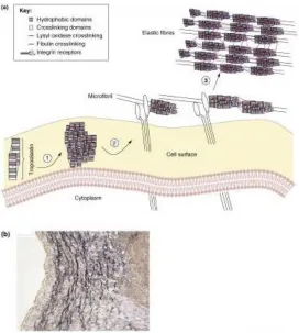
Elastin exists within a composite of numerous ECM proteins that collectively are termed
as elastic fibers.[17] Approximately 90% of the elastic fibers have been shown to be derived
from elastin.[18] Intracellularly, elastin is transcribed as a 60-70 kDa tropoelastin precursor
developmentally regulated, with highest activity during late fetal and early neonatal period
with minimal activity in adulthood due to post-transcriptional mechanisms at the
downstream developmental stages.[19, 20]Additional segmental regulation occurs during
these periods with the tissue-expression of elastin mainly being restricted to areas that
undergo repetitive stresses.[21] When translated and excreted from the cell, tropoelastin
becomes cross-linked at the cellular membrane by lysyl oxidase and the newly cross-linked
structure is termed as elastin.[17] The aggregation is aided by the numerous hydrophobic
domains which facilitate coacervation of the elastin subunits and promote the formation of
strong desmosine and isodesmosine cross-links that provide the protein with its mechanical
properties.[17, 22] The aggregates transfer to pre-existing micro-fibrils within the
extra-cellular space, whereby the major component of the micro-fibrils, fibrillin-1, serves as a
scaffold for elastin deposition.[17, 23] The assembling of the composite continues to increase
in size as the elastin coalesces. The assembly process of tropoelastin to elastin and
interaction with the pre-existing micro-fibrils is shown in Figure 2.3.
While elastin possesses a low tensile strength of approximately 4.6×10⁵ N/m2, it functions
best to provide high resilience to the artery with a contributory low stiffness.[13] Chow et
al.[24] have demonstrated that maximal elastin engagement within the medial layer is
reached at approximately 20% strain. Elastin is engaged at these low strains to allow for
slight distensibility of the arterial wall to accommodate increases in blood volume during
Figure 2.3: A. Assembly process of elastic fibers beginning at tropoelastin secretion and association with the cellular membrane where cross-linking occurs by lysyl oxidase. B. Silver stain (van Gieson) indicating that elastin is most evident within the medial layer of the vessel wall.[25] Used with permission from the Publisher.
As mentioned earlier, the medial layer contains numerous SMCs in a lamellar organization
interlaced with elastic fibers. The geometry of the tunica media aids in accommodating the
cyclic circumferential strain due to the cardiac cycle. The developing wall stress during
systole is uniformly distributed on the vessel wall due to the residual stress being greater
than zero and studies have suggested that this effect can be mainly attributed to elastin
within the medial layer.[26, 27]At low stresses, the elastic lamellae become more aligned and
taut yet the collagen alignment is more gradual and activated at much higher stresses.
However, during disease states, the underlying structure will be significantly altered.
hypertension can influence the integrity of the fibers and alternative mechanical properties
will be observed.[15, 28]
2.2 Coronary Arterial Circulation
The coronary arterial circulation is a collection of mainly muscular arteries that begin at
the sinus of Valsalva, where the ascending aorta gives rise to the right and left coronary
arteries (RCA and LCA, respectively) from its right and left sinus openings.[29] The RCA
and LCA will give rise to branches that will supply the entire myocardium. The LCA arises
from the left aortic sinus and gives rise to the left circumflex artery and the anterior
interventricular artery (left anterior descending; LAD). The RCA arises from the right
aortic sinus and progresses laterally along the right side of the heart to give rise to vessels
that mainly supply the right ventricle. In 90% of individuals, the RCA gives rise to the
posterior interventricular artery: termed a right-dominated coronary circulation. Using
arteriograms, Dodge et al.[30] indicated that normal vessel diameters of the LCA, proximal
LAD, and distal LAD are 4.5±0.5 mm, 3.7±0.4 mm, and 1.9±0.4 mm, respectivelywith
similar values obtained for the RCA and derivatives. The relative sizes between the sexes
appear to vary significantly as documented in numerous studies.[31, 32] That said, the main
deviation between individuals is the alteration in the mechanical properties of the coronary
circulation with age. Specifically, the elastin-collagen ratio decreases with age from 5.43
to 2.57 in the LCA and 3.80 to 1.72 in the RCA.[31] The decrease in the ratio suggests the
progression of stiffer arteries with age. However, ignoring the extremes of age, the typical
[33], a maximal circumferential strain reached at 20% distension[34], and a normal pressure
exposure between 50-130 mmHg.[35]
2.3 Coronary Artery Disease (CAD)
Cardiovascular disease (CVD) represents the leading cause of mortality globally from
which coronary artery disease (CAD) is the leading factor.[1, 36] The alarming trend is that
the incidence and prevalence of CVD is increasing. In the United States, it is projected that
approximately 40.5% of the population will be burdened with CVD and the associated
costs are expected to exceed $800 billion by 2030.[37] With advances in healthcare, the
morbidity of the disease and its complications are likely to parallel this trend in augmented
prevalence as individuals live longer lives.
The main instigating factor in the development of CAD is atherosclerosis, a progressive
disease characterized by gradual lumenal narrowing of blood vessels leading to
compromised distal blood flow and tissue perfusion which results in potential ischemic
complications.[38] The pathology increases in prevalence with age due to the progressive
nature of the disease and repeated exposure to mostly modifiable risk factors. A main risk
factor are increased levels of cholesterol and low-density lipoprotein (LDL), whereby the
LDL functions to bring cholesterol and phospholipids to the peripheral cells and tissues
rather than back to the liver for recycling as high-density lipoprotein facilitates.[39]
Normally, LDL circulates within the plasma at low concentrations yet can be elevated due
to a poor diet and diabetes, among other factors.[38, 40] If the endothelium is damaged, which
sub-endothelial space within the tunica intima. The resultant response, and the basis of the
pathogenesis of atherosclerosis, is the “response to injury” phenomenon which is an
immune mediated reaction to cope with the lipid insult and endothelial damage.[41] Resident
tissue macrophages within the sub-endothelial layer attempt to phagocytose and remove
the LDL. However, numerous free radicals and potential apoptotic debris are produced
which facilitate the oxidation of LDL within the layer.[38] As the macrophages repetitively
ingest the oxidized LDL, they transition in morphology to become foam cells. The
pathology and lumenal narrowing becomes exacerbated by endothelial damage and the
ability of oxidized LDL to act as a pro-inflammatory and adhesive mediator to circulating
monocytes.[38]
The tunica intima has additional functions pertaining to anti-thrombotic activities under
normal homeostatic conditions. The ECs are able to secrete numerous factors such as nitric
oxide (NO) and prostacyclin (PGI2) which attempt to mitigate adhesion and promote
vasodilation. The initial sign of endothelial damage is a decrease in the levels of NO
produced which consequently leads to lumenal narrowing which facilitates a greater
concentration of circulating clotting and inflammatory factors that can interact with the
endothelium.[42] It is the integrity of the endothelial lining that is paramount as the
sub-endothelial matrix is highly thrombogenic in vivo with collagen and von Willebrand factor
being able to interact with platelets. The damaged endothelium promotes the expression of
specific adhesion molecules that are able to attract leukocytes to the area of damage such
as P-selectin. An inflammatory cascade ensues whereby a fibrous plaque develops that
integrity of the arterial lumen.[38] The response impacts the medial SMCs and the
organization of elastin and other ECM proteins as SMC proliferation and protein
production is stimulated. The resultant disorganization of the fibers further augments the
stiffness of the blood vessel.[43] New tropoelastin may be secreted, yet it will not possess
the same arrangement and thus the mechanical properties of the arteries will be
significantly altered, especially as increased collagen is deposited due to the inflammatory
process.
The patency of the coronary arterial circulation is essential to ensure sufficient perfusion
to the myocardium. Coronary artery occlusion follows a gradual course initially
characterized by stable angina which leads to unstable angina and ultimately the potential
for myocardial infarctions. The classification of the varying states is based on the degree
of vessel occlusion. Rarely, blockages occur as a result of an embolus and are usually
associated with atherosclerotic plaques. With higher occlusion percentages, individuals
begin to complain of exertional dyspnea as the decrease in lumenal diameter compromises
myocardial perfusion resulting in the lack of sufficient oxygenation.[44] With progressive
occlusion, a myocardial infarction may occur which is characterized by a massive
compromise in cardiac output and potential conduction disarray which leads to death.
2.4 Therapeutic Interventions for CAD
The initial therapeutic approach should be centered on prophylaxis through lifestyle
modifications and pharmacological therapy. The treatment is based on attempting to reduce
of the coronary vessels and improve exercise tolerance, which is the indication for
nitro-dilators. Additionally, aspirin or clopidogrel may be recommended to ease the concern of
potential thrombi causing distal ischemia and complications. Unfortunately, the efficacy of
lifestyle modifications and pharmacological therapy suffers from poor patient
compliance.[2] More invasive interventions may be required if these techniques fail.
In the non-surgical setting, the approach is centered on revascularization of occluded
coronary vessels and can be accomplished most commonly with percutaneous coronary
interventions (PCI) which include balloon angioplasty and coronary stenting. PCI entails
the insertion of a wire or catheter, usually into the radial artery, that is guided via imaging
to the site of the coronary occlusion.[45] A meta-analysis comparing the efficacy of PCI
versus conservative pharmacological therapy in chronic stable CAD indicated that PCI fails
to offer any benefit in the management of these patients and may have additional risks.[46]
The main concerns with PCI are the potential for stent restenosis due to increased
disorganized cellular proliferation within and around the stent and the development of
thrombus formation. It has been shown that the main response to the procedure is a
significant increase in intimal thickening.[47] Balloon angioplasty has been shown to have
a 30-60% risk of re-occlusion of the vessel whereas bare metal-stents have an approximate
16-44% risk in the development of restenosis.[48] To reconcile these risks, drug-eluting
stents were developed, however complications can still occur in 3-20% of patientsdue to
allergic reactions, granulomatous inflammation, and neo-intimal thickening.[49] With the
development of more unstable ischemic inducing states, surgical interventions are
The standard surgical intervention is the coronary artery bypass graft (CABG). The
procedure is based on transferring another vessel to replace the diseased artery. Most
commonly, autografts are used such the great saphenous vein (GSV) or internal thoracic
artery. However, a common occurrence is the presence of co-morbidities that make these
vessels unusable or previous harvests have already removed them. Specifically, the GSV
is usually found to be pathological when harvested, which emphasizes the need for
consideration of other allogenic sources.[50] It is possible to harvest vessels from other
donors or animals, yet significant immune-mediated issues arise due to their inherent
thrombogenicity which has been shown to decrease the patency of the graft within a short
time-frame post-operative in many patients.[51] Unfortunately, those with CAD are likely
to have multi-vessel involvement. A recent meta-analysis comparing PCI to CABG for
those with multi-vessel disease indicated that while CABG resulted in a slightly higher
incidence of strokes, they did reduce the need for future re-vascularization and decreased
long-term mortality.[52] A possible reason for the long-term advantageous use of CABG is
that it may provide a more natural and tailorable environment for the cells to interact with.
However, for small diameter graft replacements, no ideal solution exists as of yet and
synthetic grafts produced in vitro are attempting to address this urgent clinical need.
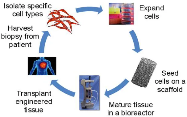
2.5 Vascular Tissue Engineering (VTE) 2.5.1 Overview
Vascular tissue engineering (VTE) attempts to mimic the native vessel architecture and
growth potential while maintaining long-term lumenal patency. In blood vessel tissue
engineering, the goal is to fabricate a tissue that is mechanically competent and biologically
responsive in a process that is highly reproducible and consistent. The creation of such an
analogue is termed a tissue engineered blood vessel (TEBV). The classical paradigm of
VTE involves the growth of cells in two-dimensional (2-D) culture and subsequent seeding
onto a three-dimensional (3-D) substrate for maturation. Ideally, maturation will occur in
a bioreactor to mimic the native hemodynamic forces the cells are exposed to in vivo. The
VTE process description for the fabrication of a TEBV is outlined in Figure 2.4.
Figure 2.4: Process description for vascular tissue engineering (VTE). The overall approach is to harvest cells from patients, expand them in culture and seed them to a 3-D scaffold for maturation and remodeling in a bioreactor.
2.5.2 Cell Source
The cells utilized for VTE should reflect those found in native vessels, being some
combination of fibroblasts, SMCs, and ECs that populate the three component vessel
layers. ECs provide potent signaling to the underlying SMCs within the vessel which can
transfer limitations of oxygen when vessel wall thickness is increased.[53] An intact
endothelium significantly reduces the thrombogenicity of the graft at the early implantation
stages – especially if seeded initially with venous ECs which have been shown to be less
thrombogenic than arterial cells.[54, 55] Additionally, EC capture and seeding can be
enhanced through the utilization of peripheral whole blood (PWB) as it contains numerous
endothelial precursor cells that possess the capacity to differentiate into ECs if the correct
milieu is provided.[56] For instance, the presence of a monolayer lining of ECs was observed
when PWB was added to a decellularized allogenic scaffold.[57] Interestingly, when a graft
is implanted into a patient, ECs have been shown to re-populate the lumenal lining in vivo
with staining confirming the original seeded cells were replaced by native cells within an
acute time-frame.[58] This suggests that vascular grafts may not necessarily need ECs when
implanted given the ability of the ECs to re-populate the graft in vivo. However, if this
were to occur, anti-platelet and anti-coagulative therapy should be provided until
endothelialization occurs for the patient to be at low risk of developing thrombosis.[59]
Given the ability of ECs to re-populate the graft in vivo,the main challenge is how to best
re-populate the graft with the cells that comprise the tunica media and adventitia.[60] Stem
cells have emerged as a viable option as a cell source due to their potential to differentiate
into SMCs. Bone-marrow mesenchymal stem cells, adipose derived stem cells, and human
umbilical cord derived stem cells have shown the ability to express vascular SMC (vSMC)
protein markers in vitro and mimic SMC-like ECM production.[61, 62] Specifically,
mesenchymal stem cells are a preferred choice due to their ability to be expanded in culture
for harvesting.[63] While promising, studies are mixed in regards to the adverse effects in
vivo with the use of mesenchymal stem cells which may limit their use until safety can be
ensured.[64] Additionally, while stem cells can be coaxed into differentiating down a vSMC
pathway, given the complexity of the process, a reasonable concern is the extent of the
differentiation.[65]
As the differentiation process has not been optimized in vitro, the simplest way to ease this
concern would be to utilize primary SMCs and fibroblasts - especially when attempting to
investigate signaling pathways within grafts. However, primary SMC lines have been
shown to have variable responses in culture that relate to the donor source.66 SMCs sourced
from older donors produce less collagen and elastin and have less proliferative capacity
than their juvenile counterparts.[66, 67] Regardless of age, primary SMCs show a limited
capacity for proliferation in vitro, where typically 10-30 population doublings occur prior
to senescenece.[67] While stem cells are more robust, there exists ways to circumvent the
growth retardation of vSMCs seen in 2-D culture. As an example, McFetridge et al. [68]
demonstrated that bioreactor systems can induce SMCs to maintain their in vivo
phenotypes. It is also possible that the utilization of higher initial seeding densities can
circumvent the need for numerous population doublings to increase ECM production.
2.5.3 Scaffolds
The ideal scaffold is expected to mimic the geometry of the tissue to be fabricated and
provide correct clues to the cells that are conducive to growth and ECM production. The
by proliferation without a cytotoxic or inhibitory effect. The ideal scaffold candidate would
therefore be native ECM itself, and this can be made possible through the use of
decellularized scaffolds. The native ECM provides a rich source of proteins such as
fibronectin that cells can interact favorably with the matrix.[69] However, decellularization
procedures usually requires the use of detergents and proteases that may disrupt the normal
architecture of the ECM and dislodge potential signaling and attachment components.[70]
The decellularization procedure generally produces variable mechanical change in the
scaffold, usually generating weaker grafts.[70] Since collagen and elastin are essential for
the mechanical properties of arteries, their use has been investigated as a comparable option
to that of decellularized scaffolds. However, it has been found that collagen itself may be
a poor scaffolding material as it loses mechanical strength and stability upon hydration in
addition to potential immune-mediated reactions and disease-risk when extracted from
animal sources.[71] Recombinant technology is attempting to produce collagen, yet the
process remains to be optimized and is not currently a viable option for VTE use.[70] A
major issue with elastin-based scaffolds are their propensity to calcify in vivo which would
mitigate the suitability of the scaffold as the mechanical properties are altered.[72] Another
approach, which would allow more consistent optimization of the required mechanical
properties, is the development of synthetic scaffolds. These scaffolds, if fine-tuned to
match the required properties of the ECM, possess high viability to be mass-produced and
available as an “off-the-shelf” option for surgical interventions.[73] Viable synthetic
scaffolds include biodegradable polymers, such as polyglycolic acid (PGA),
polycaprolactone (PCL), and derivatives synthesized via copolymerization with polymers,
in vivo.[74] The goal is to match the degradation rate in vivo with suitable ECM production
without jeopardizing the mechanical properties as this occurs.[70]
Equally important to the scaffold material is the fabrication method. Electrospinning has
emerged as a promising option for VTE due its ability to facilitate the fabrication of
nanofibrous matrices that mimic the topography of the ECM. The technique involves
subjecting a polymer solution to a high voltage which is ejected through a capillary in a
dynamic unstable manner at a controlled rate. Once ejected, the electrical force is greater
than surface tension which creates nanofibrous polymer mats.[75] The solvent is evaporated
as random fibers are deposited onto a grounded collecting plate. However, while the fibers
mimic the native ECM in morphology, it is difficult to control the exact orientation of the
fibers and a major limitation with electrospinning is a lack of suitable cell infiltration due
to small pore sizes.[76] Another technique that directly addresses this issue is utilizing
hydrogels as the scaffolding material. Hydrogels are composed of hydrophilic polymer
chains that are cross-linked allowing for the gel to swell when placed in an aqueous
environment without a loss of structure.[77] The gel matrix allows for suitable mass transfer
of nutrients and gases and additionally offers a way to ensure suitable cell encapsulation.[78]
However, a major limitation with hydrogels is their lack of mechanical integrity, especially
when utilizing natural polymers compared to synthetic options.[79] Another fabrication
method is that of solvent casting and particulate leaching (SCPL). The technique involves
dissolving a polymer in a suitable solvent and forcing the developing viscous solution
through a porogen bed to which the porogen is leached out in a solvent that does not
porosity at the macro and micro porous levels. However, SCPL offers limited control over
the orientation of the pores and the overall pore interconnectivity.[80] That said, the benefit
of SCPL is that there is a high control over overall porosity and thus cell infiltration.
Overall, SCPL offers a simple fabrication process with high tunability in a fabrication
method that is cost-effective.[81]
2.5.4 Bioreactors
The use of bioreactors allows for simulation of the native hemodynamic forces. Forces
such as shear stress on the ECs and the pulsatile nature of the blood flow can induce
signaling events. Specifically, these forces have been shown to induce growth and
maturation of the cells significantly different than that of static culture.[82] The stimulation
induces the SMCs to secrete more ECM proteins and has been associated with improved
graft mechanical properties.[83] Mechanical stimulation additionally allows vSMCs to
adopt phenotypes more akin to the in vivo environment which may further explain the
reasoning to these grafts developing higher mechanical properties and improved patency
when cultured within a bioreactor.[84, 85]
2.6 Small-Diameter VTE: Challenges and Potential Solutions
Synthetic grafts made from expanded polytetrafluoroethylene (ePTFE) and polyethylene
terephthalate (PET, Dacron) work well for large diameter vascular graft replacements
(>6 mm diameter) where the blood flow rate is high but results in small diameter coronary
artery graft applications (<4 mm diameter) have been disappointing.[86-88] The main
exacerbates the pathologies the graft attempts to alleviate. A non-autologous vascular
replacement will induce an immune response, yet the reaction may not necessarily be acute.
Furthermore, if the mechanical properties of the graft are not matched with that of the
native vasculature at the anastomotic sites, the generated turbulent flow induces an
immune-mediated response that can result in lumenal occlusion.[89] These limitations
prompted the field of VTE. Despite rapid advances made in this field, success has been
limited due to significant knowledge gaps in our ability to control and coordinate cell
phenotype and direct tissue formation, which is the ultimate goal for VTE.[90] Particularly,
stimulating and understanding elastogenesis in VTE has been proven to be difficult.[3, 11, 91]
It is understood that for a TEBV to be successful, it must incorporate elastin into the
developing tissue construct to achieve the required overall viscoelasticity of the final
tissue.[11, 92] It is possible that utilizing elastomeric scaffolds could be helpful for improving
the elasticity of the engineered tissue. Indeed, the use of elastomeric scaffolds has resulted
in more akin mechanical properties to that of the native arteries.[93, 94] There is optimism
that a cell-free tissue engineering approach can be based on the use of such scaffolds.[95]
Unfortunately, the approach relies on in vivo re-modeling and it can be expected that the
results will be variable. Additionally, this technique relies on infiltration of native cells and
an immune response to acquire the required remodeling. Yet again, this may vary from
host to host and might not be suitable for the small diameter vasculature.[96] As such, while
the synthesis of elastomeric scaffolds improves the mechanical properties, there is a need
to seed cells prior to implantation. Ideally primary human coronary SMCs (hcSMCs) that
are responsible for producing elastin and assembling it to elastic fibers within the medial
then the mechanical properties of TEBVs can be expected to increase. An avenue that can
be taken is the use of factors that can stimulate elastogenesis. However, in addition to their
impact on elastogenesis, these factors may influence the phenotype of the vSMCs.
2.7 Vascular Smooth Muscle Cell (vSMC) Culture 2.7.1 vSMC Phenotype
vSMCs represent a dynamic cell line in which their phenotype is not terminally
differentiated and rather exists as a continuum of states between synthetic and
contractile.[97] During development, vSMCs begin in a highly proliferative synthetic state
where ECM protein production is high. A transition occurs that decreases ECM production
and increases intercellular myofilaments, a switch that facilitates the required contractility
within blood vessel walls.[98] Studies have indicated that the transition can be
bi-directional.[99, 100] The phenotypic plasticity can vary along the arterial tree during normal
homeostatic conditions due to the presence of alternative populations of vSMCs.[101]
However, in vivo, the phenotypic plasticity is correlated with pathological events such as
seen in atherosclerosis whereby the usual contractile vSMCs in the medial layer can
migrate to the intima and become synthetic, promoting intimal thickening and hyperplasia
– an undesirable outcome. The continuum between synthetic and contractile is classically
characterized by the presence or absence of specific proteins.[97] The contractile state
contains minimal rough endoplasmic reticulum and ribosomes indicating that its function
is not related to extensive ECM protein synthesis.[101] When the vSMCs are in the
contractile state, they have a large volume fraction of myofilaments within their
contractile protein markers such as smooth muscle alpha actin (α-SMA), calponin,
SM-22α, and smoothelin.[101] De-differentiation into the synthetic phenotype is associated with
a loss of these markers.[101] In addition to quantitative changes in the contractile protein
markers, the modulation in phenotype is alsoassociated with alternative rearrangements
and realignments of the myofilaments.[102] Due to the lower level of protein secretion, the
contractile vSMCs are denser and more fusiform in nature, a stark contrast to the expansive,
broad, and fibroblast-like epithelioid morphology of the synthetic phenotype. When cells
de-differentiate into a synthetic phenotype, a loss of the high volume of myofilaments, a
disappearance (or reduction) of the contractile markers, and an increase in apparatuses
promoting protein production have been observed. Specifically, elastinproduction is likely
correlated with the synthetic vSMCs phenotype, yet there has been mixed results.[103, 104]
In contrast to the contractile upregulation of vSMC protein specific markers, it is thought
that the synthetic phenotype is characterized by a disappearance of the contractile markers.
However, proteins such as L-caldesmon and meta-vinculin can be used to denote the
synthetic state, yet marker specificity for the synthetic phenotype is not as specific as the
contractile state.[105]
2.7.2 vSMC Response to Microenvironments: 2-D vs. 3-D Culture
Classically, most cell culture investigations have been performed on a 2-D surface yet this
is an inaccurate representation of the in vivo environment. While 2-D culture is a quick and
well-established culture system, it only provides a simplistic and limited substrate that cells
can interact with. Within the ECM, cells are constantly exposed to varying topologies,
setting. It can be expected that cells will respond and behave differently when seeded in
2-D and 3-2-D environments. When cultured in 2-2-D, most vSMCs are known to adopt a more
synthetic phenotype compared to a contractile state usually seen in vivo.[101, 106] Yet, most
early experimental designs considered vSMCs only in a 2-D environment, one that is stiffer
than the native ECM found in blood vessels. Indeed, a major difference between 2-D and
3-D is the stiffness of the underlying substrate. Recently, Timraz et al.[107] demonstrated
that collagen-I gels with varying degrees of stiffness can influence vSMC phenotypic
differentiation and protein secretion with the synthetic phenotype being inversely
proportional to increasing the stiffness of the gels. However, the study investigated cellular
responses when encapsulated in a collagen gel and when combined with various ECM
proteins, such as fibronectin, which may influence the manner in which the cells interact
and spread within the matrix as the supplemented factor concentration is increased. The
exact experimental design for investigating how cells respond to variant stiffness in 3-D
remains to be optimized. This may be due to designs that increase the cross-linking density,
consequently increasing the propensity for cell-mediated proteolysis and subsequent
migration.[108] A commonality between how the cells sense stiffness in 2-D and 3-D is
through RhoA, a small GTPase that facilitates mechanotransduction. RhoA activation has
been shown to be upregulated in 2-D culture and in atherosclerosis.[109] When RhoA is
constitutively expressed in vSMCs in 3-D cultures, the state of vSMC differentiation has
been shown to depend on the stiffness of the substrate and produced similar cytoskeletal
effects seen in 2-D culture.[106] Upregulation of RhoA in vSMCs is associated with an
increase in contractile SMC markers in 3-D.[106] While stiffness may have an effect on the
able to sense their surroundings. Using varying matrices, Hong et al.[110] demonstrated
varying temporal differences in ECM expression in 3-D relative to 2-D with significant
variability when alternative 3-D scaffolds were used. Work by Lin et al. [111] also
demonstrated that hcSMCs seeded in 3-D expressed higher levels of elastin than in 2-D
culture. However, when TGF-β1 was utilized to increase elastin, the up-regulated
elastogenic effect was noticed only in 3-D and not in 2-D suggesting that the effect of
TGF-β1 in 2-D cultures may not translate to 3-D. Another biochemical factor that is elastogenic
but not widely studied is retinoic acid which is a vitamin A derivative. Since this thesis
focuses on retinoic acid, an expanded review is presented in the proceeding section.
2.8 Retinoids
2.8.1 Overview
Retinoids are derivatives of vitamin A that are highly active during embryogenic
development and have been shown to have a positive impact on elastogenesis. Four of the
biologically active derivatives are retinol, retinal, 9-cis retinoic acid (9cRA), and all-trans
retinoic acid (atRA).[112] While there are numerous metabolites, it is atRA which is the
carboxylic acid form that has been shown to have the most potent biological activity.[113] It
should be noted that there have been reports indicating isomerization potential between
atRA and 9cRA intracellularly.[113] As a lipophilic molecule, the atRA concentrations
within the plasma are low - ranging between 1-10 nmol/L.[113] Most in vitro experiments
with atRA utilize the relevant therapeutic and physiological concentration, corresponding
approximately to 1 μmol/L.[113] Due to its lipophilicity, a retinol binding protein (RBP)
atRA is able to diffuse past the cellular membrane and an interaction can occur with a
cellular retinoic acid binding protein (CRABP). There are two isoforms of CRABP, being
I/II, though only CRABP-I has been shown to be relevant in vSMCs.[115] Binding to the
CRABPs occur with high affinity and seems to increase the ability of atRA to be
translocated to the nucleus.[116] Within the nucleus, the complex interacts with two subsets
of nuclear receptors being the retinoic acid receptor (RAR) and retinoid X receptor
(RXR)[113], to which there are 3 isoforms of each (α, β, and γ).[117, 118] Both receptors
associate as a heterodimer that are bound to a retinoic acid response element (RARE) near
specific target genes that normally act to repress transcription. Retinoids are able to bind
to the RAR allowing for conformational changes to be induced that create positive
transcriptional effects. Both 9cRA and atRA are able to bind to the RAR yet only 9cRA
can associate directly with the RXR. However, the RXR itself has not been proven to have
physiological relevance. An overview of the metabolism and subsequent processing of
atRA is shown in Figure 2.5.
2.8.2 atRA Impact on vSMC Phenotype
The current understanding of the effect of atRA on vSMC phenotype is that an
anti-proliferative and differentiated state is induced. Interestingly, atRA is being investigated
for its role is preventing intimal thickening and restenosis following PCI.[4] The elevated
levels of contractile makers have been associated with atRA possessing potential negative
effects on growth and cellular proliferation in human tissue culture. Axel et al.[119] found
that in a dose-dependent manner, atRA decreases both proliferation and migration. The
ability to produce the latter effects may be attributed to a retinoid-inducible tissue
trans-glutaminase (ttG).[120] Recently, in vivo experiments confirm that atRA can inhibit vSMC
proliferation and promote contractile marker expression via upregulation of Kruppel-like
Factor 4 (KLF4), a zinc-finger transcription factor.[121] KLF4 activation has been shown
to activate p53 to inhibit vSMC proliferation.[122] Knockdown studies of KLF4 mitigated
the atRA mediated increase in contractile protein markers being α-SMA and SM-22α.[123]
In rat vSMCs, atRA has been shown to increase the levels of contractile markers such as
α-SMA.[124] The increase in these markers was associated with a stimulation in protein
kinase C-α activity.[124] However, it is not intracellular interference alone that atRA exerts
its anti-proliferative effect, as it has been shown to influence ECM production. Perlecan
sulfate is a large proteoglycan within the ECM that has been shown to have
anti-proliferative properties. When transgenic mice were engineered to be deficient in perlecan
sulfate expression, the atRA mediated anti-proliferative effect was depressed.[125] That
said, most of the above studies occurred in 2-D culture. The phenotype adopted by vSMCs
different responses obtained in early investigations may be due to the source of vSMCs
and the experimental design.
2.8.3 atRA Impact on Elastin Expression
The effects of atRA seems to be tissue-dependent with pleiotropic impact.[113] In terms of
elastogenesis, atRA has been shown to have potent impact on the pulmonary and arterial
circulations and is associated with dermal elasticity.[127-129] The earliest studies suggesting
atRA may increase elastin occurred in pulmonary lung fibroblasts based on observations
that elastogenesis within the alveolar septa correlated with atRA levels during brief periods
in postnatal life.[130] For the pulmonary circulation, elastin is essential for lung compliance
and proper breathing mechanics. Indeed, deletions in RARs, specifically RARγ, have
corresponded to reduced elastin and alveolar numbers.[131] Similar pathological
manifestations occur within the vasculature when atRA signaling pathways are interfered
with. Pathologies that result from low retinoid levels include an increased mortality from
CAD.[132] Within the arterial tree, the derivatives of vitamin A with the greatest elastogenic
impact are 9cRA and atRA.[133] In aortic SMCs harvested from 18-day old chick embryos
and cultured on 2-D surfaces supplemented with atRA, elastin synthesis increased in a
dose-dependent manner while cell number was decreased compared to the control.[133] This
suggests that if atRA is to be used in VTE, higher cellular seeding densities should be
2.8.4 atRA in Combination with Ascorbic Acid: Impact on Elastin
Ascorbic Acid (AA) is added to cell culture experiments due to its potent effect on collagen
synthesis in numerous cell types.[134-136] However, supplementation with AA in 2-D culture
attenuates elastogenesis in a concentration dependent manner. AA can influence
elastogenesis at both the transcriptional and post-transcriptional stages by reducing total
transcript number and tropoelastin mRNA stability.[137] There may be an interplay between
the two factors on elastogenesis in long-term cultures. While AA can increase the
mechanical properties of the engineered tissue, the resulting increase in stiffness due to a
lack of elastogenic stimulation may result in a vessel that does not mimic the in vivo
scenario.[138] While atRA was added to mitigate the loss of elasticity, the combination of
both seemed to have a complex impact on elastogenesis.
2.9 Study Rationale and Objectives
Within the VTE field, a major limitation is the inability to properly recapitulate the
mechanical properties of a native tissue. Specifically, in vitro elastogenesis has proven to
be difficult while collagen production can be sufficiently stimulated. It is necessary to
increase both collagen and elastin synthesis to ensure the engineered vessel has suitable
mechanical properties. As the goal would be to increase elastin expression, biochemical
factors can aid in stimulating elastogenesis. Specifically, we suspect that atRA can increase
elastin expression in 3-D cultures. However, as there is a need to promote collagen
synthesis too, AA and atRA may have significant effects on the phenotype of vSMCs that
between AA and atRA may lead to the engineering of functional vascular substitutes. In
view of the above, the objectives of this study were to:
i) Fabricate and characterize PCL scaffolds with suitable porosity conducive to
cellular infiltration and proliferation and
ii) Evaluate elastogenesis when hcSMCs are seeded on PCL scaffolds and treated with
Chapter 3 – Materials and Methods
3.1 Materials
ε-Polycaprolactone (PCL; Mn=80,000 Da) was used to fabricate the 3-D scaffolds and was
obtained from Sigma Aldrich (Oakville, ON, Canada). All solvents used to dissolve the
PCL pellets were obtained from Caledon Labs (Georgetown, ON, Canada) and included
chloroform (CHCl3), ethyl acetate (EtOAc), and tetrahydrofuran (THF). Ammonium
chloride (NH4Cl) was supplied by Caledon Labs and was utilized as the porogen for the
3-D scaffolds. Cell culture experiments were conducted on primary hcSMCs in Medium 231
supplied by Life Technologies (Burlington, ON, Canada) and on immortalized NIH-3T3
fibroblasts in DMEM obtained from Lonza Walkersville Inc. (Walkersville, MD, USA).
Medium 231 was supplemented with 5% penicillin G and streptomycin sulfate (P/S),
insulin-like growth factor (IGF), and basic fibroblast growth factor (bFGF). DMEM was
supplemented with 10% fetal bovine serum (FBS) and 5% P/S. All components added were
obtained from Life Technologies. Cellular washing steps were conducted in Hank’s
Balanced Salt Solution (HBSS) supplied by Life Technologies. L-ascorbic acid (AA) and
all-trans retinoic acid (atRA) were obtained from Sigma Aldrich. For cell fixation and
immuno-histochemistry, a 4% paraformaldehyde solution in diH2O was prepared from
paraformaldehyde obtained from EMD Chemicals (Gibbstown, NJ, USA).
Immunostaining utilized Alexa Fluor® 488 Phalloidin and DAPI
(4’,6-diamidino-2-phenylindole) supplied by Life Technologies. Mounting medium was obtained from
Vector Laboratories (Burlington, ON, Canada). For qPCR, initial RNA isolation occurred
Green Supermix, used in the RNA purification and cDNA synthesis respectively, were
obtained from Bio-Rad (Mississauga, ON, Canada). RNA work utilized a NanoDrop Lite
Spectrophotometer obtained from Thermo Scientific and a C1000 Touch™ thermal cycler
(CFX96™ Real-time system) supplied by Bio-Rad. Protein quantification for western
blotting was based on the construction of a standard curve utilizing bovine serum albumin
(BSA) supplied by Sigma Aldrich. Protein quantification was completed using the 660 nm
protein assay supplied by Thermo Scientific (Ottawa, ON, Canada). For western blotting,
monoclonal (α-SMA and GAPDH) primary antibodies were obtained from Santa Cruz
Biotechnology (Dallas, TX, USA) and EMD Millipore (Temecula, CA, USA) respectively.
Polyclonal primary antibody against elastin was supplied by EPC (Owens, MO, USA).
Secondary anti-rabbit and anti-goat HRP-conjugated antibodies were supplied by Santa
Cruz Biotechnology. SuperSignal® West Pico Chemiluminescent Substrate was supplied
by Thermo Scientific (Rockford, IL, USA). ChemiDoc™ XRS+ and ImageLab™ software,
obtained from Bio-Rad, were utilized to image and quantify the membranes respectively.
3.2 Methods
3.2.1 Scaffold Fabrication via Solvent Casting and Particulate Leaching (SCPL)
Tubular PCL scaffolds were fabricated using a solvent casting and particulate leaching
(SCPL) method based on previous established protocols in our laboratory.[139] Briefly,
NH4Cl particles were mechanically processed using a mortar and pestle prior to passing
through graded sieves to obtain porogen particle sizes between 180-210 μm. The collected
particles on the 180 μm sieve were re-sieved once, to ensure they fell within the desired