1071-412X/05/$08.00⫹0 doi:10.1128/CDLI.12.7.825–832.2005
Copyright © 2005, American Society for Microbiology. All Rights Reserved.
Clinical and Immunological Features of 65 Iranian Patients with
Common Variable Immunodeficiency
Asghar Aghamohammadi,* Abolhasan Farhoudi, Mostafa Moin, Nima Rezaei, Ali Kouhi,
Zahra Pourpak, Nima Yaseri, Masoud Movahedi, Mohammad Gharagozlou,
Fariborz Zandieh, Fereshteh Yazadni, Saba Arshi, Iraj MohammadZadeh,
Bahram MirSaeid Ghazi, Maryam Mahmoudi, SeyedAmir Tahaei, and Anna Isaeian
Immunology, Asthma and Allergy Research Institute, Department of Clinical Immunology of Children’s Medical Center, Tehran University of Medical Sciences, Tehran, Iran
Received 30 September 2004/Returned for modification 15 October 2004/Accepted 2 May 2005
Common variable immunodeficiency (CVID) is a primary immunodeficiency disease characterized by hy-pogammaglobulinemia and recurrent bacterial infections. The records of 65 patients with CVID (37 males and 28 females) in the age range of 24 to 537 months were reviewed. By the year 2003, 11 patients had died and seven patients could not be located. The total follow-up period was 221 patient-years. The median diagnostic delay (time between onset and diagnosis) in our patient group was 60 months. At the time of diagnosis, the baseline serum immunoglobulin G (IgG), IgM, and IgA levels were below the level normal for the patients’ age; the medians for this group were 120, 10, and 0 mg/dl, respectively. All of the patients presented with infectious diseases at the time of onset, the most common of which were otitis media, diarrhea, pneumonia, and sinusitis. Acute and recurrent infections were also found in almost all of the patients, particularly involving respiratory and gastrointestinal systems. The most common infections, before diagnosis and during follow-up, were pneumonia, acute diarrhea, acute sinusitis, and otitis media. CVID should be considered in any patient with a history of recurrent infections and decreased levels of all serum immunoglobulin isotypes.
Common variable immunodeficiency (CVID) is a heteroge-neous group of primary immunodeficiency disorders charac-terized by hypogammaglobulinemia in the absence of any rec-ognized genetic abnormality (17, 18, 26). CVID patients have decreased serum immunoglobulin G (IgG) concentrations and usually a decreased serum IgA and/or IgM concentration in the presence of normal or low numbers of circulating B cells (17, 18). Patients have recurrent bacterial infections, most notably of the upper and lower respiratory tracts and gastrointestinal tract (1, 4, 7, 11, 17, 18, 28). Symptoms of recurring infection can start at any time of life, but there are peaks of onset during 1 to 5 and 16 to 20 years of age (17, 18, 28). The major bacteria involved in nearly all of these infections are encapsulated or-ganisms such as Streptococcus pneumoniae and Haemophilus
influenzae. Mycoplasmas are another microbial agent to which
these patients are particularly susceptible (28). In addition to infectious complications, autoimmune diseases, especially au-toimmune hemolytic anemia and auau-toimmune thrombocytope-nia, are relatively common in patients with CVID (19, 53). Moreover, the incidence of lymphoma in patients with CVID is highly different from that in the healthy population (19, 39). Despite extensive investigations no unique defect has been identified for immune system abnormalities in CVID and the pathogenesis of CVID is still unknown (26). Therefore, the diagnosis of CVID is based only on a clinical history of recur-rent infections associated with hypogammaglobulinemia in the presence of a variable number of circulating B cells and genetic
exclusion of other molecularly well defined hypogammaglobu-linemias such as X-linked agammaglobulinemia (XLA) (33) and hyper-IgM syndrome, which are due to mutations of CD40 ligand (23); activation-induced cytidine deaminase (44); and X-linked lymphoproliferative disease (5, 42). Early diagnosis, management, and treatment are important, and failure to pro-vide adequate therapy results in tissue and organ damage and various complications (7, 17, 28). The standard treatment for this disorder is regular immunoglobulin replacement, by either intravenous or subcutaneous administration (2, 3, 15).
The purpose of the present study was to determine the spectrum of clinical and immunological features of Iranian patients with CVID referred to our center over a period of 20 years.
MATERIALS AND METHODS
Patients.In Iran, an Iranian Primary Immunodeficiency Registry has been
active since 1997, and 440 cases with a variety of primary immunodeficiency diseases were registered at the end of 2001 (6). Among the registered patients, the antibody deficiencies were the most common type of diagnosed immunode-ficiencies (n⫽202). In this study the charts of 65 registered patients with CVID diagnosed and treated at Children’s Medical Center were reviewed. The diag-nosis of CVID in our patients was made according to the standard criteria, including reduction of at least two serum immunoglobulin levels (serum IgG, IgA, and IgM) by 2 standard deviations from normal mean values for age (16, 43, 56). We excluded patients less than 2 years of age, because of a possible diagnosis of transient hypogammaglobulinemia. For excluding patients with a diagnosis of linked agammaglobulinemia, we used patient’s history, family history of X-linked pattern of inheritance, and very low numbers of B cells (⬍1%) as mea-sured by flow cytometry. Although occasional patients with low B-cell numbers may present as CVID when they express aBtkgene mutation, this is not a common phenomenon (33, 55).
Patients are considered related when there is a first- or second-degree family relationship.
* Corresponding author. Mailing address: Department of Clinical Immunology of Children’s Medical Center, Tehran University of Med-ical Sciences, Tehran, Iran. Phone: 98 21 693 5855. Fax: 98 21 642 8995. E-mail: [email protected].
825
on August 17, 2020 by guest
http://cvi.asm.org/
Laboratory testing.Blood samples of the patients were tested for the immu-noglobulin level on the first visit using nephelometry methods, and the results were compared with the normal range of quantitative immunoglobulin levels. Further assessment was done by obtaining complete blood counts and isohem-agglutinin titer and performing the Schick test. Before 1993, B- and T-cell subsets of patients were measured by rosette formation technique, and so for patients who were diagnosed before 1993 B- and T-cell subset measurements were re-peated by flow cytometry. Pulmonary function tests were obtained, and other procedures, such as high-resolution computed tomography and endoscopy and biopsy, were performed if medically indicated. For those who had died, the cause of death was determined by review of the death certificate.
HLA typing.HLA typing was performed using a standard
microlymphocyto-toxicity technique. Briefly, Terasaki microtiter plates (Nunc, Denmark) contain-ing various anti-HLA class I and class II antisera (Blood Transfusion Center) were seeded with 3⫻103to 4⫻103immortalized B cells. After incubation at room temperature and addition of rabbit complement, cell variability was deter-mined using 5% eosin dye (Merck, Germany) under an inverted microscope. Normal AB blood group serum was used as a negative control, and antilympho-cyte globulin and anti-HLA DR (polyspecific) antibodies were used as positive controls for HLA class I and class II microplates, respectively. Results were compared with the control group, which consisted of 85 Epstein-Barr virus-transformed B-cell lines established from healthy individuals.
Statistical methods.Data analysis was done using the SPSS statistical software
package (version 11.0). Initial testing results were used for the evaluation of immunologic values and CD markers. A linear regression to determine the association between year of disease onset and delay in diagnosis was used.
RESULTS
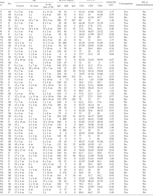
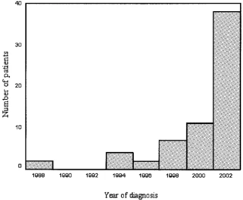
Characteristics of patients.From 1984 to 2003, there were 65 patients at Children’s Medical Center diagnosed as having CVID. There were 37 males and 28 females. The median age of patients at the time of the study was 136 months (range, 36 to 537 months) for males and 145 months (range, 24 to 504 months) for females. The median age at the time of disease onset was 24 months (range, 4 to 480 months) for males and 14 months (range, 5 to 96 months) for females, and median age at the time of diagnosis was 85 months (range, 25 to 513 months) for males and 103 months (range, 24 to 490 months) for fe-males. On average, the median diagnostic delay in our patient group was 60 months (range, 0 to 476 months) (Table 1). Statistical analysis of these data was complicated by the fact that the diagnosis had increasingly been made at an earlier age and delay in diagnosis has significantly decreased in recent years (r⫽ ⫺0.863,F⫽183.8,P⬍0.001). A reverse association was observed between year of disease onset and delay in diag-nosis (Fig. 1 and 2).
Total up period was 221 patient-years (mean follow-up, 40.7 months; range, 0 to 199 months). By the year 2003, 11 patients had died and seven patients could not be located. The prevalence of consanguineous marriages was 66.2% (in 43 fam-ilies) among our patients. Also, in 18 of these families, a history of recurrent infections in the siblings of the affected patient, with or without a documented diagnosis of immunodeficiency among them, was found, and 12 out of them had a family history of malignancy.
One of our patients at the time of diagnosis was found to have IgA deficiency but in follow-up changed to CVID (P53).
Serum immunoglobulin levels and lymphocyte studies. At the time of diagnosis, the baseline serum IgG level was 2 standard deviations (or more) below the normal level for age, and before treatment the median serum IgG level was 120 mg/dl (0 to 640 mg/dl; only one patient [P22] had an IgG level of more than 500 mg/dl), and that of other serum
immuno-globulins was 0 mg/dl for IgA (range, 0 to 235 mg/dl) and 10 mg/dl for IgM (range, 0 to 400 mg/dl). The level of IgA was under 10 mg/dl in 75.4% of patients. The serum IgM level was less than 25 mg/dl for 67.7% of patients (Table 2).
In 16 subjects (25.8%) a relative lack of CD4⫹T cells (less than 400 cells/mm3) was found. In 37 out of 65 patients
(56.9%), T-cell subset analysis, by immuno-flow cytometry, showed a reversed CD4/CD8 ratio.
All of our patients had B-cell numbers of more than 2%, and median CD19 was 10% (range, 2 to 47).
HLA classification. HLA typing with the PCR–sequence-specific primer method was done for 21 patients (14 female and seven male) and compared with that for 85 persons in the healthy control group. Expression of HLA DRB1 1303 was significantly increased in patients compared to control group (17.4% versus 0.5%, respectively; P ⫽ 0.017). HLA DQA1 0104 also was increased in patients, but the difference was not significant (4.76% versus 0.5%;P⫽0.07). Frequency of HLA DQB1 0501-3 was decreased in patients but was not significant (9.52% versus 0.22%;P⫽0.06)
Clinical manifestation. The main categories of associated diseases reported up to the time of the study included infec-tion, gastrointestinal or pulmonary disease, autoimmune dis-ease, hepatitis, granulomatous infiltrations, lymphoma, and cancer.
As seven subjects were currently unavailable but were not excluded from the total group, the frequency of these condi-tions is likely to be an underestimate. It must also be noted that the true incidence of some conditions would be particularly difficult to determine since many patients had never had biop-sies.
(i) Spectrum of infections as the presenting illnesses.
Among the 65 patients with CVID, 37 patients (56.9%) pre-sented with a form of respiratory tract infection as the first manifestation of disease, including otitis media in 16 patients (24.6%), pneumonia in 15 patients (23.1%), and sinusitis in nine patients (13.8%) (Table 3). The other presenting infec-tions included diarrhea in 18 patients (27.7%); skin involve-ment including cellulitis, omphalitis, eczema, and oral thrush in six patients (9.2%); pyelonephritis in three patients (4.6%); sepsis in one patient (1.5%) (P47); meningitis in one patient (1.5%) (P32); and osteomyelitis in one patient (1.5%) (P65). Also CVID was diagnosed in one of the patients following work-up for hepatosplenomegaly (P62) and in another case following work-up for failure to thrive (P11).
Some of our patients had two of these complications at the presenting time: four had otitis with sinusitis, two had pneu-monia and diarrhea, one had otitis and diarrhea, one had otitis and pneumonia, and one had diarrhea with oral thrush.
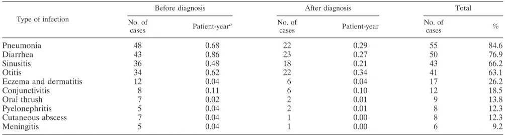
(ii) Infections in the course of disease.Acute and recurrent infections were found in almost all of our patients, particularly involving respiratory and gastrointestinal systems (Table 4). Forty-eight out of 65 patients (73.8%) had pneumonia prior to diagnosis, and of these patients, 16 developed bronchiectasis. In seven patients the diagnosis of bronchiectasis was made according to the patient’s history of chronic purulent cough, clubbing of the fingers, and X-ray findings and by excluding chronic bronchitis in them, and in 14 suspected patients high-resolution computed tomography was performed which showed bronchiectasis in nine of them.
on August 17, 2020 by guest
http://cvi.asm.org/
TABLE 1. Descriptive data for patientsa
Patient no. Sex
Age Ig level (mg/dl) CD (%)
CD4/CD8
ratio Consanguinity
FH of immunodeficiency Current At onset At the
time of Dx IgG IgM IgA CD3 CD4 CD8
P1 M 5 y 2 m 9 m 4 y 11 m 50 10 5 65.14 43.86 32.01 1.37 Yes No
P2 M 8 y 6 m 2 y 8 y 6 m 105 10 0 63.2 16 41.04 0.39 Yes No
P3 M 25 y 4 y 25 y 40 5 0 86.4 41.56 45.7 0.91 No No
P4 M 10 y 9 m 10 y 7 m 10 y 9 m 380 79 107 60 35 24 1.46 Yes No
P5 F 8 y 1 m 5 m 8 y 1 m 20 30 24 66.38 35.23 31.29 1.13 Yes No
P6 F 2 y 9 m 2 y 224 10 5 45.8 17.6 27.6 0.64 Yes No
P7 M 33 y 7 m 28 y 6 m 33 y 6 m 190 22 33 73.5 20.1 50.2 0.40 Yes No
P8 F 4 y 3 m 9 m 4 y 2 m 392 46 5 74.35 46.07 25.52 1.81 Yes No
P9 F 2 y 7 m 1 y 2 y 6 m 50 10 5 38.42 13.99 28.17 0.50 Yes No
P10 M 1 y 7 m 10 y 19 y 5 m 0 0 0 74.9 28 49 0.57 No No
P11 M 43 y 40 y 42 y 9 m 20 26 0 54 31 22.5 1.37 No No
P12 M 12 y 2 m 1 y 11 y 11 m 50 200 5 65.29 18.44 41.89 0.44 No No
P13 M 11 y 11 m 4 y 11 y 8 m 50 10 5 67.59 20.85 43.85 0.48 No No
P14 F 8 y 1 m 5 m 7 y 10 m 0 30 0 84 20.4 60.4 0.34 Yes Yes
P15 F 2 y 8 m 1 y 2 m 2 y 5 m 120 10 0 75 24 42 0.57 Yes No
P16 F 4 y 9 m 1 y 6 m 4 y 6 m 0 0 0 73.7 22.4 34 0.66 Yes No
P17 M 6 y 11 m 1 y 2 m 6 y 7 m 150 30 20 74 Yes No
P18 F 12 y 10 m 8 m 12 y 6 m 100 0 0 85.25 23.42 49.95 0.47 No No
P19 M 3 y 7 m 2 y 8 m 120 10 5 32 15 4 3.75 Yes No
P20 F 8 y 2 m 5 y 7 m 7 y 9 m 340 176 10 81.5 41.2 37.4 1.10 Yes No
P21 M 26 y 2 m 20 y 10 m 25 y 7 m 120 33 20 75.9 19.4 57.2 0.34 No No
P22 F 9 y 7 m 6 m 9 y 640 30 58 92.35 28.33 59.69 0.47 Yes No
P23 M 4 y 3 m 9 m 3 y 7 m 320 10 5 74.07 41.24 33.06 1.25 Yes No
P24 M 4 y 5 m 9 m 3 y 9 m 260 190 235 54 16.4 31.2 0.53 Yes No
P25 F 5 y 6 m 11 m 4 y 10 m 0 7.5 0 36.5 1.41 15.69 0.09 Yes No
P26 F 6 y 1 m 3 y 5 y 4 m 248 10 5 38.4 15.4 36.7 0.42 Yes No
P27 F 13 y 7 m 3 y 6 m 12 y 10 m 50 12 0 69.11 22.11 40.04 0.55 Yes No
P28 M 14 y 5 m 4 m 13 y 8 m 50 10 5 76.54 38.22 31.21 1.22 No No
P29 F 7 y 3 y 6 y 350 33 0 89.5 53 36 1.54 No No
P30 M 3 y 6 m 10 m 2 y 5 m 434 0 0 57.6 32.3 21.5 1.50 No No
P31 F 12 y 11 m 1 y 11 m 11 y 10 m 330 16 10 62.7 22.3 38.8 0.57 Yes No
P32 F 42 y 37 y 6 m 40 y 10 m 470 270 120 78 23 57 0.40 No No
P33 M 7 y 7 m 3 y 6 m 7 y 1 m 430 0 0 62.2 24.5 37.6 0.65 No No
P34 M 12 y 3 m 3 y 2 m 10 y 9 m 380 10 0 83.37 36.14 44 0.82 No Yes
P35 F 4 y 1 m 5 m 2 y 6 m 50 10 5 38.42 13.99 26.17 0.53 Yes No
P36 F 13 y 6 m 7 m 12 y 50 0 0 82.18 36.6 40.33 0.91 Yes No
P37 M 4 y 4 m 5 m 2 y 8 m 75 0 0 53 25 20 1.25 Yes No
P38 M 8 y 3 m 4 y 6 y 7 m 284 120 54 64.71 46.37 20.01 2.32 Yes No
P39 F 4 y 4 m 2 y 1 m 2 y 2 m 0 400 0 61.47 40.41 12.06 3.35 Yes No
P40 F 14 y 10 m 5 y 3 m 12 y 10 m 200 0 0 83.03 33.43 48.58 0.07 No Yes
P41 M 15 y 6 y 4 m 12 y 9 m 130 0 0 88.9 50.96 35.46 1.44 Yes No
P42 F 11 y 7 m 2 y 2 m 9 y 2 m 460 0 10 87.11 38.33 35.08 1.90 Yes No
P43 M 8 y 8 m 8 m 6 y 2 m 0 200 0 33 50 35 1.43 No No
P44 M 4 y 9 m 4 m 2 y 1 m 0 0 0 48.92 26.65 26.43 1.01 Yes No
P45 M 5 y 4 m 2 y 3 m 2 y 6 m 140 10 5 77 31 25 1.24 Yes Yes
P46 M 20 y 6 m 7 y 6 m 17 y 6 m 0 0 0 72 5 66 0.08 No No
P47 M 9 y 5 m 2 y 6 y 2 m 50 10 5 88.27 31.85 55.48 0.57 Yes No
P48 M 10 y 9 m 6 m 7 y 3 m 135 10 25 44.58 10.59 4.5 2.35 No No
P49 M 44 y 4 m 11 m 40 y 8 m 50 10 0 70.4 23.95 40.14 0.60 No No
P50 M 18 y 11 m 6 m 15 y 2 m 160 60 0 58.44 16.65 30.85 0.54 No No
P51 F 17 y 1 y 6 m 11 y 6 m 330 16 10 62.7 22.3 38.8 57.47 Yes No
P52 F 17 y 5 m 6 y 3 m 13 y 4 m 500 10 46 90.11 12.88 78.56 0.15 Yes No
P53 F 18 y 11 m 2 y 14 y 7 m 342 10 0 42.83 31.61 19.89 1.60 Yes No
P54 F 12 y 7 m 2 y 11 m 8 y 2 m 50 15 0 62.35 24.49 36.1 0.68 No No
P55 F 24 y 3 m 8 y 18 y 11 m 0 0 0 88 20.3 48.9 0.42 Yes No
P56 F 8 y 3 m 6 m 2 y 6 m 380 35 0 82.43 34.19 32.58 1.05 Yes Yes
P57 M 15 y 7 m 1 y 4 m 9 y 8 m 0 10 0 65.2 28 30 0.93 Yes No
P58 M 15 y 8 m 5 m 7 y 10 m 0 176 0 50.6 18 30 0.60 No No
P59 M 12 y 4 m 5 m 3 y 11 m 103 9 10 65 31.7 34.2 0.93 Yes No
P60 M 11 y 7 m 3 y 6 m 3 y 6 m 360 42 5 62.67 6.82 50.35 0.14 Yes No
P61 F 23 y 4 m 2 y 1 m 15 y 1 m 50 10 0 79.25 40.42 35.23 1.15 Yes No
P62 M 11 y 4 m 2 y 11 m 3 y 6 m 320 7.5 5 78.2 55.2 20 2.76 Yes No
P63 M 44 y 9 m 34 y 1 m 35 y 1 m 125 0 0 79.6 25.05 54.61 0.46 No No
P64 M 17 y 5 m 6 m 2 y 6 m 0 73 0 55 20 33 0.61 Yes Yes
P65 F 17 y 6 m 1 y 2 y 0 0 0 88.6 66.2 19 3.48 Yes No
aAbbreviations: M, male; F, female; y, year(s); m, month(s); Dx, diagnosis; FH, family history.
on August 17, 2020 by guest
http://cvi.asm.org/
Forty-one of our CVID patients (63.1%) had a history of recurrent otitis media, and 43 patients (66.2%) had sinusitis. Recurrent diarrhea was seen in 50 patients (76.9%). In 10 out of 65 patients (15.4%) unusual or opportunistic infections were seen, including oral candidiasis andPneumocystis carinii pneu-monia. Oral candidiasis developed in nine of these patients, and in another casePneumocystis cariniipneumonia was found in a 17-year-old boy with severe lack of CD4⫹T cells (less than 400 cells/mm3) (P64).
Prior to initiation of immunoglobulin administration, bacte-rial meningitis occurred in five patients. Other forms of infec-tions and their frequencies before diagnosis were as follows: septic arthritis in nine patients, chronic or recurrent conjunc-tivitis in eight patients, superficial or deep abscesses in seven patients, and pyelonephritis in six patients. The most frequent
nonspecific symptoms were hepatomegaly in 13 patients, splenomegaly in 17 patients, and lymphadenopathy in 16 pa-tients.
The most common organisms responsible for infections were
Pneumococcusspecies (30%) andEscherichia coli(20%), but
as necessary cultures were not done for many patients we cannot estimate the true incidence of each microorganism.
(iii) Gastrointestinal disease.Fifty-six patients had gastro-intestinal problems. Inflammatory bowel disease (ulcerative colitis) was seen in one patient (P41). Six other patients had significant malabsorption without any known gastrointestinal disorder. One subject had Hirschsprung disease (P60).
Twenty-five patients had liver problems, of whom three had chronic active hepatitis (P41, P52, and P64), two had acute hepatitis (both resolved without any complication) (P48 and P53), one had hepatic cyst (P32), and three had biliary duct obstruction (P20, P22, and P32). Unfortunately there was no viral testing for any of these patients. Three patients had acute appendicitis and underwent surgical removal of the appendix (P4, P57, and P61). One subject had accessory spleen (P36). One patient had mesenteric lymphadenitis (P27).
Twelve patients underwent upper endoscopy, four under-went lower endoscopy, and two patients underunder-went both. The most common endoscopic findings were as follows: colitis
FIG. 1. Distribution of diagnosis year in patients with common variable immunodeficiency (n⫽65).
FIG. 2. Association between year of disease onset and delayed di-agnosis of common variable immunodeficiency.
TABLE 2. Immunoglobulin levels and CD markers
Immunoglobulin or lymphocyte marker
Median (range) for:
Females (n⫽28) Males (n⫽37)
Immunoglobulin (mg/dl)
IgG 110 (0–640) 120 (0–434)
IgA 0 (0–120) 5 (0–235)
IgM 10 (0–400) 10 (0–200)
Lymphocyte marker (count)
CD3 1,503 (355–7,455) 1,575 (516–8,012)
CD4 667 (47–2,386) 707 (114–4,364)
CD8 823 (165–4,403) 913 (64–4,328)
CD19 245 (36–1,470) 262 (34–4,376)
TABLE 3. Frequency and spectrum of presenting illnesses
Type of disorder No. %
Otitis 16 24.6
Diarrhea 16 24.6
Pneumonia 15 23.1
Sinusitis 9 13.8
Dysentery 2 3.1
Eczema 2 3.1
Oral thrush 2 3.1
Pyelonephritis 2 3.1
Sepsis 1 1.5
Omphalitis 1 1.5
Cellulitis 1 1.5
Meningitis 1 1.5
Osteomyelitis 1 1.5
UTIa 1 1.5
FTTb 1 1.5
aUTI, urinary tract infection.
bFTT, failure to thrive.
on August 17, 2020 by guest
http://cvi.asm.org/
(66%), esophagitis (30%), and gastritis (23%). Among 12 pa-tients in whom upper gastrointestinal tract endoscopy was done villous atrophy was seen in eight patients (66.6%) and nodular lymphoid hyperplasia was seen in six (50%). Biopsies showed villous atrophy in five of six patients whose endoscopy results were normal.
(iv) Cancer.Two siblings developed Hodgkin’s disease, and they had a positive family history of this disease in four of their relatives (P34 and P40). One subject developed non-Hodgkin’s lymphoma (NHL) which was confirmed by a node biopsy re-port of pathology (P50). In all those studied these lymphomas were B cell in type.
(v) Autoimmune disease.Autoimmune disease occurred in 8 out of 65 (23%) patients. These diseases included immune thrombocytopenia (ITP) (P4, P36, and P47), hemolytic anemia (P39 and P47) or neutropenia (P5, P6, P9, P35, P41, and P64), red cell aplasia, thyroid disease (P53), and neuropathy. In patients with ITP, steroids and high-dose intravenous immu-noglobulin (IVIg) were useful and splenectomy was not nec-essary. One patient had alopecia areata (P42), and one had psoriasis (P11).
Family members with immunodeficiency.Relatives of CVID patients may also be found to be immunodeficient. In our patient group six subjects had a positive family history of im-munodeficiency (Table 1). There were two immunodeficient sibling pairs in our study with unaffected parents (P40 and P34, and P24 and P38).
Mortality.The median follow-up period in our patient group was 16.5 months (range, 0 to 186 months), during which 11 subjects died (from 3 to 179 months after their diagnosis, at age of onset of illness ranging from 2 to 97 months [median age, 57 months]). The causes of death were as follows: pneumonia in P14, P17, and P55; bowel perforation in P24; hepatic failure in P64; Hodgkin’s disease in P34; apnea and cardiac arrest in P37; low platelet and fever in P44; severe infections in P58, P16, and P19.
Excluding the seven patients who could not be located, this shows a mortality of 19% (11 of 58). If none of the seven patients who were not found had died, the mortality rate for the group would be 16.9%. Thus, the true mortality lies be-tween 16.9 and 19%. Patients who died included three females and eight males. The mean age of females at the time of death was 148 months, and that of males was 103 months. Mortality
rate was 40% after a 5-year follow-up period (5-year survival rate of 60%).
DISCUSSION
Common variable immunodeficiency is a heterogeneous pri-mary immunodeficiency disorder with variable immunological defects and clinical manifestations. In this study, 65 patients with CVID, referred to our center over a period of 20 years, were evaluated for clinical and immunological features during their follow-up in our center. There are different reports about CVID patients (17, 18, 27). The average ages at the time of diagnosis in reports by Hermans et al. in 1976, which included 50 patients (27); Cunningham-Rundles in 1989, including 103 patients (18); and Cunningham-Rundles and Bodian in 1999, including 248 cases (17), were 41.9 years, 28 years, and 31 years, respectively. In our study the mean age of patients at the time of diagnosis was 7.1 years for males and 8.6 years for females, which is somewhat less than other published data. This difference between our patients and other reports may be due to selection of our patient population from a pediatric center. However, adult patients with CVID are referred to this center also.
The median age of our patients at the time of onset of symptoms was 9 months, and the median age at the time of diagnosis was 6.1 years. An average diagnostic delay in our patient group was 5.1 years. A survey in the northwest region of England (13) showed that average delay in CVID diagnosis was 2.5 years in children and 5.5 years in adults. Also, in a study done by Cunningham-Rundles and Bodian (17) in the United States, the diagnostic delay in 248 CVID patients studied was 4 to 6 years. This delay markedly decreased over time, perhaps due to better diagnostic tools or methods; an increase in the knowledge of our physicians about primary immunodeficiency disorders, especially CVID; and the beginning of the registry program in Iran. In another study by Moin et al. of X-linked agammaglobulinemia patients in Iran, diagnostic delay also decreased over time (40).
In our patient group HLA DRB1 1303 was more frequent in CVID patients, and it may be considered a predisposing factor. In a study in 1998 (46) HLA DR3 was significantly higher in CVID patients than in the healthy population while in our study it did not show any statistically significant difference. In
TABLE 4. Frequency and spectrum of infections occurring in the course of disease
Type of infection
Before diagnosis After diagnosis Total
No. of
cases Patient-year
a No. of
cases Patient-year
No. of
cases %
Pneumonia 48 0.68 22 0.29 55 84.6
Diarrhea 43 0.86 23 0.27 50 76.9
Sinusitis 36 0.48 18 0.21 43 66.2
Otitis 34 0.62 22 0.34 41 63.1
Eczema and dermatitis 12 0.04 6 0.04 17 26.2
Conjunctivitis 8 0.11 6 0.10 12 18.5
Oral thrush 7 0.02 2 0.01 9 13.8
Pyelonephritis 5 0.04 2 0.01 8 12.3
Cutaneous abscess 7 0.04 1 0.00 8 12.3
Meningitis 5 0.04 1 0.00 6 9.2
a
Patient-year indicates risk of being infected for any individual in 1 year.
on August 17, 2020 by guest
http://cvi.asm.org/
our study none of the HLA DQB1 loci except DQB1 0501-3 was known as a predisposing or protecting factor in CVID, but in the study by Schroeder et al. (46) HLA DQB1 0201 was significantly higher in CVID patients. In a study in 1999 (22) the DQB1 0201, DQB1 0301, DQB1 0501, and DQB1 0602 alleles were not significantly different in frequency from the control group, which is compatible with our study. In a previ-ous study in Iran (9) HLA A2 (P⬍0.02) and A33 (P⬍0.001) were significantly increased in CVID patients compared to controls. A significant negative association was also evident for DR2 (P ⬍ 0.05), DR7 (P ⬍ 0.001), DR52 (P ⬍ 0.05), and DQ52 (P⬍0.05) alleles. Therefore, that study also confirmed involvement of the HLA complex in the presentation of CVID in the Iranian population.
Patients with CVID are more susceptible to recurrent infec-tions, and these infections can occur at different sites. In our study 48 out of 65 patients (73.8%) had recurrent pneumonia and 16 of them developed bronchiectasis. The same frequency was noted in a study performed by Cunningham-Rundles and Bodian (17), and the result of their study showed that 190 out of 248 patients (76.6%) with CVID had at least one episode of pneumonia prior to treatment with immunoglobulin. Also they found that 10 out of 248 patients developed bronchiectasis. In another study, done by Busse et al. (14), among 50 patients with CVID, 42 patients (84%) had at least one episode of pneumonia before diagnosis, and by using computed tomo-graphic scanning, 18 out of these 42 patients showed bronchi-ectasis. Infections are less severe in CVID patients than in XLA patients (40).
It can be concluded that respiratory infections are common medical problems in these groups of patients, and failure to provide adequate replacement therapy results in bronchiecta-sis, which may be the reason why our patients developed bron-chiectasis more often than those in other studies. Development of bronchiectasis is a serious medical problem (32, 41), and all patients with persistent purulent sputum should be assessed and managed, jointly with a chest physician, to prevent pro-gressive lung damage and to monitor functional impairment (12). Despite full replacement immunoglobulin therapy, pa-tients with chronic chest infections always require physiother-apy, antibiotic therphysiother-apy, bronchodilators, and local anti-inflam-matory agents, if progression of lung damage is to be arrested. In our study 41 out of 65 patients (63.1%) had a history of recurrent otitis media and 43 patients (66.2%) had recurrent sinusitis. It has been shown that immunologic defects have important roles in recurrent ear and sinus infections (47). A similar study done by Cunningham-Rundles and Bodian (17) showed that 243 out of 248 patients with CVID (98%) had recurrent sinusitis, otitis, or bronchitis, and according to an-other study in 1989 (18), 22% had developed chronic lung disease. Also, some studies show that resistant sinus infection can frequently be the first presenting symptom in immune deficiencies, especially antibody deficiencies (10, 38, 48), and the infections of the upper respiratory tract occur several years before the appearance of lower respiratory tract infections (34). Once the infections of the lower respiratory tract have started, the patients neglect the symptoms from the upper respiratory tract.
In our investigated group, 50 out of 65 (76.9%) patients had recurrent diarrhea. Recurrent or persistent diarrhea and/or
malabsorption may be due to infection, superinfection, food-sensitive enteropathy, autoimmune enteropathy, ulcerative co-litis, and celiac disease. As gastrointestinal pathologies were more common in Iranian CVID patients, early endoscopic study is recommended in these patients.
Opportunistic infections including oral candidiasis and
Pneumocystis cariniipneumonia were seen in 10 out of our 65
patients (15.4%). The isolation of an opportunistic agent in a child or occurrence of an unusually severe infection indicates T-lymphocyte deficiency (8). Although patients with antibody deficiencies have increased susceptibility to infection by com-mon organisms such asS.pneumoniaeandH.influenzae(12, 52) the T-lymphocyte deficiencies in some CVID patients (25) may lead some of them to develop opportunistic infections.
In addition to infection, a variety of autoimmune diseases occur in about 23% of patients with antibody deficiency (53). The most common autoimmune disorder is idiopathic throm-bocytopenic purpura, followed by autoimmune hemolytic ane-mia (53). Although cytopenias are common in all the congen-ital immune diseases, they are particularly common in the antibody defects, common variable immunodeficiency, and se-lective immunoglobulin A deficiency. In a study by Cunning-ham-Rundles (19), it has been shown that 6% of patients with common variable immunodeficiency develop ITP, and autoim-mune hemolytic anemia can occur in about 4.8% of this group of patients.
In some patients with antibody deficiencies, hematologic abnormalities were present as the first clinical manifestation; because of a major lack of awareness among clinicians, espe-cially general practitioners and pediatric and adult general physicians, about antibody deficiency disorders and their com-plications, some patients who present first with hematologic abnormalities will be missed.
Although the association between cytopenias and congenital immune deficiency is unclear, defects in T-cell regulation, cy-tokine defects, abnormal apoptosis, and abnormal production of immunoglobulins with autoimmune features are potential mechanisms.
Hypogammaglobulinemia is the main feature in CVID, and therefore the standard treatment is IVIg. Although this treat-ment has changed the spectrum of illnesses, a large number of medical complications are still seen in these patients. Previ-ously, bacterial meningitis, sepsis, and recurring pneumonia were commonly seen in hypoglobulinemic patients (18, 28, 29, 30, 50, 51, 54, 56). In our patients meningitis and sepsis oc-curred each in one patient as the presenting illness and five of our patients had meningitis prior to IVIg treatment, but no one developed sepsis when given standard amounts of IVIg and only one developed meningitis (Table 4). Overall, according to our data the occurrence of infection has decreased per case per year after IVIg therapy (Table 4).
Two siblings developed Hodgkin’s disease, and they had a positive family history of this disease in four of their relatives. One subject developed non-Hodgkin’s lymphoma which was confirmed by node biopsy report of pathology. In all those studied these lymphomas were B cell in type. The association between NHL and congenital immunodeficiency is well estab-lished, and most NHL cases appear in patients with T-cell defects (such as ataxia-telangiectasia, Wiskott-Aldrich syn-drome, and severe combined immune deficiency, as well as
on August 17, 2020 by guest
http://cvi.asm.org/
CVID) (49). In a study Cunningham-Rundles et al. estimated that females with CVID had a 438-fold-increased likelihood of developing NHL compared to the age-adjusted expected inci-dence (21).
There is an increased incidence of lymphoma and gastric carcinoma in patients with CVID (20, 24, 35, 45). However, as noted above there may have been difficulties in diagnosing lymphoma in the context of CVID, particularly before molec-ular techniques became available. Historical studies may there-fore overestimate the occurrence of lymphoma by including nonclonal proliferative lesions. There is a suspicion that the occurrence of true lymphoma may be decreasing since the introduction of higher-dose IVIg regimens, compared with the period of IVIg treatment.
Nearly all patients in this group have had stable immunode-ficiency, and serum immunoglobulin remained similarly re-duced over time. Spontaneous resolution of immunodeficiency has been noted previously, suggesting that this immunodefi-ciency is not always an intrinsically permanent B-cell defect, or that this more transient form cannot currently be distinguished from CVID (17, 31).
The mortality rate in this group is high, between 16.9 and 19% over a median follow-up period of 20 months. In the first report by Cunningham-Rundles, mortality was 22% over a 13-year period (18), and in another report it was 23 to 27% over a follow-up period of 7 years (17).
Their prior report reflected more data for a period during which intramuscular immunoglobulin was the standard treat-ment, and the second report and our results show information which includes data collected 15 to 20 years after IVIg was introduced.
While the current data suggest that IVIg has not made an impact on mortality in CVID, a number of subjects referred to this medical center have been in poor medical condition and therefore had short life expectancies. These CVID subjects, with more systemic disease, have most likely increased the overall mortality rate for this patient group. An earlier report demonstrated 22% mortality over a 13-year period, 1960 to 1973 (37); a more recent report showed a 30% mortality for a group of 240 CVID subjects followed over a 30-year period (28). It appears that mortality rates exceed that for XLA, where the mortality in one study was 17% (36). The reasons for this are unknown, although gastrointestinal disease, lym-phoma, and autoimmune disease are not commonly found in XLA patients (51). A potential explanation for these differ-ences could be immune dysregulation in CVID due to addi-tional T-cell defects that perhaps leads to addiaddi-tional medical complications.
Conclusion.The causes of CVID are unknown, and it is a heterogeneous group with antibody deficiency, accompanying immune dysregulation, T-cell deficiency, and poorly controlled inflammation leading to additional organ damage.
It is important to consider hypogammaglobulinemia in any patient with a history of recurrent infections at different organ systems, and patients should have a full assessment of immune system including measurement of serum immunoglobulin lev-els, IgG subclass levlev-els, antibody function evaluation, and B-and T-cell subset enumeration. Serum immunoglobulin levels are interpreted in relation to the normal range for age.
Diag-nostic delay results in morbidity and complications in un-treated patients.
ACKNOWLEDGMENTS
We are grateful to Lida Atarod, Akefeh Ahmadi Afshar, Nasrin Bazargan, Zahra ChavoshZadeh, Marzieh HaydarZadeh, Mohammad Nabavi, and Mohammad Hasan Bemanian, who assisted us at every step of this project. We gratefully acknowledge the efforts of Kamran Abolmaali and Tahereh AghaBagheri for their role in collecting the data. Also we thank Anahita Azimdoust of our laboratory staff and Mina Andali, Nahid Hasani, and Zahra Shobayri of our secretarial staff for their arrangements and administrative efforts.
REFERENCES
1.Aghamohammadi, A., A. Farhoudi, M. Moin, Z. Pourpak, N. Rezaei, K.
Abolmaali, M. Movahedi, M. Gharagozlou, B. M. S. Ghazi, and M.
Mah-moudi.2002. A single-center 20-year survey of infectious complication in 64
patients with common variable immunodeficiency. Med. J. Islam. Repub. Iran16:123–128.
2.Aghamohammadi, A., A. Farhoudi, M. Nikzad, M. Moin, Z. Pourpak, N.
Rezaei, M. Gharagozlou, M. Movahedi, L. Atarod, A. A. Afshar, N.
Bazar-gan, and A. R. Hosseinpoor.2004. Adverse reactions of prophylactic
intra-venous immunoglobulin infusions in Iranian patients with primary immuno-deficiency. Ann. Allergy Asthma Immunol.92:60–64.
3.Aghamohammadi, A., and A. Farhoudi.1997. Use of immunoglobulin in
patients with antibody deficiencies, p. 277–307.InA. Farhoudi (ed.), Immu-nodeficiencies in Iran, vol. 1. Shayan Nemoodar, Tehran, Iran.
4.Aghamohammadi, A., F. Jalali, A. Farhoudi, Z. Pourpak, N. Rezaei, S. H.
Khazali, and H. Hosseini Nik.2001. Ear, nose and throat manifestation in
patients with antibody deficiencies. Acta Med. Iran.39:164–168.
5.Aghamohammadi, A., H. Kanegane, M. Moein, A. Farhoudi, Z. Pourpak, M.
Movahedi, M. Gharagozlou, A. A. Zargar, and T. Miyawaki.2003.
Identifi-cation of SH2D1A mutation in a hypogammaglobulinemic male diagnosed as having common variable immunodeficiency. Int. J. Hematol.78:45–47.
6.Aghamohammadi, A., M. Moein, A. Farhoudi, Z. Pourpak, N. Rezaei, K.
Abolmaali, M. Movahedi, M. Gharagozlou, B. M. Ghazi, M. Mahmoudi, D. Mansouri, S. Arshi, N. J. Trash, H. Akbari, R. Sherkat, R. F. Hosayni, A. Hashemzadeh, I. Mohammadzadeh, R. Amin, S. Kashef, A. Alborzi, A.
Karimi, and H. Khazaei.2002. Primary immunodeficiency in Iran: first
report of the National Registry of PID in Children and Adults. J. Clin. Immunol.22:375–380.
7.Aghamohammadi, A., M. Moin, A. Farhoudi, Z. Pourpak, N. Rezaei, K.
Abolmaali, M. Movahedi, M. Gharagozlou, M. Mahmoudi, L. A. Kordestani,
and Z. Habibi.2000. The clinical spectrum of respiratory disease in patients
with primary antibody deficiency. Iran J. Allergy Asthma Immunol.1:135– 140.
8.Aghamohammadi, A., S. Strobel, V. Novelli, H. Holzel, and J. Morgan.1996.
A single center retrospective 5-year survey of infectious complication in 85 children with combined immunodeficiency. Acta Med. Iran.34:7–13.
9.Amanzadeh, A., M.-A. Shokrgozar, Z. Samadi-Bahrami, A.
Aghamoham-madi, and F. Shokri.2003. Frequency analysis of HLA antigens in Iranian
patients with common variable immunodeficiency. Arch. Iranian Med.6:16– 22.
10.Armenaka, M., J. Grizzanti, and D. L. Rosenstreich.1994. Serum
immuno-globulins and IgG subclass levels in adults with chronic sinusitis: evidence for decreased IgG3 levels. Ann. Allergy72:507–514.
11.Atarod, L., A. Raeisi, A. Aghamohammadi, A. Khodadad, A. Farhoudi, M.
Moin, Z. Pourpak, M. Gharagozlou, M. Movahedi, and N. Rezaei.2003. The
review of gastrointestinal disorders in patients with primary antibody immu-nodeficiencies during the 10-year period (1990–2000), in Children’s Medical Center. Iran J. Allergy Asthma Immunol.2:75–80.
12.Bjorkander, J., B. Blake, and L. A. Hanson.1984. Primary
hypogamma-globulinaemia: impaired lung function and body growth with delayed diag-nosis and inadequate treatment. Eur. J. Respir. Dis.65:529–536.
13.Blore J., and M. Haeney.1989. Primary antibody deficiency and diagnostic
delay. BMJ298:516–517.
14.Busse, P. J., S. Razvi, and C. Cunningham-Rundles.2002. Efficacy of
intra-venous immunoglobulin in the prevention of pneumonia in patients with common variable immunodeficiency. J. Allergy Clin. Immunol.109:1001– 1004.
15.Chapel, H. M.1994. Consensus on diagnosis and management of primary
antibody deficiencies. Consensus Panel for the Diagnosis and Management of Primary Antibody Deficiencies. BMJ308:581–585.
16.Conley, M. E., L. D. Notarangelo, and A. Etzioni.1999. Diagnostic criteria
for primary immunodeficiencies representing PAGID (Pan-American Group for Immunodeficiency) and ESID (European Society for Immunode-ficiencies). Clin. Immunol.93:190–197.
17.Cunningham-Rundles, C., and C. Bodian.1999. Common variable
on August 17, 2020 by guest
http://cvi.asm.org/
deficiency: clinical and immunological features of 248 patients. Clin. Immu-nol.92:34–48.
18.Cunningham-Rundles, C.1989. Clinical and immunologic analyses of 103
patients with common variable immunodeficiency. J. Clin. Immunol.9:22– 33.
19.Cunningham-Rundles, C.2002. Hematologic complications of primary
im-mune deficiencies. Blood Rev.16:61–64.
20.Cunningham-Rundles, C., P. Lieberman, G. Hellman, and R. S. K.
Cha-ganti. 1991. Non-Hodgkin lymphoma in common variable
immunodefi-ciency. Am. J. Hematol.37:69–74.
21.Cunningham-Rundles, C., F. P. Siegal, S. Cunningham-Rundles, and P.
Lieberman.1987. Incidence of cancer in 98 patients with common varied
immunodeficiency. J. Clin. Immunol.7:294–299.
22.De La Concha, E. G., M. Fernandez-Arquero, A. Martinez, F. Vidal, P. Vigil,
L. Conejero, M. C. Garcia-Rodriguez, and G. Fontan.1999. HLA class II
homozygosity confers susceptibility to common variable immunodeficiency (CVID). Clin. Exp. Immunol.116:516–520.
23.Farrington, M., L. S. Grosmair, S. Nonomyamas, S. H. Fischer, D.
Hollen-baugh, J. A. Ledbetter, R. J. Neolle, A. Auffo, and H. D. Ochs.1994. CD40
ligand expression is defective in a subset of patient with common variable immunodeficiency. Proc. Natl. Acad. Sci. USA91:1099–1103.
24.Filipovich, A. H., A. Mathur, D. Kamat, J. H. Kersey, and R. S. Shapiro.
1994. Lymphoproliferative disorders and other tumours complicating immu-nodeficiencies. Immunodeficiency5:91–112.
25.Fischer, M. B., I. Hauber, H. Eggenbauer, V. Thon, E. Vogel, E. Schaffer, J.
Lokaj, J. Litzman, H. M. Wolf, J. W. Mannhalter, et al.1994. A defect in
early phase of T-cell receptor mediated T-cell activation in patients with common variable immunodeficiency. Blood84:4234–4241.
26.Hammarstrom, L., I. Vorechovsky, and D. Webster.2000. Selective IgA
deficiency (SIgAD) and common variable immunodeficiency (CVID). Clin. Exp. Immunol.120:225–231.
27.Hermans, P. E., J. A. Diaz-Buxo, and J. D. Stobo1976. Idiopathic late-onset
immunoglobulin deficiency: clinical observation in 50 patients. Am. J. Med.
61:221–237.
28.Hermaszewski, R. A., and A. D. B. Webster.1993. Primary
hypogamma-globulinemia: a survey of clinical manifestations and complications. Q. J. Med.81:31–42.
29.Hill, L. E.1971. Clinical features of hypogammaglobulinemia. Med. Res.
Counc. (G. B.) Spec. Rep. Ser.310:9–34.
30.Janeway, C. A., L. Apt, and D. Gitlin.1953. Agammaglobulinemia. Trans.
Assoc. Am. Phys.66:200–202.
31.Johnson, M. L., L. G. Keeton, Z. B. Zhu, J. E. Volanakis, M. D. Cooper, and
H. W. Schroeder.1997. Age related changes in serum immunoglobulins in
patients with familial IgA deficiency and common variable immunodefi-ciency (CVID). Clin. Exp. Immunol.108:477–483.
32.Kainulainen, L., M. Varpula, K. Liippo, E. Svedstrom, J. Nikoskelainen, and
O. Ruuskanen. 1999. Pulmonary abnormalities in patients with primary
hypogammaglobulinemia. J. Allergy Clin. Immunol.104:1031–1036.
33.Kanegane, H., S. Tsukada, T. Iwata, T. Futanai, K. Nomura, J. Yamamoto,
T. Yoshida, K. Agematsu, A. Komiyama, and T. Miyawaki.2000. Detection
of Bruton’s tyrosine kinase mutations in hypogammaglobulinaemic males registered as common variable immunodeficiency (CVID) in the Japanese Immunodeficiency Registry. Clin. Exp. Immunol.120:512–517.
34.Karlsson, G., B. Petruson, J. Bjorkander, and L. A. Hanson.1985. Infections
of the nose and paranasal sinuses in adult patients with immunodeficiency. Arch. Otolaryngol.111:290–293.
35.Kinlen, L. J., A. D. Webster, A. G. Bird, R. Haile, J. Peto, J. F. Soothill, and
R. A. Thompson.1985. Prospective study of cancer in patients with
hypoga-mmaglobulinaemia. Lanceti:263–266.
36.Lederman, H. M., and J. A. Winkelstein.1985. X-linked
agammaglobuline-mia: an analysis of 96 patients. Medicine64:145–156.
37.Longhurst, H. J., C. O’Grady, G. Evans, C. De Lord, A. Hughes, J.
Cave-nagh, and M. R. Helbert. 2002. Anti-D immunoglobulin treatment for
thrombocytopenia associated with primary antibody deficiency. J. Clin. Pathol.55:64–66.
38.May, A., S. Zielen, C. von Ilberg, and A. Weber.1999. Immunoglobulin
deficiency and determination of pneumococcal antibody titers in patients with therapy-refractory recurrent rhinosinusitis. Eur. Arch. Otorhinolaryn-gol.256:445–449.
39.Mellemkjaer, J., L. Hammarstrom, V. Andersen, et al.2002. Cancer risk
among patients with IgA deficiency or common variable immunodeficiency and their relatives: a combined Danish and Swedish study. Clin. Exp. Im-munol.130:495–500.
40.Moin, M., A. Aghamohammadi, A. Farhoudi, Z. Pourpak, N. Rezaei, M.
Movahedi, M. Gharagozlou, B. M. Ghazi, A. Zahed, K. Abolmaali, M.
Mah-moudi, L. Emami, and M. Bashashati.2004. X-linked agammaglobulinemia:
a survey of 33 Iranian patients. Immunol. Investig.33:81–93.
41.Newson, T., A. J. Chippindale, and A. J. Cant.1999. Computed tomography
scan assessment of lung disease in primary immunodeficiencies. Eur. J. Pe-diatr.158:29–31.
42.Nistala, K., K. C. Gilmour, T. Cranston, E. G. Davies, D. Goldblatt, H. B.
Gaspar, and A. M. Jones.2001. X-linked lymphoproliferative disease, three
atypical cases. Clin. Exp. Immunol.126:126–130.
43.Pacheco, S. E., and W. T. Shearer.1994. Laboratory aspects of immunology.
Pediatr. Clin. N. Am.41:623–655.
44.Revy, P., T. Muto, Y. Levy, F. Geissmann, A. Plebani, O. Sanal, N. Catalan,
M. Forveille, R. Dufourcq-Labelouse, A. Gennery, I. Tezcan, F. Ersoy, H. Kayserili, A. G. Ugazio, N. Brousse, M. Muramatsu, L. D. Notarangelo, K.
Kinoshita, T. Honjo, A. Fischer, and A. Durandy.2000. Activation-induced
cytidine deaminase (AID) deficiency causes the autosomal recessive form of the hyper-IgM syndrome (HIGM2). Cell102:565–575.
45.Sander, C. A., L. J. Medeiros, L. M. Weiss, T. Yano, M. C. Sneller, and E. S.
Jaffe.1992. Lymphoproliferative lesions in patients with common variable
immunodeficiency. Am. J. Surg. Pathol.16:1170–1182.
46.Schroeder, H. W., Jr., Z. B. Zhu, R. E. March, R. D. Campbell, S. M. Berney,
S. A. Nedospasov, R. L. Turetskaya, T. P. Atkinson, R. C. Go, M. D. Cooper,
and J. E. Volanakis.1998. Susceptibility locus for IgA deficiency and
com-mon variable immunodeficiency in the HLA-DR3, -B8, -A1 haplotypes. Mol. Med.4:72–86.
47.Sethi, D. S., J. A. Winkelstein, H. Lederman, and M. C. Loury.1995.
Im-munologic defects in patients with chronic recurrent sinusitis: diagnosis and management. Otolaryngol. Head Neck Surg.112:242–247.
48.Shapiro, G. G., F. S. Virant, C. T. Furukawa, W. E. Pierson, and C. W.
Bierman.1991. Immunologic defects in patients with refractory sinusitis.
Pediatrics87:311–316.
49.Smith, M. S., A. D. Webster, A. P. Dhillon, G. Dusheiko, R. Boulton, K.
Savage, K. Rolles, and A. K. Burroughs.1995. Orthotopic liver
transplanta-tion for chronic hepatitis in two patients with common variable immunode-ficiency. Gastroenterology108:879–884.
50.Sneller, M. C., W. Strober, E. Eisenstein, J. S. Jaffee, and C.
Cunningham-Rundles.1993. New insights into common variable immunodeficiency. Ann.
Intern. Med.18:720–730.
51.Spickett, G. P., J. Farrant, M. E. North, J. Zhang, L. Morgan, and A. D. B.
Webster 1997. Common variable immunodeficiency: how many diseases.
Immunol. Today18:325–328.
52.Stiehm, E. R., T. W. Chin, A. Haas, and A. G. Peerless.1986. Infectious
complications of primary immunodeficiencies. Clin. Immunol. Immuno-pathol.40:69–86.
53.Webster, A. D. B., T. A. E. Platts-Mills, G. Janossy, M. Morgan, and G. L.
Asherson.1981. Autoimmune blood dyscrasias in five patients with
hypo-gammaglobulinaemia: response of neutropenia to vincristine. J. Clin. Immu-nol.1:113–118.
54.Webster, A. D. B., D. Taylor-Robinson, P. M. Furr, and G. L. Asherson.1987.
Mycoplasmal (ureaplasma) septic arthritis in hypogammaglobulinemia. Br. Med. J.1:478–479.
55.Weston, S. A., M. L. Prasad, C. G. Mullighan, H. Chapel, and E. M. Benson.
2001. Assessment of male CVID patients for mutations in the Btk gene: how many have been misdiagnosed? Clin. Exp. Immunol.124:465–469.
56.World Health Organization.1997. Primary immunodeficiency diseases.
Re-port of a WHO Scientific Group. Clin. Exp. Immunol.109:1–28.