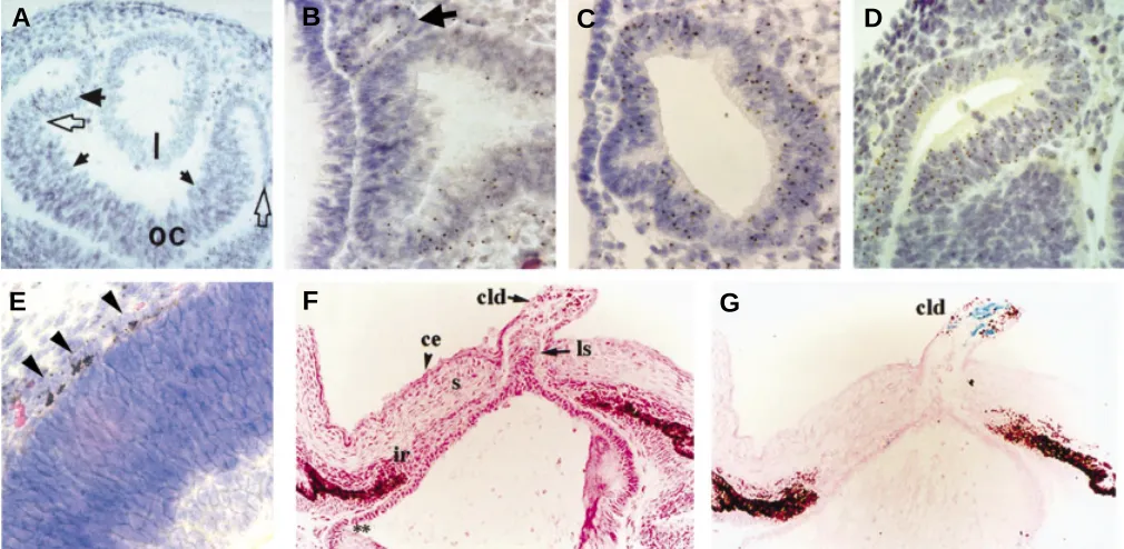
Analysis of mouse eye development with chimeras and mosaics
Full text
Figure


Related documents
mg, 1.20 mmol) was added to the mixture and the consumption of the metallated reagent was checked by GC-analysis of hydrolyzed reaction aliquots. The reaction mixture was stirred for
METHODS: A literature search was undertaken to identify studies that assessed the cost- effectiveness of telehealth interventions for chronic cardiovascular diseases and chronic
Despite the efforts of implementing presumptive tax system, Zimbabwe revenue Authority missed its annual tax collection by 6% and 7% in 2014 and 2015 respectively,
We identified and validated biomarkers to (1) predict disease severity in children with pneumonia, (2) predict blood culture positivity, (3) predict probable bacterial etiology
Currently, the transsphenoidal approach for removal of pituitary tumour is widely used and is achieved by the otorhinolaryngologist providing easy access to the sphenoid sinus..
Information on patient demography, type of diabetes, cardiovascular risk factors (BP, lipids, BMI and smoking history), glycaemic control (HbAlc and FBS), renal function
Male and female adult outpatients > 18 years of age, with mild to moderate uncomplicated essential hypertension (mean sitting diastolic blood pressure (DBP) > 95 mmHg and <
Diverticular disease of the right colon is not uncommon in Asian populations. Among Japanese these diverticula account for more than half of all colonic diverticula on

