Original Article
Effects of hepatocyte growth factor anti-sense
oligodeoxynucleotides or met D/D genotype on
mouse molar crown morphogenesis
RÉGINE SCHMITT1, JEAN-LUC FAUSSER2, HERVÉ LESOT1 and JEAN-VICTOR RUCH*,1
1 INSERM U424, Institut de Biologie Médicale, Faculté de Médecine, Strasbourg, France and 2 Institut d’Embryologie, Faculté de Médecine, Strasbourg, France.
ABSTRACT Hepatocyte growth factor (HGF) is considered to be one of the mediators of epithelio-mesenchymal interactions during early organogenesis and to be also involved in the development of murine molars. In the developing tooth, HGF is expressed in the cells of the dental papillae, and c-Met, its receptor, in the cells of dental epithelia. In order to study the functional role played by HGF in tooth development, we tested the effects of HGF translation arrest by anti-sense phosphorothioate oligodeoxynucleotides on E-14 molars cultured in vitro. We also analyzed the histo-morphogenesis and crown cytodifferentiation of transgenic met E-14 molars cultured in vitro. 3D reconstructions revealed perturbations of the cusp pattern. However, histo-morphogenesis and crown cytodiffer-entiation were normal at the histological level.
KEY WORDS:
Mouse molar, hepatocyte growth factor, c-Met, oligodeoxynucleotides, in vitro.
0214-6282/2000/$20.00
© UBC Press Printed in Spain
www.ehu.es/ijdb
*Address correspondence to: Professeur Jean-Victor Ruch. INSERM U424, Institut de Biologie Médicale, Faculté de Médecine, 11, rue Humann, 67085 Strasbourg Cedex, France. Tel.: 03 88 35 87 61. Fax: 03 88 25 78 17. e-mail: Ruch@odont3.u-strasbg.fr
Abbreviations used in this paper: HGF, hepatocyte growth factor; ODNs, oligodeoxynucleotides; IDE, inner dental epithelium; ODE, outer dental epithelium; M1, first lower molar; M2, second lower molar; 3D, three dimensional.
Introduction
The hepatocyte growth factor (HGF), a member of the family of neurotrophic factors, is a heterodimer with a 69 kDa α-chain and a 34 kDa β-chain, bound together by a single disulfide bond (Zarnegar and Michalopoulos, 1989). A single receptor for HGF has been identified as the product of the proto-oncogene c-Met, and consists of a transmembrane protein containing a tyrosine kinase (Bottaro et al., 1991; Naldini et al., 1991a,b). The c-Met/HGF receptor is a heterodimeric molecule composed of an extracellular 50 kDa α -chain disulfide linked to a transmembranous 145 kDa β-chain. The cytoplasmic portion of the β-chain contains the catalytic domain and critical sites for the regulation of its kinase activity (Park et al., 1987; Vigna et al., 1994). The biological effects of HGF are mediated by autophosphorylation of the c-Met tyrosine kinase on two carboxyl-terminal tyrosines (Ponzetto et al., 1996). Mutation of both tyrosine residues in the mouse genome (met D/D) is lethal and causes defects identical to the phenotype of c-met null mutants (Maina et al., 1996). Inactivation of the hgf or met genes in the mouse causes embryonal lethality between E12.5 and E15.5 (Schmidt et al., 1995; Uehara et al., 1995).
During embryogenesis, met is expressed in the epithelial compo-nent of various organs, while hgf is expressed in the adjacent mesenchyme (Sonnenberg et al., 1993). Extensive studies have
established that HGF mediates epithelio-mesenchymal interac-tions. HGF is a paracrine mediator in these interactions (Rubin et al., 1991; Galimi et al., 1993; Matsumoto and Nakamura, 1996a, b, 1997). HGF has the ability to elicit a variety of responses in cultured cells, especially cells of epithelial origin. These responses include mitogenesis (Nakamura et al., 1987; Defrances et al., 1992; Balkovetz and Lipschutz, 1999) motogenesis (Stoker et al., 1987; Weidner et al., 1990, 1991; Lee et al., 1999) and morphogenesis (Stern et al., 1990; Tabata et al., 1996).
described the temporospatial distribution of HGF and c-Met during tooth germ development. These authors showed that HGF trans-lation arrest with anti-sense treatment on mouse tooth germs cultured in chemically-defined and serum-free medium produced an abnormal histostructure in which the enamel organ was sur-rounded by a thin layer of dentin and the dental papilla appeared “inside-out” compared to control and sense-treated explants.
In this paper, we repeated HGF translation arrest experiments and also cultured the molars from mouse embryos with, met D/D met D/+ met +/+ genotypes. The transgenic met abrogation altered
the cusp pattern and reduced the growth of the cervical loop. Terminal odontoblast and ameloblast differentiation was normal.
Results
Oligodeoxynucleotide (ODN) treatment of E-14 molars
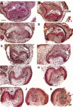
Fifty-four molars were cultured in the presence of sense ODNs. After 2 days of culture, sense-treated explants showed the pres-ence of the primary enamel knot (EK1) (Fig. 1A). The inner and outer dental epithelia (IDE, ODE) and the stratum intermedium developed. The stellate reticulum demonstrated the presence of some lacunae. After 4 and 6 days of culture, the cervical loop progressed in an apical direction and a tooth-specific bell devel-oped (Fig. 1 C,E). After 6 days, polarizing odontoblasts were observed (Fig. 1E). After 8 and 10 days of culture, predentin-dentin was secreted and polarizing preameloblasts were present (Fig. 1 G,I).
Fifty-seven teeth were cultured in vitro in the presence of anti-sense ODNs. After 2, 4, 6 and 8 days of culture, no histological differences existed when compared to the sense ODNs treated teeth. Histogenesis of the enamel organ and the progression of the cervical loop had occurred (Fig. 1 B,D,F,H). After 8 days, functional odontoblasts secreted predentin-dentin (Fig. 1H) and polarizing ameloblasts were observed (Fig. 1 H,J,K). After 10 days of culture, one explant demonstrated the presence of functional odontoblasts in the absence of an IDE (Fig. 1K). Both the sense and anti-sense ODNs treatments led to limited growth of the teeth (compare Figs. 1 and 3).
In vitro culture of the E-14 transgenic molars
The genotyping of embryos born from the mating of met D/+ mice revealed the occurrence of the three anticipated genotypes met D/D, met D/+ and met +/+ (Fig. 2). The PCR procedure resulted in the amplification of fragments of different length visualized on agarose gel (Fig. 2). Three distinct genotypes existed: the 500 bp fragments corresponded to the homozygotes met D/D, the 350 bp
Fig. 2. Met expression in transgenic embryos revealed by polymerase chain reaction (PCR).Lanes 1-8, 11-16, 20-21, 24-25, 29-31, 33, 35-36, correspond to Met D/+ embryos with 350 and 500 base pair PCR products.
Lanes 20 and 36 correspond to female and male adult Met D/+ respec-tively. Lanes 9, 22, 26-28, 32, 34 correspond to Met D/D embryos with 500 base pair PCR products. Lanes 10, 17-19, 23 correspond to Met +/+ embryos with 350 base pair PCR products. (M) molecular marker (DNA/ EcoRI, Hind III. From the top to the bottom: 21226, 4268, 2027, 1904, 1548, 1375, 947, 831 and 546 bp).
which the enamel organs appeared surrounded by a thin layer of dentin were observed for E-14 ICR mice molars cultured in vitro for 14 days.
Using the same phosphorothioate sense and anti-sense oligodeoxynucleotides as Tabata et al. (1996), but extending only slightly the culture and treatment period to the period of time during which in vitro cultured E-14 mouse molars normally achieve their histo-morphogenesis (Schmitt et al., 1999), we observed normal crown morphogenesis and cytodifferentiation at the histological level. In only one of the specimens cultured and treated for 10 days with anti-sense oligodeoxynucleotides, we observed the presence of functional odontoblasts (secreting predentin-dentin), no longer in contact with IDE cells. The constant physiological coexistence of normally distributed preodontoblasts-odontoblasts and preameloblasts (IDE cells) during the shorter culture periods and the role of the IDE in odontoblasts differentiation (Ruch, 1998) leads us to assume that in the molar cultured for 10 days, the IDE disappeared as a secondary event. Both the sense and anti-sense oligodeoxynucleotides probably had slight cytotoxic effects lead-ing to reduced growth. The anti-sense treatment strategy imple-mented by Tabata et al. (1996) is not completely identical with ours: we introduced the ODN’s in agar solidified medium and cannot exclude a possible, albeit unlike, limitation of the distribution of the ODN’s leading eventually to restricted concentrations at cellular level and incomplete HGF translation arrest. Complete loss of the targeted protein should be documented.
fragments to the homozygotes met +/+ and the 350 and 500 bp fragments to the heterozygotes met D/+. Seven met D/D, twenty-two met D/+ and five met +/+ embryos were obtained.
Histological observations
After 6 or 7 days in vitro, all the cultured first lower molars (met +/+ n=10 teeth, met D/+ n=42 teeth, met D/D n=12 teeth) demon-strated the presence of well-defined dental papillae and enamel organs. Normal gradients of odontoblast terminal differentiation were observed and polarizing ameloblasts were present (Fig. 3 A-F). met D/+ and met D/D molars demonstrated short cervical loops (Fig. 3F). One of twelve met D/D explants demonstrated fused M1 and M2 (Fig. 3 E,F).
3D reconstructions
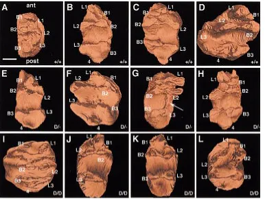
Four randomly selected right or left met +/+ molars cultured for 6 or 7 days were analyzed by means of 3D reconstructions. Two anterior, two median and two posterior cusps were visible (Fig. 4 A-D). Cusp 4 appeared in the posterior part of three teeth and was separated from the cusps L3 and B3 in front by a third posterior transverse cleft (Fig. 4 B-D). A curved, nearly closed, crest linked the four anterior and median cusps (Fig. 4 A-D). The teeth were subdivided by a deep median transverse fissure into a larger anterior portion including four cusps (L1, B1, B2, L2) and a smaller posterior one including cusps L3, B3 and 4 (Fig. 4 B-D).
Four randomly selected left or right met D/+ molars cultured for 6 or 7 days were reconstructed. Crowns of the left and right molars comprised six (Fig. 4G) or seven (Fig. 4 E,F,H) cusps: two series of three buccal and three lingual cusps, essentially of a paired nature and eventually one single posterior cusp. One left molar demonstrated a supernumerary ridge linking elongated L2 and L3 (Fig. 4G).
Four randomly selected left or right met D/D molars cultured for 6 or 7 days were reconstructed. The left and right crowns demon-strated variable cusp morphology. One left tooth cultured for 7 days demonstrated a normal cusp pattern with seven cusps. A normal curved, nearly closed, crest linked the four anterior and median cusps (Fig. 4I). For the three other teeth cultured for 6 days, B1 was small and was often fused with L1 (Fig. 4 J-L). In some teeth, cusp B3 also was rudimentary (Fig. 4 J,K). The crest linking the four anterior and median cusps had a variable configuration: either the crest just linked the median cusps (Fig. 4 J,K); or an abnormal crest linked L2, L1, B1 and B2 but not L2 and B2 (Fig. 4L); or the crest that normally joined B1, L1 and L2 was nearly absent (Fig. 4 J,K).
Discussion
During embryogenesis, HGF supports organogenesis and mor-phogenesis of various organs including liver, kidney, lung, gut, mammary gland and skeletal system (Matsumoto and Nakamura, 1996a, b). HGF appears also to be involved in tooth morphogen-esis. Tabata et al. (1996) documented the expression of HGF by dental mesenchyme from early cap to mid bell-stage of tooth development, and the expression of c-Met, the cognate receptor, by dental epithelial cells from cap to advanced bell-stage. These authors also analyzed the effects of HGF translation arrest by means of anti-sense oligodeoxynucleotides, and suggested that the imbalance between the proliferation activities of the IDE and dental mesenchyme caused by HGF translation arrest could lead to abnormal tooth morphogenesis. Abnormal tooth structures in
To avoid the possibility of such technical limitation and get more information on the putative role of HGF during tooth histo-morpho-genesis, we cultured in vitro E-14 molars from transgenic met D/D embryonic mice. These mice express a severe loss of function phenotype which results in embryonic lethality and recapitulates the defects of met and hgf null mutants (Maina et al., 1996). The c-Met receptor is the only known receptor for HGF (Park et al., 1987; Bottaro et al., 1991; Naldini et al., 1991a, b; Vigna et al., 1994; Ponzetto et al., 1996; Matsumura et al., 1998; Kajihira et al., 1999). The cap-stage, E-14 molars, from met +/+, met D/+, met D/D embryos had similar morphological features as observed under the stereomicroscope, and most of the isolated M1 tissues included the primordium of M2.
Having cultured embryonic teeth in a panel of different culture media (Schmitt et al., 1999 and references therein) we concluded that in our hands the most “physiological” crown morphology was expressed when molars were grown on agar solidified medium containing 20% of fetal calf serum. In these conditions, E-14 molars developed a normal cusp pattern, the cusps containing post-mitotic odontoblasts and ameloblasts after 6 days in vitro. We also observed that longer culture periods could eventually lead to histological alterations. For these reasons the molars of the transgenic mice were cultured on agar solidified medium for 6-7 days. Furthermore the 3D reconstructions provided an as-sessment of the entire tooth morphology allowing for avoidance of possible misinterpretation of single sections. The histo-morpho-logical analysis of these molars revealed a more or less apparent shortening of the cervical loop for met D/+, and met D/D molars. The 3D reconstructions of cultured met +/+, met D/+ and met D/ D molars revealed significant alterations of the cusp pattern and morphology for the met D/D molars, which could be related to
discrete alterations in cell proliferation kinetics. Since HGF has the ability to elicit motogenesis (Stoker et al., 1987; Weidner et al., 1990; Balkovetz, 1998; Maffe and Comoglio, 1998; Lee et al., 1999), the abrogation of HGF activity could also interfere with cell migration, which has been suggested to play an important role in cuspidogenesis. Indeed, Coin et al. (1999) have shown that non-cycling inner dental epithelium cells initially associated with the signaling primary enamel knot underwent sequential segregation and that this led to the formation of distinct groups of cells, each one corresponding to a particular developing cusp. Coin et al. (1999) suggested an essential role of these cells in cusp forma-tion. Incomplete segregation of these cells could lead to the fusion of cusps and to abnormal crest formation. On the other hand, the exceptional fusion of M1 and M2 could be a culture artifact. The use of a genetically engineered mouse model, rather than our anti-sense strategy confirms that HGF is involved in the crown morphogenesis. Further challenge is to understand how and when HGF works knowing the signaling activity of the enamel knots, and the implication of cell migration, cell proliferation, cell death, cell-cell and cell-matrix interactions during cusp formation.
Materials and Methods
Tissues
Normal E-14 embryos were obtained by mating Swiss mice. The morning of the appearance of the vaginal plug was designated as day 0 of embryonic development. First right lower molars (M1) were dissected on day 14 of gestation (E-14). E-14 met +/+, met D/+and met D/D embryos were obtained by mating CD1 met D/+ mice. The morning of the appear-ance of the vaginal plug was designated as day 0 of embryonic develop-ment. First right and left lower molars (M1) were dissected.
Oligodeoxynucleotides
Phosphorothioate oligodeoxynucleotides (ODNs) for the cDNA se-quence of mouse HGF, a 15 mer 5’CAT GCT TGC AGT TCG 3’, were used according to Tabata et al. (1996). The corresponding control sense ODNs was 5’CGA ACT GCA AGC ATG 3’. All the ODNs were prepared by IGBMC (Strasbourg, France).
Organ culture
The molars of Swiss mouse were cultured for 2, 4, 6, 8 or 10 days on 100 µl of semi-solid medium per microwell (Nunclon delta SI, Nunc, Denmark). The medium consisted of BGJ-B (Gibco, Fitton Jakson modified) supple-mented with ascorbic acid (0,18 mg/ml, Merck), L-Glutamine (2 mM, Seromed), kanamycin (0.1 mg/ml, Gibco) and Difco agar (0.5%). During the first two days culture, transferrin was added. Sense or anti-sense ODNs were added with culture medium at 30 µM and changed every other day together with the culture medium. The E-14 molars of met +/+, met D/+and met D/D embryos were cultured for 6 or 7 days on 2 ml of semi-solid medium per Petri dish (Nunc, Roskilde, Denmark; 35x10 mm). The medium con-sisted of BGJ-B (Gibco, Fitton Jakson modified) supplemented with ascor-bic acid (0.18 mg/ml, Merck), L-Glutamine (2 mM, Seromed), fetal calf serum (20%, Boehringer Bioproducts), kanamycin (0.1 mg/ml, Gibco) and Difco agar (0.5%). The teeth were incubated and grown at 37°C in a humidified atmosphere of 5% CO2 in air and the medium was changed every two days.
Genotyping
Extraction of DNA
DNA samples were extracted from embryonic skin using the standard proteinase K digestion as described by Sambrook et al. (1989) with minor modifications. Briefly, met D/+ mice were mated and embryos collected. While molars from each embryos were cultured, small pieces of skin (~16 mm2) were suspended in 700 µl of extraction buffer (Tris 50 mM pH 8.0;
EDTA 100 mM; SDS 0.5%; proteinase K: 0.25 mg/ml), and incubated at 55°C overnight. The samples were then heated at 95°C for 10 min and mixed with an equal volume of absolute ethanol and 0.1 volume of sodium acetate 3M. After centrifugation (6 min at 8500 g) the pellets were washed in 700 µl of ethanol 70% and centrifuged 6 min at 8500 g. The pellets were then air dried and resuspended in sterile double-distilled water. The concentration of the DNA was measured by absorption at 260 nm. Amplification conditions
The polymerase chain reaction (PCR) was employed to generate double-stranded DNA fragments. Each amplification was performed in the presence of 20 mM Tris HCl pH 8.4, 50 mM KCl, 2 mM MgCl2 (Gibco BRL), 200 µg BSA, 0.7 µM of primer 610 and 655, 1.4 µM of primer 611, 1.5 U of Taq DNA polymerase (Perkin Elmer Cetus), 200 µM of each dNTP (Boehringer Mannheim) and 100 ng of template DNA in a volume of 40 µl. The following set of primers was employed.
P610: 5'-AGGATTGATCATTGGTGCGGTC-3' P611: 5'-CATCTCTGTAGTTGGACTTACAC-3' and P655: 5'-CAGCTCATTCCTCCCACTC-3'.
Reactions were done in a Perkin Elmer Cetus DNA thermocycler 480 as follows: pre-denaturation (3 min at 94°C). Eight cycles of denaturation (1 min at 94°C), annealing (1 min at 60°C), extension (1 min at 72°C) and then, 35 cycles of: denaturation (1 min at 94°C), annealing (1 min at 54°C), extension (1 min at 72°C), followed by a final extension step (10 min at 72°C).
Amplification products were electrophoresed on 1.8 % agarose gels in TBE buffer (Tris base 87 mM, boric acid 89 mM, EDTA 2 mM, pH 8.0) in the presence of 1 µg/ml of ethidium bromide. Electrophoresis was performed at 200 mA for 4 h in TBE. Gels were than examined and photographed under UV light with a Polaroid system. Embryos were classified as follows: met +/+ corresponds to a 350 bp fragment; met D/D to a 500 bp fragment and met +/D to a 350 and 500 bp fragments.
Histology
Teeth were fixed in Bouin-Hollande fluid and embedded in paraffin wax. Five µm serial sections were stained with Mallory’s stain.
3D reconstructions
Drawings of the contours of the mesenchyme and of the inner dental epithelium of the molars were made at 5 µm intervals from the serial histological sections using a Zeiss Jenaval microscope equipped with a drawing chamber. The digitalization of the serial drawings was achieved using a Hamamatsu C2400 camera connected to a digital imaging system. The digitalization of the serial drawings and correlation of successive images (Olivo et al., 1993) have been previously described (Lesot et al., 1996). Software packages allowing image acquisition and treatment were developed and adapted to this work. Three-dimensional images were generated using a volume rendering program (Sun Voxel, Sun Microsystems).
Acknowledgements
The met D/+ mice were kindly provided by Dr. C. Prunotto and Pr. C. Ponzetto. We wish to thank Dr A. J. Smith for critical reading of this manuscript, Dr. D. Montagnon, Mr. F. Ruffenach and Mr. A. Ackermann for technical help. This research was partially financed by the International Human frontier Science Program (grant TG-558/95 M) and by the Fondation Dentaire de France (UB/SS 500144-98002598).
References
BALKOVETZ, D.F. (1998). Hepatocyte growth factor and madin-darby canine kidney cells: in vitro models of epithelial cell movement and morphogenesis. Microsc. Res. Tech. 43: 456-463.
BALKOVETZ, D.F. and LIPSCHUTZ, J.H. (1999). Hepatocyte growth factor and the kidney: it is not just for the liver. Int. Rev. Cytol. 186: 225-260.
BEI, M. and MAAS, R. (1998). FGFs and BMP4 induce both Msx1-independent and Msx1-dependent signaling pathways in early tooth development. Development. 125: 4325-4333.
BOTTARO, D.P., RUBIN, J.S., FALETTO, D.L., CHAN, A.M., KMIECIK, T.E., VANDE WOUDE, G.F. and AARONSON, S.A. (1991). Identification of the hepatocyte growth factor receptor as the c-Met proto-oncogene product. Science 251: 802-804.
COIN, R., LESOT, H., VONESCH, J.L., HAIKEL, Y. and RUCH, J.V. (1999). Aspects of cell proliferation kinetics of the inner dental epithelium during mouse molar morphogenesis: a reappraisal of the role of the enamel knot area. Int. J. Dev. Biol. 43: 261-267.
DEFRANCES, M.C., WOLF, H.K., MICHALOPOULOS, G.K. and ZARNEGAR, R. (1992). The presence of hepatocyte growth factor in the developing rat. Develop-ment 116: 387-395.
FAUSSER, J.L., SCHLEPP, O., ABERDAM, D., MENEGUZZI, G., RUCH, J.V. and LESOT, H. (1998). Localization of antigens associated with adherens junctions, desmosomes and hemidesmosomes during murine molar morphogenesis. Differ-entiation 63: 1-11.
GALIMI, F., BRIZZI, M.F. and COMOGLIO, P.M. (1993). The hepatocyte growth factor and its receptor. Stem Cells 11: 22-30.
GAUNT, W. (1955). The development of the molar pattern mouse. Acta Anat. 24: 249-268.
GAUNT, W. (1961). The development of the molar pattern of the golden hamster (Mesocricetus Auratus W.), together with a re-assessment of the molar pattern of the mouse (Mus Musculus). Acta. Anat. 45: 219-251.
KAJIHARA, T., OHNISHI, T., ARAKAKI, N., SEMBA, I. and DAIKUHARA, Y. (1999) Expression of hepatocyte growth factor/scatter factor and c-Met in human dental papilla and fibroblasts from dental papilla. Arch. Oral Biol. 44: 135-147.
LEE, K.K., WONG, C.C., WEBB, S.E., TANG, M.K., LEUNG, A.K., KWOK, P.F., CAI, D.Q. and CHAN, K.M. (1999). Hepatocyte growth factor stimulates chemotactic response in mouse embryonic limb myogenic cells in vitro. J. Exp. Zool. 283: 170-180.
LESOT, H., PETERKOVA, R., SCHMITT, R., MEYER, J.M., VIRIOT, L., VONESCH, J.L., SENGER, B., PETERKA, M. and RUCH, J.V. (1999). Initial features of the inner dental epithelium histo-morphogenesis in the first lower molar in mouse. Int. J. Dev. Biol. 43: 245-54.
LESOT, H., VONESCH, J.L., PETERKA, M., TURECKOVA, J., PETERKOVA, R. and RUCH, J.V. (1996). Mouse molar morphogenesis revisited by 3D reconstruction. II. Spatial distribution of mitoses and apoptosis in cap to bell staged first and second upper molar teeth. Int. J. Dev. Biol. 40: 1017-1031.
MAAS, R. and BEI, M. (1997). The genetic control of early tooth development. Crit. Rev. Oral. Biol. Med. 8: 4-39.
MAFFE, A. and COMOGLIO, P.M. (1998). HGF controls branched morphogenesis in tubular glands. Eur. J. Morphol. 36: 74-81.
MAINA, F., CASAGRANDA, F., AUDERO, E., SIMEONE, A., COMOGLIO, P.M., KLEIN, R. and PONZETTO, C. (1996). Uncoupling of Grb2 from the Met receptor in vivo reveals complex roles in muscle development. Cell 87: 531-542.
MATSUMOTO, K. and NAKAMURA, T. (1996a). Heparin functions as a hepatotrophic factor by inducing production of hepatocyte growth factor. Biochem. Biophys. Res. Commun. 227: 455-461.
MATSUMOTO, K. and NAKAMURA, T. (1996b). Emerging multipotent aspects of hepatocyte growth factor. Biochem. 119: 591-600.
MATSUMOTO, K. and NAKAMURA, T. (1997). Hepatocyte growth factor (HGF) as a tissue organizer for organogenesis and regeneration. Biochem. Biophys. Res. Commun. 239: 639-644.
MATSUMURA, T., TABATA, M.J., WAKISAKA, S., SAKUDA, M. and KURISU, K. (1998). Ameloblast-lineage cells of rat tooth germs proliferate and scatter in response to hepatocyte growth factor in culture. Int. J. Dev. Biol. 42: 1137-1142.
NAKAMURA, T., NAWA, K., ICHIHARA, A., KAISE, N. and NISHINO, T. (1987). Purification and subunit structure of hepatocyte growth factor from rat platelets. FEBS Lett. 224: 311-316.
NALDINI, L., VIGNA, E., NARSIMHAN, R.P., GAUDINO, G., ZARNEGAR, R., MICHALOPOULOS, G.K. and COMOGLIO, P.M. (1991b). Hepatocyte growth factor (HGF) stimulates the tyrosine kinase activity of the receptor encoded by the proto-oncogene c-Met. Oncogene 6: 501-504.
NALDINI, L., WEIDNER, K.M., VIGNA, E., GAUDINO, G., BARDELLI, A., PONZETTO, C., NARSIMHAN, R.P., HARTMANN, G., ZARNEGAR, R. and MICHALOPOULOS, G.K. (1991a). Scatter factor and hepatocyte growth factor are indistinguishable ligands for the MET receptor. EMBO J. 10: 2867-2878.
OLIVO, J.C., IZPISUA-BELMONTE, J.C., TICKLE, C., BOULIN, C. and DUBOULE, D. (1993). Reconstruction from serial sections: a tool for developmental biology. Application to Hox genes expression in chicken wing buds. Bioimaging 1: 151-158.
PARK, M., DEAN, M., KAUL, K., BRAUN, M.J., GONDA, M.A. and VANDE WOUDE, G.F. (1987). Sequence of MET proto-oncogene cDNA has features characteristic of the tyrosine family of growth factor receptors. Proc. Natl. Acad. Sci. USA 84: 6379-6383.
PETERKOVA, R., TURECKOVA, J., LESOT, H., VONESCH, J.L., PETERKA, M. and RUCH, J.V. (1997). Bone morphogenetic proteins (BMPs) and tooth develop-ment. Trends in Glycosci. Glycotechnol. 9: 253-265.
PONZETTO, C., ZHEN, Z., AUDERO, E., MAINA, F., BARDELLI, A., BASILE, M.L., GIORDANO, S., NARSIMHAN, R. and COMOGLIO, P. (1996). Specific uncou-pling of GRB2 from the Met receptor. Differential effects on transformation and motility. J. Biol. Chem. 271: 14119-14123.
RUBIN, J.S., CHAN, A.M., BOTTARO, D.P., BURGESS, W.H., TAYLOR, W.G., CECH, A.C., HIRSCHFIELD, D.W., WONG, J., MIKI, T. and FINCH, P.W. (1991). A broad-spectrum human lung fibroblast-derived mitogen is a variant of hepato-cyte growth factor. Proc. Natl. Acad. Sci. USA 88: 415-419.
RUCH, J.V. (1995). Tooth crown morphogenesis and cytodifferentiations: candid questions and critical comments. Connect. Tissue Res. 31: 1-8.
RUCH, J.V. (1998). Odontoblast commitment and differentiation. Biochem. Cell. Biol. 76: 923-938.
SAMBROOK, J., FRITSCH, E.F. and MANIATIS, T. (1989). Molecular cloning. A laboratory manual. (2nd edition). Cold Spring Harbor, New York.
SCHMIDT, C., BLADT, F., GOEDECKE, S., BRINKMANN, V., ZSCHIESCHE, W., SHARPE, M., GHERARDI, E. and BIRCHMEIER, C. (1995). Scatter factor/ hepatocyte growth factor is essential for liver development. Nature 373: 699-702.
SCHMITT, R., LESOT, H., VONESCH, J.L. and RUCH, J.V. (1999). Mouse odontogenesis in vitro: the cap-stage mesenchyme controls individual molar crown morphogenesis. Int. J. Dev. Biol. 43: 255-260.
SHARPE, P.T. (1995). Homeobox genes and orofacial development. Connect. Tissue Res. 32: 17-25.
SLAVKIN, H.C. and DIEKWISCH, T. (1996). Evolution in tooth developmental biology: of morphology and molecules. Anat. Rec. 245: 131-150.
SONNENBERG, E., MEYER, D., WEIDNER, K.M. and BIRCHMEIER, C. (1993). Scatter factor/hepatocyte growth factor and its receptor, the c-met tyrosine kinase, can mediate a signal exchange between mesenchyme and epithelia during mouse development. J. Cell Biol. 123: 223-235.
STERN, C.D., IRELAND, G.W., HERRICK, S.E., GHERARDI, E., GRAY, J., PERRYMAN, M. and STOKER, M. (1990). Epithelial scatter factor and develop-ment of the chick embryonic axis. Developdevelop-ment 110: 1271-1284.
STOKER, M., GHERARDI, E., PERRYMAN, M. and GRAY, J. (1987). Scatter factor is a fibroblast-derived modulator of epithelial cell mobility. Nature 327: 239-242.
TABATA, M.J., KIM, K., LIU, J.G., YAMASHITA, K., MATSUMURA, T., KATO, J., IWAMOTO, M., WAKISAKA, S., MATSUMOTO, K., NAKAMURA, T., KUMEGAWA, M. and KURISU, K. (1996). Hepatocyte growth factor is involved in the morpho-genesis of tooth germ in murine molars. Development 122: 1243-1251.
THESLEFF, I., VAAHTOKARI, A., VAINIO, S. and JOWETT, A. (1996). Molecular mechanisms of cell and tissue interactions during early tooth development. Anat. Rec. 245: 151-161.
TUCKER, A.S., MATTHEWS, K.L. and SHARPE, P.T. (1998). Transformation of tooth type induced by inhibition of BMP signaling. Science 282: 1136-1138.
UEHARA, Y., MINOWA, O., MORI, C., SHIOTA, K., KUNO, J., NODA, T. and KITAMURA, N. (1995). Placental defect and embryonic lethality in mice lacking hepatocyte growth factor/scatter factor. Nature 373: 702-705.
VIGNA, E., NALDINI, L., TAMAGNONE, L., LONGATI, P., BARDELLI, A., MAINA, F., PONZETTO, C. and COMOGLIO, P.M. (1994). Hepatocyte growth factor and its receptor, the tyrosine kinase encoded by the c-MET proto-oncogene. Cell. Mol. Biol. 40: 597-604.
WEIDNER, K.M., ARAKAKI, N., HARTMANN, G., VANDEKERCKHOVE, J., WEINGART, S., RIEDER, H., FONATSCH, C., TSUBOUCHI, H., HISHIDA, T. and DAIKUHARA, Y. (1991). Evidence for the identity of human scatter factor and human hepatocyte growth factor. Proc. Natl. Acad. Sci. USA 88: 7001-7005.
WEIDNER, K.M., BEHRENS, J., VANDEKERCKHOVE, J. and BIRCHMEIER, W. (1990). Scatter factor: molecular characteristics and effect on the invasiveness of epithelial cells. J. Cell. Biol. 111: 2097-2108.
WEISS, K.M., STOCK, D.W. and ZHAO, Z. (1998). Dynamic interactions and the evolutionary genetics of dental patterning. Crit. Rev. Oral Biol. Med. 9: 369-398.
YOSHIBA, K., YOSHIBA, N., ABERDAM, D., MENEGUZZI, G., PERRIN-SCHMITT, F., STOETZEL, C., RUCH, J.V. and LESOT, H. (1998). Expression and localiza-tion of laminin-5 subunits during mouse tooth development. Dev. Dynamics 211: 164-176.
ZARNEGAR, R. and MICHALOPOULOS, G.R. (1989). Purification and biological characterization of human hepatopoietin A, a polypeptide growth factor for hepatocytes. Cancer Res. 49: 3314-3320.