Interactions between dorsal-ventral patterning genes lmx1b,
engrailed-1 and wnt-7a in the vertebrate limb
HAIXU CHEN and RANDY L. JOHNSON*
Program in Genes and Development, Department of Biochemistry and Molecular Biology, University of Texas, MD Anderson Cancer Center, Houston, USA
ABSTRACT The vertebrate limb has characteristic morphological features that distinguish dorsal and ventral regions. For example, humans and most other mammals have nails on the dorsal surface of their digits, while the ventral surface is covered by skin or footpads. Internally, there is a high degree of organization along the dorsal-ventral axis. Extensor muscles are generally located dorsally while flexor muscles are generally located ventrally. The skeleton has subtle differences that allow for attachment of these muscles and distinct pools of motor neurons innervate either dorsal or ventral muscles. How is this complex arrangement of tissues generated? Recent studies have identified a molecular cascade of three factors that govern early events in dorsal-ventral limb patterning. Two of these factors, engrailed-1 and wnt-7a are expressed in the dorsal and ventral ectoderm respectively. The function of engrailed-1 is to repress the expression of wnt-7a in the ventral limb bud ectoderm. The third factor, a LIM-homeodomain transcription factor, lmx1b is induced in dorsal mesenchyme by wnt-7a and it is both necessary and sufficient to specify dorsal limb pattern. In this report, we examine genetic interactions between wnt-7a, engrailed-1, and lmx1b by analyzing the phenotypes of mice that are double mutants for lmx1b and either wnt-7a or engrailed-1. These studies indicate that lmx1b is the only target of wnt-7a and engrailed-1 that is of consequence for dorsal-ventral patterning. Moreover, this genetic analysis suggests that lmx1b plays additional roles in anterior-posterior patterning and growth that were not previously appreciated.
KEY WORDS: limb, lmx1b, engrailed-1, wnt-7a, development
0214-6282/2002/$25.00
© UBC Press Printed in Spain www.ijdb.ehu.es
*Address correspondence to: Dr. Randy Johnson. Box 117, Department of Biochemistry and Molecular Biology, University of Texas, MD Anderson Cancer Center, 1515 Holcombe Blvd., Houston, TX 77030, USA. Fax: +1-713-791-9478. e-mail: rjohnson@odin.mdacc.tmc.edu
Abbreviations used in this paper: AER, apical ectodermal ridge; BMP, bone morphogenetic protein; FGF, fibroblast growth factor; ZPA, zone of polarizing activity.
Introduction
The developing vertebrate limb is amenable to genetic and embryological manipulation and has long been used as a model system to understand pattern formation (Johnson and Tabin, 1997). Vertebrate limbs initially form as buds that protrude at thoracic and lumbar levels to form the forelimb and hindlimb buds. Initially, they are composed of a single columnar epithelium encompassing a loose mesenchyme. The limb bud mesenchyme receives dual contribu-tions from the lateral plate mesoderm and the somitic mesoderm. The lateral plate mesoderm contributes to the connective and skeletal elements of the limb while the muscle of the limb derives solely from somite-derived myoblasts that migrate into the limb bud.
An important step in vertebrate limb pattern is the specification of positional information along the anterior-posterior, dorsal-ventral, and proximal distal axes (Capdevila and Izpisua Belmonte, 2001). Anterior-posterior and proximal-distal patterning is mediated largely by two signaling centers in the early limb bud, the zone of polarizing activity (ZPA) and the apical ectodermal ridge (AER). These signaling
centers secrete sonic hedgehog in the case of the ZPA and fibroblast growth factors (FGFs) wnt proteins and bone morphogenetic pro-teins (BMPs) in the case of the AER that in turn modulate anterior-posterior and proximal distal patterning respectively. The AER is located at the distal end of the limb bud at the interface of dorsal and ventral ectoderm, and is necessary and sufficient for limb bud outgrowth.
ventral fate is a default. Subsequently, the dorsal-ventral polarity is transferred to the ectoderm prior to limb bud outgrowth resulting in the expression of wnt-7a in the presumptive dorsal limb bud ectoderm and engrailed-1 in the ventral limb bud ectoderm. In the next step, wnt-7a, induces the expression of the LIM-homeodomain transcription factor lmx1b specifically in the dorsal mesenchyme of the limb bud. Gain of function experiments in the chick indicated that lmx1b is sufficient to specify dorsal limb pattern in ventral limb mesenchyme (Riddle et al., 1995; Vogel et al., 1995). Subse-quently, it was shown through gene targeting in the mouse that lmx1b activity is necessary to specify dorsal limb pattern (Chen et al., 1998).
The availability of mouse mutants for lmx1b (Chen et al., 1998), wnt-7a (Parr and McMahon, 1995) and engrailed-1 (Loomis et al., 1996) has allowed us to explore interactions between these genes by examining the phenotypes of lmx1b/wnt-7a and lmx1b/engrailed-1 double mutant mice. Comparison of the limb phenotypes of single versus double mutants reveals complex genetic interactions that affect both the dorsal-ventral and anterior-posterior axes. Additionally, this analysis reveals roles for lmx1b in limb bud growth and patterning that were not previously observed in lmx1b single mutants.
Results
Dorsal Ventral Patterning in lmx1b/wnt-7a and lmx1b/ engrailed-1 Double Mutants
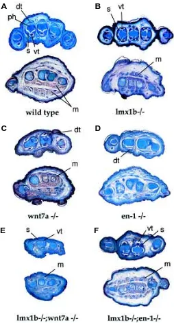
Lmx1b and wnt-7a homozygotes display a dorsal-to-ventral conversion of the distal limb bud (Chen et al., 1998; Parr and McMahon, 1995). These transformations are most obvious from sections of newborn hindlimbs (Fig. 1). Characteristic features of dorsal-ventral polarity at the level of the digits are the shape of the phalanges, the presence of sesamoid bones, the morphology of tendons, and the arrangement of muscles (Fig. 1A). In the wild-type situation the phalanges have a ventral narrowing and a smooth dorsal surface. Located at sites of ventral tendon attach-ment are sesamoid bones, small derivatives of the ventral tendons. The ventral tendons exhibit a characteristic rounded appearance, while the dorsal tendons are flattened. Finally, at a more proximal level, the dorsal limb is devoid of muscle, while the ventral limb has a complex muscle pattern. In lmx1b homozygotes (Fig. 1B), the limb is mirror-symmetric with a double ventral pattern as assayed by skeleton, tendon, and muscle morphology. Wnt-7a mutants exhibit similar dorsal-to-ventral transformation (Fig. 1C), although the transformation is not as complete as seen in the lmx1b mutants. As expected lmx1b/wnt-7a double mutant limbs display a dorsal-ventral phenotype similar to that seen in the lmx1b single mutants (Fig. 1E).
In contrast to either lmx1b or wnt-7a mutants, engrailed-1 mutant mice exhibit a ventral-to-to dorsal conversion of limb tissues (Loomis et al., 1996). Sections through the distal hindlimbs of engrailed-1 mutants reveal that the characteristic pattern of ventral skeletal and tendon elements is converted to a dorsal phenotype (Fig. 1D). Since lmx1b homozygous mutants display an opposite ventralizing phenotype we could determine the epistatic relationship between lmx1b and engrailed-1 by generating the double mutants. Lmx1b/engrailed-1 double mutant limbs (Fig. 1F) have a phenotype very similar to that seen in lmx1b single mutants suggesting that engrailed-1 is epistatic to lmx1b with respect to dorsal-ventral limb patterning.
Fig. 1. Histology of wild-type and mutant newborn hindlimbs. In all panels, dorsal is up and ventral is down. (A) Sections through the distal phalanges (upper section) and plantar region (lower section) reveal morpho-logical distinctions between the dorsal and ventral portions of the limb. The ventral tendons (vt) have a rounded appearance while the dorsal tendons (dt) have a flattened appearance. The phalanges (ph) have a rounded dorsal surface and a pointed ventral surface that is in close apposition to the sesamoid bones (s). In the plantar region, muscle (m) is present only ventrally. (B) Lmx1b-/- hindlimbs are symmetric with respect to dorsal-ventral polarity. Note the presence of sesamoid bones and dorsal-ventral tendons in the dorsal portion of the limb as well as dorsal muscle masses in a duplicated ventral arrangement. (C) Wnt-7a -/- hindlimbs exhibit a similar appearance to that of lmx1b mutants. (D) Engrailed-1 mutants have the opposite appearance with dorsal tendons in the ventral limb and absence of muscle. (E) Lmx1b/wnt-7a double homozygotes have a similar appearance to either single mutant but the overall size is smaller. (F) Lmx1b/engrailed-1 mutants exhibit dorsal-to-ventral transformations very similar to that of lmx1b single mutants.
A
B
C
D
anterior connection the humerus. In addition, elbow joint has failed to separate the humerus and the radius completely, leading to the fusion of the long bones to form a single element. In contrast, engrailed-1 appears to suppress the lmx1b phenotype (Fig. 2E). In lmx1b/engrailed-1 double homozygotes, the ulna is restored to full size and with variable penetrance an extra digit is observed.
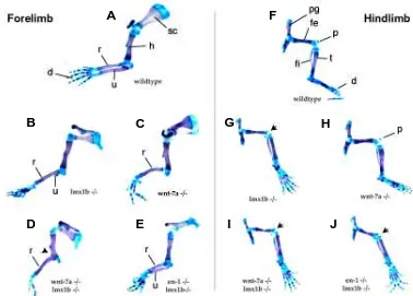
The hindlimb phenotype of lmx1b mutants and of double mu-tants is not as pronounced as the forelimb phenotype. However, several interesting features can still be noted. The wild-type hindlimb pattern is shown in Fig. 2F. At the most proximal region is the pelvic girdle followed by the femur in the upper leg. The patella, a derivative of the dorsal patellar tendon, is located at a dorsal position inbetween the femur and the fibula and tibia. Digits are located at the distal end of the hindlimb. Lmx1b mutant hindlimbs (Fig. 2G) exhibit a similar overall structure to wild-type hindlimbs. Prominent alterations include the absense of a patella, a straight-ening of the fibula and an abmormal flexure of the foot, most likely due to a dorsal-ventral patterning defect in the ankle region. In contrast to what was observed for the forelimbs, lmx1b/wnt-7a (Fig. 2I) and lmx1b/engrailed-1 double mutant (Fig. 2J) hindlimbs are very similar to the lmx1b single mutants, indicating that there are no observable downstream interactions between these genes in hindlimbs.
Analysis of hoxd11 Expression in Single and Double Mutants The skeletal phenotypes of lmx1b/wnt7a and lmx1b/engrailed-1 mutants suggest that anterior-posterior patterning is affected in these mutants. To gain insight into whether these skeletal defects are preceded by alterations in the anterior-posterior patterning Skeletal Phenotypes of lmx1b/wnt-7a and lmx1b/engrailed
Double Mutants
To assess the effects of removing either wnt-7a or engrailed-1 in a lmx1b mutant background on skeletal patterning, we examined alcian blue/alizarin red skeletal preparations of fore-limbs and hindlimbs dissected from newborn pups (Fig. 2). Wild-type fore-limbs are depicted in Fig. 2A. The characteristic skeletal elements that can be seen are the scapula, located at the most proximal portion of the limb, a single long bone, the humerus, in the upper arm, two long bones in the forearm region (also called the zeugopod), the radius and the ulna, followed by the digits at the most distal region. The ulna marks the posterior side of the limb and the radius marks the anterior side of the limbs. In the lmx1b single and double mutants, the forelimbs are more severely affected than the hindlimbs and their phenotype is described below. As reported previously (Chen et al., 1998), lmx1b mutant forelimbs (Fig. 2B) have a variable loss of the unla, but retain five digits. Similarly, wnt-7a (Parr and McMahon, 1995) mutants (Fig. 2C) also have variable ulnar loss and in addition usually lack the posterior-most digit. In contrast, the skeletal pattern of the engrailed-1 mutant is relatively normal with respect to digit number and ulnar morphology (data not shown).
In the lmx1b mutant background, wnt7a significantly enhances the lmx1b forelimb skeletal phenotype (Fig. 2D). The number of digits is reduced to two and with full penetrance, there is a single bone in the zeugopodal region. The identity of the digits is difficult to determine, but they probably corresponds to the anterior-most digits judging from their morphology. Similarly, the single long bone in the zeugopodal region is likely to be radius because of the
A
F
B
C
D
E
G
H
I
J
Fig. 2. Skeletal analysis of wild-type and mutant limbs. Panels A-E are new-born forelimb preparations and panels F-J are newborn hindlimb preparations. (A) Wild-type forelimb. (B) Lmx1b mu-tant forelimb. Note the near-absence of the radius. (C) Wnt-7a mutant forelimb. Note the absence of the ulna in this specimen and the reduction in digit num-ber. (D) Lmx1b/wnt-7a double mutant forelimb. Note the further reduction in digit number and the fusion of the radius to the humerus (arrowhead). (E) Lmx1b/ engrailed-1 double mutant forelimb. Note the presence of a relatively normal ulna. (F) Wild-type hindlimb. (G) Lmx1b mutant hindlimb. Note that the fibula is much straighter than the wild-type and articulates with the ankle region rather than with the tibia as in wild-type; the tibia is thinner, the patella is absent (arrow), and the digits are rotated to be in plane with the fibula and tibia. (H) Wnt-7a mutant. The phenotype of the hindlimb is very similar to the wild-type. Note that the presence of the patella, the shape and insertion point of the
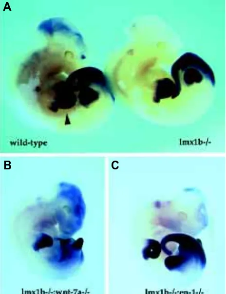
pathway, we examined hoxd11 (Dolle et al., 1989; Izpisua-Belmonte et al., 1991) in single and double mutant limb buds by whole-mount in situ hybridization. In wild-type embryos (Fig. 3A), hoxd11 is expressed in forelimb buds in separate proximal and distal domains. Within each domain, there is stronger expression of hoxd11 in the posterior portions of the limb bud. In lmx1b single mutants there is no evidence of reduction or alteration of hoxd11 expression. Suprisingly, in the lmx1b/wnt-7a double mutant, the expression of hoxd11 is similar to that of either the lmx1b or wnt-7a mutant (data not shown), even though the limbs are significantly reduced in size relative to either the wnt-7a or lmx1b single mutants. Perhaps as a consequence of this smaller size, the proximal and distal domains of hoxd11 expression are not as distinguished relative to the wild-type. No significant alterations in the expression of hoxd11 were in engrailed-1 mutants but in lmx1b/ engrailed-1 double mutants, a fusion of the proximal and distal domains of hoxd11 expression was observed.
Discussion
The final form of the vertebrate limb is sculpted through a series of inductive events that pattern the three cardinal limb axes (Capdevila and Izpisua Belmonte, 2001; Johnson and Tabin, 1997). The anterior-posterior axis is specified at least in part through the action of the secreted signaling molecule sonic hedge-hog. The proximal-distal axis is defined through the antagonism of the AER (mediated by wnt and fgf signaling) and signals emanating from the proximal limb region. The dorsal-ventral limb axis is determined through a series of reciprocal epithelial-mesenchymal interactions between limb bud ectoderm and mesenchyme. In order for the proper shape and function of the adult limb to be achieved, these patterning processes must be coordinated so that positional information along all three axes is seamlessly inte-grated. We have some knowledge of the pathways that link patterning along the cardinal axes, but our understanding is far from complete.
Known components of dorsal-ventral limb patterning include three factors: wnt-7a expressed in the dorsal ectoderm, engrailed-1 expressed in the ventral ectoderm, and lmx1b expressed in the dorsal mesenchyme. Previous studies (Cygan et al., 1997; Loomis et al., 1996; Loomis et al., 1998; Parr and McMahon, 1995; Riddle et al., 1995; Yang and Niswander, 1995) have integrated these genes into a pathway in which engrailed-1 suppresses the expres-sion of wnt-7a in the ventral ectoderm and wnt-7a induced the expression of lmx1b in the dorsal mesenchyme. Although this simple pathway serves as a useful intellectual framework for understanding dorsal-ventral limb pattern, the situation in the embryo is actually much more complex (Chen and Johnson, 1999). Wnt-7a cannot be the only factor regulating dorsal expression of lmx1b because in wnt-7a mutants, lmx1b expression is only lost from the distal anterior limb mesenchyme (Cygan et al., 1997; Loomis et al., 1998). In addition, wnt-7a is thought to mediate integration of dorsal-ventral and anterior-posterior patterning through regulation of levels of sonic hedgehog expression (Parr and McMahon, 1995; Yang and Niswander, 1995). Likewise, engrailed-1 affects both dorsal-ventral polarity and AER position-ing (Loomis et al., 1996). Similarly, lmx1b mutants display a reduction in the unla along with dorsal-to-ventral conversions of the limb mesenchyme (Chen et al., 1998). Hence, while these three genes, lmx1b, engrailed-1 and wnt-7a may have a predominant role in dorsal-ventral patterning, it is clear that they must have additional roles in patterning of the other limb axes.
The availability of lmx1b, wnt-7a and engrailed-1 mutants has allowed us to test whether the effects of these genes on anterior-posterior and dorsal-ventral patterning occurs via parallel or linear pathways. The simplest and most straightforward phenotype that we observe is that lmx1b/engrailed-1 mutants display a double-ventral phenotype. Since lmx1b and engrailed-1 mutants have opposite effects on dorsal-ventral polarity with lmx1b mutants being ventralized and engrailed-1 mutants being dorsalized, we can conclude that lmx1b acts downstream of engrailed-1 to control dorsal cell fates. This is in accord with the simple model for dorsal-ventral limb patterning outlined above. However, we can make another strong conclusion from this experiment: lmx1b is the only relevant target for engrailed-1 regulation with respect to dorsal-ventral patterning. A secondary conclusion is that lmx1b is the only relevant target for wnt-7a in the dorsal mesenchyme with respect
Fig. 3. Expression of hoxd11 in single and double mutant embryos. (A) Whole-mount in situ hybridization of wild-type (left) and lmx1b mutant (right) E 11.5 embryos. Hoxd11 is expressed in the forelimb bud (arrow-head) in a proximal and distal domain. This pattern is unaltered in lmx1b mutants. (B) Hoxd11 expression in lmx1b/wnt-7a double mutants. The two domains of expression are now fused into a single domain that retains posterior restriction. Note that the forelimb is much smaller than wild-type or lmx1b single mutants and has a posterior truncation. (C) Hoxd11 expression in lmx1b/engrailed-1 double mutants. The forelimb bud has a relatively normal shape bud and exhibits a single domain of hoxd11 expression. Note that this embryo is at a slightly earlier stage than the embryos shown in panel (A).
A
to dorsal cell fate specification. This conclusion is supported by our observation that the wnt-7a/lmx1b double mutant phenotype re-sembles each single mutant with respect to dorsal-ventral polarity. The situation with respect to the functions of lmx1b, wnt-7a and engrailed-1 on other limb axes is more complex. Wnt-7a regulates the expression of sonic hedgehog, but apparently this effect is not mediated through the action of lmx1b because lmx1b single mutants do not have a reduction in sonic hedgehog expression nor do they display digit loss. However, both wnt-7a and lmx1b mutants exhibit a variable loss of the ulna. In the case of wnt-7a mutants, this was interpreted to be due to a reduction of sonic hedgehog expression. Do lmx1b and wnt-7a affect ulnar development by distinct parallel pathways? The phenotype of the lmx1b/wnt7a double mutants suggests that this is the case. With complete penetrance, the ulna is lost in these double mutants. It is difficult to determine whether this is simply an additive phenotype or a synergistic phenotype, but in either case it suggests that lmx1b regulates a novel pathway within the limb mesenchyme that controls posterior limb development. What might that pathway be? The unexpected observation that loss of lmx1b enhances the digit phenotype of wnt-7a mutants suggests that lmx1b may be involved a general aspect of posterior limb bud growth. One possibility is that lmx1b acts to modulate the ability of cells to receive growth promoting signals. A potential mechanism for this could be produc-tion of extracellular matrix factors that facilitate growth factor signaling. Pertinent to this argument are the observations that in the eye and kidney, lmx1b regulates the composition of the extracellular matrix (Morello et al., 2001; Pressman et al., 2000). Perhaps this function of lmx1b could be conserved in most or all tissues in which its activity is required.
In contrast to the enhancement of the anterior-posterior defects seen in the lmx1b/wnt-7a mutants, engrailed-1 suppresses the ulnar defect in lmx1b mutants. What might be the mechanism leading to this unexpected observation? Engrailed-1 mutants have an ex-panded ventral AER leading to a broadened expression of fgf-8 and presumably other factors that are produced by the AER. Perhaps this enhanced signaling by the AER makes up for defects in specifica-tion and/or growth of ulnar precursors in the lmx1b mutants. In this regard, it is interesting to note that lmx1b expression is restriced to the dorsal mesenchyme of the limb while engrailed-1 expression is limited to the ventral ectoderm. Hence it is difficult to imagine how engrailed-1 dependent factors interact with lmx1b-dependent fac-tors. Perhaps this information is integrated in at the distal part of the limb where the dorsal mesenchyme and the ventral ectoderm are fairly closely apposed. Indeed, it has been proposed that dorsal-ventral positional information is specified within this distal mesen-chyme (Akita, 1996). The determination of the exact mechanism of engrailed-1 suppression of the lmx1b ulnar phenotype will require a detailed knowledge of the pathways regulated by engrailed-1 and lmx1b.
Materials and Methods
Mice were housed in a conventional colony and genotyped by PCR using DNA extracted from tail biopsies according to the methods outlined in Chen et al. (1998) for lmx1b, Parr and McMahon (1995) for wnt-7a, and Loomis et al. (1996) for engrailed-1. Timed pregnant matings were set
between lmx1b/wnt7a and lmx1b/engrailed-1 double heterozygotes and the date of plug set as 0.5 dpc. Newborn mice were sacrificed and skeletal preparations prepared according to Chen et al. (1998). For histological studies, newborn forelimbs or hindlimbs were immersed in Bouin’s fixative overnight and processed for paraffin sectioning. Seven micron sections were stained with Mallory’s trichrome according to Pressman et al. (2000). In situ hybridization was carried out according to Chen et al. (1998).
Acknowledgements
We thank Andrew McMahon for providing wnt-7a mutant mice, Alexandra Joyner for providing engrailed-1 mutant mice, and Dennis Duboule for providing the hoxd11 in situ probe.
References
AKITA, K. (1996). The effect of the ectoderm on the dorsoventral pattern of epidermis, muscles and joints in the developing chick leg: a new model. Anat Embryol (Berl) 193, 377-86.
CAPDEVILA, J. AND IZPISUA BELMONTE, J. C. (2001). Patterning mechanisms controlling vertebrate limb development. Annu Rev Cell Dev Biol 17, 87-132.
CHEN, H. AND JOHNSON, R. L. (1999). Dorsoventral patterning of the vertebrate limb: a process governed by multiple events. Cell Tissue Res 296, 67-73.
CHEN, H., LUN, Y., OVCHINNIKOV, D., KOKUBO, H., OBERG, K. C., PEPICELLI, C. V., GAN, L., LEE, B. AND JOHNSON, R. L. (1998). Limb and kidney defects in Lmx1b mutant mice suggest an involvement of LMX1B in human nail patella syndrome. Nat Genet 19, 51-5.
CYGAN, J. A., JOHNSON, R. L. AND MCMAHON, A. P. (1997). Novel regulatory interactions revealed by studies of murine limb pattern in Wnt-7a and En-1 mutants. Development 124, 5021-32.
DOLLE, P., IZPISUA-BELMONTE, J. C., FALKENSTEIN, H., RENUCCI, A. AND DUBOULE, D. (1989). Coordinate expression of the murine Hox-5 complex homoeobox-containing genes during limb pattern formation. Nature 342, 767-72.
IZPISUA-BELMONTE, J. C., TICKLE, C., DOLLE, P., WOLPERT, L. AND DUBOULE, D. (1991). Expression of the homeobox Hox-4 genes and the specification of position in chick wing development. Nature 350, 585-9.
JOHNSON, R. L. AND TABIN, C. J. (1997). Molecular models for vertebrate limb development. Cell 90, 979-90.
LOOMIS, C. A., HARRIS, E., MICHAUD, J., WURST, W., HANKS, M. AND JOYNER, A. L. (1996). The mouse Engrailed-1 gene and ventral limb patterning. Nature 382, 360-3.
LOOMIS, C. A., KIMMEL, R. A., TONG, C. X., MICHAUD, J. AND JOYNER, A. L. (1998). Analysis of the genetic pathway leading to formation of ectopic apical ectodermal ridges in mouse Engrailed-1 mutant limbs. Development 125, 1137-48.
MORELLO, R., ZHOU, G., DREYER, S. D., HARVEY, S. J., NINOMIYA, Y., THORNER, P. S., MINER, J. H., COLE, W., WINTERPACHT, A., ZABEL, B. et al. (2001). Regulation of glomerular basement membrane collagen expression by LMX1B contributes to renal disease in nail patella syndrome. Nat Genet 27, 205-8.
PARR, B. A. AND MCMAHON, A. P. (1995). Dorsalizing signal Wnt-7a required for normal polarity of D-V and A-P axes of mouse limb. Nature 374, 350-3.
PRESSMAN, C. L., CHEN, H. AND JOHNSON, R. L. (2000). LMX1B, a LIM homeodomain class transcription factor, is necessary for normal development of multiple tissues in the anterior segment of the murine eye. Genesis 26, 15-25.
RIDDLE, R. D., ENSINI, M., NELSON, C., TSUCHIDA, T., JESSELL, T. M. AND TABIN, C. (1995). Induction of the LIM homeobox gene Lmx1 by WNT7a establishes dorsoventral pattern in the vertebrate limb. Cell 83, 631-40.
VOGEL, A., RODRIGUEZ, C., WARNKEN, W. AND IZPISUA BELMONTE, J. C. (1995). Dorsal cell fate specified by chick Lmx1 during vertebrate limb develop-ment. Nature 378, 716-20.