Defects of the body plan of mutant embryos lacking Lim1,
Otx2 or Hnf3
β
activity
SIMON J. KINDER
1, TANIA E. TSANG
1, SIEW-LAN ANG
2, RICHARD. R BEHRINGER
3and PATRICK P.L. TAM*
,1 1Embryology Unit, Children’s Medical Research Institute, Wentworthville, Australia,2Institute of Genetics and Molecular and Cellular Biology, Illkirch, Strasbourg, France and
3Department of Molecular Genetics, MD Anderson Cancer Center, University of Texas, Houston, Texas, USA
ABSTRACT The orientation of the anterior-posterior (A-P) axis was examined in gastrula-stage Hnf3β, Otx2 and Lim1 null mutant embryos that display defective axis development. In situ hybridization analysis of the expression pattern of genes associated with the posterior germ layer tissues and the primitive streak (T, Wnt3 and Fgf8) and anterior endoderm (Cer1 and Sox17) revealed that the A-P axis of mutant embryos remains aligned with the proximo-distal plane of the gastrula. Further analysis revealed that cells which express Chrd activity are either absent in Hnf3β mutant embryos or localised in heterotopic sites in Lim1 and Otx2 null mutants. Lim1-expressing cells are present in the Hnf3βmutant embryo albeit in heterotopic sites. In all three mutants, Gsc-expressing cells are missing from the anterior mesendoderm. These findings suggest that although some cells with organizer activity may be present in the mutant embryo, they are not properly localised and fail to contribute to the axial mesoderm of the head. By contrast, in T/T mutant embryos that display normal head fold development, the expression domains of organizer, primitive streak and anterior endoderm genes are regionalised correctly in the gastrula.
KEY WORDS: axis alignment, gastrulation, mouse, mutant.
0214-6282/2001/$25.00 © UBC Press
Printed in Spain
www.ijdb.ehu.es
*Address correspondence to: Patrick P.L. Tam. Embryology Unit, Children’s Medical Research Institute, Locked Bag 23, Wentworthville, NSW 2145, Australia. FAX: +61-2-9687-2120. e-mail: [email protected]
Abbreviations used in this paper: A, anterior; AVE, anterior visceral endoderm; D, dorsal; Dist, distal; dpc, days post coitum; ICM, inner cell mass; L, left; P, posterior; Prox, proximal; R, right; V, ventral; VE, visceral endoderm.
Introduction
In the Zebrafish, Xenopus and chick embryo, the orientation and polarity of the embryonic axes can be traced back to asymmetrical features of the embryo before the onset of gastrulation (Yamanaka et al., 1998; Harland and Gerhard, 1997; Bachvarova et al., 1998; Izpisua-Belmonte et al., 1993). Recently, embryological studies in the mouse blastocyst have revealed that the polar body formed at fertilization remains tethered to the trophectodermal descendant of a blastomere during pre-implantation development. In the blasto-cyst, the polar body is localized asymmetrically in regards to the inner cell mass (ICM), and its position marks the animal-vegetal plane of the blastocyst that lies orthogonally to the embryonic-abembryonic axis (Gardner, 1997). Fate mapping studies have demonstrated that ICM cells located at the animal pole (towards the polar body) contribute descendants to the visceral endoderm (VE) that resides in a more distal location in the 5.5-day embryo than those derived from ICM cells at the vegetal pole away from the polar body (Weber et al., 1999). By the early-gastrula stage, the visceral endodermal cells derived from the two poles of the ICM are distributed unevenly to the prospective anterior and posterior sides of the embryo (Weber et al., 1999). Although a relationship between the animal-vegetal axis of the blastocyst and any of the three
The endodermal cells originating from different parts of the ICM are not only spatially segregated in the visceral endoderm, they are also organized in coherent clonal populations. By contrast, de-scendants of ICM cells in the epiblast intermingle extensively. The breakdown of the clonal coherence may be caused by the segre-gation of clonally related cells during the epithelialization of the epiblast and the separation of the mitotic descendants of epiblast cells as they alter cell-cell and cell-basal lamina contacts during cell proliferation (Gardner and Cockroft, 1998). This difference in the coherence of cell populations between the epiblast and the endo-derm raises the possibility that, if an earlier asymmetry in the blastocyst is critical to the specification of the embryonic polarity that emerges in the postimplantation embryo, the regionalised information would have to be perpetuated by the tissue with the least disruption in the spatial relationship of clonally derived cell populations. This would implicate the primitive endoderm and not the epiblast as the key tissue responsible for driving the patterning of the embryo.
During gastrulation, visceral endoderm cells continue to be displaced towards the anterior and proximal sides of the embryo by the newly formed definitive endoderm (Lawson et al., 1986; Lawson and Pedersen, 1987; Tam and Beddington, 1992). Concomitantly, the proximal epiblast which is fated to be extraembryonic meso-derm and amnionic ectomeso-derm is displaced posteriorly towards the primitive streak (Lawson et al., 1991; Kinder et al., 1999), and more distal epiblast moves to this position (Quinlan et al., 1995). Migra-tion of more distal epiblast to a more proximal locaMigra-tion may occur
in concert with the migration of the mesoderm and definitive endoderm (Parameswaran and Tam, 1995, Tam et al., 1997; reviewed by Tam and Behringer, 1997), though such coordinate movement of the two germ layers has not yet been shown.
In the mouse, the loss of Lim1 or Otx2 function results in the truncation of the anterior body axis (Shawlot and Behringer, 1995; Ang et al., 1996; Acampora et al; 1995; Matsuo et al., 1995). Analy-sis of the expression of molecular markers has revealed defects in the patterning of the visceral endoderm in the Lim1 mutant embryo, where the Cer1-expressing endoderm (presumptive AVE) is localized ectopically in the distal region of the gastrula (Shawlot et al., 1998). The localization of the Cer1-expressing endoderm in the Otx2 mutant embryo varies from distal to anterior sites (Biben et al., 1998). In the Cripto null mutant which shows no neural axis morphogenesis, Cer1-, Hesx1- and Hex-expressing endodermal cells remain in a distal position and are not found in the AVE (Ding et al., 1998). These findings suggest that the distal endo-derm may behave like the AVE for the specification of certain anterior neural characteristics (Thomas and Beddington, 1996). However, despite the in-duction of BF1 activity in the distal epiblast, no head morphogenesis or development of the A-P embry-onic axis is found in Cripto null mutant embryos which fail to gastrulate (Ding et al., 1998). In the Wnt3 null mutant, the presence of Lim1- and Cer1-expressing cells in the anterior endoderm is simi-larly insufficient for initiation of anterior ment (Liu et al., 1999). Induction of head develop-ment therefore needs more than a functional AVE and has been shown to require the synergistic interaction of organizer-derived tissues and the anterior endoderm and epiblast tissue of the early Fig. 1. Expression of
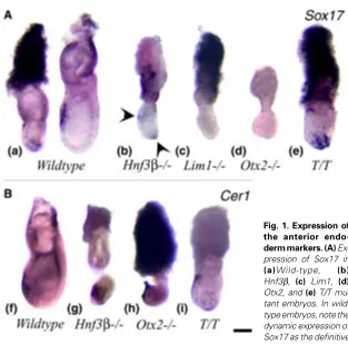
the anterior endo-derm markers. (A) Ex-pression of Sox17 in (a)Wild-type, (b) Hnf3β, (c) Lim1, (d) Otx2, and (e) T/T mu-tant embryos. In wild-type embryos, note the dynamic expression of Sox17 as the definitive endoderm is displaced from the distal posterior region to the anterior. Arrowheads in (b) mark the sites of Sox17 expression in the endoderm of the distal and anterior regions of the Hnf3β mutant embryo. (B) Expression of Cer1 in (f) Wild-type, (g) Hnf3β, (h) Otx2, and (i) T/T mutant embryos. Note the widespread Cer1 expression in the anterior and distal region in the Hnf3β mutant (g). Anterior is to the left and distal is down. Scale bar, 200 µm.
TABLE 1
EXPRESSION PATTERN OF ANTERIOR ENDODERM MARKERS (Sox17 AND Cer1) IN 7.5-DAY Hnf3ß-/-, Otx2-/-, Lim1-/- AND T/T
MUTANT EMBRYOS
Gene Mutant Expression pattern (N)
Sox17 Hnf3β-/- Distal endoderm (4/5) Anterior endoderm (2/5)
Lim1-/- No expression (6/6)
Otx2-/- No expression (3/4)
Localized to a clump of loose endoderm-like
tissue at the distal tip (1/4)
T/T Distal and anterior endoderm (6/7) Anterior endoderm only (1/7, late gastrula)
Cer1 Hnf3β-/- Distal endoderm (7/8) Proximal tissues (4/8) Anterior endoderm (3/8)
Lim1-/- Weak or no expression (12/14) Shawlot et al., 1998 Distal endoderm (2/14) Shawlot et al., 1998
Otx2-/- Anterior and distal germ layer tissues (3/3) and Biben et al., 1998.
T/T Anterior and distal endoderm (5/5)
tion of the A-P axis and the presence of the organizer-derived tissues in gastrula-stage mutant embryos. In parallel, we have examined the expression of the same set of genes in the T/T mutant embryo which forms head structures but fails to develop the trunk (Chesley et al., 1935).
Results
Ectopic localization of the anterior endoderm
The expression of Cer1 and Sox17 was examined in wild-type and mutant embryos at 7.5 days post coitum (dpc). In wild-type embryos, Cer1 and Sox17 are first expressed in the AVE in the early-streak stage embryo (Belo et al., 1997; Biben et al., 1998; Shawlot et al., 1998; J. Gad and K. Bolton, per. comm.). During gastrulation, the expression of both genes shows a dynamic pattern that appears to reflect the formation and movement of the definitive endoderm to the prospective foregut (Figs. 1 a,f). In Hnf3ß mutant embryos Sox17 expression was frequently found in the distal endoderm but sometimes also in the anterior endoderm (Fig. 1b, arrowheads). No Lim1 mutant embryos showed any Sox17 expression (Fig. 1c). Similarly most Otx2 mutant embryos showed no Sox17 expression (Fig. 1d) except one in our series which displayed expression in a loose cluster of endoderm-like cells at the distal tip of the egg cylinder (data not shown, Table 1). In T/T mutant embryos, Sox17- expressing cells were found in the anterior and distal endoderm (Fig. 1e), but mutant embryos ap-peared developmentally retarded compared to the wild-type littermates.
Previous studies have shown that Cer1 is expressed correctly in the anterior endoderm of the 7.5-day Hnf3ß mutant embryo (Klingensmith et al., 1999). In the eight Hnf3ß mutant embryos
studied, Cer1 expression in the endoderm was found to be variable from being in the distal region to the proximal-anterior region (Fig. 1g, Table 1). In Otx2mutant embryos Cer1 was expressed in an anterior and distal domain, as previously described (Fig. 1h, Biben et al., 1998), while in a few Lim1 mutants, Cer1 expression is restricted to the distal endoderm (Table 1; Shawlot et al., 1998). In T/T mutant embryos Cer1-expressing cells were found in the anterior and distal endoderm (Fig. 1i).
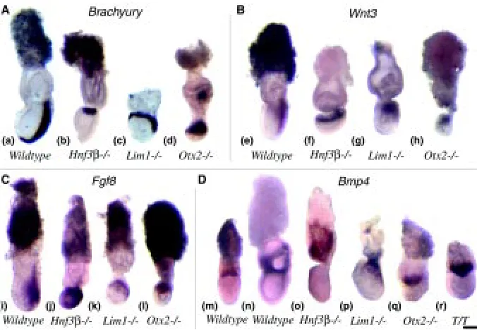
Because of the retarded development of mutant embryos, it is difficult to determine whether the Sox17- and Cer1-expressing population in the endoderm represents the AVE, or the definitive endoderm or both. If the expressing cells are representative of the AVE, the detection of distal localization in some Hnf3β mutant embryos may indicate the failure of the re-orientation of the A-P embryonic axis. However, some Hnf3β mutant embryos seemed to display the anterior expression pattern of both genes that is characteristic of the younger embryo. Alternatively, if the express-ing cells are precursors of the definitive endoderm, their distal location could reflect retarded migration or abnormal placement of the prospective foregut endoderm. The abnormal localization of Sox17-and Cer1-expressing endoderm may be correlated with the absence of foregut in the Hnf3β mutant embryo (Ang et al., 1994). However, the lack of Sox17 expression in the Otx2 and Lim1 mutant embryos is difficult to reconcile with the formation of foregut-like structures in these embryos (Shawlot and Behringer, 1995; Ang et al., 1996). Cer1 however is expressed in all mutant embryos, but is either distally localised (Lim1 or Otx2) or scattered in Hnf3β null mutant embryos. Taking together the findings of Cer1 and Sox17 expression and disregarding the nature of mutant endodermal population that may express these genes, it seems that the A-P axis may be aligned incorrectly in Hnf3β, Lim1 Fig. 2. Expression of molecular markers of the primitive streak and extraembryonic tissues. Expression of (A) T, (B) Wnt3, (C) Fgf8 and (D) Bmp4 in Wild-type, Hnf3β, Otx2, Lim1 and T/T mutant embryos. Note the expression of T, Wnt3 and Fgf8 in the proximal region of the mutant embryos, and (p) the ectopic expression of Bmp4 in the embryonic mesoderm in the Lim1 mutant. Anterior is to the left and distal is down. Scale bar, 200 µm.
mouse gastrula (Tam and Steiner, 1999). In the Hnf3β mutant embryo, a rudimentary neural axis with proper A-P molecular sub-divisions is formed despite the absence of any recognisable organiser or axial mesendoderm. However, tissues resembling the AVE are found in the mutant gastrula-stage embryo (Klingensmith et al., 1999), suggesting that there may be sufficient A-P organising activity for the initial patterning of embryonic tissues. The analysis of the de-velopmental potential of chimaeras that com-prise mutant and wild-type embryonic cells reveals that, in addition to the normal AVE function, Lim1, Otx2 or Hnf3ß activity is also required in other germ layer tissues for ante-rior development (Shawlot et al., 1999; Dufort et al., 1998; Rhinn et al., 1998, 1999).
orienta-and Otx2 mutants and that the anterior pole of the axis has remained in the distal region of the embryo.
Localization of the primitive streak
The localization of the primitive streak was examined in mutant embryos by the expression of T, Wnt3 and Fgf8. In the wild-type late-streak (7.5 dpc) embryo, T expression marks the full extent of the primitive streak on the posterior aspect of the gastrula and the axial mesendoderm anterior to node (Fig. 2a). As previously reported for the Hnf3ß, Lim1 and Otx2 mutant embryos (Table 2), the primitive streak did not extend the full length of the posterior side of the embryo. In many mutant embryos, T expression was localized to the proximal epiblast cells of the gastrula (Figs. 2 b-d; Table 2). This is reminiscent of the proximal localization of T activity in the wild-type pre-gastrula embryo (Thomas and Beddington, 1996), suggesting the germ layer tissues are formed at an abnor-mal site in the mutant embryo.
Similar to the T gene, Wnt3 is expressed uniformly in the proximal epiblast of the pre-gastrula embryo and subsequently in the posterior half of the primitive streak (Liu et al., 1999, Fig. 2e). In Hnf3ß, Lim1 and Otx2 mutant embryos, Wnt3 expression was restricted to the proximal region of the gastrula and showed very little distal extension on the posterior side of the embryo (Figs. 2 f-h).
Fgf8 is expressed first in the epiblast adjacent to the prospective site of the primitive streak in the pre-streak embryo. During gastrulation it continues to be expressed in the epiblast adjacent to and in the posterior 2/3 of the primitive streak (Crossley and Martin,
1995; Fig. 2i). In Hnf3ß, Lim1 and Otx2 null mutant embryos, Fgf8 expression was greatly expanded in the proximal and posterior regions that encompass more than half of the embryo (Fig. 2 j-l). However, there was no discrete localization of Fgf8 activity to any primitive streak structure, although stronger expression was seen in the proximal epiblast.
The localization of primitive streak markers to the proximal region of the Hnf3ß, Lim1 and Otx2 mutant embryos reveals a previous unnoticed defect in the organization of the A-P body plan. Recent work has shown that the location at which a cell passes through the streak as well as the time of ingression correlates with the final destination of the cell within the A-P axis (Kinder et al., 1999). The incorrect positioning of the streak in the proximal region of the epiblast in all three mutant embryos may result in different positional cues being perceived by cells as they ingress through the streak compared with those in the normal primitive streak. This may lead to incorrect localization of the mesodermal and endodermal cells in the A-P axis, the lack of segmental pattern in the paraxial mesoderm of the Hnf3β null mutant and the loss of anterior axial mesendoderm in the Lim1 and Otx2 mutant embryos.
Primitive streak localization to the proximal region of the mutant gastrula raises the question whether the proper inductive interac-tion exists between the extraembryonic tissues and the epiblast. In the mouse, one member of the BMP family Bmp4, is expressed in the extraembryonic ectoderm on the boundary with the proximal Fig. 3. Expression of Chrd and Lim1 in mutant embryos. (A) Expression
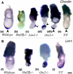
of Chrd (arrowheads) in Wild-type, Hnf3β, Otx2, Lim1 and T/T mutant embryos. (d) Two specimens of Otx2 mutant embryo showing (i) proximal and (ii) distal and the anterior midline expression of Chrd. (B) Expression of Lim1 in Wild-type, Hnf3β, Otx2 and T/T mutant embryos. Anterior is to the left and distal is down. Scale bar, 200 µm.
TABLE 2
EXPRESSION OF PRIMITIVE STREAK AND MESODERM MARKERS, T, Wnt3 AND Fgf8, AND Bmp4 IN 7.5-DAY Hnf3ß-/-, Otx2-/-,
Lim1-/-AND T/T MUTANT EMBRYOS
Gene Mutant Expression pattern (N)
T Hnf3β-/- Proximal epiblast (3/7)
Proximal epiblast and slight distal extension on the posterior side (1/7), and Ang and Rossant, 1994 Posterior side of the embryo (3/7)
Lim1-/- Proximal epiblast (1/2)
Posterior side of the embryo (1/2), and Shawlot and Behringer, 1995
Otx2-/- Proximal epiblast (2/3) Posterior side of the embryo (1/3), and Ang et al., 1996
Posterior side, node and anterior midline (Acampora et al., 1995)
Wnt3 Hnf3β-/- Proximal epiblast (3/5) Posterior side of the embryo (2/5)
Lim1-/- Proximal epiblast (3/3)
Otx2-/- Posterior side of the embryo (2/3) Proximal epiblast (2/3)
Fgf8 Hnf3β-/- Expanded domain in the proximal region (3/3)
Lim1-/- Proximal region of the embryo (3/4) No expression (1/4)
Otx2-/- Proximal region of the embryo (5/5)
Bmp4 Hnf3β-/- Distal extraembryonic tissues adjacent to the epiblast (4/4) Extraembryonic mesoderm and amnion (Klingensmith et al., 1999)
Lim1-/- Distal extraembryonic tissues adjacent to the epiblast and in the proximal germ layer tissues in the embryo (3/3)
Otx2-/- Distal extraembryonic tissues adjacent to the epiblast (3/3)
T/T Distal extraembryonic tissues adjacent to the epiblast (5/5)
epiblast (Fig. 2m) and later in the extraembryonic mesoderm and the chorionic tissues of the gastrula embryo (Fig. 2n; Waldrip et al., 1998). Bmp4 signalling activity has been shown to be essential for the formation of the mesoderm (Winnier et al., 1995; Mishina et al., 1995) and for lineage speci-fication of proximal epiblast cells (Lawson et al., 1999). In the Hnf3ß and Otx2 mutant gastrulae, Bmp4 expression persisted in the extraembryonic tissue adjacent to the epiblast (Table 2; Figs 2 o,q), similar to that in the wild-type early gastrula (Fig. 2m). In more advanced Hnf3ß mutant em-bryos, Bmp4 was expressed in the extraembryonic mesoderm and the chorionic ectoderm (data not shown; Klingensmith et al., 1999). In the early-streak Lim1 mutant embryo, Bmp4 was expressed in the extraembryonic ectoderm as in the wild-type embryo (data not shown). However, in gastrula-stage embryos, Bmp4 expression was not
re-mesendoderm immediately anterior to the node (Fig. 3a). In the Hnf3β mutant, no Chrd expression was found in the 7.5-day gastrula (Fig. 3b) but weak and diffuse expression has been observed in the posterior epiblast in the early gastrula (Klingensmith et al., 1999). The lack of Chrd and also Nog expression is consistent with the absence of any morphologically distinct node in the Hnf3ß mutant embryo (Ang and Rossant, 1994; Dufort et al., 1998; Klingensmith et al., 1999). In the Lim1mutant embryo, Chrd expression was localized to the posterior epiblast but at variable sites (Table 3; Fig. 3c). Previous studies on Otx2 mutant embryos have shown that organizer genes such as Chrd (Rhinn et al., 1998) and Hnf3β (Ang et al., 1996) are correctly localized in the distal region of the gastrula embryo. However, in one of the four mutant embryos studied in the present series, Chrd was localized to an expanded domain in the proximal region of the embryo (Table 3: Fig. 3 d,i). This demonstrates that in a minority of Otx2 mutants the organizer may be ectopically localized. Embryos that displayed distal localization of Chrd activity also showed expression in the anterior axial tissues (Fig. 3d ii). In the T/T mutant Chrd was localised to the node region and anterior midline as in wild-type embryos (Fig. 3e).
In view of the variable regionalization of Chrd activity among the three types of mutant embryos, we next studied the differentiation of the axial mesendodermal derivatives in the late-streak and early-somite stage embryo. T expressing cells were absent from the anterior midline in Hnf3ß, Lim1 and Otx2 mutant embryos (Fig. 2 b-d; Table 2; Acampora et al., 1995; Ang and Rossant, 1994). Expression of Lim1 in the anterior mesendoderm was also absent in the Hnf3ß mutant (Fig. 3g; Ang and Rossant, 1994; Table 3), but was present in Otx2 mutant embryos (Ang et al., 1996; Fig. 3h). T/ T mutants showed normal Lim1 expression in the node and anterior midline (Fig. 3i).
The absence of T-expressing cells in the anterior midline of the Hnf3ß, Lim1 and Otx2 mutant embryos raises the question of whether the more rostral segment of the axial mesendoderm that does not express T activity may be present. Contained in this segment of axial tissue is the prechordal mesoderm that charac-teristically expresses Gsc activity (Fig. 4a arrowhead; Belo et al., 1998). Surgical ablation of the axial mesendoderm from the 7.5 dpc wild-type embryo produces a phenocopy of the truncated neural tube found in the Otx2 and Lim1 null mutant embryos Fig. 4. Expression of Gsc and Shh in the axial mesendoderm. Expression of Gsc (arrowheads) in the early-somite stage (a) Wild-type, (b) Hnf3β, (c) Lim1, (d) Otx2 and (e) T/ T mutant embryos. (f) Expression of Shh in the midline of the Lim1 mutant embryo. Anterior is to the left (a-d, f) or up (e). Scale bar, 400 µm.
stricted to the extraembryonic tissues but was expressed ectopically in the proximal embryonic germ layer tissues (Fig. 2p). In the T/T mutant embryo Bmp4 expression was present in the extraembryonic tissues adjacent to the epiblast (Fig. 2r). These results show that in all four mutant embryos, BMP4 signalling that might mediate the inductive interaction between the epiblast and the extraembryonic tissues may have persisted for much longer than during normal development. Whether this may lead to abnormal regionalization or morphogenetic activity of the primitive streak is not known. Our preliminary results show that in the Lim1 mutant embryo, the differentiation of tissue lineages that are specified during gastrulation such as the germ cells, allantoic mesoderm and lateral and intermediate mesoderm are affected (Tsang et al., submitted). It is not known if the defective differentiation of these lineages may be associated with the ectopic Bmp4 activity in the primitive streak.
The localization of the organizer and its derivatives
The expression of Chrd was examined to reveal the localization of the organizer in mutant embryos. In the wild type embryo, Chrd is first expressed in the anterior primitive streak of mid-streak gastrula (data not shown) and later in the node and the axial
TABLE 3
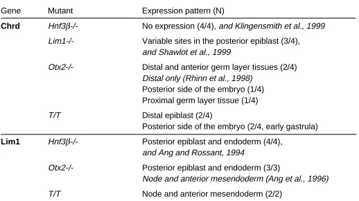
EXPRESSION OF Chrd AND Lim1 IN ORGANIZER-DERIVED TISSUE IN 7.5-DAY Hnf3ß-/-, Otx2-/-, Lim1-/- AND T/T MUTANT EMBRYOS
Gene Mutant Expression pattern (N)
Chrd Hnf3β-/- No expression (4/4), and Klingensmith et al., 1999
Lim1-/- Variable sites in the posterior epiblast (3/4), and Shawlot et al., 1999
Otx2-/- Distal and anterior germ layer tissues (2/4) Distal only (Rhinn et al., 1998) Posterior side of the embryo (1/4) Proximal germ layer tissue (1/4)
T/T Distal epiblast (2/4)
Posterior side of the embryo (2/4, early gastrula)
Lim1 Hnf3β-/- Posterior epiblast and endoderm (4/4), and Ang and Rossant, 1994
Otx2-/- Posterior epiblast and endoderm (3/3)
Node and anterior mesendoderm (Ang et al., 1996)
T/T Node and anterior mesendoderm (2/2)
(Camus et al., 2000), thus pointing to a critical role of the mesendoderm in head development.
Gsc expression has been examined in the Hnf3ß, Otx2, and Lim1 at early gastrulation. Gsc-expressing cells were found mainly in the proximal epiblast of the early-streak embryo (Table 4). We have examined Gsc expression in 8.5 dpc mutant embryos. Gsc was weakly expressed in only one of ten Hnf3ß mutant embryos studied, and was not localised to the midline (Table 4; Fig. 4b). The weak expression of Gsc in the Hnf3β mutant embryo suggests that some prechordal mesoderm might have been derived from the Gsc-expressing epiblast in the early gastrula. This contrasts with the absence of Lim1, Shh and T-expressing tissues (Ang and Rossant, 1994) that are normally found in the more caudal axial mesendoderm in the wild-type embryo. However, the expression of Gsc (Table 4) and Lim1 activity (Fig. 3g, Table 3) in the Hnf3ß gastrula may be sufficient for the molecular patterning of the neural axis. No Lim1 mutant embryos that formed a truncated head were found to express Gsc activity but Shh expressing cells were found in the midline tissues posterior to the level of axis truncation (Fig. 4 c,f). Gsc-expressing cells are found in the anterior axial tissues in some Otx2 null mutant embryos but, like the Hnf3ß mutant embryo, the
expres-sion was weak (Fig. 4d). T/T embryos that form normal head structures displayed normal anterior Gsc expression (Fig. 4e).
Discussion
Abnormal gastrula morphology is associated with aberra-tions of primitive streak activity and morphogenetic tissue movement
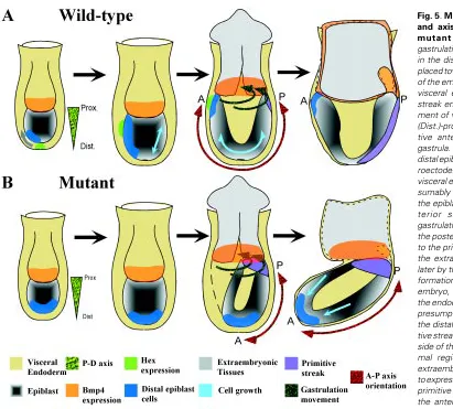
Our in situ hybridization analysis of T, Wnt3 and Fgf8 activity has shown that these genes are expressed in the proximal region and not restricted to the posterior aspect of the mutant gastrula-stage embryo. The persistence of expression of these genes in the proximal germ layers is mirrored by that of Bmp4 in the extraembryonic tissues juxtaposed to the epiblast. Bmp4 is ex-pressed in the distal part of the extraembryonic ectoderm of the pre-streak embryo, where Sox2 and Sox3 are also expressed, and as gastrulation proceeds it is expressed in the developing extraembryonic mesoderm and amnion (Lawson et al., 1999). In wild type embryos, T and Wnt3 are expressed early in the proximal region of the epiblast at the pre-streak stage, which is fated to form extraembryonic tissues (Lawson et al., 1991). Later expression is Fig. 5. Morphogenetic tissue movement and axis orientation in wild-type and
mutant embryos. (A) During
restricted to the posterior aspect of the embryo and finally to the primitive streak of the gastrula (Thomas et al., 1998; Liu et al., 1999). Fgf8 expression is detected first in the prospective posterior region of the proximal epiblast and later found in the primitive streak and the nascent mesoderm (Crossley and Martin, 1995). The expression pattern of T, Wnt3, Fgf8 and Bmp4 in the mutant embryo is therefore reminiscent of the pre-gastrulation wild-type pattern. This might be accounted for by the delay in development of the mutant embryo such that the preceding expression pattern has persisted until a normal expression pattern can be subse-quently established. However, this is unlikely because, although mutant embryos examined at 7.5 dpc may be variable in their developmental stages, they have already initiated gastrulation and normal morphology is never restored. This contrasts with the ability of wild-type embryos to regulate growth and morphogenesis in a compensatory response to tissue deficiency before the onset of gastrulation (Snow and Tam, 1979; Power and Tam, 1994). The heterotopic expression of these genes in the mutant embryo may instead be indicating the abnormal localization of the primitive streak. In the wild-type embryo, Bmp4 activity in the extraembryonic ectoderm is required for the specification of cell fate in the proximal epiblast (Lawson et al., 1999), therefore it is possible that BMP4 signalling has an overriding influence in the specification of the mesoderm in the proximal epiblast. Bmp4 null mutants lack primor-dial germ cells and mesoderm of the posterior body (Lawson et al., 1999) and in the absence of Bmpr1 activity, the embryo fails to proceed with gastrulation (Mishina et al., 1995). The persistence of Bmp4 expression in the extraembryonic tissues located closest to the epiblast may have confined the primitive streak to the proximal region of the mutant embryo. This ectopic localization of the primitive streak might have resulted in the precocious recruitment of the proximal epiblast to the germ layers. In normal embryos, cells are recruited first from the posterior-proximal epiblast to the extraembryonic mesoderm of the yolk sac, followed later by the more anterior-proximal population to the ectoderm of the amnion and the allantoic mesoderm (Lawson et al., 1991; Kinder et al.,
1999). The precise fate of cells in the proximal epiblast of mutant embryos is not known, but the constriction of the embryo at the border of the epiblast and the extraembryonic tissue may be caused by the premature depletion of cells from the anterior region of the proximal epiblast to the extraembryonic mesoderm.
In some mutant embryos, the expression of Cer1 or Sox17 is localized predominantly in the distal region of the gastrula. Both are normally expressed in the AVE early, and later in the definitive endoderm of the prospective foregut. It is therefore not possible to distinguish, based on the distal localization of gene activity, whether this is due to the failure of the AVE precursors to relocate to the anterior region of the gastrula, or the abnormal congregation of the foregut endoderm in the distal region. The mouse Cer1 gene has been shown to encode anti-BMP activity by animal cap assay (Biben et al., 1998). The localization of such activity to the AVE may be essential for the modulation of BMP activity emanating from the extraembryonic ectoderm, as well as in the positioning of the primitive streak and the organizer (Camus and Tam, 1999). The distal localization of Cer1 and Sox17-expressing endodermal cells in mutant embryos may result in a lack of modulation of BMP signalling activity in the proximal region of the embryo. Conse-quently, the primitive streak and the organizer cannot be regional-ised correctly in the mutant gastrula. The present findings of abnormal expression pattern of genes associated with the primitive streak and organizer are consistent with this hypothesis.
During normal development, the endoderm in the distal and posterior region of the pre-streak and early-streak embryo is displaced towards the anterior and proximal region by the definitive endoderm recruited from the primitive streak during gastrulation (Lawson et al., 1986; Lawson and Pedersen, 1987; Tam and Beddington, 1992). The morphogenetic movement of the definitive endoderm appears to be in concert with that of the mesoderm (Tam et al., 1997; reviewed by Tam and Behringer 1997). Analysis of the movement of clonal descendants of the epiblast reveals that some cells in the proximal epiblast are displaced towards the primitive streak (Lawson et al., 1991; Kinder et al., 1999) and their place is filled by cells that were previously localized in the anterior and distal region of the epiblast (Lawson et al., 1991; Quinlan et al., 1995). The persistent localization of Cer1 and Sox17-expressing endo-derm in the distal region of the mutant embryo suggests that there is a general lack of anterior-proximal displacement of the endo-derm and the underlying germ layer tissues. This may be brought about by the lack of germ layer generation in the posterior region of the embryo as a result of the proximal localization of the primitive streak or by defects in the proliferation of epiblast cells. The failure to replenish the anterior-proximal epiblast population that is preco-ciously recruited for gastrulation and the elevated apoptosis of the distal epiblast (as shown in the Hnf3β mutant embryo, Manova et al., 1998) may have led to the formation of the constriction in the mutant gastrula (Fig. 5).
Defective organizer differentiation impacts on head development The detection of Cer1 and Sox17-expressing cells in mutant embryos suggests that the AVE population may be present in the distal region of the gastrula. In the Criptomutant embryo, the distally localized AVE seems to be associated with the induction of neural gene activity in the distal ectoderm but the embryo never forms a proper neural axis (Ding et al., 1998). However, in the absence of the organizer or primitive streak derivatives, AVE TABLE 4
EXPRESSION OF Gsc AND Shh IN THE AXIAL MESENDODERM IN 7.5- AND 8.5-DAY Hnf3ß-/-, Otx2-/-, Lim1-/- AND T/T
MUTANT EMBRYOS
Gene Mutant Expression pattern (N)
Gastrula-stage Early-somite-stage
Gsc Hnf3β-/- Proximal germ layer tissues Anterior tissue clumps (1/10) (Ang and Rossant, 1994) No expression (9/10)
Lim1-/- Proximal germ layer tissues No expression (10/10) (Shawlot and Behringer, 1995)
Otx2-/- Proximal germ layer tissues (1/4) Anterior midline tissues (4/13) Ang et al., 1996 No expression (9/13) No expression (3/4) Ang et al., 1996
T/T Not known Anterior mesendoderm (2/2)
Shh Hnf3β-/- Not known Gut endoderm only (Ang and Rossant, 1994)
Lim1-/- Not known Axial mesendoderm and gut endoderm (2/3) Gut endoderm only (1/3)
Otx2-/- Not known Gut endoderm only (Acampora et al., 1995)
alone is not sufficient to induce anterior neural development in the Cripto and Wnt3 null mutant embryos (Ding et al., 1998; Liu et al., 1999). No morphologically recognizable organizer such as the node or any localized expression of Gsc that may indicate the formation of the early gastrula organizer are found in the Hnf3ß or Lim1mutant embryos (this study, Shawlot and Behringer, 1995; Klingensmith et al., 1999) although diffuse Gsc expression is seen in the Hnf3ß mutant epiblast at the early streak stage (Ang and Rossant., 1994). Cells that show Lim1 activity are found in a diffuse domain in the epiblast of the Hnf3ß mutant embryo. The Gsc- or Lim1-expressing cells and their descendants might have provided the necessary organizing activity for the formation of the rudimentary neural axis in this mutant. Chrd-expressing cells are present but are not always properly regionalised in the Otx2 and the Lim1 mutant embryo. However, Gsc-expressing anterior mesendoderm that is associated with the rostral midline of the head fold and later with the prospective forebrain is absent in all three mutant embryo strains. Ablation of the Gsc-expressing rostral axial mesendoderm has been shown to result in the abnormal morphogenesis of the forebrain and midbrain (Camus et al., 2000). The absence of a defined organizer and the loss of one of its derivatives, the anterior axial mesendoderm, may be the cause of the anterior truncation in the Lim1 mutant embryo and the failure of head morphogenesis in the Hnf3ß mutant. Results of this study therefore highlight the critical requirement of the morphogenetic function as well as the proper regionalization of the activity of the gastrula organizer and the AVE for the formation of the AP axis.
Materials and Methods
Otx2(Ang et al., 1996), Hnf3ß (Ang and Rossant, 1994), Lim1(Shawlot and Behringer, 1995), and T/T(Chesley, 1935) mutant mice were used for this study. Embryos were collected at 7.5 and 8.5 dpc from heterozygous female mice of each strain that were crossed to heterozygous males of the respective mutant strain. Otx2, Hnf3ß and Lim1 null mutant embryos were identified by their characteristic phenotype and in some cases confirmed by genotyping. For T/T mutant embryos, which do not display any abnormal phenotype at early stages, all embryos in the litter were processed for in situ hybridization using a mixed reagent that contained the riboprobe for T and the gene of interest. Embryos that showed no T hybridization signal were assumed of T/T genotype, when the majority of their littermates showed positive results in the same in situ hybridization experiment.
The protocol for in situ hybridization followed that of Wilkinson and Nieto (1993) with the following modifications. The Ampliscribe kit (Epicentre Technologies, Madison, Wisconsin, USA) was used in conjunction with Dig-11-UTP (Roche, Mannheim, Germany) to synthesize RNA probes, SDS was used instead of CHAPS in the hybridization solution and post-hybridization washes. Only 5x SSC was used in the post-hybridization solution, while post hybridization washes were at 70ºC and excluded formamide. No
RNA digestion was performed after hybridization. Following in situ hybridi-zation embryos were photographed using a SPOT 2 digital camera and figures were prepared with Photoshop 5.5 and Illustrator 8.0.
Acknowledgments
The authors would like to thank G. Martin, B. Herrmann, B. Hogan, P. Koopman, and Y. Kanai for the gift of riboprobes, and Bruce Davidson and Peter Rowe for reading the manuscript. Our work is supported by Human Frontier Science Program (grant to SLA, RRB, PPLT and Hiroshi Sasaki), the National Health and Medical Research Council (NHMRC) of Australia, the Ramaciotti Foundation and Mr James Fairfax. P.P.L.T. is a NHMRC Principal Research Fellow.
References
ACAMPORA, D., MAZAN, S., LALLEMAND, Y., AVANTAGGIATO, V., MAURY, M., SIMEONE, A. and BRULET, P. (1995). Forebrain and midbrain regions are deleted in Otx2-/- mutants due to a defective anterior neuroectoderm specification during gastrulation. Development 121: 2379-3290
ANG, S-L., and ROSSANT, J. (1994). HNF3β is essential for node and notochord formation in mouse development. Cell 78: 561-574.
ANG, S-L., JIN, O., RHINN, M., DAIGLE, N., STEVENSON, L. and ROSSANT, J. (1996). A targeted mouse Otx2 mutation leads to severe defects in gastrulation and formation of axial mesoderm and to deletion of rostral brain. Development 122: 243-252.
BACHVAROVA, R.F., SKROMME, I. and STERN, C.D. (1998). Induction of primitive streak and Hensen’s node by the posterior margin in the early chick embryo. Development 125: 3521-3534.
BEDDINGTON R.S.P. and ROBERTSON, E.J. (1999). Axis development and early asymmetry in mammals. Cell 96: 195-209.
BEDDINGTON, R.S. and ROBERTSON, E.J. (1998). Anterior patterning in the mouse. Trends Genet. 14: 277-284.
BELO, J.A., BOUWMEESTER, T., LEYNS, L., KERTESZ, N., GALLO, M., FOLLEITE, M. and DE ROBERTIS, E.M. (1997). Cerberus-like is a secreted factor with neuralising activity expressed in the anterior primitive endoderm of the mouse gastrula. Mech. Dev. 68: 45-57.
BIBEN, C., STANLEY, E., FABRI, L., KOTECHA, S., RHINN, M., DRINKWATER, C., LAH, M., WANG, C.C., NASH, A., HILTON, D., ANG, S-L., MOHUN, T. and HARVEY, R.P. (1998). Murine Cerberus homologue mCer-1: a candidate anterior patterning molecule. Dev. Biol. 194: 135-151.
CAMUS, A. and TAM, P.P.L. (1999). The organizer of the gastrulating mouse embryo. Curr. Topics Dev. Biol. 45:117-153.
CAMUS, A., DAVIDSON, B.D., BILLIARDS, S., KHOO, P-L., RIVERA-PEREZ, J.A., WAKAMIYA, M., BEHRINGER, R.R. and TAM, P.P.L. (2000). The morphogenetic role of midline mesendoderm and ectoderm in the development of the forebrain and midbrain of the mouse embryo. Development (in press).
CHESLEY, P. (1935). Development of the short-tailed mutant in the house mouse. J. Exp. Zool. 70: 429-454.
CROSSLEY, P.H. and MARTIN, G.R. (1995). The mouse Fgf8 genes encodes a family of polypeptides and is expressed in regions that direct outgrowth and patterning in the developing embryo. Development 121: 439-451.
DING, J., YANG, L., YAN, Y-T., CHEN, A., DESAI, N., WYNSHAW-BORIS, A., and SHEN, M.M. (1998). Cripto is required for correct orientation of the anterior-posterior axis in the mouse embryo. Nature 395: 702-706.
DUFONT, D., SCHWARTZ, L., HARPAL, K., and ROSSANT, J. (1998). The transcrip-tion factor HNF3β is required in the visceral endoderm for normal primitive streak morphogenesis. Development 125: 3015-3025.
GARDNER, R.L. (1997). The early blastocyst is bilaterally symmetrical and its axis of symmetry aligned with the animal-vegetal axis of the zygote in the mouse. Development 124: 289-301.
GARDNER, R.L. and COCKROFT, D.L. (1998). Complete dissipation of coherent clonal growth occurs before gastrulation in the mouse epiblast. Development 125: 2397-2402.
HARLAND, R. and GERHART, J. (1997). Formation and function of Spemann’s organiser. Annu. Rev. Cell Dev. Biol. 13: 611-667.
IZPISUA-BELMONTE, J.C., DE ROBERTIS, E.M., STOREY, K.G. and STERN, C.D. (1993). The homeobox gene goosecoid and the origin of organiser cells in the early chick blastoderm. Cell 27: 645-659.
KINDER, S.J., TSANG, T.E., QUINLAN, G.A., HADJANTONAKIS, A.K., NAGY, A. and TAM, P.P.L. (1999). The orderly allocation of mesodermal cells to the extraembryonic structures and the anteroposterior axis during gastrulation of the mouse embryo. Development 126: 4691-4701.
KLINGENSMITH, J., ANG, S-L., BACHILLER, D., and ROSSANT, J. (1999). Neural induction and patterning in the mouse in the absence of the node and its derivatives. Dev. Biol. 216: 535-549.
LAWSON, K.A., DUNN, N.R., ROELEN, B.A.J., ZEINSTRA, L.M., DAVIS, A.M., WRIGHT, C.V.E., KORVING, J.P.W.F.M., and HOGAN, B.L.M. (1999). Bmp4 is required for the generation of primordial germ cells in the mouse embryo. Genes and Development 13: 424-436.
LAWSON, K.A., MENESES, J.J. and PEDERSON, R.A. (1986). Cell fate and cell lineage in the endoderm of the presomite mouse embryo studied with an intracellular tracer. Dev. Biol. 115: 325-339.
LAWSON, K.A., MENESES, J.J. and PEDERSON, R.A. (1991). Clonal analysis of epiblast fate during germ layer formation in the mouse embryo. Development 113: 891-911.
LIU, P., WAKIMIYA, M., SHEA, M.J., ALBRECHT, U., BEHRINGER, R.R., and BRADLEY, A. (1999). Requirement for Wnt3 in vertebrate axis formation. Nature Genetics 22: 361-365.
MANOVA, K., TOMIHARA-NEWBEWBERGER, C., WANG, S., GODELMAN, A., KALANTRY, S., WITTY-BLEASE, K., DE LEON, V., CHEN, W.S., LACY, E., and BACHVAROVA, R.F. (1998) Apoptosis in mouse embryos: Elevated levels in pregastrulae and in the distal anterior region of gastrulae of normal and mutant mice. Dev. Dyn. 213: 293-308.
MATSUO, I., KURATANI, S., KIMURA, C., TAKEDA, N. and AIZAWA, S. (1995). Mouse Otx2 functions in the formation and patterning of rostral head. Genes Dev. 9: 2646-2658.
MISHINA, Y., SUZUKI, A., UENO, N. and BEHRINGER, R.R. (1995). Bmpr encodes a type I bone morphogenetic protein receptor that is essential for gastrulation during mouse embryogenesis. Genes Dev. 9: 3027-3037.
PARAMESWARAN, M. and TAM, P.P.L. (1995). Regionalisation of cell fate and morphogenetic movement of the mesoderm during mouse gastrulation. Dev. Genet. 17: 16-28.
POWER, M.A. and TAM, P.P.L. (1993). Onset of gastrulation, morphogenesis and somitogenesis in mouse embryos displaying compensatory growth. Anat. Embryol (Berl) 187: 493-504.
QUINLAN, G.A., WILLIAMS., E.A., TAN, S.S. and TAM, P.P.L. (1995). Neuroecto-dermal fate of epiblast cells in the distal region of the mouse cylinder: implication for body plan organisation during early embryogenesis. Development 121: 87-98.
RHINN, M., DIERICH, A., LE MEUR, M. and ANG, S-L. (1999). Cell autonomous and non-cell autonomous functions of Otx2 in patterning rostral brain. Develop-ment 126: 4295-4304.
RHINN, M., DIERICH, A., SHAWLOT, W., BEHRINGER, R.R., LE MEUR, M., and ANG, S-L. (1998). Sequential roles for Otx2 in visceral endoderm and neuroec-toderm fore forebrain and midbrain induction and specification. Development 125: 845-856.
SHAWLOT , W. and BEHRINGER, R.R. (1995). Requirement for Lim1 in head organiser function. Nature 374: 425-430.
SHAWLOT, W., DENG, J.M. and BEHRINGER, R.R. (1998). Expression of the mouse Cerberus related gene, Cerr1, suggests a role in anterior neural induction and somitogenesis. Proc. Natl. Acad. Sci. USA 95: 6198-6203.
SHAWLOT, W., WAKAMIYA, M., KWAN, K.M., KANIA, A., JESSELL, T.M. and BEHRINGER, R.R. (1999). Lim1 is required in both primitive streak-derived tissues and visceral endoderm for head formation in the mouse. Development 126: 4925-4932.
SNOW, M.H.L. and TAM, P.P.L. (1979). Is compensatory growth a complicating factor in mouse teratology? Nature 279: 555-557.
TAM, P.P.L. and BEDDINGTON, RS.P. (1992). Establishment and organisation of germ layers in the gastrulating mouse. Ciba Found. Symp. 165: 27-41.
TAM, P.P.L. and BEHRINGER, R.R. (1997). Mouse gastrulation: formation of a mammalian body plan. Mech. Dev. 68: 3-25.
TAM, P.P.L., STEINER, K.A., ZHOU, S.X. and QUINLAN, G.A. (1997). Lineage and functional analyses of the mouse organiser. Cold Spring Harb. Symp. Quant. Biol. 62: 135-144.
TAM. P.P.L. and STEINER, K.A. (1999). Anterior patterning by synergistic activity of the early gastrula organiser and the anterior germ layer tissues of the mouse embryo. Development 126: 5171-5179.
TSANG, T.E., KHOO, P.-L., JAMIESON, R.V., ZHOU, S.X., ANG, S.-L., BEHRINGER, R.R. and TAM, P.P.L. (2001). The allocation and differentiation of mouse primordeal germ cells. Int. J. Dev. Biol. (submitted).
THOMAS , P.O. and BEDDINGTON, R.S.P. (1996). Anterior primitive endoderm may be responsible for patterning the anterior neural plate in the mouse embryo. Curr. Biol. 6: 1487-1496.
THOMAS, P.O., BROWN, A. and BEDDINGTON, R.S.P. (1998). Hex: a homeobox gene revealing peri-implantation asymmetry in the mouse embryo and as early transient marker of endothelial cell precursors. Development 125: 85-94.
WALDRIP, W.R., BIKOFF, E.K., HOODLESS, P.A., WRANA, J.L. and ROBERTSON, E.J. (1998). Smad2 signalling in extraembryonic tissues determines anterior-posterior polarity of early mouse embryo. Cell 92: 797-808.
WEBER, R.J., PERDERSON, R.A., WIANNY, F., EVANS, M.J., and ZERNIKA-GOETZ, M. (1999). Polarity of the mouse embryo is anticipated before implan-tation. Development 126: 5591-5598.
WILKINSON, D.G., and NIETO, M.A. (1993) Detection of messenger RNA by in situ hybridization to tissue sections and wholemounts. Methods Enzymol. 225: 361-373.
WINNIER, G., BLESSING, M., LABOSKY, P.A. and HOGAN, B.L. (1995). Bone morphogenetic protein-4 is required for mesoderm formation and patterning in the mouse. Genes Dev. 9: 2105-2116.