A UK-centric history of studies on the mouse t-complex
0214-6282/2000/$20.00 © UBC Press
Printed in Spain
www.lg.ehu.es/ijdb
*Address for reprints: CRC Centre for Cell and Molecular Biology, Institute of Cancer Research, Chester Beatty Laboratories, 237 Fulham Road, London SW3 6JB, United Kingdom. Fax: 44 0171 351 3325. e-mail: [email protected]
KEITH R. WILLISON
1*and
MARY F. LYON
21CRC Centre for Cell and Molecular Biology, Institute of Cancer Research,
Chester Beatty Laboratories, London and 2MRC Mammalian Genetics Unit, Harwell, Didcot, Oxfordshire, United Kingdom
Early work on discovery of the t-complex and its peculiar
properties
Early work on the mouse t-complex was well reviewed by Grüneberg (1943), then reader in genetics at University College, London. The Brachyury or T-locus was discovered by Dobrovolskaia-Zavadskaia in Paris in 1927. Animals heterozy-gous for T, T/+, have a short tail and homozygotes, T/T, die prenatally. In 1932, however, Dobrovolskaia-Zavadskaia reported on three lines of tailless mice which had been obtained by crossing T/+ animals with mice derived from the wild (Dobrovolskaia-Zavadskaia, 1927). Each of the tailless lines bred true, and in 1935 it was suggested that each line constituted a balanced lethal system in which T/t animals were tailless and both T/T and t/t died prenatally. Later, two of the three lines were proved to carry the same recessive lethal, called t0, and the other a different lethal, called t1. Genetically t0/t1 animals were viable and normal-tailed. However, t0/t1males were sterile. In early breeding studies, T/t0 and T/t1 females transmitted T and t normally in equal ratios to their offspring. However, both T/t0 and T/t1 males gave a large excess of young carrying t0 or t1 over the expected Mendelian value of 50%. The abnormal ratios were not due to misclassification of T and t, nor to reduced viability of T-carrying young. Thus, T/t males transmit the t-allele with an abnormally high ratio. Reviewing the work at this stage, Grüneberg made the prescient suggestion that the properties of t-alleles (as
they were then known) could be due to inversions or to other structural changes. Further properties of the t-complex were discovered soon after. Dunn and Caspari found crossover-sup-pression between T and the nearby locus of Fu in mice heterozy-gous for t (Dunn and Caspari, 1945), and in balanced lethal T/t stocks Dunn found repeat "mutations" to other t’s (Dunn and Gluecksohn-Schoenheimer, 1950), either lacking lethality or with a new complementing lethal. t-alleles were found commonly in wild mouse populations. Most naturally occurring t’s were reces-sive lethals but some were viable when homozygous; however, homozygous males were sterile. Dunn and his co-workers sug-gested that t’s were maintained in the wild by their high transmis-sion from heterozygous males.
Elucidation of genetics of transmission ratio distortion
and male sterility
The involvement of British geneticists in studies of the t-complex began when Carter and Phillips found a t, termed t6, in a laboratory
Abbreviations used in this paper: T, Brachyury; to, t1, etc, t-haplotype chromo-somes; Fu, fused; tf, tufted; A-factor, abnormal ratio (later named re-sponder/Tcrt); Tcd, t-complex distorter; MHC, major histocompatibility
stock of mice (Carter and Phillips, 1950). Soon after, Lyon (1956) found a spontaneous recessive mutation, tufted, tf, which affected the appearance of the coat and mapped near the Brachyury locus. It thus provided a visible marker of crossing-over in the neighbourhood of T. Lyon and Phillips used the tufted mutant to show that the crossover-suppression due to t6 extended at least to the locus of tf (Lyon and Phillips, 1959). They were able to show for the first time that "mutation" of t-alleles was due to crossing-over in the suppressed region, since they obtained a "mutant" lacking the lethality of t6 and carrying the tf marker. Lyon and Meredith carried this work further by studying a number of "mutant" or recombinant haplotypes derived from t6 in a stock of Ttf/t6 + x Ttf/t6 + (Lyon and Meredith, 1964). There were two main types: those which retained the property of interaction with T to give taillessness, but had lost the lethality and were carrying the tf marker; and the complemen-tary type which had lost the T-interaction property but retained the lethality and did not carry tf. The non-lethal type could be further subdivided into those with a normal transmission ratio and those with an abnormally low (less than 50%) ratio. Until this time the view had been that the various recessive lethals exhibited by t-haplotypes were allelic with Brachyury. This work showed for the first time that t-haplotypes occupied a length of chromatin and that the various properties mapped to different points. The T-interaction factor mapped close to Brachyury, whereas the lethal factor of t6 mapped
close to tf. Since all the mutant haplotypes had lost the high transmission ratio in males, this suggested that this ratio depended on factors at more than one point.
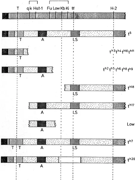
Lyon and Mason obtained further insight into transmission ratio and male sterility by making various combinations, in cis and trans, of the recombinant alleles (Lyon and Mason, 1977). They postu-lated a factor, which they called the A-factor (for abnormal ratio), which lay between the T-interaction and lethal factors, and which was required for an abnormal ratio. When present alone, the A-factor resulted in an abnormally low ratio. Combinations of haplotypes carrying the A-factor with various other haplotypes could lead to an increased ratio. Lyon and Mason also postulated a factor for male sterility carried in haplotypes which also carried the lethal factor (Fig. 1). Lyon later obtained further recombinant haplotypes from t6, including some central haplotypes, having the
putative A-factor only, and also studied other recombinant haplotypes derived from them (Lyon, 1984). Studies of numerous combinations of these haplotypes led to further insights into ratio distortion and male fertility. It was suggested that ratio distortion was produced by the action of distorter genes on a responder. The responder in the central part of the t-complex corresponded to the previous A-factor, and when present alone was transmitted at a low frequency. At least three distorter genes, Tcd-1, -2 and –3, in the proximal, distal and central regions respectively, acted additively, either in cis or in trans, to raise the transmission ratio of the responder. Heterozygosity for the responder was necessary for an abnormal ratio, and if the distorters were present without the t-form of the responder Tcrt, the transmission ratio was normal. When the responder was heterozygous and all three distorters were heterozygously present, in either cis or trans configuration, which-ever chromosome carried the Tcrt was transmitted at high ratio. Further evidence suggested that male sterility was due to homozy-gosity of the distorter genes (Lyon, 1986). Sterility did not require the presence of the Tcrt. Indeed, in the absence of the responder, distorters could impair fertility even when only heterozygous rather than homozygous (Lyon, 1987). The interpretation made was that the distorters acted in concert to produce a harmful effect on sperm carrying the wild-type allele of the responder, and that sperm carrying the t-allele of the responder Tcrt were relatively resistant to this harmful action. When the distorters were homozygous, the harmful effect was so severe that the resistance of the Tcrtwas overcome and sperm carrying it too were harmed. Thus, when the distorters and responder were heterozygous, sperm with Tcrt preferentially took part in fertilization, giving high transmission of Tcrt. When the distorters were homozygous, few or no sperm could fertilise and sterility resulted.
This interpretation fits with the work of others on the reproduc-tive physiology of the effects of the t-complex. Seitz and Bennett (Seitz and Bennett, 1985) and Olds-Clarke and Peitz (Olds-Clarke and Peitz, 1985) showed by different methods that the high transmission ratio was not due to superiority of t-bearing sperm but to a deleterious effect on wild-type meiotic partners of t-sperm. The numbers of t- and wild-type sperm are equal at the time of mating and at deposition of sperm in the uterus (Silver and Olds-Clarke, 1984). Thus, the abnormality lies in the function rather than in the formation of sperm bearing the t-chromosome.
Implicit in Lyon’s explanation of the effects on transmission ratio and sterility was the concept that different partial t-haplotypes had different lengths and positions of t-chromatin. Early work on the t
-Fig. 1. Lyon and Mason’s interpretation of t-complex ratio factors. The interpretation is based on the haplotype t6 and partial haplotypes derived
complex was hampered by lack of sufficient genetic markers to test these matters. This changed with the advent of molecular cloning and the isolation of DNA markers. Apart from those in the MHC region which is located within the t-complex, some of the first available markers of the t-complex region were developed by microdissection of the chromosome (Röhme et al., 1984). Study of RFLVs between t and wild-type for some of these markers con-firmed Lyon’s postulates of the lengths of the various partial haplotypes (Fox et al., 1985).
Physical structure of t-haplotypes
The properties of t-haplotypes were studied for two decades at the Sloan-Kettering Memorial Cancer Hospital in New York, USA, in the groups of Bennett and Artzt, themselves scientific progeny of L.C. Dunn. Silver and Artzt (1981) showed, by use of a t-haplotype with a mutant tf marker (tw12tf), that crossing-over occurs
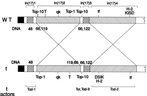
freely in homozygous t-chromatin, indicating chromosome rear-rangements in t-complex chromosomes as compared to wild-type chromosome 17’s of laboratory mice. These US investigators first defined the distal inversion in the MHC complex (Shin et al., 1983), and a UK group identified a second, proximal inversion (Herrmann et al., 1986). Later, Silver and colleagues characterised two other inversions (Hammer et al., 1989); thus four inversions comprise the full extent of the t-complex, two large and two small (early work reviewed in Silver, 1985) (Fig. 2).
Using DNA probes from the two large proximal and distal inversions, our groups showed, by in situ hybridisation, the location of the t-complex with respect to the Giemsa banding pattern of chromosome 17 (Lyon et al., 1986) and visualised the inversions on t-chromosomes marked by translocations (Lyon et al., 1988). Thus, by the end of the 1980’s it was known that the t-complex occupied around 30Mbps of chromosome 17, covering several chromosomal bands that had undergone various independent rearrangement events during its evolution. Furthermore, it had become clear that all the isolates of t-chromosomes, from wild mouse populations all over Europe, Asia and the continental US, and including both subspecies M. m. domesticus and M.m. muscu-lus, had such similar physical structures that they all derived from a single founding population.
Rogers, at the MRC Laboratory of Molecular Biology, Cam-bridge, studied extensively the MHC complex of t-haplotypes using many DNA markers which became available in the early 1980’s (Rogers et al., 1985). This DNA-sequence-based approach also suggested that t-haplotypes were much more similar to one an-other than had been previously surmised from serological analysis of the MHC. This work addresses the important but still unsolved question of whether particular gene families may evolve at different rates depending upon their chromosomal positions.
Relation of properties of t-complex to each other
For many years the nature of the embryonic lethality pheno-types of t-haplopheno-types divided investigators into those who pro-posed that the t-complex encoded a large developmental control region, and others (Silver, 1985) including ourselves, who thought the accumulation of different lethal genes was a by-product of transmission ratio distortion. Briefly, the argument supporting this second view runs as follows: t-haplotypes are maintained in
populations of wild-mice by high transmission from heterozygous males. This requires at least four factors distributed throughout the 30Mbps of the t-complex and thus the inversions confer a selective advantage in holding these factors together, since recombination is suppressed. However, the same factors which give high trans-mission lead to male sterility in homozygotes; therefore, lethals which have arisen at random during t-chromosome evolution are strongly selected for, since prenatal lethality of homozygotes is less deleterious than presence of live sterile males in breeding populations. Now 16 different lethals are known which are distrib-uted throughout the t-complex. Although even to this day none has been molecularly cloned and characterised, it would seem most unlikely that they could share any common functions or regulatory interactions. Moreover, detailed analysis of other mouse chromo-some regions indicates that finding 16 recessive lethal loci within a 30Mbp stretch is well within the bounds of expectation.
Structure of partial t- haplotypes
The availability of many DNA markers, combined with the understanding of the four inversions, enabled the elucidation of the structure of partial t-haplotypes and their mode of origin. The majority are formed by homologous recombination in the small regions between the inversions, particularly between the proximal inversion In(17)2 and the middle inversion In(17)3. However, some are formed by crossing-over within an inversion. Owing to the disparate orientation of the DNA between t and wild-type this results in duplication and/or deletion of small stretches of chroma-tin (reviewed in Silver, 1985; Artzt et al., 1991). Groups in the UK have particularly studied the proximal partial haplotypes of the t9 group (Bucan et al., 1987; Crossley and Little, 1991; Barclay et al., 1996). These haplotypes, comprising t4, t9, tw18 and tks1, have arisen by crossing-over between a complete t-haplotype and wild-type in the distal inversion. All members of the group are recessive prenatal lethals which impede gastrulation and do not complement
each other. The crossovers which gave rise to them occurred near the proximal breakpoint of In(17)4. The DNA up to the start of In(17)4 is derived from the t-complex, the proximal part of In(17)4 is deleted and the distal part is duplicated, there being one t-complex and one wild-type copy. The deleted region is at least 3.3Mbp in length (Barclay et al., 1996) and includes a number of zinc finger sequences (Crossley and Little, 1991). Although the deletion is presumed to be the cause of the prenatal lethality, the exact gene(s) responsible have not been identified. Other haplotypes have been formed by crossing-over within inversion In(17)2. Wild-type chromatin carries an inverted duplication in this region, with one duplicated copy in the same orientation as the t-complex copy (Herrmann et al., 1987). Some partial haplotypes have arisen through crossing-over in this region. Two particularly interesting haplotypes are TtOrl and TtLub2. These have both arisen by cross-ing-over within the proximal inversion, and each has duplicated and deleted sequences which are complementary in the two haplotypes (Sarvetnick et al., 1986).
Spermatogenesis
The enigma of the effects of t-complex genes on spermatoge-nesis proved a great impetus to the discovery and characterisation of genes expressed in the germ cells of the testis (Willison and Ashworth, 1987). Since transmission ratio distortion is most likely to be a consequence of perturbations in the function of genes expressed post-meiotically, laboratories became interested in the control of haploid gene expression. Silver and colleagues had discovered, by 2-D gel electrophoresis, several polypeptide spots expressed in adult testis germ cells which were mapped to the t-complex: TCP-1 to TCP-9. TCP-1 exists as a more acidically
migrating polypeptide species in t-haplotypes, TCP-1A, compared to wild-type chromo-somes, TCP-1B (Silver et al., 1979). In 1979, cDNA cloning techniques were developing to the extent that it seemed feasible to isolate tissue-expressed sequences by probing li-braries with 32P-labelled probes made from
total or size-fractionated mRNA populations and Willison’s laboratory attempted and even-tually succeeded in cloning Tcp-1 cDNA (Willison et al., 1986). A set of several hun-dred testis-expressed genes were initially recovered, some of which were expressed in haploid spermatids (Dudley et al., 1984). A number of these haploid-expressed genes were mapped onto mouse chromosomes. Using probes enriched for Tcp-1 mRNA, we found a cDNA clone, pB1.4, which mapped to the t-complex and was shown to encode TCP-1B. We sequenced and characterised the Tcp-1a and Tcp-1b genes in detail (Willison et al., 1986). Willison’s group at the Institute of Cancer Research (ICR) showed that Tcp-1 mRNA (Haffner, Tcp-1988) and protein was expressed in all spermatids of Thp/+ testes and, since Thp is deleted for Tcp-1, the group postulated that the products of haploid ex-pressed genes are shared between meiotic
Fig.3. Brachyury bound to DNA. Ribbon diagram of the T-domain dimer bound to DNA. Depicted are residues 39-221 of both monomers (strands and loops: red; helices: yellow) and the 24-mer DNA duplex (blue). Labels mark strands (A-G) of the central core of the monomers, which is a seven-stranded β-barrel related to an immunoglobulin fold. View perpendicular to the DNA axis with the dyad vertical. Reprinted from Nature with permission.
partners (Haffner, 1988; Willison et al., 1988). Braun showed, by driving human growth hormone expression in spermatids with the mouse protamine-1 gene promoter, that spermatids shared gene products by diffusion of molecules throughout the syncytium of spermatogenic cells (Braun et al., 1989). These experiments provided molecular proof of the concept of shared meiotic gene expression derived from genetic and physiological experiments with t-haplotypes. Many laboratories characterised t-complex en-coded genes expressed during spermatogenesis; 117c3 (Rappold et al., 1987), the Tcp-10 gene family (Schimenti et al., 1988), the Tctex series (Huw et al., 1995) and Tcp-11 (Hosseini et al., 1994), but their functions remain undetermined. Despite the increased understanding of the genetic basis of t-complex transmission ratio distortion at the phenotypic level, the molecular identity of the genes involved remains unknown and provides a challenge for the future. By detailed study of DNA markers in partial t-haplotypes with and without the t-complex responder Tcrt , the position of the responder has been accurately mapped to an interval of ~40kb (reviewed by Snyder and Silver, 1991). In this region there is a gene termed Tcr10b which is meiotically expressed and is differentially spliced in t-complex DNA (Cebra-Thomas et al., 1991). This gene seemed a strong candidate for the responder. However, both Tcr10b transgenes (Snyder and Silver, 1991) and targeted mu-tagenesis of Tcr10b failed to provide evidence for such a function and the responder remains unidentified (Ewulonu et al., 1996).
thus a possible candidate for Tcd2. However, all that is known of the location of Tcd2 is that it lies in the distal inversion, and since this is relatively large there may be several other candidate genes to be found. A candidate for Tcd1 in the proximal region of the complex showed overexpression of mRNA in t-haplotypes, but a normal amount of protein with some amino-acid changes (O’Neill and Artzt, 1995). Lyon found that a deletion of wild-type chromatin in the Tcd1 region had an effect like that of Tcd1t, suggesting that Tcd1t was an amorph or hypomorph (Lyon, 1992). This makes O’Neill and Artzt’s candidate gene unlikely but does not exclude it, as the protein could be non-functional. On the other hand, a candidate for Tcd3, named Tcte2, found by Braidotti and Barlow (1997) is indeed deleted in t-haplotypes. This makes it a somewhat stronger candidate for Tcd3 than a gene previously found by Rappold et al. (1987). Interestingly, Tcte2 is also a candidate for a hybrid-sterility gene. Three such genes, Hst1, Hst4, and Hst6, map in the t-complex region (Pilder et al., 1991, 1993). Hurst has suggested that the distorted transmission of t-haplotypes has arisen by the suppression of hybrid-sterility barriers (Hurst, 1993). As the t- form of Tcte2 involves a deletion, this could mean that it is deleting a hybrid-sterility gene. Thus, the identification of the t-complex responder and distorter genes remains an outstanding problem which could potentially throw light not only on the mecha-nism of transmission ratio distortion itself, but also on sperm function and on hybrid-sterility.
Evolution of t-complex
Although there is no compelling evidence yet linking TCP-1 or, indeed, any of the TCP proteins (now TCP-1- TCP-11) directly to Tcr or Tcd function, further study on Tcp-1 has illuminated many areas of genetics and biochemistry. Sequencing Tcp-1 genes provided important insight into the evolution of the t-chromosomes. The comparison of RFLVs and the DNA sequences of Tcp-1 from laboratory strains, t-haplotypes and wild-caught mice showed that all the t-haplotypes carried identical Tcp-1a genes, leading Willison (Willison et al., 1986) to suggest that all t-haplotypes had radiated from a common population of mice very recently, possibly only 7000 years’ ago, concomitant with human migration from the fertile crescent. Later, DNA sequencing of introns from the Tcp-1 gene supported this idea that t-chromosomes originated very recently, and introgressed into Mus musculus and Mus domesticus (Morita et al., 1992). Again, characterisation of the human TCP-1 gene and related sequences provided insight into the syntenic relationship between mouse chromosome 17 and human chromosome 6. A long-standing controversy over the existence or otherwise of a human equivalent of the mouse t-complex was settled by the demonstration that human TCP-1 mapped to the long-arm of chromosome 6 very distant from, and furthermore across the centromere from, the human MHC and other linked markers; the 30Mbp t-complex region is fragmented in humans (Willison et al., 1987).
Developmental genetics
The most important contribution of t-complex studies to devel-opmental genetics is indisputably the molecular cloning of the Brachyury gene, T, by Herrmann and colleagues (Herrmann et al., 1990) at the National Institute for Medical Research (NIMR)
laboratory, in London. Brachyury is a DNA binding transcription factor which specifies mesoderm formation not only in mouse, but in all vertebrates. Brachyury is the first member of a gene family, T-box genes, which are master-controlling genes in mesoderm induction and organ rudiment specification. The crystallographic structure of the T domain-DNA complex has revealed a new way in which a protein can recognize DNA (Fig. 3) (Muller and Herrmann, 1997). Reduction in Brachyury function is the cause of the short-tail phenotype in T/t-animals, but it still remains to be understood how T interacts with products of t-haplotype factors to cause absence of tails altogether.
An allele of Brachyury, Thp, was known to carry a second mutation called Tme, ‘T maternal effect’. For many years this mutation was merely a curiosity, but subsequent research has shown it to be an early example of an imprinted genetic locus. Imprinted genes are those whose expression properties are de-pendent upon whether they are maternally or paternally inherited. The fine mapping of the t-complex and knowledge of the syntenic relationship between mouse chromosome 17 and human chromo-some 6 led Barlow et al. (1991) to identify the IgfII receptor (Igf2r) as the imprinted gene lying within the Thp deletion and responsible for the Tme effect (Barlow et al., 1991).
Protein folding
Remarkably, the study of TCP-1 has opened up a new area of research in eukaryotic cell and molecular biology. TCP-1 has turned out to be one of the subunits of the eukaryotic molecular chaperone named the Chaperonin Containing TCP-1, CCT, which is present in the cytosol of all eukaryotes. CCT is active in the folding pathways of newly synthesised actins and tubulins through nucleotide binding and ATP-dependent release of folding interme-diates of these substrates after their synthesis on the ribosome. Willison used monoclonal antibodies raised against TCP-1 (Willison et al., 1989) to purify TCP-1 biochemically initially from testis. Willison’s group at the Institute of Cancer Research (ICR) discov-ered TCP-1 to be one of the subunits of a large heteromeric protein complex (Lewis et al., 1992) which had many biochemical proper-ties in common with the chaperonin of E. coli, GroEL. TCP-1 is also weakly related in sequence to GroEL reflecting, in the main, the conservation of the nucleotide binding sites (Lewis et al., 1992; Kim
Fig.4. Average images of nucleotide-free murine CCT. (A) Side view;
(B) Front view. The side view shows the characteristic 4-stripe appearance of chaperonins, although CCT shows differences to GroEL in mass distri-bution. The front view reveals the 8 subunits of the CCT ring, one of which is TCP-1. Figure adapted from Llorca et al. (1998).
et al., 1994). This group went on to isolate the mouse genes for the 8 subunits of CCT (Kubota et al., 1994; Kubota and Willison, 1997) which are all related in sequence to Tcp-1. These 8 mouse genes have been named Ccta-Cctq and are each orthologous to the corresponding yeast gene, CCT1-CCT8 (Kubota and Willison, 1997). Because CCT is a very large protein complex with a molecular weight of 950 kDa, it can be visualised under the electron microscope (Fig. 4) (Llorca et al., 1998).
It can be seen that CCT is composed of two back-to-back rings having 8 subunits in each ring and Willison’s group has developed a model for the order of arrangement of the subunits within a single ring (Liou et al., 1998). Work is in progress to determine the function of each different CCT subunit in binding and folding of the interme-diates of actin and tubulin. The mechanism of protein folding is one of the outstanding problems in modern biology, and further studies on CCT will illuminate further how proteins fold in eukaryotic cytosol (Willison, 1999).
Summary
The t-complex of the mouse was discovered over 70 years’ ago and British geneticists have been involved in studies on this fascinating region of the mouse genome for 50 years. As a classical genetic system, t-complex studies have illuminated the nature of inversion polymorphism and recombination suppression, the behaviour of non-Mendelian factors and transmission ratio distor-tion, and genetic imprinting. This work helped to establish the t -complex region on chromosome 17 as one of the best mapped and characterised mouse chromosomes, and provided the framework for pioneering positional cloning and genome analysis approaches. Brachyury, T, was the first mouse gene isolated by positional cloning and this was a direct consequence of the genetic and structural analysis of the t-complex. Tcp-1 was discovered and subsequently molecularly cloned by the application of novel 2D-PAGE technology and differential cDNA cloning in conjunction with genetic analysis of partial t-haplotypes. Characterisation of the DNA- binding transcription factor T has been a vital and unique contribution to vertebrate developmental genetics. TCP-1 is a component of the chaperonin containing TCP-1, CCT, an abso-lutely conserved protein folding machine present in the cytosol of all eukaryotes. We look forward to the complete DNA sequencing of the 30MBp t-complex within the next few years. Then perhaps the major outstanding problem of the components and mechanism of male transmission ratio distortion will be elucidated.
KEY WORDS:
t-complex, spermatogenesis, mouse genetics, protein
folding
Note added in proof: Herrmann and colleagues have recently shown that
the t-complex responder is a novel protein kinase probably involved in
regulating sperm motility [(see Herrmann, B.G. et al., (1999); Nature 402:
141-146 and accompanying News and Views article by Willison, K.R.
(1999) Nature 402: 131-132]. This discovery will now allow the study of the
molecular cell biological processes which cause transmission ratio distortion
Acknowledgements
We thank Bernhard Herrmann for helpful comments on the manuscript and Christoph Meuller for providing Figure 3. Keith R. Willison and Mary F. Lyon have been in receipt of long-term funding from the Cancer Research Campaign and the Medical Research Council, respectively.
References
ARTZT, K., BARLOW, D., DOVE, W.F., FISCHER-LINDAHL, K., KLEIN, J., LYON, M.F. and SILVER, L.M. (1991). Maps of chromosome 17; first report. Mammal. Genome 1: 5-29.
BARCLAY, J., KING, T.F., CROSSLEY, P.H., BARNARD, R.C., LARIU, Z., LEHRACH, H. and LITTLE, P.F.R. (1996). Physical analysis of the region deleted in the tw18 allele of the mouse tcR-4 complementation group. Genomics
36: 39-46.
BARLOW, D.P., STOGER, R., HERRMANN, B.G., SAITO, K. and SCHWEIFER, N. (1991). The mouse insulin-like growth factor type-2 receptor is imprinted and closely linked to the Tme locus. Nature349: 84-87.
BRAIDOTTI, G. and BARLOW, D.P. (1997). Identification of a male meiosis-specific gene, Tcte2, which is differentially spliced in species that form sterile hybrids with laboratory mice and deleted in t-chromosomes showing meiotic drive. Dev. Biol. 186: 85-99.
BRAUN, R.E., BEHRINGER, R.R., PESCHON, J.J., BRINSTER, R.L. and PALMITER, R.D. (1989). Genetically Haploid Spermatids are Phenotypically Diploid. Nature337: 373-376.
BUCAN, M., HERRMAN, B.G., FRISCHAUF, A.-M., BAUTCH, V.L., BODE, V., SILVER, L.M., MARTIN, G.R. and LEHRACH, H. (1987). Deletion and duplica-tion of DNA sequences is associated with the embryonic lethal phenotype of the
t9 complementation group of the mouse t- complex. Genes Dev. 1: 376-385.
CARTER, T.C. and PHILLIPS, R.J.S. (1950). Three recurrences of mutants in the house mouse. J. Hered. 41: 252.
CEBRA-THOMAS, J.A., DECKER, C.L., SNYDER, L.C., PILDER, S.H. and SIL-VER, L.M. (1991). Allele- and haploid- specific product generated by alternative splicing from a mouse t complex responder locus candidate. Nature 349: 239-241.
CROSSLEY, P.H. and LITTLE, P.F.R. (1991). A cluster of related zinc finger protein genes is deleted in the mouse embryonic lethal mutation tw18.Proc. Natl. Acad.
Sci. USA 88: 7923-7927.
DOBROVOLSKAIA-ZAVADSKAIA, N. (1927). Sur la mortification spontanée de la queue chez la souris nouveau-née et sur l’existence d’un caractère (facteur) héréditaire "non-viable". C.R. Soc. Biol. (Paris) 97: 114-116.
DUDLEY, K., POTTER, J., LYON, M.F. and WILLISON, K.R. (1984). Analysis of male sterile mutations in the mouse using haploid stage expressed cDNA probes. Nucleic Acids Res. 12: 4281-4293.
DUNN, L.C. and CASPARI, E. (1945). A case of neighboring loci with similar effects.
Genetics 30: 543-568.
DUNN, L.C. and GLUECKSOHN-SCHOENHEIMER, S. (1950). Repeated mutations in one area of a mouse chromosome. Proc. Natl. Acad. Sci. USA 36: 233-237. EWULONU, U.K., SCHIMENTI, K., KUEMERLE, B., MAGNUSON, T. and
SCHIMENTI, J. (1996). Targeted mutagenesis of a candidate t complex re-sponder gene in mouse t haplotypes does not eliminate transmission ratio distortion. Genetics144: 785-792.
FOX, H.S., MARTIN, G.R., LYON, M.F., HERMANN, B., FRISCHAUF, A.-M., LEHRACH, H. and SILVER, L.M. (1985). Molecular probes define different regions of the mouse t-complex. Cell 40: 63-69.
GRÜNEBERG, H. (1943). The Genetics of the Mouse, 1st, ed. Cambridge Univer-sity Press. Pp. xii + 412.
HAFFNER, R. (1988). The expression of the t-complex polypeptide-1 (Tcp-1), in spermatogenesis and embryogenesis. Academic Dept., London, University of London, 238, PhD.
HAMMER, M.F., SCHIMENTI, J. and SILVER, L. (1989). Evolution of Mouse Chromosome 17 and the Origins of Inversions Associated With t-Haplotypes.
Proc. Natl. Acad. Sci. USA 86: 3261-3265.
HERRMANN, B., BUCAN, M., MAINS, P.E., FRISCHAUF, A.-M., SILVER, L.M. and LEHRACH, H. (1986). Genetic analysis of the proximal portion of the mouse t
complex: Evidence for a second inversion with t haplotypes. Cell44: 469-476. HERRMANN, B.G., BARLOW, D.P. and LEHRACH, H. (1987). A large inverted duplication allows homologous recombination between chromosomes het-erozygous for the proximal t complex inversion. Cell48: 813-825.
HERRMANN, B.G., LABEIT, S., POUSTKA, A., KING, T.R. and LEHRACH, H. (1990). Cloning of the T gene required in mesoderm formation in the mouse.
HOSSEINI, R., RUDDY, S., BAINS, S., HYNES, G., MARSH, P., PIZZEY, J. and DUDLEY, K. (1994). The mouse t-complex gene, Tcp-11 is under translational control. Mech. Dev. 47: 73-80.
HURST, L.D. (1993). A model for the mechanism of transmission ratio distortion and for t-associated hybrid sterility. Proc. R. Soc. Lond. (Biol.) 253: 83-91. HUW, L.Y., GOLDSBOROUGH, A.S., WILLISON, K. and ARTZT, K. (1995). Tctex2:
a sperm tail surface protein mapping to the t-complex. Dev. Biol. 170: 183-194. KIM, S., WILLISON, K.R. and HORWICH, A.L. (1994). Cytosolic chaperonin subunits
have a conserved ATPase domain but diverged polypeptide-binding domains.
Trends Biochem. Sci.19: 543-548.
KUBOTA, H. and WILLISON, K.R. (1997). Introduction and 8 sections on CCT for Guidebook to Molecular Chaperones. (Ed. Gething M-J.). Sambrook/Tooze publications at Oxford University Press, Oxford, UK.
KUBOTA, H., HYNES, G., CARNE, A., ASHWORTH, A. and WILLISON, K. (1994). Identification of six Tcp-1-related genes encoding divergent subunits of the TCP-1-containing chaperonin. Curr. Biol. 4: 89-99.
LEWIS, V.A., HYNES, G.M., ZHENG, D., SAIBIL, H. and WILLISON, K. (1992). T-complex polypeptide-1 is a subunit of a heteromeric particle in the eukaryotic cytosol [see comments]. Nature358: 249-52.
LIOU, K.F., MCCORMACK, E.A. and WILLISON, K.R. (1998). The Chaperonin containing TCP-1 (CCT) displays a single-ring mediated disassembly and reas-sembly cycle. Biological Chem. Hoppe-Seyler, 379: 311-319.
LYON, M.F. (1956). Hereditary hair loss in the tufted mutant of the house mouse. J. Hered. 47: 101-103.
LYON, M.F. (1984). Transmission ratio distortion in mouse t-haplotypes is due to multiple distorter genes acting on a responder locus. Cell 37: 621-628. LYON, M.F. (1986). Male sterility of the mouse t-complex is due to homozygosity of
the distorter genes. Cell 44: 357-363.
LYON, M.F. (1987). Distorter genes of the mouse t-complex impair male fertility when heterozygous. Genet. Res. 49: 57-60.
LYON, M.F. (1992). Deletion of mouse t-complex distorter-1 produces an effect like that of the t-form of the distorter. Genet. Res. 59: 27-33.
LYON, M.F. and MASON, I. (1977). Information on the nature of t-haplotypes from the interaction of mutant haplotypes in male fertility and segregation ratio. Genet. Res. Camb. 29: 255-266.
LYON, M.F. and MEREDITH, R. (1964). The nature of t-alleles in the mouse. I, II, III.
Heredity 19: 301-312, 313-325, 327-330.
LYON, M.F. and PHILLIPS, R.J.S. (1959). Crossing-over in mice heterozygous for t -alleles. Heredity 13: 23-32.
LYON, M.F., ZENTHON, J., EVANS, E.P., BURTENSHAW, M.D., DUDLEY, K. and WILLISON, K.R. (1986). Location of the t complex on mouse chromosome 17 by
in situ hybridization with Tcp-1. Immunogenetics 24: 125-127.
LYON, M.F., ZENTHON, J., EVANS, E.P., BURTENSHAW, M.D. and WILLISON, K.R. (1988). Extent of the mouse t complex and its inversions shown by in situ
hybridization. Immunogenetics 27: 375-382.
LLORCA, O., SMYTH, M.G., MARCO, S., CARRASCOSA, J.L., WILLISON, K.R. and VALPUESTA, J.M. (1998). ATP binding induces large conformational changes in the apical and equatorial domains of the eukaryotic chaperonin containing TCP-1 complex. J. Biol. Chem. 273: 1-4.
MAZARAKIS, N.D., NELKI, D., LYON, M.F., RUDDY, S., EVANS, E.P., FREEMONT, P. and DUDLEY, K. (1991). Isolation and characterization of a testis-expressed developmentally regulated gene from the distal inversion of the mouse t-complex.
Development III: 561-571.
MORITA, T., KUBOTA, H., MURATA, K., NOZAKI, M., DELARBRE, C., WILLISON, K., SATTA, Y., SAKAIZUMI, M., TAKAHATA, N., GACHELIN, G. and MATSUSHIRO, A. (1992). Evolution of the mouse t haplotype: recent and worldwide introgression to Mus musculus. Proc. Natl. Acad. Sci. USA89: 6851-6855.
MULLER, C.W. and HERRMANN, B.G. (1997). Crystallographic structure of the T domain-DNA complex of the Brachyury transcription factor. Nature389: 884-888.
O’NEILL, M.J. and ARTZT, K. (1995). Identification of a germ-cell-specific transcrip-tional repressor in the promoter of Tctex-1. Development 121: 561-568. OLDS-CLARKE, P. and PEITZ, B. (1985). Fertility of sperm from t/+ mice: evidence
that +-bearing sperm are dysfunctional. Genet. Res. 47: 49-52.
PILDER, S.H., HAMMER, M.F. and SILVER, L.M. (1991). A novel mouse chromo-some 17 hybrid sterility locus: implications for the origin of t haplotypes. Genetics 129: 237-246.
PILDER, S.H., OLDS-CLARKE, P., PHILLIPS, D.M. and SILVER, L.M. (1993). Hybrid sterility-6; a mouse t complex locus controlling sperm flagellar assembly and movement. Dev. Biol.159: 631-642.
RAPPOLD, G.A., STUBBS, L., LABEIT, S., CRKVENJAKOV, R.B. and LEHRACH, H. (1987). Identification of a testis-specific gene from the mouse t-complex next to a CpG-rich island. EMBO J. 6: 1975-1980.
ROGERS, J.H., LYON, M.F. and WILLISON, K.R. (1985). The arrangement of H-2 class I genes in mouse t haplotypes. J. Immunogenet. 12: 151-165.
RÖHME, D., FOX, H., HERRMANN, B., FRISCHAUF, A.-M., EDSTROM, J.-E., MAINS, P., SILVER, L.M. and LEHRACH, H. (1984). Molecular clones of the mouse t complex derived from microdissected metaphase chromosomes. Cell 36:
783-788.
SARVETNICK, N., FOX, H.S., MANN, E., MAINS, P.E., ELLIOTT, R.W. and SILVER, L.M. (1986). Nonhomologous pairing in mice heterozygous for a t- haplotype can produce recombinant chromosomes with duplications and deletions. Genetics 113: 723-734.
SCHIMENTI, J., CEBRA-THOMAS, J.A., DECKER, C.L., ISLAM, S.D., PILDER, S.H. and SILVER, L.M. (1988). A candidate gene family for the mouse t complex responder (tcr) locus responsible for haploid effects on sperm function. Cell55: 71-78.
SEITZ, A.W. and BENNETT, D. (1985). Transmission ratio distortion of t haplotypes is due to interactions between meiotic partners. Nature 313: 143-144. SHIN, H.-S., FLAHERTY, L., ARTZ, K., BENNETT, D. and RAVETCH, J. (1983).
Inversion in the H-2 complex in t-haplotypes of mice. Nature306: 380-383. SILVER, L.M. (1985). Mouse t haplotypes. Annu. Rev. Genet. 19: 179-208. SILVER, L.M. and ARTZT, K. (1981). Recombination suppression of mouse
t-haplotypes is due to chromatin mismatching. Nature 290: 68-70.
SILVER, L.M. and OLDS-CLARKE, P. (1984). Transmission ratio distortion of mouse t haplotypes is not a consequence of wild-type sperm degeneration. Dev. Biol. 105:
250-252.
SILVER, L.M. ARTZT, K. and BENNETT, D. (1979). A major testicular cell protein specified by a mouse T/t complex gene. Cell17: 275-284.
SNYDER L.C. and SILVER L.M. (1991). The mouse t complex responder locus.
Genome Analysis, vol 3, Genes and Phenotypes. (ed. K.E. Davies), Cold Spring Harbor Press. pp. 37-57.
WILLISON, K.R. (1999).Composition and Function of the Eukaryotic Cytosolic Chaperonin Containing TCP-1., Molecular Chaperones and Folding Catalysts. In
Regulation, Cellular Function and Mechanisms., pp. 555-571, B. Bukau, 1st, ed. Harwood Academic Publishers.
WILLISON, K. and ASHWORTH, A. (1987). Mammalian spermatogenic gene expres-sion. Trends Genet. 3: 351-355.
WILLISON, K.R., DUDLEY, K. and POTTER, J. (1986). Molecular cloning and sequence analysis of a haploid expressed gene encoding t complex polypeptide 1. Cell44: 727-738.
WILLISON, K., KELLY, A., DUDLEY, K., GOODFELLOW, P., SPURR, N., GROVES, V., GORMAN, P., SHEER, D. and TROWSDALE, J. (1987). The human homo-logue of the mouse t-complex gene, TCP1, is located on chromosome 6 but is not near the HLA region. EMBO J. 6: 1967-1974.
WILLISON, K., LEWIS, V., ZUCKERMAN, K.S., CORDELL, J., DEAN, C., MILLER, K., LYON, M.F. and MARSH, M. (1989). The t-complex polypeptide 1 (TCP-1) is associated with the cytoplasmic aspect of Golgi membranes. Cell57: 621-632. WILLISON, K., MARSH, M. and LYON, M.F. (1988). Biochemical evidence for sharing