Original Article
A cystatin-related gene, testatin/cresp, shows male-specific
expression in germ and somatic cells from the initial stage
of murine gonadal sex-differentiation
YASUHIKO KANNO1,2, MASARU TAMURA1,3, SHINICHIRO CHUMA1,3, TAKAYUKI SAKURAI1,3, TAKEO MACHIDA2 and NORIO NAKATSUJI1*
1Mammalian Development Laboratory, National Institute of Genetics, Mishima, Japan, 2Department of Regulation Biology, Faculty of Science, Saitama University, Urawa, Japan, and 3Program for Promotion of Basic Research Activities for Innovative Biosciences (PROBRAIN), Japan
ABSTRACT Sex-differentiation in mammals initiates at mid-gestation when the differentiation of Sertoli cells is triggered by the expression of the testis-determining gene, Sry. However, little is known about the succeeding germ-soma interaction that directs the sex-differentiation of germ cells. We carried out subtraction and differential screening between male and female gonads at 13.5 dpc (days post coitum). A novel cystatin-related gene was identified and named cresp (cystatin-related expressed in Sertoli and spermatogonia), and has recently been reported independently under the name testatin (Töhönen et al., 1998). The presumed amino acid sequence of testatin/cresp showed considerable homology to the cystatin family, but it lacked a few critical amino acid residues for the cysteine-protease inhibitory activity. A 0.7 kb RNA was detected by northern blotting specifically in the fetal and adult testes from 11.5 dpc and expression increased between 11.5 dpc and 12.5 dpc. Using RT-PCR analysis, the testatin/cresp mRNA was first detectable at 9.5 dpc in both male and female embryos but it was maintained only in the male. In females, the expression became weaker at 11.5 dpc and was undetectable after 12.0 dpc. In situ hybridization and immunohistochemical analyses, as well as single cell RT-PCR analysis, showed that the testatin/ cresp mRNA was localized specifically in both the (pro)spermatogonia and Sertoli cells in the testis from 12.5 dpc to adult. Thus, expression of the testatin/cresp gene is upregulated in male gonads but downregulated in females immediately after the initiation of sex-differentiation, suggesting roles in the early developmental cascade of testis such as the germ-soma interaction.
KEY WORDS: testis development, sex differentiation, testatin/cresp, spermatogonia, Sertoli cells
0214-6282/99/$15.00 © UBC Press
Printed in Spain
www.ehu.es/ijdb
*Present and corresponding address for reprints: Department of Development and Differentiation, Institute for Frontier Medical Sciences, Kyoto University, Kyoto 606-8507, Japan. FAX: 81-75-751-3808. e-mail: [email protected]
(Address until 31 Dec.,1999: Mammalian Development Laboratory, National Institute of Genetics, 1111 Yata, Mishima, 411-8540 Japan. FAX: 81-559-81-6828. e-mail: [email protected])
Introduction
Mammalian sex differentiation depends on the presence or absence of the Y chromosome. The testis determining gene on the Y chromosome, Sry, initiates the embryonic gonads to differentiate into the testis (Koopman et al., 1991). Precisely controlled expres-sion of Sry at 10.5-11.5 dpc (days post coitum) in somatic cells of the male indifferent gonad triggers their differentiation into Sertoli cell precursors (Palmer and Burgoyne, 1991). These cells then commence secretion of Müllerian inhibiting substance (MIS), which induces regression of the Müllerian duct (Josso et al., 1998; Lane and Donahoe, 1998). Also, interstitial cells in the testis differentiate
into Leydig cells, which produce testosterone and promote Wolf-fian duct development (Parker et al., 1999). Without the proper expression of Sry, gonadal somatic cells differentiate as in the female, that is, into follicle cells and theca cells (Berta et al., 1990; Jager et al., 1990).
finally into the posterior body wall and genital ridge at 11.5 dpc (Wylie et al., 1985). Germ cells continue to proliferate mitotically after arrival at the genital ridge until 12.5 or 13.5 dpc. At 12.5 dpc, genital ridges show the first signs of sexual dimorphism with testicular cord formation in the males. Then, PGCs in the testis are mitotically arrested by 15.5 dpc to become prospermatogonia, while those in the ovary enter into meiosis from 13.5 to 15.5 dpc to become oocytes (McLaren, 1994). Previous studies have indi-cated that such sex-differentiation of germ cells is directed by the sex of the gonadal somatic cells and not by that of germ cells themselves (McLaren, 1995).
During the early development of fetal testes, it has been re-ported that several transcription factors, such as Sry, Sox9, WT1 and Ad4BP/SF-1, play important roles (reviewed in Swain and Lovell-Badge, 1999). However, little is known about genes acting
in the developmental cascade under the control of these transcrip-tional regulators. For example, there is almost no information about the molecules involved in the somatic-germ cell interaction that directs the sex-differentiation of germ cells.
We therefore attempted to isolate such genes involved in the gonadal development and differentiation by using subtraction and differential screening between male and female fetal gonads at 13.5 dpc (Tamura et al., manuscript in preparation). A novel gene found in our screen showed specific expression in the testis from the initial stages of sex-differentiation. It was related to the cystatin family and had the highest similarity with the CRES (cystatin-related epididymal specific) gene (Cornwall et al., 1992). After we named it cresp and sent the sequence data to the DNA data bank (DDBJ data base, accession no. AB017157), a paper reporting the same gene, named testatin, was published (Töhönen et al., 1998).
B
A
Our present study reports both confirming and new results regarding the expression of testatin/cresp, including data that appear to contradict with the published report.
Results
Identification of a novel gene by subtractive cloning
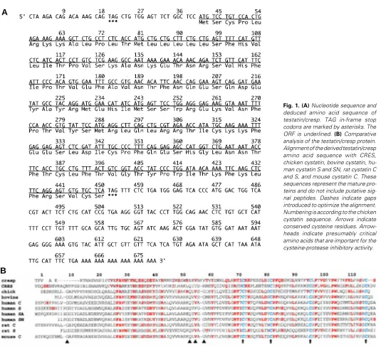
To identify novel genes that have sex-specific expres-sion patterns during gonad development, an 11.5-13.5 dpc mouse gonadal cDNA library was screened with male-specific subtracted probes. From such screening, a cDNA containing a 678bp insert was isolated and sequenced in both directions. Figure 1A shows the nucleotide sequence and deduced amino acid sequence of testatin/cresp. An open reading frame was identified and it had an upstream in-frame TAG stop codon at nucleotides 19-21, an ATG initiator at 40-42, and a TAG stop codon at 451-453.
Nucleotide and protein databases were searched with these nucleotide and amino acid sequences. Although overall sequence homology at the nucleotide level was not found, minimal homology with members of the cystatin family was recognized in several regions of the amino acid sequence. The highest similarity was found with CRES, and we named this novel gene cresp (cystatin-related expressed in Sertoli and spermatogonia). Figure 1B shows the alignment of the derived amino acid sequences of testatin/cresp when compared with sequences of chicken, human, bovine, rat, and mouse cystatin family genes. There were a number of discrete regions conserved be-tween testatin/cresp and cystatins, such as the cysteine residues in the carboxyl-terminal portion (indicated by
number. Analysis of these staged samples revealed that the expression level was maintained until the 20-somite stage and decreased to an undetectable level in female gonads by the 24-somite stage, which corresponds to 12.0 dpc (Fig. 3B; Table 1).
Expression analysis using in situ hybridization
Cellular localization of the testatin/cresp mRNA was examined further by in situ hybridization. Paraffin sections of fetal and postnatal testes were used for the ordinary in situ hybridization or double-stained with digoxigenin-labeled antisense testatin/cresp RNA and anti-SSEA-1 monoclonal antibody or TRA98 monoclonal antibody.
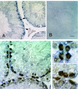
In adult testes, significant labeling appeared to localize to both Sertoli cells and spermatogonia (Fig. 4A), although Töhönen et al. (1998) implied that testatin mRNA was expressed only in Sertoli cells. Double-staining with the germ cell-specific TRA98 antibody (Fig. 4C) indicated that not all the spermatogonia were labeled, and that all the labeled spermatogonia were located at the most peripheral region, close to the basal lamina of the seminiferous tubules. We recognized many spermatogonia that showed distinct labeling of both the testatin/cresp mRNA and TRA 98 antibody (Fig. 4D). We also detected the testatin/cresp-negative spermatogonia (Fig. 4E). Approximately one fourth of the spermatogonia labeled with the TRA98 antibody were double-labeled cells. Negative controls using the sense probe showed no significant labeling (Fig. 4B).
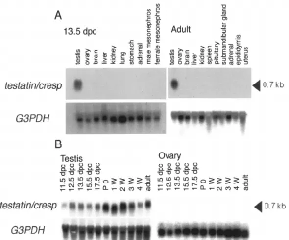
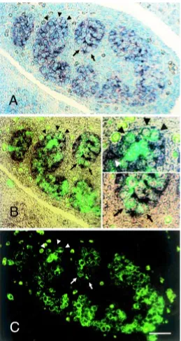
We also labeled fetal gonads at 12.5 dpc. The hybridization signals were observed specifically within the developing testis cord (Fig. 5A). Double labeling with the germ cell-specific anti-SSEA-1 Fig. 2. Northern blotting analysis of testatin/cresp.(A) Tissue-specific expression pattern of testatin/cresp. Total RNA isolated form a variety of male and female tissues of 13.5 dpc fetuses and adults were loaded. A 0.7 kb RNA was detected only in the testis. (B) Developmental time course of testatin/cresp mRNA expression. Total RNA isolated from the prenatal and postnatal gonads were loaded. The mRNA expression level increased from 11.5 to 12.5 dpc in the male, while no hybridization signal was detected in female samples. The blots were then stripped and rehybridized with the G3PDH probe as a loading control.
arrows in Fig. 1B), which were conserved among the cystatin family. Since all the cystatins are secreted proteins, they all possess a hydrophobic signal peptide at the amino-terminal end, which was also present in testatin/cresp. However, the presumed protein product of testatin/cresp lacks a few critical amino acids that are thought to be important for the cysteine-protease inhibitory activity. These sites include G at 9, Q at 53, V at 55 and G at 57 as indicated by arrowheads in Figure 1B (Bode et al., 1988).
Testatin/crespmRNA expression
To determine the expression pattern of testatin/cresp mRNA among various tissues, Northern blotting was performed using RNAs from a wide variety of fetal and adult tissues. A 0.7 kb transcript was detected specifically in fetal and adult testes (Fig. 2A). To examine expression during gonadal development, RNAs were isolated from fetal and postnatal gonads at various ages (Fig. 2B). Low-level expression of testatin/cresp expression was ob-served in the male urogenital ridge at 11.5 dpc, whereas a higher level of expression was observed in the testis from 12.5 dpc to adult. Expression was not detected in female gonads.
antibody showed that the testatin/cresp mRNA was expressed in somatic cells and also in approximately 20-25% of germ cells (Fig. 5A-C). Male gonads at 11.5 dpc also showed similar expression signals on germ and somatic cells (data not shown). There was no significant labeling for the testatin/cresp mRNA in female gonads at any stages (data not shown).
Figure 6 shows the expression pattern of cystatin C, a member of the cystatin family, in the embryo and adult testes. Its mRNA expression pattern was clearly different from that of the testatin/cresp mRNA. Cystatin C was expressed not only in the testis cord but also in the mesonephric region at 12.5 dpc (Fig. 6A). It was expressed in most cells inside the seminiferous tubules, including the Sertoli cells and spermatocytes, in adult testis (Fig. 6B). Interestingly, the labeling was stronger on the inner layer containing spermatocytes and much weaker on the peripheral spermatogonia, exhibiting an almost complementary pattern to the testatin/cresp.
Expression analysis using single-cell RT-PCR
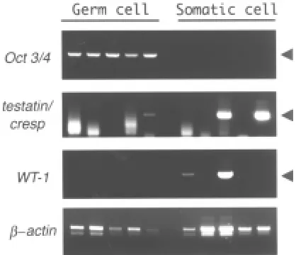
Single-cell RT-PCR analysis was carried out to confirm the double-labeling results indicating testatin/cresp mRNA expres-sion in both the somatic and germ cells in male gonads. We analyzed mRNA obtained from individual germ or somatic cells that had been picked-up from dissociated 12.5 dpc male gonads after vital staining with the anti-SSEA-1 and anti-alkaline
phos-phatase antibodies (Fig. 7). We detected the testatin/cresp ampli-fication product in some of the germ cells that also expressed the germ cell-specific Oct3/4 mRNA. We performed such RT-PCR experiment three times using 10~20 embryos from two batches and obtained similar results. In total, 3 out of 16 germ cells gave a detectable signal representing testatin/cresp mRNA expres-sion, while 6 out of 16 somatic cells were positive for testatin/ cresp. Such ratio of the testatin/cresp positive germ cells was comparable to that in the in situ hybridization. The observed expression pattern of WT1 in somatic cells was caused by the fact that these somatic cells included both the Sertoli and interstitial Fig. 4. In situ hybridization analysis of the adult testis.(A) In situ hybridization revealed labeling in the round cells in the extreme periphery of the seminiferous tubules and radially elongated Sertoli cells (arrow-heads). (B) Control labeling with the sense probe. (C) Double-staining with digoxigenin-labeled anti-sense testatin/cresp RNA and TRA98 monoclonal antibody. Arrows indicate spermatogonia labeled with TRA98 but not with the hybridization signals. (D,E) Magnified views of the double-labeled spermatogonia in the upper-left part of (C) and spermatogonia indicated by arrows in (C). Bar, 50 µm.
TABLE 1
RT-PCR ANALYSIS OF TESTATIN/CRESP EXPRESSION IN FEMALE GONADS DURING THE EARLY SEX-DIFFERENTIATION PERIOD
number of tail somites 17 18 19 20 21 22 23 24 25
testatin/cresp
positive/total sample 3/3 4/4 4/4 4/4 5/6 3/5 1/5 0/5 0/5
Number of testatin/cresp positive individuals per total samples are indicated. Em-bryos with 18 somites and 24 somites correspond to 11.5 dpc and 12.0 dpc, respectively.
cells. Previous studies (Mundlos et al., 1993) reported that WT1 was expressed in the Sertoli and peripheral cells but not in the germ cells of the embryonic testis. Also, there was no evidence that all the Sertoli cells expressed the WT1 gene.
Discussion
The testatin/cresp gene described in this report showed homol-ogy with type II cysteine proteinase inhibitors (cystatins). Four highly conserved cysteine residues showed exact alignment with other member of the cystatin family. These four cysteines govern the overall conformation of the cystatins through formation of two disulfide bonds (Grubb et al., 1984). In addition, testatin/cresp has a putative signal peptide and a predicted cleavage site at the same relative position as that of the cystatins. These similarities suggest that the testatin/cresp protein has a conformational resemblance with the cystatins and that it is a secreted protein. Unlike the cystatins, however, testatin/cresp does not contain highly con-served amino acid residues that are presumed to be necessary for the cysteine-protease inhibitory activity. This fact suggests a relatively remote relationship with the known cystatins, and it may represent a member of a new family of cystatins. The testatin/cresp protein therefore, may either inhibit still unknown cysteine pro-teases or bind to proteins that are not cysteine proteinases.
Cysteine proteases, members of the papain superfamily, com-prise a group of proteolytic enzymes that cleave peptide bonds by the use of a catalytic cysteine residue and they are inhibited by the cystatins (Barrett et al., 1986). Such cysteine proteases are probably involved in the protein catabolism (Kominami et al., 1986) and the proteolytic processing of prohormones and proenzymes (Taugner et al., 1985; Marks et al., 1986). These proteases are present in most living organisms and are thought to have medical importance (Barrett et al., 1986). On the other hand, the cystatins are present in most tissues as well as biological fluids such as saliva, tears, urine, plasma, cerebrospinal fluid and seminal plasma (Abrahamson et al., 1986; Tavera et al., 1990). In the testis, the cysteine protease cathepsin L (Elicson-Lawrence et al., 1991) and its inhibitor cystatin C (Tavera et al., 1990) are secreted from Sertoli cells. It has been suggested that cathepsin L and cystatin C have interactive roles in the adherence of germ cells to Sertoli cells and subsequent formation of the intercellular junctions (Murk et al., 1997).
In addition to its unusual structure among the cystatins, the expression pattern of testatin/cresp was different from any other members of the cystatin family. Northern blotting and RT-PCR analyses demonstrated that expression of the testatin/cresp mRNA was almost confined exclusively to the male gonad and expression increased immediately after the initiation of testis differentiation at 11.5-12.5 dpc, while it decreased to an undetectable level in the female gonad during the same period. Our detailed in situ hybridiza-tion and immunohistochemical analyses demonstrated that the testatin/cresp mRNA was localized both in the germ and Sertoli cells in the fetal and adult testes. Such highly restricted expression is in marked contrast to the wider expression of cystatin C and other members of the cystatin family in the testis and other tissues as observed in the present and previous studies (Cole et al., 1989).
Töhönen et al. (1998) reported expression of testatin/cresp in Sertoli cells and its precursors from 11.5 dpc, and they suggested that this gene is located downstream of Sry or Sox9 during early events of the sex-differentiation in testis somatic cells. Our present results indicated expression of the testatin/cresp in both the germ
and somatic cells in the male gonad from 11.5 dpc, and also in embryos of both sexes at earlier developmental stages. At 9.5 dpc, the urogenital ridge is being formed from the intermediate meso-derm, while the PGCs are migrating through the hindgut region. Fig. 5. In situ hybridization analysis of the fetal testis. A section of a male gonad at 12.5 dpc, double-stained with digoxigenin-labeled antisense testatin/cresp RNA and anti-SSEA-1 monoclonal antibody and FITC-labeled second antibody, counter stained with methylgreen. Photographs of (A)
Fig. 6. In situ hybridization of the cystatin C mRNA. (A) In the male embryos at 12.5 dpc, cystatin C mRNA signals were present within the testis cord in the gonad (upper-left part) and the attached mesonephros. (B) Labeling of the adult testis revealed hybridization signals on most cells inside the seminiferous tubules including the spermatocyte layer (arrowheads) but excluding the spermatogonia (arrows) located at the extreme periphery. (C) A negative control hybridized with the sense probe. Scale bars indicate 50 µm.
Although testatin/cresp expression was first detected by RT-PCR at 9.5 dpc, it was not clear which cells expressed this gene. In spite of many trials, we could not detect the expression by in situ hybridization at these earlier stages, perhaps because of a lower expression level.
Expression of the testatin/cresp mRNA was not detected in all the germ cells at 12.5 dpc when analyzed by the single cell RT-PCR method. Also, not all the spermatogonia were labeled by in situ hybridization. It is not clear whether such results represent varia-tion in the expression level among cells or presence of heteroge-neous sub-populations. More detailed analyses are necessary to clarify these matters.
Expression of Sox-9 starts at around 10.0 dpc in both the male and female, and it is upregulated in the testis and downregulated in the ovary at 11.5 dpc (Morais da Silva et al., 1996), thus taking a very similar course to testatin/cresp expression. Also, expression
of Ad4BP/SF-1, a transcription regulator important for steroidgenic cell development (Ingraham et al., 1994), starts at 9.5 dpc in somatic cells of the gonadal rudiment (Ikeda et al., 1994; Hatano et al., 1996), and it is sex-dependent during gonad development (Hatano et al., 1994). Thus, these two genes may be involved in the transcriptional regulation of testatin/cresp.
It has been known that PGCs arriving at the urogeni-tal ridge undergo a major change in cellular phenotype (Donovan et al., 1986; Ffrench-constant et al., 1991). More recently, it was shown that expression of the mouse vasa homolog starts after they arrive at the genital ridges (Fujiwara et al., 1994). Germ cells at these stages take different developmental pathways depending on the sex of the gonadal somatic cells, more precisely, Sertoli precursor cells (McLaren, 1995). PGCs in the female gonad enter into meiosis, while those in the male gonad go into mitotic arrest. Thus, interactions of the PGCs and gonadal somatic cells are very important for sex-differentiation of germ cells. The upregulation of testatin/cresp expression in the male gonad and its downregulation in the female takes place immediately before such sex-differentiation of the germ cells, suggesting an important function at these stages. Also, since testatin/cresp is expressed in both the germ and somatic cells at the same critical period of early testis differentiation, it may be involved in the interaction between the germ and somatic cells. For example, it is possible that the testatin/cresp protein may be involved in the processing of signaling molecules in collaboration with a cysteine protease.
Materials and Methods
Animals and embryos
ICR strain mice were purchased from Japan SLC, Inc. Embryos were obtained from mating of the mice maintained in a room with light/dark periods between 8 am and 8 pm. Noon of the day of appearance of the vaginal plug was designated as 0.5 dpc. For more accurate staging of em-bryos from 11.5 to 12.5 dpc, the number of tail somites posterior to the hind limb bud was counted as described by Hacker et al. (1995). The genetic sex of phenotypically sex-indifferent embryos was determined by genomic PCR of the Ube-1 gene on the X and Y chromosomes (Chuma and Nakatsuji, submitted for publication). The protocol was designed so that samples from male embryos gave two bands from the X and Y chromosomes and those of females gave a single band from the X chromosome. We isolated genital ridges or gonads at 11.5 dpc or later stages, urogenital ridges at 10.5 dpc, posterior trunk regions between the forelimb bud and tail at 9.5 dpc, or the posterior embryo proper at 8.5 dpc, respectively.
Library screening
DNA sequencing
The nucleotide sequences of clones were determined by using the dye-terminator cycle sequencing kit (Perkin-Elmer), and nucleotide data was analyzed with the Blast program (Altschul et al., 1990).
Northern blotting analysis
Total RNAs were extracted from mouse tissues by ISOGEN following the manufacturers protocol (Nippon Gene). Ten µg of total RNA was fractionated on 1.3% formaldehyde-agarose gels, and transferred to Hybond-N+ (Amersham). After prehybridization, membranes were
hy-bridized at 65° in Church buffer with about 105 cpm 32 P-labeled probes.
After hybridization, membranes were washed in 2x SSC – 0.1% SDS two times for 10 min at RT, 0.5x SSC – 0.1% SDS for 15 min at 65°, and 0.1xSSC – 0.1% SDS for 15 min at 65°. 32P-labeled probes were prepared
using Strip-EZ random priming system (Ambion). The full length testatin/ cresp insert and G3PDH PCR product were used as templates for probes.
RT-PCR
cDNAs were synthesized from total RNAs with oligo (dT)12-16 and Superscript II RNase H- reverse transcriptase following the manufacturers
protocol (Gibco BRL). One µg of total RNAs was used for the reverse transcription reaction in a 20 µl of reverse transcription mixture. One µl of the cDNAs were used as templates for 35 cycles of PCR (94°, 60 sec → (95°, 60 sec; 60°, 60 sec; 72°, 60 sec) → 72°, 60 sec) for testatin/cresp, and 25 cycles of (94°, 60 sec → (95°, 60 sec; 60°, 60 sec; 72°, 90 sec) → 72°, 60 sec) for G3PDH. The following primers were used for PCR:
testatin/cresp-F, 5’-CACAGTGGAATTTGCCGTGA-3’; testatin/cresp-R, 5’-AAGGTAACCCCTTCACGGGAT-3’; G3PDH-F, 5’-TGAAGGTCGGTGTGAACGGATTTGGC-3’; G3PDH-R, 5’-CATGTAGGCCATGAGGTCCACCAC-3’.
In situ hybridization and immunohistochemistry
Mouse embryos were fixed with 10% formaldehyde in PBS. Five µm paraffin sections were prepared, and mounted on silane-coated glass slides (Matsunami). In situ hybridization was performed as described by Saito et al. (1996). Then, immunohistochemical staining was carried out by using an anti-SSEA-1 monoclonal antibody (American Research Products) or a TRA98 monoclonal antibody (gift from Dr. Y. Nishimune,
Osaka Univ.). The TRA98 antibody labels spermatogonia and primary spermatocytes, although staining of the latter appear less intense than the former, but not any somatic cells in the testis (Nishimune et al., personal communication). FITC-labeled secondary antibody was used for the SSEA-1 staining. Biotinylated secondary antibody and Vectastain ABC Elite kit (Vector laboratories) were used for the TRA98 staining. If necessary, the specimens were counter-stained with methylgreen.
Single cell RT-PCR
Male gonads at 12.5 dpc were dissociated by digestion with a 0.05% trypsin and filtration. Dissociated cells were incubated at 4°C for 30 min in the culture medium containing anti-SSEA-1 and anti-calf intestinal alkaline phosphatase antibodies (Sigma). Then, such cells were washed with PBS-two times, and suspended in the medium with fluorescein-conjugated 2nd antibody. Cell suspensions were observed with a fluorescence micro-scope. The double-labeled germ cells, or the double negative and adherent somatic cells, were picked-up by using a glass micropipette and a microma-nipulator (Eppendorf). mRNAs from each cell were separately purified using the Dynabeads mRNA DIRECT kit (Dynal), followed by solid phase-reverse transcription using Superscript II RNase H- reverse transcriptase.
Since the cDNAs were attached to particles, the same templates were used for several times of PCR amplification serially. Expression of Testatin/ cresp, β-actin (Alonso et al., 1986), Oct 3/4 and WT1 was analyzed using the following PCR primers.
We thank Prof. Y. Nishimune for the TRA98 antibody, and Dr. T. Saito and Dr. T. Tada for valuable discussion. This work was supported by a grant from Program for Promotion of Basic Research Activities for Innovative Biosciences (PROBRAIN), by the Special Coordination Funds for Promoting Science and Technology from the Ministry of Science and Technology, and by Grant-in-Aids from the Ministry of Education, Sci-ence, Sports and Culture.
References
ABRAHAMSON, M., SALVESEN, G., BARRETT, A.J. and GRUBB, A. (1986). Isolation of six cysteine proteinase inhibitors from human urine. J. Biol. Chem. 261: 11282-11289.
ALONSO, S., MINTY, A., BOURLET, Y. and BUCKINGHAM, M. (1986). Comparison of three actin-coding sequences in the mouse; evolutionary relationships between the actin genes of warm-blooded vertebrates. J. Mol. Evol. 23: 11-22. ALTSCHUL, S.F., GISH, W., MILLER, W., MYERS, E.W. and LIPMAN, D.J. (1990).
Basic local alignment search tool. J. Mol. Biol. 215: 403-410.
BARRETT, A.J., FRITZ, H., GRUBB, A., ISEMURA, S., JARVINEN, M., KATUNUMA, N., MACHLEIDT, W., MULLER-ESTERI, W., SASKI, M. and TURK, V. (1986). Nomenclature and classification of the proteins homologous with the cysteine proteinase inhibitor cystatin. Biochem. J. 236: 312-317.
BERTA, P., HAWKINS, J.R., SINCLAIR, A.H., TAYLOR, B.L. and GRIFFITHS, P.N. (1990). Genetic evidence equating SRY and the testis-determing factor. Nature 348: 448-450.
BODE, W., ENGH, R., MUSIL, D., THIELE, U., HUBER, R., KARSHIKOV, A., BRZIN, J., KOS, J. and TURK, V. (1988). The 2.0 A x-ray crystal structure of egg white cystatin and its possible mode of interaction with cysteine proteases. EMBO J. 7: 2593-2599.
COLE, T., DIKSON, P.W., ESNARD, F., AVERILL, S., RISBRIDGER, G.B., GAUTHIER, F. and SCHREIBER, G. (1989). The cDNA structure and expression analysis of the genes for the cysteine proteinase inhibitor cystatin C and for β2-macroglobulin in rat brain. Eur. J. Biochem. 186: 35-42.
CORNWALL, G.A., ORGEBIN-CRIST, M-C. and HANN, S.R. (1992). The CRES gene: a unique testis-regulated gene related to cystatin family is highly restricted
in its expression to the proximal region of the mouse epididymis. Mol. Endocrinol. 6: 1653-1664.
MORAIS DA SILVA, S., HACKER, A., HARLEY, V., GOODFELLOW, P., SWAIN, A. and LOVELL-BADGE, R. (1996). Sox9 expression during gonadal develop-ment implies a conserved role for the gene in testis differentiation in mammals and birds. Nature Genet. 14: 62-68.
DONOVAN, P.J., SCOTT, D., CAIRNS, L.A., HEASMAN, J. and WYLIE, C.C. (1986). Migratory and postmigratory mouse primordial germ cells behave differently in culture. Cell 44: 831-838.
ELICSON-LAWRENCE, M., ZABLUDOFF, S. and WRIGHT, W.W. (1991). Cyclic protein 2, a secretory product of rat Sertoli cells, is the proenzyme form of cathepsin L. Mol. Endocrinol. 5: 1789-1798.
FFRENCH-CONSTANT, C., HOLLINGSWORTH, A., HEASMAN, J. and WYLIE, C.C. (1991). Response to fibronectin on mouse primordial germ cells before, during and after migration. Development 113: 1365-1373.
FUJIWARA, Y., KOMIYAMA, T., KAWABATA, H., SATO, M., FUJIMOTO, H., FURUSAWA, M. and NOCE, T. (1994). Isolation of a DEAD-family protein gene that encodes a murine homolog of Drosophila vasa and its specific expression in germ cell lineage. Proc. Natl. Acad. Sci. USA 91: 12258-12262.
GINSBURG, M., SNOW, M.H.L. and McLAREN, A. (1990). Primordial germ cells in the mouse embryo during gastrulation. Development 110: 521-528. GRUBB, A., JESSON, O., GUDMUNDSSON, G., ARNASON, A., LOFBERG, H.
and MALM, J. (1984). Abnormal metabolism of τ-trace alkaline microprotein: the basic defect in hereditary cerebral hemorrhage with amyloidosis. New Engl. J. Med. 311: 1547-1549.
HACKER, A., CAPEL, B., GOODFELLOW, P.N. and LOVELL-BADGE, R. (1995). Expression of Sry, the mouse sex determing gene. Development 121: 1603-1614.
HATANO, O., TAKAKUSU, A., NOMURA, M. and MOROHASHI, K. (1996). Identifica-tion origin of adrenal cortex and gonad revealed by expression profiles of Ad4BP/SF-1. Genes Cells 1: 663-671.
HATANO, O., TAKAYAMA, A., IMAI, T., WATERMAN, M.R., TAKAKUSU, A., OMURA, T. and MOROHASHI, K. (1994). Sex-dependent expression of a transcription factor, Ad4BP, regulating steroidogenic P-450 genes in the go-nads during prenatal and postnatal rat development. Development 120: 2787-2797.
IKEDA, Y.W., SHEN, H.A., INGRAHAM, H.A. and PARKER, K.L. (1994). Develop-mental expression of mouse steroidgenic factor 1 an essential regulator of the steroid hydroxylase. Mol. Endocrinol. 8: 654-662.
INGRAHAM, H.A., LALA, D.S., IKEDA, Y., LUO, X., SHEN, W.H., NACHTIGAL, M.W., ABBUD, R., NILSON, J.H. and PARKER, K.L. (1994). The nuclear receptor steroidogenic factor 1 acts at multiple levels of reproducrive axis. Genes Dev. 8: 2302-2312.
JAGER, R.J., ANVRET, M., HALL, K. and SCHERER, G. (1990). A human XY female with a frame shift mutation in the candidate testis-determining gene SRY. Nature 348: 452-454.
JOSSO, N., RACINE, C., DI CLEMENTE, N., REY, R. and XAVIER, F. (1998). The role of anti-mullerian hormone in gonadal development. Mol. Cell. Endocrionol. 145: 3-7.
KANEKO-ISHINO, T., KUROIWA, Y., MIYOSHI, N., KOHDO, T., SUZUKI, R., YOKOYAMA, M., VIVILLE, S., BARTON, S.C., ISHINO, F. and SURANI, M.A.
(1995). Peg1/Mest imprinted gene on chromosome 6 identified by cDNA subtraction hybridization. Nature Genet. 11: 52-59.
KOMINAMI, E., OHSHITA, T. and KATUNUMA, N. (1986). Functional shares of cathepsins B, H, and L in autophagy and heterophagy. In Cysteine proteinases and their inhibitors. (Ed. Turk V.). Walker de Gruyter, Berlin, pp. 229-238. KOOPMAN, P., GUBBY, J., VIVIAN, N., GOODFELLOW, P. and LOVELL-BADGE,
R. (1991). Male development of chromosomally female mice transgenic for Sry. Nature 351: 117-121.
LANE, A.H. and DONAHOE, P.K. (1998). New insight into mullerian inhibiting substance and its mechanism of action. J. Endocrinol. 158: 1-6.
MARKS, N., BERG, M.J. and BENUCK, M. (1986). Preferential action of rat brain cathepsin B as peptidyl dipeptidase converting pro-opioid oligopeptides. Arch. Biochem. Biophys. 249: 489-499.
McLAREN, A. (1994). Germ line and soma: Interactions during early mouse development. Semin. Dev. Biol. 5: 43-49.
McLAREN, A. (1995). Germ cells and germ cell sex. Philos. Trans. R. Soc. Lond. [Biol.] 350: 229-233.
MUNDLOS, S., PELLETIER, J., DARVEAU, A., BACHMANN, M., WINTERPACHT, A. and ZABEL, B. (1993). Nuclear localization of the protein encoded by the Wilms’ tumor gene WT1 in embryonic and adult tissues. Development 119: 1329-1341.
MURK, D., ZHU, L.J., SILVERSTRINI, B., LEE, W.M. and CHENG, C.Y. (1997). Interactions of proteases and protease inhibitors in Sertoli-germ cell cocultures preceding the formation of specialized Sertoli-germ cell junctions in vitro. J. Androl. 18: 612-622.
PALMER, S. and BURGOYNE, P.S. (1991). In situ analysis of fetal, prepubertal and adult XX-XY chimaeric mouse testis: Sertoli cells are predominantly, but not exclusively, XY. Development 112: 265-268.
PARKER, K.L., SCHEDL, A. and SCHIMMER, B.P. (1999). Gene interactions in gonadal development. Annu. Rev. Physiol. 61: 417-433.
SAITO, T., LO, L., ANDERSON, D.J. and MIKOSHIBA, K. (1996). Identification of novel paired homeodomain protein related to C. elegans unc-4 as a potential downstream target of MASH1. Dev. Biol. 180: 143-155.
SWAIN, A. and LOVELL-BADGE, R. (1999). Mammalian sex deremination: a molecular drama. Genes Dev. 13: 755-767.
TAUGNER, R., BUHRLE, C.P., NOBILING, R. and KIRSCHKE, H. (1985). Coexist-ence of renin and cathepsin B in epitheloid cell secretory granules. Histochem-istry 83: 103-108.
TAVERA, C., PROVOT, D., GIROLAMI, J.P., LEUNG-TACK, J. and COLLE, A. (1990). Tissue and biological fluid distribution of cysteinee proteinase inhibitor: rat cystatin C. Biol. Chem. H-S. 371: 187-192.
TÖHÖNEN, V., ÖSTERLUND, C. and NORDQVIST, K. (1998). Testatin: a cystatin-related gene expressed during early testis development. Proc. Natl. Acad. Sci. USA 95: 14208-14213.
WYLIE, C.C., STOTT, D. and DONOVAN, P.J. (1985). Primordial germ cell Migration. In Developmental Biology (Ed. L.W. Browder), Vol.2, Plenum, New York. pp. 433-448.