The role of stem cell factor and of alternative c-kit gene
products in the establishment, maintenance and
function of germ cells
CLAUDIO SETTE, SUSANNA DOLCI, RAFFAELE GEREMIA and PELLEGRINO ROSSI*
Dipartimento di Sanita’ Pubblica e Biologia Cellulare, Sezione di Anatomia, Universita’ di Roma “Tor Vergata”, Rome, Italy.
ABSTRACT The c-kit gene plays a fundamental role during the establishment, the maintenance and the function of germ cells. In the embryonal gonad the c-kit tyrosine kinase receptor and its ligand Stem Cell Factor (SCF) are required for the survival and proliferation of primordial germ cells. In the postnatal animal, c-kit/SCF are required for the production of the mature gametes in response to gonadotropic hormones, i.e. for the survival and/or proliferation of the only proliferating germ cells of the testis, the spermatogonia, and for the growth and maturation of the oocytes. Finally, a truncated c-kit product, tr-kit, specifically expressed in post-meiotic stages of spermatogenesis and present in mature spermatozoa, causes parthenogenetic activation when microinjected into mouse eggs, suggesting that it might play a role in the final function of the gametes, fertilization.
KEY WORDS:
c-kit, stem cell factor, spermatogenesis, oogenesis, fertilization.
0214-6282/2000/$20.00 © UBC Press
Printed in Spain
www.ehu.es/ijdb
*Address correspondence to: Pellegrino Rossi, Dipartimento di Sanità Pubblica e Biologia Cellulare, Sezione di Anatomia, Universita’ di Roma “Tor Vergata”, Via O. Raimondo 8, 00173, Rome, Italy. TEL: 39 06 7259 6272. FAX: 39 06 7259 6268. e-mail: [email protected]
Abbreviations used in this paper: SCF, Stem Cell Factor; SH2, SRC Homology 2; SH3, SRC Homology 3; PGCs, Primordial Germ Cells; tr-kit, truncated c-kit; PLC, Phospholipase C; IP3, Inositol 1,4,5 Triphosphate.
The effect on gametogenesis of mutations at the w and
sl locus in mice
Mutations in the W (dominant White spotting) and Sl (Steel) loci in the mouse cause defects in pigmentation, anemia and sterility (Russell, 1979). These defects are due to the lack of stem cells belonging to three lineages: melanocytes, hematopoietic cells and germ cells. Although W and Sl mice show the same phenotype, some fundamental differences had led to hypothesize the exist-ence of genes with different functions in the two loci (Russell, 1979). Bone marrow transplants from a wild type into a W recipient is able to rescue its anemic phenotype, while it is completely ineffective in a Sl mouse. On the other hand, hematopoietic stem cells from a Sl mouse are functional when transplanted in a wild type recipient, whereas hematopoietic stem cells from a W mouse are not (Russell, 1979). These observations suggested that the defect carried by the W mutations was intrinsic to the stem cells, whereas the defects carried by the Sl mutations derived from the environment surrounding the stem cells, which was supposed to play a fundamental function in their development and differentia-tion. Thus, the phenotype observed in the W and Sl mice was considered the result of the lack of proliferation and migration of stem cells originating the three lineages (Russell, 1979).
Molecular cloning of the genes responsible for the W and Sl mutations has confirmed the initial hypothesis. The proto-oncogene c-kit is located on mouse chromosome 5 and encodes a
transmem-brane tyrosine-kinase receptor that belongs to the family of the PDGF receptor (Besmer et al., 1986; Yarden et al., 1987; Qiu et al., 1988). A series of studies led to its identification as the product of the W locus (Chabot, et al., 1988; Geissler et al., 1988). The parallelism between the W and Sl phenotype led to the hypothesis the Sl locus contained the gene encoding the growth factor specifically recognized by the c-kit receptor. This hypothesis was indeed confirmed with the cloning of the cDNA of a new pleiotropic growth factor in the Sl locus which was able to bind and activate c-kit. The multiple names given to this growth factor (MGF=Mast Cell Growth Factor; SLF=Steel Factor; KL=Kit ligand; SCF=Stem Cell Factor) reflect its many functions (Witte et al., 1990; Besmer, 1991; Williams et al., 1992).
The proto-oncogene c-kit
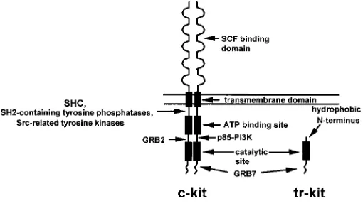
receptors is organized in 5 immunoglobulin-like domains and is devoted to ligand recognition and binding. The transmembrane region, a short hydrophobic stretch of amino acids, anchors the receptor to the plasma membrane. The intracellular region con-tains the kinase domain of the receptors required for transduction of the signal conveyed by the cognate ligands. The kinase domain of this class of receptors is characteristically split in two regions by an interkinase sequence which separates the ATP binding pocket from the phosphotransferase catalytic site (Yarden et al., 1987; Qiu et al., 1988). The juxtamembrane region, the interkinase region and the short carboxy terminal tail that follows the phosphotransferase catalytic site play an important role in the c-kit-mediated signaling. Indeed, upon ligand binding, dimerization of the receptor and activation of the intrinsic tyrosine kinase activity of the receptor occur. The consequent autophosphorylation of several tyrosine residues in the non-catalytical regions of the intracellular domain creates docking sites for specific src-homol-ogy 2 (SH2) containing proteins (Fig. 1). These proteins are either phosphorylated by c-kit or serve as adaptor molecules that bring other substrates in close proximity (Lev et al., 1991; Herbst et al., 1991; Rottapel et al., 1991). Several signal transducers which interact directly with the autophosphorylated c-kit receptor have been identified, such as the p85 subunit of phosphatidylinositol 3-kinase (PI3K; Serve et al., 1994), SH2-containing tyrosine phosphatases (Kozlowski et al., 1998), src-related kinases (Price et al., 1997), JAK2 (Brizzi et al., 1994), STAT1 (Deberry et al., 1997), and adaptor proteins such as SOCS1 (De Sepulveda et al., 1999), SHC (Lennartsson, 1999), GRB2 and GRB7 (Thommes et al., 1999). Less well defined is the interaction with other transduc-ers such as the Ras-GTPase activating protein and phospholipase C γ−1 (PLCγ1; Herbst et al., 1995) or the Tec kinase (Tang et al., 1994). These transducers regulate different cell-specific responses such as proliferation, survival and motility, either by acting directly or after activation of downstream pathways culminating in the activation of protein kinase C (PKC; Blume-Jensen, 1994), AKT (Blume-Jensen et al., 1998), and mitogen-activated protein kinases
(MAPKs) such as extracellular-signal-regulated kinases 1/2 (ERK1/ 2; Hemesath et al., 1998), or Jun-kinase (JNK; Timokhina et al., 1998).
The c-kit gene encodes at least three different mRNAs, with three corresponding protein variants (Reith et al., 1991; Rossi et al., 1992). Two of these protein variants derive from a 5.5 kb mRNA and can be distinguished for the in-frame insertion of four amino acids (Gly-Asn-Asn-Lys) in the extracellular region of the receptor, due to alternative splicing. The only functional difference reported between these two isoforms is the increased basal level of autophosphorylation of the shorter form. However, it is not known whether these two isoforms show a different tissue specific expres-sion pattern, or whether the shorter form, which displays a higher level of autophosphorylation and is constitutively more active, represents a signal for the cells expressing it (Reith et al., 1991). The third protein variant is encoded by an alternative mRNA of 3.2 kb derived from alternative promoter usage and shows an ex-tremely specific pattern of expression. Indeed, this shorter mRNA is only expressed in post-meiotic germ cells of the testis (Sorrentino et al., 1991; Rossi et al., 1992; Albanesi et al., 1996) and it encodes a truncated form of the cytoplasmic region of the c-kit receptor (Rossi et al., 1992). This truncated variant of c-kit has been named “tr-kit” (Sette et al., 1997), and its possible function during male gamete development and function will be discussed later (Fig. 1).
Stem cell factor
The c-kit ligand, Stem Cell Factor (SCF), is produced in a soluble and a membrane bound form, resulting from alternative splicing around the exon 6 of the gene. The soluble form of SCF derives from proteolytic cleavage of a membrane bound precursor by a not yet identified protease (Williams et al., 1992). The membrane bound form is originated by a splicing process that bypasses exon 6, which encodes for a sequence of 28 extracellular amino acids containing the proteolytic cleavage site. Both forms are expressed in a variety of tissues, and the ratio between the two can vary from tissue to tissue and in different developmental stages. Human SCF is translated with a 25 amino acids leader sequence, followed by a 185 amino acid extracellular sequence, a 27 amino acid transmembrane region and a 30 amino acid intrac-ellular region. Murine SCF shows 83% homology with human SCF. However activation of c-kit by SCF appears to be highly species-specific (Fleischman, 1993; Williams et al., 1992).
The molecular characterization of a Sl mutation led to the hypothesis that the soluble and membrane bound forms of SCF serve different functions. Brannan and colleagues (1991) showed that the Steel Dickie mutation results in a SCF protein which lacks the transmembrane and cytoplasmic regions. However, the puri-fied recombinant SCF Dickie has the same biological activity as the wild type soluble SCF. In spite of this, homozygous Sl-Dickie mice lack pigmentation, are sterile and severely anemic, displaying all the hallmarks of a c-kit/SCF lack of function. This observation suggests that the transmembrane form of SCF plays a fundamen-tal role during the establishment and the maintenance of the hematopoietic, melanocytic and germinal stem cells (Flanagan et al., 1991; Brannan et al., 1991). In support to this hypothesis are observations obtained with cell lines stably transfected with either the soluble or the transmembrane forms of SCF. Toksoz et al. (1992) have shown that transmembrane SCF is able to support
long-term growth of human hematopoietic progenitor cells in vitro, whereas soluble SCF shows only a transient effect. Dolci et al. (1991) and Matsui et al. (1991) have also shown that transmem-brane SCF is required for primordial germ cell survival in vitro. These observations suggest that the transmembrane form of SCF is absolutely required during embryonal development for the correct establishment of the three stem cell lineages that give rise to melanocytes, germ cells and hematopoietic cells.
The role of scf/c-kit in the establishment of the male and
female germ cell line
In the mouse embryo, primordial germ cells (PGCs) originate from a limited number of cells that separate from the extra-embryonal mesoderm at the stage of 7-7.5 days post coitum (dpc). PGCs begin to migrate towards the gonadal ridges from the base of the allantoid and begin to actively proliferate in coincidence with the expression of c-kit (Manova et al., 1991) to reach a number of approximately 25.000 at 13.5 dpc. (see for review: De Felici e Pesce, 1994). Once reached the gonadal ridges, PGCs continue to proliferate before entering a process of differentiation that will be completed at a later time with the formation of mature gametes. The end of their proliferative process coincides with the end of c-kit expression on these cells (Manova et al., 1991). W and Sl mice have a drastically reduced number of PGCs in the gonadal ridges, which probably results from the reduced proliferation and survival of these cells as well as the failure to migrate from the base of the allantoid. Several lines of evidence from in vitro studies support the hypothesis that alterations of the SCF/c-kit system are responsible for these defects in vivo. It has been shown that PGCs are only able to survive and proliferate in vitro in co-culture with nurse cells that produce SCF, and that the transmembrane form of the growth factor is much more efficient than the soluble form (Godin et al., 1991; Dolci et al., 1991; Matsui et al., 1991). More recent experi-ments have shown a synergy of effects between SCF and other growth factors such as the leukemia inhibitory factor (LIF), and molecules that induce a rise in intracellular cAMP levels, such as the pituitary adenylate cyclase activating polypeptides (PACAPs), to support PGCs survival in vitro (Dolci et al., 1993; De Felici et al., 1993; Pesce et al., 1996). Furthermore, in the absence of nurse cells, PGCs undergo an apoptotic process in vitro, which can be
synergistically prevented or attenuated by the presence of SCF and/or LIF in the culture medium (Pesce et al., 1993). In this context, it is interesting to notice that, while c-kit is expressed by the PGCs during their migration toward the gonadal ridges, in situ hybridization and immunohistochemical experiments have shown that SCF is expressed by the cells present along the migratory path of PGCs (Matsui et al., 1990). Finally, in vitro experiments have also shown a role for SCF/c-kit in the adhesion PGCs to layers of nurse cells (Matsui et al., 1991; Pesce et al., 1997).
Once they reach the gonadal ridges, PGCs begin a process of differentiation that will eventually lead to the production of mature gametes. The SCF/c-kit system plays a fundamental role also during the development of the male and female gonads, by supporting the production and survival of mature germ cells.
The role of scf/c-kit in the proliferation and/or survival of
male mitotic germ cells
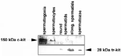
In the adult mouse testis, the c-kit receptor is mainly expressed by spermatogonia (Manova et al., 1990; Sorrentino et al., 1991) and by Leydig cells (Motro et al., 1991). Type A spermatogonia are the cell type in which maximal levels of both c-kit RNA and protein are observed, followed by type B spermatogonia (Manova et al., 1990; Sorrentino et al., 1991; Yoshinaga et al., 1991). Indeed, the full length c-kit receptor can be easily immunoprecipitated from rat spermatogonia (Dym et al., 1995), it can be readily localized in mouse spermatogonia by either indirect immunofluorescence (Albanesi et al., 1996) or by western blot analysis (Fig. 2), and it has been detected by immunohistochemical methods also in hamster and monkey spermatogonia (von Schonfeldt et al., 1999). Moreo-ver, the c-kit receptor mRNA and protein are expressed also in human spermatogonia (Natali et al., 1992; Sandlow, 1996). The expression of the mRNA for the full length c-kit receptor has also been demonstrated in mouse spermatocytes, albeit at lower levels, but it ceases completely during meiosis and it is absent in post-meiotic cells (Sorrentino et al., 1991; Rossi et al., 1992). Indeed, in spermatocytes the c-kit protein either is not present (Manova et al., 1990; Yoshinaga et al., 1991; von Schonfeldt et al., 1999) or, at least, is barely detectable (Fig. 2; Vincent et al., 1998).
The expression of c-kit in the only proliferating cells during spermatogenesis, the spermatogonia, has led to the hypothesis that the SCF/c-kit interaction is required for the proliferation and/or survival of these cells. Further support to this hypothesis comes from the observation that, in mice, both the soluble and membrane-bound form of SCF are expressed by the nurse cells of spermato-gonia, the Sertoli cells (Rossi et al., 1991, 1993; Manova et al., 1993; Tajima et al., 1991a; Marziali et al., 1993), whose intimate contact with the germ cells is necessary for their survival and differentiation. The selective site of testicular SCF expression in the Sertoli cell has also been confirmed in the rat (Munsie et al., 1997) and in humans (Sandlow et al., 1996). The expression of the mRNA for SCF is induced by the pituitary hormone FSH (follicle stimulating hormone) in pre-puberal mouse Sertoli cells cultured in vitro, through an increase in cAMP levels (Rossi et al., 1991, 1993). Stage-dependent induction of SCF mRNA expression by FSH has also been observed in the adult rat testis (Yan et al., 1999), and the maximal levels of SCF mRNA induction are observed in stages of the seminiferous epithelium which show the maximal sensitivity to FSH stimulation, and in which type A spermatogonia are actively
dividing. Interestingly, the soluble and membrane forms of SCF are differentially expressed during testis development. Sertoli cells from prepuberal mice mainly express the mRNA encoding for the transmembrane form, while the mRNA encoding for the soluble form is expressed at higher levels later, in coincidence with the beginning of the spermatogenic process, and the two transcripts are expressed at equivalent levels in the adult testis (Rossi et al., 1993). Moreover, FSH and/or cAMP analogs, beside increasing SCF mRNA levels, also modify the splicing pattern of the two isoforms in cultured mouse Sertoli cells in favour of the mRNA encoding for the soluble form (Rossi et al., 1993). In agreement with these observations is the finding that the highest levels of the transmembrane form of SCF are detected immunohistochemically in stages VII-VIII of the mouse seminiferous epithelium, and that, at these stages, a peculiar pattern of expression over the whole Sertoli cell membrane, rather than just in proximity of the basal layer of germ cells, is observed (Vincent et al., 1998). The equiva-lent stages in rat testis are the less sensitive to FSH stimulation in the adult testis (Parvinen, 1982). This pattern of expression could be physiologically relevant, since it has been reported that the transmembrane form of SCF, but not the soluble form, is required for mouse germ cells adhesion to Sertoli cells in culture (Marziali et al., 1993): it is conceivable that the transmembrane form of SCF might be involved in the progression through the blood-testis barrier of mitotic germ cells entering the first meiotic prophase at stages VII-VIII. Recently, it has been reported that the splicing pattern of SCF mRNA in Sertoli cells might also be influenced by local changes of pH in the seminiferous epithelium (Mauduit et al., 1999). FSH and/or cAMP regulation of SCF expression in rat Sertoli cells is due, at least in part, to transcriptional activation (Yan et al., 1999), and cAMP-responsive regions of the SCF promoter have been identified in the 5’ flanking regions of the human (Taylor et al., 1996), rat (Jiang et al., 1997) and mouse (P. Grimaldi et al., manuscript in preparation) SCF genes.
Early studies performed in W mutant mice suggested that c-kit plays a critical role in the differentiation from type A to type B spermatogonia (Koshimizu et al., 1991). Moreover, a peculiar Steel mutation, Sl17H, resulting in a splicing defect in the SCF cytoplasmic tail, in the homozygous condition induces sterility in males but not females, due to loss of spermatogonia during postnatal development (Brannan et al., 1992).
The first evidence that c-kit plays an important role in mitotically dividing male germ cells came from the observation that addition of the soluble form of SCF to in vitro cultured male germ cells from 7-8 day old mice at mitotic stages of differentiation stimulates DNA synthesis selectively in type A, but not in type B spermatogonia (Rossi et al., 1993). SCF-induced thymidine incorporation in cul-tured spermatogonia reflects DNA replication, rather than DNA repair, since it is completely abolished by aphidicolin, which specifically inhibits DNA polymerase α and arrests cells in early S-phase (Geremia et al., 1994). Selective induction of DNA synthesis in type A spermatogonia by soluble SCF has been confirmed using an in vitro tissue culture system for stage-defined seminiferous tubules of the adult rat testis (Hakovirta et al., 1999). The important role of the SCF/c-kit system in the maintenance of the germ cell line in the mouse post-natal testis is also suggested by in vivo and in vitro experiments in which the interaction between SCF and c-kit was blocked by an antibody directed against the extracellular region of the receptor. Under these conditions, c-kit expressing
type A spermatogonia, but not c-kit negative spermatogonial stem cells, are depleted (Yoshinaga et al., 1991) and unable to prolifer-ate (Tajima et al., 1994), and differentiating spermatogonia show increased levels of apoptosis (Packer et al., 1995). The role of soluble SCF in promoting survival and/or proliferation of rat and porcine spermatogonia has also been reported (Allard et al., 1996; Dirami et al., 1999).
SCF can trigger both mitogenic and anti-apoptotic
re-sponses
It has been argued that stimulation of DNA synthesis per se does not necessarily mean that SCF acts as a bona fide mitogenic factor, i.e. that SCF stimulates entry into the mitotic cell cycle of dividing type A spermatogonia. According to this view, the stimu-lation of spermatogonial DNA synthesis by SCF could simply reflect SCF action as a survival factor that prevents programmed cell death in cells which are intrinsically committed to proliferate (Packer et al., 1995; Hakovirta et al., 1999). However, appear-ance of actively proliferating type A1-A4 spermatogonia coincides with re-expression of the c-kit receptor, which is not expressed in post-natal spermatogonial stem cells, i.e. type A0 spermatogonia (Manova et al., 1990; Tajima et al., 1994; Dym, 1994). Moreover, we have recently found that SCF does prevent apoptosis induced by growth factor deprival in cultured mouse spermatogonia, but this effect is more evident in type B spermatogonia (S. Dolci et al., manuscript in preparation), rather than in type A1-4 spermatogo-nia, the cell type in which DNA synthesis is selectively stimulated (Rossi et al., 1993; Yan et al., 1999). This might imply that the soluble form of SCF sustains the proliferation of differentiating type A spermatogonia, and, at the same time, it does not stimulate proliferation of type B spermatogonia, but rather it prevents their cell death before they enter the meiotic stages of differentiation, when c-kit expression is down-regulated. The basis for the differ-ent cellular response in type B with respect to type A spermato-gonia, might depend on their lower levels of c-kit expression (Sorrentino et al., 1991). Indeed, in cultured c-kit expressing mast cells, higher doses of soluble SCF are required to trigger G1/S transition and promoting cell cycle progression than those re-quired for suppressing apoptosis (Yee et al., 1994). Moreover, it is known that transient versus sustained stimulation of
lar transducers by activated tyrosine kinase receptors causes differential responses (proliferation versus growth arrest, or in-duction of apoptosis versus promotion of survival and differentia-tion) and that the specific cellular response depends on the cell cycle situation of the responding cell, i.e. whether it is in a quiescent state or actively proliferating (Marshall, 1995; Blagosklonny, 1999). It is possible that in vivo these role are played differentially by the soluble and the transmembrane form of SCF, which could release signals of different strength to mitotic germ cells (transient versus sustained stimulation), as they do in hematopoietic precursors (Toksoz et al., 1992). In support to this hypothesis are studies performed in cryptorchid testes of mice heterozygous for the Steel Dickie mutation, which only express a soluble, but not a transmembrane form of SCF: proliferation of type A1-4 spermatogonia was apparently unaffected in surgically reversed cryptorchid testes of these animals, suggesting that soluble SCF is sufficient for their divisions, but a dramatic reduc-tion in type B spermatogonia was observed, with consequent impairment of spermatogenesis (Tajima et al., 1991b).
The fact that SCF can be both a mitogenic and an anti-apoptotic stimulus in the same cell type has been clearly demon-strated in c-kit expressing cells of the hematopoietic lineage. In mast cells, activation of the PI3K/Rac/JNK pathway is required for SCF-induced proliferative response, whereas inhibition of JNK accompanies SCF-mediated protection from radiation-induced apoptosis (Timokhina et al., 1998). A key role is probably played by PI3K activation in both situations, since PI3K also activates AKT, which is required to block the apoptotic pathway (Franke et al., 1997). Indeed c-kit-mediated inhibition of apoptosis in a hematopoietic cell line is coupled to PI3K-dependent activation of AKT (Blume-Jensen et al., 1998), and selective PI3K inhibitors block the anti-apoptotic effects of SCF in germ cells during fetal oogenesis (Morita et al., 1999). Moreover, SCF stimulation of mast cells is accompanied by a proliferative response, but also by simultaneous induction of SOCS1 expression; SOCS1, in turn, binds to the autophosphorylated c-kit receptor, to the GRB2 adaptor protein, and to the VAV GEF protein, with consequent block of both the Ras/Raf/MEK/ERK and Rac/JNK pathways, which are required for the mitogenic response (De Sepulveda et al., 1999). It has been proposed that c-kit-mediated induction of SOCS1 in mast cells suppresses the mitogenic potential of c-kit while maintaining SCF-dependent anti-apoptotic signals, and that SOCS1 is an inducible switch which modulates the SCF-mediated proliferative response in favour of a cell survival re-sponse (De Sepulveda et al., 1999).
More detailed studies on signal transduction pathways stimulated by c-kit activation in proliferating spermatogonial cells and their following effects on progression of the cell cycle and prevention of apoptosis are needed to establish whether SCF acts on these cells as a mitogenic or survival factor, or both, and whether it might also influence their differentiation into meiotic spermatocytes.
Possible roles of scf/c-kit in female and male meiotic
germ cells
Expression of the c-kit receptor ceases in fetal oocytes around the time they enter the prophase of the first meiotic division, but it resumes in post-natal oocytes, and is present at all the stages of their development (Manova et al., 1990; Horie et al., 1991; Yoshinaga
et al., 1991). Similarly to what has been described for Sertoli cells in the testis, granulosa cells surrounding the oocytes produce SCF (Manova et al., 1993). SCF expression in the female gonad is also under the control of gonadotropins (Motro and Bernstein, 1993; Ismail et al., 1996) and it has been proposed as a growth factor required for growth and maturation of the oocytes (Manova et al., 1993; Packer et al., 1994). The importance of SCF in post-natal oogenesis is also suggested by studies in panda and Steel-contrasted mutant mice, carrying DNA rearrangements located > 100 kb 5' of SCF-coding sequences, which lead to tissue-specific effects on SCF mRNA expression (Bedell et al., 1995). In both these mutants, SCF expression and spermatogenesis in the post-natal mutant testis is normal, but decreased SCF mRNA expres-sion causes sterility in females only, by affecting the initiation and maintenance of ovarian follicle development. SCF has also been recently proposed to play a role in the maintenance of meiotic arrest in the oocyte. In vitro experiments indicate that soluble SCF delays spontaneous meiotic resumption in mouse oocytes, ob-served as germinal vesicle breakdown (GVBD), and this effect is blocked by anti-c-kit antibodies (Ismail et al., 1996). Moreover, the same authors have shown that microinjection of anti-sense c-kit RNA lowers the expression levels of c-kit in mouse oocytes and increases their ability to reenter meiotic progression (Ismail et al., 1997). The c-kit receptor is also expressed in ovulated oocytes (Manova et al., 1990; Horie et al., 1991; Yoshinaga et al., 1991), that are arrested at metaphase of the second meiotic division under the action of proto-oncogene c-mos (Colledge et al., 1994; Hashimoto et al., 1994), but the function of c-kit and/or SCF at this stage is unknown.
et al., 1998; von Schonfeldt et al., 1999), even though trace amounts might be present on their membrane, as indicated by western blot analysis with an antibody recognizing the intracellular domain (Fig. 2) and by the observation that they can be enriched by FACS separation using antibodies directed against the extracel-lular domain (Vincent et al., 1998). The transmeiotic progression observed in vitro might reflect one of the two above mentioned possibilities: either the c-kit receptor is re-expressed in late pach-ytene spermatocytes just before the meiotic divisions, or a more complex interaction occurs, involving distinct c-kit positive cells (such as c-kit positive spermatogonia) present in the starting germ cell population used in these studies. Both possibilities agree with the observation that artificial activation of ERK1 in in vitro cultured pachytene spermatocytes leads to G2/M transition of the first meiotic division, suggesting that an unidentified extracellular signal from the seminiferous tubule environment actually regulates male meiotic progression (Sette et al., 1999).
An alternative form of c-kit (tr-kit) is specifically
ex-pressed during spermiogenesis
The 5.5 kb c-kit mRNA is not expressed during the haploid phase of spermatogenesis (Sorrentino et al., 1991; Rossi et al., 1992). Nevertheless, the full length c-kit receptor has been claimed to be present in isolated mouse, rat and human spermatozoa, based on immunofluorescence and/or western blot analysis using an anti-c-kit antibody directed against the extracellular region of the receptor (Sandlow et al., 1996; Feng et al., 1997). The same authors also reported the localization of c-kit in the acrosomal region of spermatids and spermatozoa, and its possible involve-ment in the acrosome reaction of the sperm in vitro (Feng et al., 1998). These data are in conflict with previous observations that the c-kit receptor protein is not detectable in haploid germ cells by immunocytochemistry on testis sections using the same anti-c-kit antibody (Yoshinaga et al., 1991). Using a different antiserum, directed against the intracellular portion of the c-kit receptor,
immunoreactivity was shown in both spermatogonia and spermatids in permeabilized sections of mouse, hamster and monkey testis (von Schonfeldt et al., 1999). Notwithstanding that this antiserum is directed against the cytoplasmic domain of the c-kit receptor, spermatogonia were purified by magnetic cell sorting using such antibodies, probably because partial damage of the spermatogo-nial membrane allows their specific adsorption. However, spermatids were not enriched by this fractionation method, suggesting that, whereas the c-kit receptor is present in the spermatogonial mem-brane, a cytoplasmic c-kit immunoreactive protein, rather than a SCF-responsive membrane receptor, must be present in spermatids of all those species (von Schonfeldt et al., 1999).
Indeed, the expression of the mRNA for the c-kit receptor is drastically reduced in premeiotic cells (spermatocytes) and is virtually absent in the postmeiotic spermatids (Sorrentino et al., 1991). However, Northern blot analysis of germ cells at different developmental stages has shown the presence of two alternative mRNA, of 3.2 and 2.3 kb, encoded by the c-kit gene in the haploid cells of the mouse testis (Sorrentino et al., 1991). Similar alterna-tive transcripts have also been detected in the adult rat testis (Orth et al., 1996). The two alternative spermatid-specific c-kit tran-scripts originate in 16th intron of the mouse c-kit gene, and contain all the exons downstream of it (Rossi et al., 1992). These alterna-tive c-kit mRNA encode for a truncated form of the receptor, called tr-kit (Sette et al., 1997), with an ORF that starts in the intron 16 and encodes for 12 hydrophobic amino acids followed by the last 190 carboxy terminal residues of the c-kit receptor (Fig. 1). Thus, tr-kit lacks the extracellular portion of the receptor, the transmembrane region, the ATP binding site and the interkinase sequence of the full-length c-kit. Tr-kit contains the phosphotransferase domain and the cytoplasmic tail of c-kit, where several molecular interac-tions with signaling proteins take place (Herbst et al., 1995; Sette et al., 1997; Thommes et al., 1999). In vitro transcription experi-ments with nuclear extracts from round spermatids have sug-gested the presence of an alternative promoter in the 16th intron of the c-kit gene (Albanesi et al., 1996). Indeed, transgenic mice in
Fig. 4. Schematic representation of possible mechanisms through which spermatozoa can trigger egg activation at fertilization. After egg-sperm binding, a tyrosine-kinase receptor or a G protein-coupled receptor on the egg plasma membrane is stimulated and activates either the
which the expression of the reporter gene LacZ is under the control of this intronic promoter have demonstrated the specific expres-sion of β-galactosidase in the testis (Albanesi et al., 1996). A more detailed analysis of the testes from these transgenic mice has revealed specific expression of the reporter gene uniquely in haploid germ cells. Indeed, β-galactosidase was expressed in a stage-specific manner, starting from step 8 of spermiogenesis, i.e. the haploid phase of spermatogenesis (Albanesi et al., 1996). Thus, it appears that this intronic promoter of the c-kit gene is only active in the late stages of spermatogenesis, suggesting a role for this truncated c-kit protein variant either during spermatid differen-tiation or for the function of mature sperm.
tr-kit is expressed in spermatids and spermatozoa and
induces the activation of metaphase II arrested oocytes
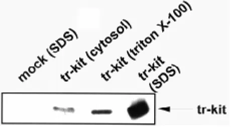
A truncated c-kit protein of ~30 kDa has actually been detected in mouse testis cells using an antibody raised against the last 13 amino acids at the carboxy terminus of the c-kit receptor (Albanesi et al., 1996). As shown in Fig. 2, this protein, named tr-kit, is first expressed in round spermatids, it accumulates in elongated spermatids and it is present in testicular and epididymal sperma-tozoa (Albanesi et al., 1996; Sette et al., 1997), whereas no trace of the ~150 kDa full-length c-kit receptor is detectable in haploid germ cells. A cytoplasmic c-kit immunoreactive protein has also been detected immunohistochemically in permeabilized hamster and monkey spermatids (von Schonfeldt et al., 1999). We also found that a ~30 kDa c-kit immunoreactive protein is detectable by western blot in rat and human spermatozoa (unpublished observa-tions). In the mature mouse sperm, tr-kit accumulates in the post-acrosomal region and in the midpiece (Sette et al., 1997), which are proximal to the site of the sperm that fuses with the egg membrane at fertilization (Yanagimachi, 1994). Indirect immunofluorescence analysis on permeabilized spermatozoa localizes tr-kit in the residual sperm cytoplasm (Sette et al., 1997). However, western blot analysis indicates that the majority of this protein is not present in the soluble fraction of spermatozoa, but it is found mostly in the Triton-X100 insoluble material, and the same subcellular distribu-tion of tr-kit is observed in COS cells transfected with a tr-kit expression vector (Fig. 3). It is possible that the N-terminal hydro-phobic residues present in tr-kit and encoded by c-kit intronic sequences (Fig. 1) are involved in its stable interaction with the particulate compartment. Interestingly, microinjection of a recombinant tr-kit expressed in the cytosolic fraction of tr-kit transfected COS cells is able to activate mouse oocyte arrested at metaphase II after ovulation (Sette et al., 1997, 1998). The amount of recombinant tr-kit required for such activation is lower than the amount of tr-kit present in a single sperm (Sette et al., 1998). Furthermore, oocyte activation follows the same pathway and timing described for the physiological activation at fertilization, with Ca2+-mediated cortical granule exocytosis, completion of meiosis and extrusion of the second polar body, and decrease in MAPK activity, with the consequent formation of a parthenogenetic pronu-cleus (Sette et al., 1997, 1998). These results have led to the hypothesis that tr-kit is a sperm factor released from the sperm into the oocyte at fertilization to trigger egg activation. Indeed, the molecular basis of egg activation at fertilization in the mouse are still unknown. Ovulated mouse oocytes are arrested at metaphase of the second meiotic division, with the chromosomes aligned at
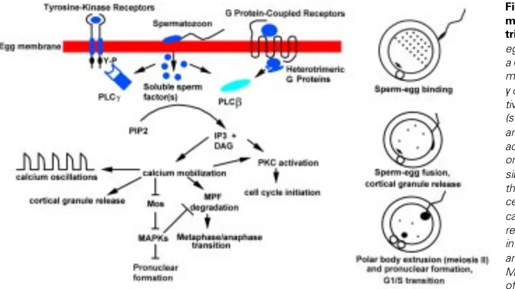
the equator of the meiotic spindle located near one pole of the cell. Sperm-egg fusion triggers a series of intracellular Ca2+ oscillations which are thought to cause the completion of meiotic division and the extrusion of the second polar body (Kline and Kline, 1992; Whitaker and Swann, 1993). Although the mechanism has not been elucidated, three hypotheses have been proposed to explain sperm-induced egg activation (Fig. 4):
1) a sperm-membrane ligand activates a tyrosine kinase receptor on the egg membrane;
2) a sperm-membrane ligand activates a G-protein-coupled receptor on the egg membrane;
3) the sperm releases cytosolic or particulate factors in the egg cytoplasm that trigger egg activation.
The first two hypotheses rely on experiments showing that microinjection of mRNA for receptors of either type allows activa-tion of oocytes by the corresponding ligand (Yim et al., 1994; Shilling et al., 1994). However, the presence and functionality of such receptors in mouse oocytes has not been demonstrated. The third hypothesis is supported by experiments in which sperm cytosolic extracts from several species have been injected into ovulated oocytes and shown to induce Ca2+ oscillations and all the events characteristic of fertilization. (Swann, 1990; Homa e Swann, 1994; Dozortev et al., 1995; Sette et al., 1997; Wu et al., 1997; Stricker, 1997). It is also known that sperm-egg fusion precedes the onset of Ca2+ oscillations in mouse eggs, supporting the hypothesis that a cytosolic sperm factor is released in the egg cytoplasm and triggers egg activation (Lawrence et al., 1997). Mobilization of intracellular Ca2+ is probably achieved by activation of one or more PLC isoenzymes and the consequent rise in IP3 intracellular levels (Miyazaki et al., 1992, 1993; Dupont et al., 1996). Interestingly, tr-kit-induced mouse oocyte activation is suppressed by co-injection of the SH3 domain of PLCγ1 and tr-kit is able to induce the tyrosine phosphorylation and activation of PLCγ1 when co-expressed in a heterologous system (Sette et al., 1998). We could not demonstrate a direct physical interaction between tr-kit and PLCγ1 (Sette et al., 1998). Moreover, the finding that the SH3, rather than the SH2 domain of PLCγ1 is required for tr-kit induced egg activation, led us to hypothesize that intermedi-ate adaptor proteins are required to elicit tr-kit mediintermedi-ated stimulation of the enzyme. Since tr-kit lacks the ATP-binding site (Rossi et al., 1992), it should lack intrinsic tyrosine kinase activity, and thus it should be unable to autophosphorylate or to phosphorylate tyro-sine residues on downstream target proteins. The observation that microinjection of tr-kit mRNA is equally efficient in eliciting egg activation as the recombinant protein expressed in COS cells (Sette et al., 1997), suggests that, if posttranslational modifica-tions, such as phosphorylation, are required for tr-kit function, they can be achieved in the egg cytoplasm. Thus, it is possible that a distinct tyrosine kinase present in the egg cytoplasm phosphorylates tr-kit, thus allowing PLCγ1 stimulation and consequent egg activa-tion.
Studies are underway to dissect the molecular mechanisms by which tr-kit elicits activation of PLCγ1 and to establish whether this truncated product of the c-kit gene is one of the sperm factors that plays a physiological role at fertilization in the mouse and in other mammalian species.
genetically modified mice. We are performing the genetic ablation through homologous recombination (Capecchi, 1989) of the intronic promoter sequences which drive tr-kit specific expression during spermiogenesis (Albanesi et al., 1996). This was an obligate choice, since we know that normal protocols of replacement of exonic sequences shared with the full-length c-kit gene product would have just generated W mutant mice with no germ cells in their embryonal gonads (Russell, 1979).
Acknowledgements
We would like to thank Drs. Cristina Albanesi, Paola Grimaldi, Marco Barchi, Silvia Di Agostino, Manuela Pellegrini, and Andrea Bianchini who contributed to the work from our laboratory described in this paper, and Drs. Vincenzo Sorrentino, Laura Pozzi, Marco Giorgio, Arturo Bevilacqua and Franco Mangia who collaborated with us in several aspects of the described research. This work was supported by CNR Targeted Project “Biotechnol-ogy” (R.G.), by CNR grant 98.00509.CT04 (P.R.), by grants from Ministero per l’Universita’ e la Ricerca Scientifica e Tecnologica (P.R. and R.G.), and by Agenzia Spaziale Italiana.
References
ALBANESI, C., GEREMIA, R., GIORGIO, M., DOLCI, S., SETTE, C. and ROSSI, P. (1996). A cell- and developmental stage-specific promoter drives the expression of a truncated c-kit protein during mouse spermatid elongation. Development 122: 1291-1302.
ALLARD, E.K., BLANCHARD, K.T. and BOEKELHEIDE, K. (1996). Exogenous stem cell factor (SCF) compensates for altered endogenous SCF expression in 2,5-hexanedione-induced testicular atrophy in rats. Biol. Reprod. 55: 185-193.
BEDELL, M.A., BRANNAN, C.I., EVANS, E.P., COPELAND, N.G., JENKINS, N.A. and DONOVAN, P.J. (1995). DNA rearrangements located over 100 kb 5' of the Steel (Sl)-coding region in Steel-panda and Steel-contrasted mice deregulate Sl expression and cause female sterility by disrupting ovarian follicle development. Genes Dev 9: 455-470.
BESMER, P., MURPHY, J.E., GEORGE, P.C., QIU, F.H., BERGOLD, P.J., LEDERMAN, L., SNYDER, H.W. Jr., BRODEUR, D., ZUCKERMAN, E.E. and HARDY, W.D. (1986). A new acute transforming feline retrovirus and relationship of its oncogene v-kit with the protein kinase gene family. Nature 320: 415-421.
BESMER, P. (1991). The kit ligand encoded at the murine steel locus: a pleiotropic growth and differentiation factor. Curr. Opin. Cell. Biol. 3: 939-946.
BLAGOSKLONNY, M.V. (1999). A node between proliferation, apoptosis, and growth arrest. Bioessays 21: 704-709.
BLUME-JENSEN, P., RONNSTRAND, L., GOUT, I., WATERFIELD, M.D. and HELDIN, C.H. (1994). Modulation of Kit/stem cell factor receptor-induced signaling by protein kinase C. J. Biol. Chem. 269: 21793-21802.
BLUME-JENSEN, P., JANKNECHT, R. and HUNTER, T. (1998). The kit receptor promotes cell survival via activation of PI 3-kinase and subsequent Akt-mediated phosphorylation of Bad on Ser136. Curr. Biol. 8: 779-782.
BRANNAN, C.I., LYMAN, S.D., WILLIAMS, D.E., EISENMAN, J., ANDERSON, D.M., COSMAN, D., BEDELL, M.A., JENKINS, N.A. and COPELAND, N.G. (1991). Steel-Dickie mutation encodes a c-kit ligand lacking transmembrane and cyto-plasmic domains. Proc. Natl. Acad. Sci. USA 88: 4671-4674.
BRANNAN, C.I., BEDELL, M.A., RESNICK, J.L., EPPIG, J.J., HANDEL, M.A., WILLIAMS, D.E., LYMAN, S.D., DONOVAN, P.J., JENKINS, N.A. and COPELAND, N.G. (1992). Developmental abnormalities in Steel17H mice result from a splicing defect in the steel factor cytoplasmic tail. Genes Dev. 6: 1832-1842.
BRIZZI, M.F., ZINI, M.G., ARONICA, M.G., BLECHMAN, J.M., YARDEN, Y. and PEGORARO, L. (1994). Convergence of signaling by interleukin-3, granulocyte-macrophage colony-stimulating factor, and mast cell growth factor on JAK2 tyrosine kinase. J. Biol. Chem. 269: 31680-31684.
CAPECCHI, M.R. (1989). Altering the genome by homologous recombination. Sci-ence 244: 1288-1292.
CHABOT, B., STEPHENSON, D.A., CHAPMAN, V.M., BESMER, P. and BERNSTEIN, A. (1988). The proto-oncogene c-kit encoding a transmembrane tyrosine kinase receptor maps to the mouse W locus. Nature 335: 88-89.
COLLEDGE, W.H., CARLTON, M.B.L., UDY, G.B., and EVANS, M.J. (1994). Disrup-tion of c-mos causes parthenogenetic development of unfertilized mouse eggs. Nature 370: 65-68.
DE FELICI, M., DOLCI, S. and PESCE, M. (1993). Proliferation of mouse primordial germ cells in vitro: a key role for cAMP. Dev. Biol. 157: 277-280.
DE FELICI, M. and PESCE, M. (1994). Growth factors in mouse primordial germ cell migration and proliferation. Prog. Growth Factor Res. 5: 135-143.
DE SEPULVEDA, P., OKKENHAUG, K., ROSE, J.L., HAWLEY, R.G., DUBREUIL, P. and ROTTAPEL, R. (1999). Socs1 binds to multiple signaling proteins and suppresses steel factor-dependent proliferation. EMBO J. 18: 904-915.
DEBERRY, C., MOU, S. and LINNEKIN, D. (1997). Stat1 associates with c-kit and is activated in response to stem cell factor. Biochem. J. 327: 73-80.
DIRAMI, G., RAVINDRANATH, N., PURSEL, V. and DYM, M. (1999). Effects of stem cell factor and granulocyte macrophage-colony stimulating factor on survival of porcine type A spermatogonia cultured in KSOM. Biol. Reprod. 61: 225-230.
DOLCI, S., WILLIAMS, D.E., ERNST, M.K., RESNICK, J.L., BRANNAN, C.I., LOCK, L.F., LYMAN, S.D., BOSWELL, H.S. and DONOVAN, P.J. (1991). Requirement for mast cell growth factor for primordial germ cell survival in culture. Nature 352: 809-811.
DOLCI, S., PESCE, M. and DE FELICI, M. (1993). Combined action of stem cell factor, leukemia inhibitory factor, and cAMP on in vitro proliferation of mouse primordial germ cells. Mol. Reprod. Dev. 35: 134-139.
DOZORTSEV, D., RYBOUCHKIN, A. DE SUTTER, P., QIAN, C. and DHONT, M. (1995). Human oocyte activation following intracytoplasmic injection: the role of the sperm cell. Hum. Reprod. 10: 403-407.
DUPONT, G., MCGUINNESS, O.M., JOHNSON, M.H., BERRIDGE, M.J. and BORGESE, F. (1996). Phospholipase C in mouse oocytes: characterization of β and γ isoforms and their possible involvement in sperm-induced Ca2+ spiking.
Biochem. J. 316: 53-591.
DYM, M. (1994). Spermatogonial stem cells of the testis. Proc. Natl. Acad. Sci. USA 91: 11287-11289.
DYM, M., JIA, M.C., DIRAMI, G., PRICE, J.M., RABIN, S.J., MOCCHETTI, I. and RAVINDRANATH N. (1995). Expression of c-kit receptor and its autophosphorylation in immature rat type A spermatogonia. Biol. Reprod. 52: 8-19.
FENG, H., SANDLOW, J.I. and SANDRA, A. (1997). Expression and function of the c-kit proto-oncogene protein in mouse sperm. Biol. Reprod. 57: 194-203.
FENG, H., SANDLOW, J.I. and SANDRA, A. (1998). The c-kit receptor and its possible signaling transduction pathway in mouse spermatozoa. Mol. Reprod. Dev. 49: 317-326.
FLANAGAN, J.F., CHAN, D.C. and LEDER, P. (1991). Transmembrane form of the kit ligand growth factor is determined by alternative splicing and is missing in the Sld mutant. Cell 64: 1025-1035.
FLEISCHMAN, R.A. (1993). From white spots to stem cells: the role of the Kit receptor in mammalian development. Trends Genet. 9: 285-290.
FRANKE, T.F., KAPLAN, D.R. and CANTLEY, L.C. (1997). PI3K: downstream AKTion blocks apoptosis. Cell 88: 435-437.
GEISSLER, E.N., RYAN, M.A. and HOUSMAN, D.E. (1988). The dominant-white spotting (W) locus of the mouse encodes the c-kit proto-oncogene. Cell 55: 185-192.
GEREMIA, R., ALBANESI, C., DOLCI, S., GIUSTIZIERI, L., GRIMALDI, P., GRIPPO, P., ORLANDO, P.A., PISCITELLI, D. and ROSSI, P. (1994). C-kit receptor function and regulation by SLF in the post-natal testis. In Frontiers in Endocrinology, vol. 5, Cell and Molecular Biology of the Testis (Eds. M.L. Dufau, A. Fabbri and A. Isidori), pp. 189-198. Ares-Serono Symposia.
GODIN, I., DEED, R., COOKE, J., ZSEBO, K., DEXTER, M. and WYLIE, C.C. (1991). Effects of the steel gene product on mouse primordial germ cells in culture. Nature 352: 807-809.
HAKOVIRTA, H., YAN, W., KALEVA, M., ZHANG, F., VANTTINEN, K., MORRIS, P.L., SODER, M., PARVINEN, M. and TOPPARI, J. (1999). Function of stem cell factor as a survival factor of spermatogonia and localization of messenger ribonucleic acid in the rat seminiferous epithelium. Endocrinology 140: 1492-1498.
HEMESATH, T.J., PRICE, E.R., TAKEMOTO, C., BADALIAN, T. and FISHER, D.E. (1998). MAP kinase links the transcription factor Microphthalmia to c-Kit signaling in melanocytes. Nature 391: 298-301.
HERBST, R., LAMMERS, R., SCHLESSINGER, J. and ULLRICH, A. (1991). Substrate phosphorylation specificity of the human c-kit receptor tyrosine kinase. J. Biol. Chem. 266: 19908-19916.
HERBST, R., SHEARMAN, M.S., JALLAL, B., SCHLESSINGER, J. and ULLRICH, A. (1995). Formation of signal transfer complexes between stem cell and platelet-derived growth factor receptors and SH2 domain proteins in vitro. Biochemistry 34: 5971-5979.
HOMA, S.T., and SWANN, K. (1994). A cytosolic sperm factor triggers calcium oscillations and membrane hyperpolarization in human oocytes. Hum. Reprod. 9: 2356-2361.
HORIE, K., TAKAKURA, K., TAII, S., NARIMOTO, K., NODA, Y., NISHIKAWA, S., NAKAYAMA, H., FUJITA, J. and MORI, T. (1991). The expression of the c-kit protein during oogenesis and early embryonic development. Biol. Reprod. 45: 547-552.
ISMAIL, R.S., OKAWARA, Y., FRYER, J.N. and VANDERHYDEN, B.C. (1996). Hormonal regulation of the ligand for c-kit in the rat ovary and its effects on spontaneous oocyte meiotic maturation. Mol. Reprod. Dev. 43: 458-469.
ISMAIL, R.S., DUBE, M. and VANDERHYDEN, B.C. (1997). Hormonally regulated expression and alternative splicing of kit ligand may regulate kit-induced inhibition of meiosis in rat oocytes. Dev. Biol. 184: 333-342.
KLINE, D. and KLINE, J.T. (1992). Repetitive calcium transients and the role of calcium in exocytosis and cell cycle activation in the mouse egg. Dev. Biol. 149: 80-89.
KOSHIMIZU, U., SAWADA, K., TAJIMA, Y., WATANABE, D. and NISHIMUNE, Y. (1991). White-spotting mutations affect the regenerative differentiation of testicu-lar germ cells: demonstration by experimental cryptorchidism and its surgical reversal. Biol. Reprod. 45: 642-648.
KOZLOWSKI, M., LAROSE, L., LEE, F., LE, D.M., ROTTAPEL, R. and SIMINOVITCH, K.A. (1998). SHP-1 binds and negatively modulates the c-Kit receptor by interac-tion with tyrosine 569 in the c-Kit juxtamembrane domain. Mol. Cell. Biol. 18: 2089-2099.
LAWRENCE, I., WHITAKER, M. and SWANN, K. (1997). Sperm-egg fusion is the prelude to the initial Ca2+ increase at fertilization in the mouse. Development 124:
233-241.
LENNARTSSON, J., BLUME-JENSEN, P., HERMANSON, M., PONTEN, E., CARLBERG, M. and RONNSTRAND, L. (1999). Phosphorylation of shc by src family kinases is necessary for stem cell factor receptor/c-kit mediated activation of the Ras/MAP kinase pathway and c-fos induction. Oncogene 18: 5546-5553.
LEV, S., GIVOL, D. and YARDEN. Y. (1991). A specific combination of substrates is involved in signal transduction by the kit-encoded receptor. EMBO J. 10: 647-654.
MANOVA, K., NOCKA, K., BESMER, P. and BACHVAROVA, R.F. (1990). Gonadal expression of c-kit encoded at the W locus of the mouse. Development 110: 1057-1069.
MANOVA, K. and BACHVAROVA, R.F. (1991). Expression of c-kit encoded at the W locus of mice in developing embryonic germ cells and presumptive melanoblasts. Dev. Biol. 146: 312-324.
MANOVA, K., HUANG, E.J., ANGELES, M., DE LEON, V., SANCHEZ, S., PRONOVOST, S.M., BESMER, P. and BACHVAROVA, R.F. (1993). The expres-sion pattern of the c-kit ligand in gonads of mice supports a role for the c-kit receptor in oocyte growth and in proliferation of spermatogonia. Dev. Biol. 157: 85-99.
MARSHALL, C.J. (1995). Specificity of receptor tyrosine kinase signaling: transient versus sustained extracellular signal-regulated kinase activation. Cell 80: 179-185.
MARZIALI, G., LAZZARO, D. and SORRENTINO, V. (1993). Binding of germ cells to mutant Sld Sertoli cells is defective and is rescued by expression of the transmem-brane form of the c-kit ligand. Dev. Biol. 157: 182-190.
MATSUI, Y., ZSEBO, K.M. and HOGAN, B.L. (1990). Embryonic expression of a hematopoietic growth factor encoded by the Sl locus and the ligand for c-kit. Nature 347: 667-669.
MATSUI, Y., TOKSOZ, D., NISHIKAWA, S., NISHIKAWA, S., WILLIAMS, D., ZSEBO, K. and HOGAN, B.L. (1991). Effect of Steel factor and leukemia inhibitory factor on murine primordial germ cells in culture. Nature 353: 750-752.
MAUDUIT, C., CHATELAIN, G., MAGRE, S., BRUN, G., BENAHMED, M. and
MICHEL, D. (1999). Regulation by pH of the alternative splicing of the stem cell factor pre-mRNA in the testis. J. Biol. Chem. 274: 770-775.
MIYAZAKI, S., YUZAKI, M. NAKADA, K. SHIRAKAWA, H. NAKANISHI, S. NAKADE, S. and MIKOSHIBA, K. (1992). Block of Ca2+ wave and Ca2+ oscillation by
antibody to the inositol 1,4,5-triphosphate receptor in fertilized hamster eggs. Science 257: 251-255.
MIYAZAKI, S., SHIRAKAWA, H. NAKADA, K. and HONDA, Y. (1993). Essential role of the inositol 1,4,5-triphosphate receptor/Ca2+ release channel in Ca2+ waves and
Ca2+ oscillations at fertilization of mammalian eggs. Dev. Biol. 158: 62-78.
MORITA, Y., MANGANARO, T.F., TAO, X.J., MARTIMBEAU, S., DONAHOE, P.K. and TILLY, J.L. (1999). Requirement for phosphatidylinositol-3'-kinase in cytokine-mediated germ cell survival during fetal oogenesis in the mouse. Endocrinology 140: 941-949.
MOTRO, B., VAN DER KOOY, D., ROSSANT, J., REITH, A. and BERNSTEIN, A. (1991). Contiguous patterns of c-kit and steel expression: analysis of mutations at the W and Sl loci. Development 113: 1207-1221.
MOTRO, B. and BERNSTEIN, A. (1993). Dynamic changes in ovarian c-kit and Steel expression during the estrous reproductive cycle. Dev. Dyn. 197: 69-79.
MUNSIE, M., SCHLATT, S., DEKRETSER, D.M. and LOVELAND, K.L. (1997). Expression of stem cell factor in the postnatal rat testis. Mol. Reprod. Dev. 47: 19-25.
NATALI, P.G., NICOTRA, M.R., SURES, I., SANTORO, E., BIGOTTI, A. and ULLRICH, A. (1992). Expression of c-kit receptor in normal and transformed human non-lymphoid tissues. Cancer Res. 52: 6139-6143.
ORTH, J.M., JESTER, W.F. Jr. and QIU, J. (1996). Gonocytes in testes of neonatal rats express the c-kit gene. Mol. Reprod. Dev. 45: 123-131.
PACKER, A.I, HSU, Y.C., BESMER, P. and BACHVAROVA, R.F. (1994). The ligand of the c-kit receptor promotes oocyte growth. Dev. Biol. 161: 194-205.
PACKER, A.I., BESMER, P. and BACHVAROVA, R.F. (1995). Kit ligand mediates survival of type A spermatogonia and dividing spermatocytes in postnatal mouse testes. Mol. Reprod. Dev. 42: 303-310.
PARVINEN, M. (1982). Regulation of the seminiferous epithelium. Endocr. Rev. 3: 404-417.
PESCE, M., FARRACE, M.G., PIACENTINI, M., DOLCI, S. and DE FELICI, M. (1993). Stem cell factor and leukemia inhibitory factor promote primordial germ cell survival by suppressing programmed cell death (apoptosis). Development 118: 1089-1094.
PESCE, M., CANIPARI, R., FERRI, G.-L., SIRACUSA, G. and DE FELICI, M. (1996). Pituitary adenylate cyclase-activating polypeptide (PACAP) stimulates adenylate cyclase and promotes proliferation of mouse primordial germ cells. Development 122: 215-221.
PESCE, M., DI CARLO, A. and DE FELICI, M. (1997). The c-kit receptor is involved in the adhesion of mouse primordial germ cells to somatic cells in culture. Mech. Dev. 68: 37-44.
PRICE, D.J., RIVNAY, B., FU, Y., JIANG, S., AVRAHAM, S. and AVRAHAM, H. (1997). Direct association of Csk homologous kinase (CHK) with the diphosphorylated site Tyr568/570 of the activated c-KIT in megakaryocytes. J. Biol. Chem. 272: 5915-5920.
QIU, F., RAY, P., BARKER, P.E., JHANWAR, S., RUDDLE, F.H. and BESMER, P. (1988). Primary structure of c-kit: relationship with the CSF-1/PDGF receptor kinase family - oncogenic activation of v-kit involves deletion of extracellular domain and C terminus. EMBO J. 7: 1003-1011.
REITH, A.D., ELLIS, C., LYMAN, S.D., ANDERSON, D.M., WILLIAMS, D.E., BERNSTEIN, A. and PAWSON, T. (1991). Signal transduction by normal isoforms and W mutant variants of the Kit receptor tyrosine kinase. EMBO J. 10: 2451-2459.
ROSSI P., ALBANESI C., GRIMALDI P. and GEREMIA R. (1991). Expression of the mRNA for the ligand of c-kit in mouse Sertoli cells. Biochem. Biophys. Res. Commun. 176: 910-914.
ROSSI P., MARZIALI G., ALBANESI C., CHARLESWORTH A., GEREMIA R. and SORRENTINO V. (1992). A novel c-kit transcript, potentially encoding a truncated receptor, originates within a kit gene intron in mouse spermatids. Dev. Biol. 152: 203-207.
ROSSI, P., SETTE, C. and GEREMIA, R. (1999). Role of distinct c-kit gene products in spermatogenesis and fertilization. In The Male Gamete: from Basic Science to Clinical Applications. (Ed. C. Gagnon), pp. 11-21. Cache River Press, Vienna, IL.
ROTTAPEL, R., REEDIJK, M., WILLIAMS, D.E., LYMAN, S.D., ANDERSON, D.M., PAWSON. T. and BERNSTEIN, A. (1991). The Steel/W transduction pathway: kit autophosphorylation and its association with a unique subset of cytoplasmic signaling proteins is induced by the Steel factor. Mol. Cell. Biol. 11: 3043-3051.
RUSSELL, E.S. (1979). Hereditary anemias of the mouse: a review for geneticists. Adv. Genet. 20: 357-459.
SANDLOW, J.I., FENG, H.L., COHEN, M.B. and SANDRA, A. (1996). Expression of c-KIT and its ligand, stem cell factor, in normal and subfertile human testicular tissue. J. Androl. 17: 403-408.
SERVE, H., HSU, Y.C. and BESMER, P. (1994). Tyrosine residue 719 of the c-kit receptor is essential for binding of the P85 subunit of phosphatidylinositol (PI) 3-kinase and for c-kit-associated PI 3-kinase activity in COS-1 cells. J. Biol. Chem. 269: 6026-6030.
SETTE, C., BEVILACQUA, A., BIANCHINI, A., MANGIA, F., GEREMIA, R. and ROSSI, P. (1997). Parthenogenetic activation of mouse eggs by microinjection of a truncated c-kit tyrosine kinase present in spermatozoa. Development 124: 2267-2274.
SETTE, C., BEVILACQUA, A., GEREMIA, R. and ROSSI, P. (1998). Involvement of phospholipase C γ1 in mouse egg activation induced by a truncated form of the c-kit tyrosine kinase present in spermatozoa. J. Cell Biol. 142: 1063-1074.
SETTE, C., BARCHI, M., BIANCHINI, A., CONTI, M., ROSSI, P. and GEREMIA, R. (1999). Activation of the mitogen-activated protein kinase ERK1 during meiotic progression of mouse pachytene spermatocytes. J. Biol. Chem. 274: 33571-33579.
SHILLING, F.M., CARROLL, D.J. MUSLIN, A.J. ESCOBEDO, J.A. WILLIAMS, L.T. and JAFFE, L.A. (1994). Evidence for both tyrosine kinase and G protein-coupled pathways leading to starfish egg activation. Dev. Biol. 162: 590-599.
SORRENTINO, V., GIORGI, M., GEREMIA, R., BESMER, P. and ROSSI, P. (1991). Expression of the c-kit proto-oncogene in the murine male germ cells. Oncogene 6: 149-151.
STRICKER, S.A. (1997). Intracellular injections of a soluble sperm factor trigger calcium oscillations and meiotic maturation in unfertilized oocytes of a marine worm. Dev. Biol. 186: 185-201.
SWANN, K. (1990). A cytosolic sperm factor stimulates repetitive calcium increases and mimics fertilization in hamster eggs. Development 110: 1295-1302.
TAJIMA, Y., ONOUE, H., KITAMURA, Y. and NISHIMUNE, Y. (1991a). Biologically active kit ligand growth factor is produced by mouse Sertoli cells and is defective in SId mutant mice. Development 113: 1031-1035.
TAJIMA, Y., SAKAMAKI, K., WATANABE, D., KOSHIMIZU, U., MATSUZAWA, T. and NISHIMUNE, Y. (1991b). Steel-Dickie (Sld) mutation affects both maintenance and differentiation of testicular germ cells in mice. J. Reprod. Fertil. 91: 441-449.
TAJIMA, Y., SAWADA, K., MORIMOTO, T. and NISHIMUNE, Y. (1994). Switching of mouse spermatogonial proliferation from the c-kit receptor-independent type to the receptor-dependent type during differentiation. J. Reprod. Fertil. 102: 117-122
TANG, B,, MANO, H., YI, T. and IHLE, J.N. (1994). Tec kinase associates with c-kit and is tyrosine phosphorylated and activated following stem cell factor binding. Mol. Cell. Biol. 14: 8432-8437.
THOMMES, K., LENNARTSSON, J., CARLBERG, M. and RONNSTRAND, L. (1999). Identification of Tyr-703 and Tyr-936 as the primary association sites for Grb2 and Grb7 in the c-Kit/stem cell factor receptor. Biochem. J. 341: 211-216.
TIMOKHINA, I., KISSEL, H., STELLA, G. and BESMER, P. (1998). Kit signaling through PI 3-kinase and Src kinase pathways: an essential role for Rac1 and JNK activation in mast cell proliferation. EMBO J. 17: 6250-6262.
TOKSOZ, D., ZSEBO, K.M., SMITH, K.A., HU, S., BRANKOW, D., SUGGS, S.V., MARTIN, F.H. and WILLIAMS, D.A. (1992). Support of human hematopoiesis in long-term bone marrow cultures by murine stromal cells selectively expressing the membrane-bound and secreted forms of the human homologue of the steel gene product, stem cell factor. Proc. Natl. Acad. Sci. USA 89: 7350-7354.
ULLRICH, A. and SCHLESSINGER, J. (1990). Signal transduction by receptors with tyrosine kinase activity. Cell 61: 203-212.
VINCENT, S., SEGRETAIN, D., NISHIKAWA, S., NISHIKAWA, S.I., SAGE, J., CUZIN, F. and RASSOULZADEGAN, M. (1998). Stage-specific expression of the Kit receptor and its ligand (KL) during male gametogenesis in the mouse: a Kit-KL interaction critical for meiosis. Development 125: 4585-4593.
VON SCHONFELDT, V., KRISHNAMURTHY, H., FOPPIANI, L. and SCHLATT, S. (1999). Magnetic cell sorting is a fast and effective method of enriching viable spermatogonia from Djungarian hamster, mouse, and marmoset monkey testes. Biol. Reprod. 61: 582-589.
WHITAKER, M. and SWANN, K. (1993). Lighting the fuse at fertilization. Development 117: 1-12.
WILLIAMS, D.E., DE VRIES, P., NAMEN, A.E., WIDMER, M.B. and LYMAN, S.D. (1992). The Steel factor. Dev. Biol. 151: 368-376.
WITTE, O.N. (1990). Steel locus defines new multipotent growth factor. Cell 63: 5-6.
WU, H., HE, C.L. and FISSORE, R.A. (1997). Injection of a porcine sperm factor triggers calcium oscillations in mouse oocytes and bovine eggs. Mol. Reprod. Dev. 46: 176-189.
YAN, W., LINDERBORG, J., SUOMINEN, J. and TOPPARI, J. (1999). Stage-specific regulation of stem cell factor gene expression in the rat seminiferous epithelium. Endocrinology 140: 1499-1504.
YANAGIMACHI, R. (1994). Mammalian fertilization. In The Physiology of Reproduction (Eds. E. Knobil and J.D. Neill), pp. 189-317. Raven Press Ltd, New York.
YARDEN, Y,, KUANG, W.J., YANG-FENG, T., COUSSENS, L., MUNEMITSU, S., DULL, T.J., CHEN. E., SCHLESSINGER. J., FRANCKE. U. and ULLRICH, A. (1987). Human proto-oncogene c-kit: a new cell surface receptor tyrosine kinase for an unidentified ligand. EMBO J. 6: 3341-3351.
YEE, N.S., PAEK, I. and BESMER, P. (1994). Role of kit-ligand in proliferation and suppression of apoptosis in mast cells: basis for radiosensitivity of white spotting and steel mutant mice. J. Exp. Med. 179: 1777-1787.
YIM, D.L., OPRESKO, L.K., WILEY, H.S. and NUCCITELLI., R. (1994). Highly polarized EGF receptor tyrosine kinase activity initiates egg activation in Xenopus. Dev. Biol. 162: 41-55.