Echocardiographic wall-to-cavity ratios in
hypertrophic cardiomyopathy and other types of
cardiac hypertrophy.
Anita M Devlin
ProQuest Number: U642907
All rights reserved
INFORMATION TO ALL USERS
The quality of this reproduction is dependent upon the quality of the copy submitted.
In the unlikely event that the author did not send a complete manuscript
and there are missing pages, these will be noted. Also, if material had to be removed,
a note will indicate the deletion.
uest.
ProQuest U642907
Published by ProQuest LLC(2016). Copyright of the Dissertation is held by the Author.
All rights reserved.
This work is protected against unauthorized copying under Title 17, United States Code.
Microform Edition © ProQuest LLC.
ProQuest LLC
789 East Eisenhower Parkway
P.O. Box 1346
Statement of conjoint work
I hereby acknowledge that a proportion of the data presented in this thesis was collected jointly with Dr I
Ostman-Smith. I believe this statement to be a fair reflection o f my personal contribution.
The contributions to the control data from 262 normal subjects were approximately equal as were the
contributions to the disease population. Approximately 70% of the data from the first-degree family
members, 85% of the data from the athletes and 100% o f the data from the hypertensive group was
contributed by the thesis author.
Wall thickness data from magnetic resonance imaging (MRI) studies was contributed by Dr N Moore and
left ventricular mass measurements were collected jointly. The echocardiographic measurements used for
comparison were made by the thesis author.
In the published material submitted in support of this thesis, the data presentation, the reasoning behind
ABSTRACT
Background - Left ventricular (LV) hypertrophy is diagnosed by increased LV wall thickness or increased LV mass. A single cut-off for echocardiographic wall thickness (>15mm in adults) is commonly used
irrespective of gender. This approach has yielded an inexplicable excess in male prevalence of 2-3:1 for
hypertrophic cardiomyopathy (HCM) in population based studies. Children's cardiac measurements are
referenced to body surface area by nomograms or regression equations which take no account o f LV
cavity size. LV mass or mass index is a continuous variable in the normal population with no absolute
diagnostic cut-off for hypertrophy, whilst published normal values can be seen in hypertrophic
cardiomyopathy. Consequently, a need exists for a simpler echocardiographic method to diagnose and
sequentially monitor LV hypertrophy at all ages irrespective of gender, which may also help to
differentiate between aetiologies.
Objectives - To describe a new echocardiographic method for assessing LV hypertrophy which is applicable at all ages and is independent o f gender.
Methods - 262 normal subjects, 41 patients with hypertrophic cardiomyopathy, 52 first degree relatives from families with familial hypertrophic cardiomyopathy, 26 athletes and 16 patients with hypertension
underwent echocardiography. M-mode diastolic and systolic ratios of LV wall thickness to LV diameter
were calculated. A subset of 10 patients with HCM had LV wall thickness and mass measurements
performed using magnetic resonance imaging (MRI) for comparison with echocardiography.
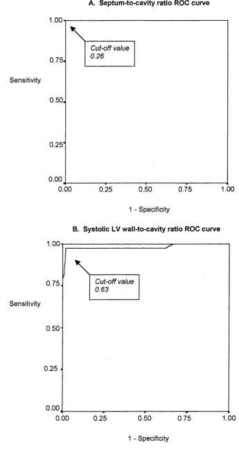
Results - Diastolic and systolic wall-to-cavity ratios show little variation with age and are independent of gender. In adults diastolic ratios are independent of body surface area. A septum-to-cavity ratio of >0.26
identified all HCM patients (100% sensitivity) and no control subjects (0% false positives). A systolic LV
wall-to-cavity ratio of > 0.63 performed equally well in subjects > 1 year. Septum-to-posterior wall
thickness ratio >1.3 performed less well (90% sensitivity, 10% false positive rate). A septum-to-cavity
ratio >0.26 identified all first-degree relatives with an abnormal phenotype suggestive of gene carriage
with no obvious false positives. It was also more successful than traditional measures at dividing relatives
in to affected and unaffected subjects in the proportions expected for autosomal dominant inheritance.
Athletes with marked cardiac hypertrophy showed a 7.7% false positive rate for the diagnosis of HCM
using diastolic septum-to-cavity ratio >0.26, but are recognised as showing physiological hypertrophy by
the presence o f normal systolic function (systolic LV wall-to-cavity ratios). Hypertensive cardiac
hypertrophy is also identified by a septum-to-cavity ratio > 0.26. Systolic LV wall-to-cavity ratios are
significantly lower than in subjects with HCM as are other measures of systolic function.
MRI and echocardiography show good agreement between LV wall thickness measurements, particularly
o f the anterior interventricular septum. The agreement between the assessments o f LV mass was poor.
Conclusions - Septum-to-cavity ratio and systolic LV wall-to-cavity ratio can diagnose LV hypertrophy at all ages, are useful in the diagnosis o f and screening for HCM and can help differentiate physiological
from pathological hypertrophy. MRI and echocardiography are complimentary techniques in the
TABLE OF CONTENTS
L INTRODUCTION.
...9
1.1 Hypertrophie Cardiomyopathy...9
1.11 History and nomenclature...9
1.12 Pathology... 10
1.13 Natural history...13
1.14 Clinical features... 18
1.15 Diagnostic investigations...21
1.16 Treatment... 32
1.17 Inheritance and genetics...37
1.2 The normal heart...43
1.21 Post-mortem studies... 43
1.22 Angiography...43
1.23 Echocardiography...43
1.3 Left ventricular hypertrophy... 50
1.31 Electrocardiography... 50
1.32 Echocardiography...51
1.4 Physiological cardiac hypertrophy... 52
1.41 Electrocardiogram (EGG)... 52
1.42 Echocardiography...52
1.5 Hypertensive cardiac hypertrophy...
56
1.51 Electrocardiography (EG G)... 56
1.52 Echocardiography...56
1.6 Magnetic resonance imaging in hypertrophic cardiomyopathy... 59
1.7 Aims of the thesis...60
2
SUBJECTS AND METHODS.
...61
2.1 Echocardiography... 61
2.11 Subjects... 61
2.12 M ethods... 62
2.2 Magnetic Resonance imaging... 63
2.21 Subjects... 63
3. RESULTS
...65
3.1 The Control Population...65
3.11 Left ventricular measurements... 65
3.12 Left ventricular wall-to-cavity ratios... 65
3.13 Gender comparisons...73
3.14 Influence o f body surface area...75
3.2 Patients with hypertrophic cardiomyopathy (HCM)...77
3.21 Left ventricular measurements...77
3.22 Left ventricular wall-to-cavity ratios...79
3.3 First degree relatives from kindreds with hypertrophic cardiomyopathy... 86
3.31 Septum-to-cavity ratio... 86
3.32 Other wall-to-cavity ratios...89
3.4 The athletic population...92
3.41 Left ventricular measurements...92
3.42 Left ventricular wall-to-cavity ratios...94
3.5 Subjects with hypertension... 97
3.51 The group with hypertension and the control group... 97
3.52 Subjects with hypertension and those with H C M ... 101
3.6 Magnetic resonance imaging (MRI) and echocardiography as imaging modalities in
hypertrophic cardiomyopathy...108
3.61 Wall thickness measurements... 108
3.62 Left ventricular mass (LVM )... 112
4. DISCUSSION.
...113
4.1 The normal population...113
4.11 M-mode echocardiography...113
4.12 Normal ranges...113
4.13 Gender variation... 116
4.14 Body surface area (BSA) variation... 116
4.2 The diagnosis of hypertrophic cardiomyopathy (HCM)...118
4.21 Diagnosis in childhood...120
4.3 Screening for hypertrophic cardiomyopathy... 121
4.31 Echocardiographic screening... 121
4.33 Genetic screening... 126
4.34 Benefits of screening... 128
4.4 Physiological left ventricular hypertrophy... 131
4.5 Cardiac hypertrophy secondary to hypertension... 135
4.51 Hypertensive subjects and normal subjects... 135
4.52 Hypertensive subjects and subjects with HCM...137
4.6 Magnetic resonance imaging in the assessment of subjects with HCM...140
BIBLIOGRAPHY.
...143
APPENDIX
...172
Publications in support of thesis...172
Devlin AM, Moore NR, Ostman-Smith I. A comparison o f MRI and echocardiography in
hypertrophic cardiomyopathy. British Journal o f Radiology 1999;72:258-64.
Devlin AM, Ostman-Smith I. Diagnosis o f hypertrophic cardiomyopathy and screening for the
phenotype suggestive of gene carriage in familial disease: a simple echocardiographic procedure.
Journal o f Medical Screening 2000;7:82-90.
Ostman-Smith I, Devlin AM. A simple method for assessing the regression or progression of
ventricular hypertrophy in the growing child and adult: the value o f left ventricular wall-to-cavity
LIST OF FIGURES
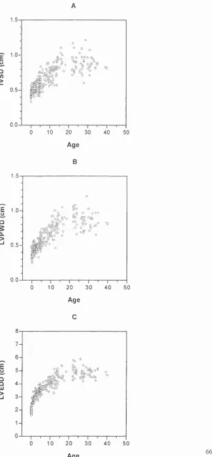
FIGURE 1 A-E. LEFT VENTRICULAR MEASUREMENTS IN THE NORMAL POPULATION 66
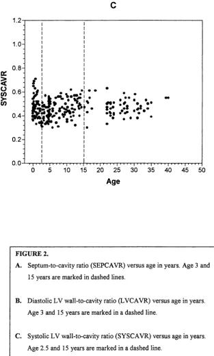
FIGURE 2 A-C. WALL-TO-CAVITY RATIOS AND AGE IN THE NORMAL POPULATIO N 69
FIGURE 3. GENDER COMPARISONS OF CONVENTIONAL MEASUREMENTS AND WALL-TO
CAVITY RATIOS IN ADULTS... 74
FIGURE 4. BODY SURFACE AREA (BSA) AND WALL-TO-CAVITY RATIOS IN A D U LTS 75
FIGURE 5. LEFT VENTRICULAR MEASUREMENTS IN ADULT HCM PATIENTS COMPARED
WITH THE CONTROL GROUP... 78
FIGURE 6 A-D. FREQUENCY DISTRIBUTION OF WALL-TO -CAVITY RATIOS IN THE HCM
GROUP COMPARED TO THE CONTROL GROUP...80
FIGURE 7 A-D. RECEIVER OPERATOR CHARACTERISTIC (ROC) CURVES FOR
WALL-TO-CAVITY RATIOS... 83
FIGURE 8. SEPTUM-TO-CAVITY RATIOS IN FIRST-DEGREE RELATIVES OF SUBJECTS WITH
HCM COMPARED WITH THE CONTROL POPULATION...87
FIGURE 9. LEFT VENTRICULAR MEASUREMENTS IN ADULT ATHLETES AND CONTROL
SUBJECTS... 93
FIGURE 10. SEPTUM-TO-CAVITY RATIOS AND SYSTOLIC LV WALL-TO-CAVITY RATIOS IN
ATHLETES AND CONTROL SUBJECTS... 95
FIGURE 11. LEFT VENTRICULAR MEASUREMENTS IN HYPERTENSIVE SUBJECTS AND
CONTROL SUBJECTS... 98
FIGURE 12 A-D. WALL-TO-CAVITY RATIOS IN HYPERTENSIVE SUBJECTS AND THOSE
WITH H C M ... 102
FIGURE 13 A-C. MEASURES OF SYSTOLIC FUNCTION IN HYPERTENSIVE SUBJECTS AND
THOSE WITH H CM ...105
FIGURE 14 A-D. BLAND-ALTMAN PLOTS COMPARING WALL THICKNESS MEASUREMENTS
MADE BY MRI AND ECHOCARDIOGRAPHY... 109
FIGURE 15. BLAND-ALTMAN PLOT SHOWING THE INTEROBSERVER VARIABILITY OF THE
LIST OF TABLES
TABLE 1. RELATIONSHIP BETWEEN AGE AND WALL-TO-CAVITY RATIOS... 68
TABLE 2. REFERENCE RANGES FOR WALL-TO-CAVITY RATIOS IN SELECTED AGE GROUPS
...71
TABLE 3. GENDER DIFFERENCES IN CARDIAC MEASUREMENTS IN ADULTS... 73
TABLE 4. WALL-TO-CAVITY RATIOS AND BODY SURFACE AREA IN ADULTS...75
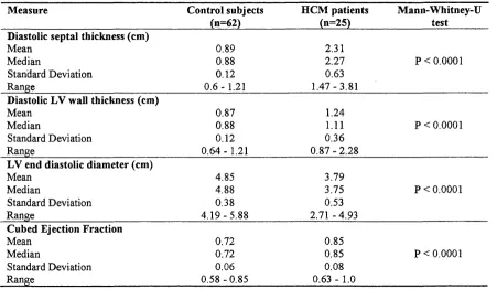
TABLE 5. CARDIAC MEASUREMENTS ADULT HCM PATIENTS AND CONTROLS 77
TABLE 6. WALL-TO-CAVITY RATIOS IN CONTROL SUBJECTS AND HCM PATIENTS 79
TABLE 7. RESULTS OF THE APPLICATION OF THE DERIVED CUT-OFF VALUES IN THE
DISEASE AND CONTROL POPULATIONS...82
TABLE 8. WALL-TO-CAVITY RATIOS IN FIRST DEGREE RELATIVES OF SUBJECTS WITH
HCM WITH ESTIMATED SENSITIVITY AND FALSE POSITIVE RATES...90
TABLE 9. LEFT VENTRICULAR MEASUREMENTS IN ATHLETES AND CONTROL SUBJECTS
... 92
TABLE 10. WALL-TO-CAVITY RATIOS IN ATHLETES AND CONTROL SUBJECTS... 94
TABLE 11. DIAGNOSTIC CRITERIA FOR HCM APPLIED TO THE ATHLETIC POPULATION... 96
TABLE 12. LEFT VENTRICULAR MEASUREMENTS IN HYPERTENSIVE SUBJECTS AND
CONTROL SUBJECTS... 97
TABLE 13. WALL-TO-CAVITY RATIOS IN HYPERTENSIVE SUBJECTS AND ADULT CONTROL
SUBJECTS... 99
TABLE 14. DIAGNOSTIC CRITERIA FOR HCM APPLIED TO THE HYPERTENSIVE
POPULATION...100
TABLE 15. MEASURES OF SYSTOLIC FUNCTION IN HYPERTENSIVE SUBJECTS AND
SUBJECTS WITH H CM ... 104
TABLE 16. MEASURES OF SYSTOLIC FUNCTION IN SUBJECTS WITH HYPERTENSION AND
HCM PATIENTS WITH SEPTAL THICKNESS MEASUREMENTS OF 15-20 m m ... 107
TABLE 17. DEGREES OF BIAS AND LIMITS OF AGREEMENT BETWEEN MEASUREMENTS
MADE BY MRI AND ECHOCARDIOGRAPHY... I l l
TABLE 18. COMPARISON OF ECHOCARDIOGRAPHIC MEASUREMENTS USED TO DIAGNOSE
1. IN T R O D U C T I O N
1.1 Hypertrophie Cardiomyopathy
1.11 History and nomenclature
It is thought that the first observation of myocardial disease, which can be interpreted as hypertrophic
cardiomyopathy (HCM), was made in the middle of the 19* century in France (1, 2). A century later in the
1950s there remained no classification or framework for the cardiomyopathies, which were frequently
considered alongside myocarditis even when the myocarditis was known to have a specific inflammatoiy
aetiology (3). In 1957 Brigden published his St Gyres lecture on "Uncommon myocardial diseases; the
non coronary cardiomyopathies" and was one of the first to employ the term "cardiomyopathy" (4). In the
same year Brock published the first clinical account o f hypertrophic cardiomyopathy (5) which was
followed in 1958 by Teare's systematic description of the disease characterised by "asymmetrical
hypertrophy o f the heart" with a non-dilated ventricular cavity (6). Further characterisation o f the disease
was performed by Goodwin (7) who attempted a first descriptive title for the disorder o f "obstructive
cardiomyopathy" which was subsequently modified as follows: "Cardiomyopathy - an acute, subacute or
chronic disorder of heart muscle o f unknown or obscure aetiology, often with associated endocardial or
sometimes with pericardial involvement but not atherosclerotic in origin" (8).
During the first 15 years following Teare's description of HCM (6), the obstructive features of the disease
associated with massive ventricular hypertrophy and impaired diastolic function had been recognised (7,
9, 10) and this was reflected in the nomenclature. Terms such as idiopathic hypertrophic subaortic stenosis
(11), hypertrophic obstructive cardiomyopathy (10, 12) and muscular or hypertrophic subaortic stenosis
(9, 13) were employed. However, the idea of obstruction as an essential component o f the disease was
called in to question by Criley et al (13) when the outflow tract gradients in a series of patients with HCM
were located in the ventricle itself, in areas which emptied early such as the apex and the trabecular
spaces. In addition, Goodwin's group noted that as the disease became more severe the signs of
obstruction tended to disappear which is unlike the picture in true obstructive outflow tract disease o f the
left ventricle (14). Furthermore, vyith the development of echocardiography large numbers o f patients were
subsequently identified in whom obstruction was absent (15). These observations led to the omission of
the word obstructive from the definition.
The asymmetric nature of the hypertrophy seen in the disorder was then emphasised and was characterised
by Henry et al (16) as the pathognomonic feature of the disease by the observation that the ratio of
diastolic septal thickness o f the left ventricle to the diastolic thickness o f the left ventricular free wall was
relatives o f index cases, implying that HCM was a disease with autosomal dominant inheritance (15). This
led to the term asymmetric septal hypertrophy or "ASH" being used to describe the disease entity of HCM.
In 1975 Goodwin called for agreement in nomenclature (17) the need for which was reinforced by Maron
and Epstein (18) who listed no fewer than 58 terms by which hypertrophic cardiomyopathy had become
known. This was partly because the disease was characterised simultaneously by many groups who
assigned it a different name and partly because information about and concepts of the disease evolved over
time and became incorporated in to the nomenclature. However, since the most consistent feature of the
disease, that of a hypertrophied heart with an non-dilated left ventricular cavity in the absence o f a
systemic disease that could itself produce hypertrophy, there was a consensus to refer to the disease as
Hypertrophic Cardiomyopathy (HCM) (18). This term had already been developed and employed by those in the United Kingdom for some years (19) and remains consistent with the current title endorsed by the
joint task force o f the World Health Organisation and International Society and Federation o f Cardiology
(20).
1.12 Pathology
The interventricular septum is formed from the apical in-folding of a unichamber structure. At that point
the septum is approximately twice as thick as either the left or right ventricular free wall. Subsequent
remodelling during cardiogenesis allows the septum to become a left ventricular structure in both
configuration and dimension (21) except for the outer surface, which is formed by the right ventricular
trabeculae. The hallmark of HCM is the finding o f left ventricular hypertrophy (LVH), particularly o f the
interventricular septum, with a normal sized or small left ventricular cavity (22). In 1958 Teare
emphasised the asymmetric nature of the left ventricular (LV) hypertrophy in his initial morphologic
description of HCM (6). Menges and others added quantitation o f ventricular septal thickness
measurement to the necropsy diagnosis and found that the increased ratio o f septal to free LV wall
thickness separated those subjects with HCM from those with other cardiac diseases and normal hearts
(23, 24). In addition to asymmetric ventricular hypertrophy without dilatation, morphological observations
include endocardial thickening in the outflow portion o f the ventricle caused by the impact o f the anterior
mitral valve leaflet (mirror image of the leaflet), enlargement of the atria, thickening o f the mitral valve
leaflets and areas of fibrosis in the ventricular wall (22, 25).
HCM is associated with the presence of mutations in the genes coding for contractile proteins and this is
discussed in detail later (chapter 1.17). The resulting microscopic appearance o f the cardiac muscle cells
o f the septum and elsewhere is one of greatly hypertrophied fibres with a characteristic disorganised
arrangement in which adjacent cells are arranged obliquely and perpendicularly to one another. There is
sarcomeres (6, 26, 27) which is seen irrespective of the presence o f outflow tract obstruction in vivo (26-
28). The myocardial pathology o f infants, children and adults with asymmetric septal hypertrophy (ASH)
has been compared to subjects of a similar age with normal hearts and those with other types of congenital
or acquired heart disease (29, 30). In the normal heart the cardiac muscle cells are rectangularly shaped
and arranged in parallel. Some small foci o f disorganised cardiac cells were observed but occupy
approximately 2% of septal tissue sections (30). The transverse diameters of the muscle cells in the
ventricular septum and LV wall in one study (29) ranged from 5-1 Sp.. Myocardium from the septum and
free wall o f hearts with congenital defects other than ASH shows hypertrophied normally arranged cells
with transverse diameters o f 6-27p. In those with HCM extensive muscle cell disorganisation is seen
occupying a mean o f 53% o f septal tissue sections (30). The cells show abnormal size and arrangement
with diameters o f 10-70p (27, 29). The disorganisation assumes two patterns: 1) foci where adjacent cells
are arranged perpendicularly and obliquely and 2) small groups or bundles of cardiac muscle cells that are
interlaced in various directions among larger and more normally arranged groups o f cells resulting in a
swirling appearance (27, 29). These regions of disorganised myocardial architecture are not confined to
greatly thickened portions of the left ventricular wall and were present in regions o f normal or only mildly
increased thickness (31). These changes reflect the abnormalities seen in all age groups.
In the normal heart the collagen matrix consists o f an orderly arrangement of thin "spring-like" perimysial
coils which run parallel to the long axis of the muscle cells. These coils are connected through transversely
oriented struts to a network o f pericellular weaves encircling the sarcolemna o f individual muscle cells
(30). In one study (30), total collagen volume in septal tissue sections of the normal heart was found to be
4% and was evenly distributed throughout all parts of the septum. Conversely in the hearts from subjects
with HCM there was a marked increase in number and thickness of all fibre components of the collagen
matrix (30). The perimysial fibres were thickened and had lost the coiled configuration and the pericellular
weave formed dense networks o f thickened fibres which showed a disorganised pattern when compared to
normal hearts. Mean total collagen volume in the septal tissue sections was 16.7% and was sited in
interstitial rather than perivascular tissue. The expanded size of the collagen matrix bore no relation to any
morphologic or clinical variable nor was it related to the degree of myocyte disorganisation, suggesting
that it was an independent and primary abnormality in HCM (30). This is supported by the observation of
an increased collagen matrix even in infants dying at less than 5 months o f age. Interestingly those with
hypertensive heart disease showed no areas of increased myocyte disorganisation, no change in
morphology o f the perimysial coils although they were thicker and appeared more abundant. The total
collagen volume was increased above normal occupying 8.2% of the septal tissue sections but was still far
less than in the hearts with HCM (30).
As well as an increase in interstitial connective tissue elements (32) replacement fibrosis of variable
severity and distribution is described including scarring which may be extensive (32, 33) or transmural
HCM at post-mortem (33, 34). The walls of these vessels show increased smooth muscle, collagen, elastic
fibres and mucoid deposits in the intima or media, which frequently narrow the lumen. Clusters o f these
abnormal vessels are seen within or at the margins of sizeable areas of fibrosis or collagen including large
areas o f transmural infarction (30, 33).
The extent to which myocardial fibre disarray and asymmetric septal hypertrophy were pathognomonic of
HCM was called in to question by Bulkley et al (35) who studied 215 hearts including those o f normal
embryos, foetuses, children and adults, and those with congenital and acquired disease. Disproportionate
septal thickening was present in all embryos and in some abnormal hearts particularly those with severe
right ventricular hypertrophy. Small areas of myocardial fibre disarray were present in all hearts, including
normal hearts, at the junction of interventricular septum and ventricular free wall. Extensive areas were
found in hearts with semilunar valve atresia with intact ventricular septum and in the inflindibulum of a
proportion o f those with tetralogy of Fallot. Disarray was also described at the borders o f human
myocardial infarcts (35) and in experimental myocardial infarction (36). It is noteworthy that abnormal
myocardial fibre disarray was not present in the developing embryo or foetus despite the presence of
disproportionate septal thickening and was not found in those hearts with mitral atresia with ventricular
septal defect or in combined aortic and mitral atresia. It was also absent in the adult hearts with aortic
stenosis. Therefore the presence of left ventricular outflow tract obstruction alone does not appear to
provide the substrate for the development of the disorganised muscle fibre appearances. The substrate
appears to be the presence of a hypertrophied ventricle with an extremely small cavity such as that found
in outflow tract obstruction with intact septum. It was speculated therefore that the fibre disarray in HCM
and aortic or pulmonary valve atresia with intact ventricular septum might result fi'om the loss of
direction-orientated wall stresses due to systolic cavity obliteration during cardiac development (37).
Consideration was also given to whether this feature represented a primary genetic abnormality but this
was difficult to argue as the various disorders in which it occurred demonstrated no other unifying
features. Olsen (22) suggested that additional histological/histochemical features such as bizarre nuclei,
perinuclear halos and perinuclear pools of glycogen were which helpful in distinguishing HCM from other
forms o f hypertrophy.
Primary structural abnormalities of the mitral valve are also well recognised in HCM and have been
identified both pathologically (38) and echocardiographically (39). Examination o f almost 100 valves
removed at surgery or post-mortem showed alterations in size, shape and morphologic characteristics (38).
The abnormalities included increased valve area of up to twice normal and was caused primarily by
elongation o f the leaflets or segments of the leaflets o f either the anterior or posterior leaflet or both. Some
patients with normal sized mitral valves may show anomalous insertion o f papillary muscle directly in to
1.13 Natural history
Prevalence
There have been many studies estimating the prevalence o f HCM in a given population. One of the earlier
investigations involved M-mode echocardiography of 3000 offspring of the original Framingham cohort
(41) and found echocardiographic markers o f HCM in 0.3%. A large cohort o f 12841 Japanese workers
(42) underwent electrocardiography in the first instance followed by echocardiography in a subset of 12%.
The reported prevalence o f HCM in this subset was 0.17%. The use of electrocardiography as the primary
screening method could have resulted in an underestimation of the prevalence in that study. A more recent
community-based population study of those referred for echocardiography because o f a suspicion of
cardiac disease demonstrated a prevalence of 0.5% (43) whereas a population based study in Olmsted
County, Minnesota (44) described an age and sex adjusted prevalence of only 0.02% for HCM. However,
the latter study was a disease surveillance analysis o f patients who had come to medical attention for
HCM rather than identification of subjects by population screening which could easily lead to an
underestimate o f the true prevalence.
A more recent assessment o f the population prevalence of HCM was established in the CARDIA study of
4111 subjects age range 23-35 years (45) and was found to be 0.2%. This prevalence is supported by the
figure of 0.29% from a very large study where 15 137 echocardiograms were performed in 64 rural
communities at the request o f the primary care physician for the purpose o f excluding cardiovascular
abnormalities (46). The CARDIA study showed that the recognition of HCM by morphological criteria
yields up to 10 times the number of positive cases than those based on symptomatic presentation and
substantiates that most young people identified with HCM have no symptoms. Interestingly, HCM was
found to be 2.9 times more common in men than in women in this study (45) for which the cut-off for the
diagnosis was a maximum absolute LV wall thickness measurement of >15mm irrespective of sex. An
epidemiological study in children showed a similar male preponderance (47). Since there is no ready
explanation for the skew in the sex-related prevalence in what has been shown to be an autosomal
dominant disorder, it might be concluded that the diagnostic criteria employed were not of sufficient
sensitivity to detect affected females.
Adult disease
McKenna et al (48) retrospectively reviewed the clinical, electrocardiographic and haemodynamic features
of 254 patients of all ages diagnosed and followed-up at the Royal Postgraduate Medical School for 1-23
years with a mean o f 6 years. 58 (23%) had died, 32 suddenly and 6 with heart failure. In those aged 15-45
years at diagnosis there was a 2.5% annual mortality rate. Discriminant analysis showed that young age
(14 years or less), syncope at diagnosis, positive family history o f sudden death from HCM and severe
dyspnoea at last examination best predicted future sudden death. However, 38% o f the patients who died
high incidence o f sudden death was seen in earlier studies, one from the same group which included 119
patients of which 30 died, 19 suddenly (49) and another multicentre study by Shah et al (50) of 190
patients with a left ventricular outflow tract gradient at rest or with provocation, of whom 49 died, 26
suddenly. All suggested that the annual mortality rate in adults was in the region o f 2-4% (50, 51) and that
HCM was thus, not a benign disease.
Over time however, it has come to be appreciated that most published studies have come from a small
number o f tertiary institutions expert in the study and management of the disease and therefore subject to
biases in patient referral (52). Since patients with severe symptoms or those at high risk for sudden cardiac
death have been preferentially referred to these centres, the outlook reported in the literature has perhaps
been skewed towards the more dismal end o f the spectrum (53). More recently, studies have been
conducted in regional cohorts not referred for specialist care. They have shown that many patients can
remain asymptomatic (54) and have an annual mortality rate as low as 0.6-1.3% (54-57) with a normal
overall life expectancy (57).
The difficulty remains in the identification o f those most at risk o f sudden death. This is complicated by
the phenotypic heterogeneity and by a prevalence which means that few centres have a disease population
of sufficient size to ascertain risk factors in subgroups (58). These centres are subject to referral biases and
tend to see the more severe cases. Almost half o f the studies on HCM from a previous decade which were
published in 3 American and 1 British journal came from only two centres and >90% of the patients
reported to have severe symptoms also came from these institutions (59). Some have found that in certain
groups the history and particularly changes in symptoms were better predictors of poor prognosis than any
haemodynamic or electrocardiographic measurements (3). However, whilst the documentation of changes
in symptomatology appeared to be quite specific in identifying a high-risk group it is insufficiently
sensitive. This point was illustrated by Maron et al (60) who described a subgroup of families in whom
early sudden death occurred -with considerable frequency and referred to this as "malignant" hypertrophic
cardiomyopathy. 18/31 subjects who died suddenly were <25 years old and in 15 subjects this was the first
manifestation o f cardiac disease (60).
Since then work has shown that a high risk group can be identified on the basis o f electrophysiological
factors (61), a family history o f sudden death (60) and more recently guidelines on risk factors and risk
stratification for sudden cardiac death in patients with hypertrophic cardiomyopathy have been published
(58). Factors thought to suggest a high risk o f sudden death are young age at diagnosis (48, 62, 63),
history o f aborted sudden death (58), strong family history o f sudden death (60), syncope (62, 64) and the
presence of arrhythmias including occult conduction disease (58, 65). More recent studies suggested that
in addition, atrial fibrillation, a basal outflow tract gradient of > 30mmHg and an LV wall thickness o f
history, are largely helpfiil in those already known to have the disease. The challenge remains however, to
identify subjects before their clinical presentation with cardiac symptoms such as sudden death.
Childhood disease
HCM was recognised as cause of morbidity and mortality in adults in the 1960s and 70s (6, 8, 19, 49, 51)
but in children little clinical, haemodynamic, echocardiographic information or natural history was known
before 1976 (28). HCM had been observed at the post-mortem studies of infants demonstrating that this
disease may be present at birth or indeed in utero (29, 66). It had also been described in those less than 6
months o f age where it was responsible for the death of 4 infants in the first five months o f life (28, 29).
This suggested that the abnormal cells are present at or near the time o f birth and lent support to the idea
that these morphological features are an expression of a genetic defect (29). Unfortunately, when overt
clinical manifestations o f the disease such as heart failure and marked ventricular hypertrophy are evident
in infancy the prognosis is particularly unfavourable and such infants rarely survive beyond the first two
years o f life (66). In one study, 9/11 such infants died in the first year o f life despite medical or surgical
therapy (66).
The study by Maron et al (28) of children with HCM showed that of the children with clinical signs of
cardiac disease, 60% (21/35) clinically deteriorated or died over a mean follow-up period o f 7.4 years (1-
16 years). Sudden death occurred in 31% (11/35) over the same period giving an annual mortality rate of
4%. 3 children who died were completely asymptomatic at the time of death and a fiirther child had
suffered a single syncopal episode 16 months prior to death. The occurrence o f sudden death showed no
relationship to age at onset of symptoms, functional status, electrocardiographic abnormalities or left
ventricular ejection or upstroke time. Three other children, siblings o f children in the study, who were
known to but not formerly evaluated by the group, died without any overt evidence o f cardiac disease but
the diagnosis o f HCM was confirmed at necropsy (28). The mortality in this study by Maron et al was in
agreement with previous studies in adults (49, 50) but somewhat higher than that reported by other studies
o f children (49, 67). The most concerning feature o f the study was the observation that sudden death in
children and siblings o f affected children occurred without prior warning and could not be linked to
symptoms or functional status.
A later study by McKenna et al (48) confirmed the concerning nature of HCM in childhood compared to
adult disease by showing an annual mortality rate of 5-6% which was higher than that described for adults
(48-50). McKenna's study (48) found that patients diagnosed in childhood were usually asymptomatic and
had an unfavourable family history. 41% of the patients diagnosed at < 14 years old died over the mean
follow-up period of 6 years compared to 12-14% o f those diagnosed > 14 years old. 4/11 o f those
diagnosed in childhood had one or more first-degree relatives who died with HCM and 7/11 suffered
offspring from families with "malignant" HCM as described by Maron et al (60). Even in more recent
studies describing a more benign natural history o f HCM in adults, those individuals identified with
disease in childhood showed significantly reduced survival compared with a similar non-diseased
population cohort (57). The need to seek to identify children affected by HCM is therefore clear since they
often fail to identify themselves before the occurrence o f a catastrophic cardiac event. Therefore any
methods which assist in the diagnosis of the disease in childhood may help to prevent such sudden deaths
by allowing medical or surgical intervention such as the treatment of arrhythmias.
In 1986 Maron et al (68) undertook a study to determine whether the pattern o f left ventricular
hypertrophy in HCM is always established at or shortly after birth or if the process is an evolving one
which develops and progresses during childhood. Serial echocardiograms of 39 children <15 years old
with a family history or morphologic evidence of the disease were followed over a mean period o f 4 years.
The presence and progression of the disease in childhood was assessed by extrapolation from the
nomograms for normal myocardial wall thickness corrected for age and BSA devised by Henry et al (69).
The study produced further worrying results with regard to this disease in childhood and adolescence.
22/39 showed a substantial increase in left ventricular wall thickness. 17 had shown pre-existing
hypertrophy which had increased and 5 previously normal subjects had become abnormal, to the extent of
having wall thickness measurements of 18-28mm in three cases. In the 22 subjects with disease
progression the maximal wall thickness at the start of the study was 8-27mm and at follow-up was 14-
46mm, with increases o f > 10mm in 14 subjects. These changes occurred at different periods in childhood
but most frequently in the adolescent years. 14/22 were asymptomatic throughout the period o f follow-up
and none who were asymptomatic at the start became symptomatic. Those with symptoms were affected
by fatigue and dyspnoea on exertion, chest pain, light-headedness or syncope and had no noteworthy
increase in these symptoms. Thus, marked increases in the magnitude and distribution o f hypertrophy may
occur in childhood and children with morphologically normal hearts may rapidly become abnormal
without the development of symptoms. Serial evaluation o f the grooving child at risk o f developing HCM
is clearly necessary for which accurate methods for individualised, sequential assessment are essential.
Athletes
In 1992 the Hypertrophic Cardiomyopathy Association launched a campaign to raise awareness o f HCM
in association with the National Sports Medicine Institute following the death from the condition of the
son o f a well known footballer (70). Athletes are particularly vulnerable as HCM is the commonest cause
o f death in this group (71), which predominantly involves children and young adults (72). Liberthson (72)
performed a combined analysis of 9 papers examining the causes of sudden death in those aged 1-30 years
and found a rate o f 1.3-8.5 per 100 000 patient years and in 2/3 a specific cardiac cause o f death was
identified. When extrapolated, this observation predicts that several thousand young Americans die
identified HCM as the commonest cause. In another study (62) 70% o f patients with hypertrophic
cardiomyopathy who died suddenly did so before the age o f 30 years and 40% died during or after
exertion. 54% of individuals were free of functional limitation and no clinical or morphologic variable
could be shown to reliably identify patients at risk o f sudden death. Once again, it would appear that the
majority o f these subjects require to be identified since they do not present with symptoms.
A study by Maron et al (71) examined the causes o f sudden and unexpected death in 29 highly conditioned
competitive athletes aged 13-30 years. Sudden death occurred during or just after severe exertion on the
athletic field in 22/29 athletes and structural cardiovascular abnormalities were identified at necropsy in
28/29 athletes and in 22 were thought to be the cause o f death. The abnormalities included anomalous
origin of the left coronary artery, idiopathic concentric left ventricular hypertrophy, coronary heart disease
and ruptured aorta. However, the most common underlying pathology was HCM, which was present in at
least 14 subjects.
8/29 athletes had experienced transient symptoms including syncope in three, presyncope in one, chest
pain in two, periodic mild fatigue in one and mild fatigue, presyncope and palpitations in one. In two of
these subjects and five other asymptomatic athletes cardiovascular disease had been suspected by an
examining physician during life. In the latter five cardiovascular disease had been suspected because of a
heart murmur in two, a family history of heart disease in one, ventricular tachycardia on an exercise
electrocardiogram (EGG) during a routine preparticipation athletic examination in one and during
evaluation for the Marfan syndrome in one. 6/7 athletes suspected o f having cardiovascular disease during
life turned out to have HCM. One was correctly diagnosed following the sudden death o f his brother from
the disease. The diagnoses made in the other five athletes during life were "normal athlete heart" in three,
ventricular septal defect in one and Wolff-Parkinson-White syndrome in one. All were permitted to
continue participation in competitive athletics and tragically died subsequently in practise or during
competition. These deaths must surely be considered as potentially preventable and subsequently
guidelines have been produced concerning the eligibility for competition of athletes with cardiovascular
abnormalities (73).
HCM was re-affirmed as the commonest cause o f death during exercise in those less than 30 years in a
recent study from France (74). This study illustrates that it is not only athletes who partake of regular
training who are at risk but also those with HCM who happen to participate in occasional exercise. It also
reinforces the need for forensic autopsy in these cases so that other members o f the family might be
alerted to potential dangers surrounding exercise. These studies demonstrate the importance of correctly
differentiating between physiological hypertrophy or "normal athletes heart", other cardiac lesions and
HCM and show the need for more accurate and reliable methods for this purpose than were available to
1.14 Clinical features
Examination
Before the advancements in technology, which permitted such techniques as cardiac catheterisation, the
clinician relied on the bedside examination to make the diagnosis of HCM. The diagnosis largely rested on
the presence o f three cardinal signs: the presence o f a late onset ejection systolic murmur audible at the
apex, the left sternal edge and occasionally the base, a jerky quality to the arterial pulse due to the rapid
upstroke and rapid decline o f the impulse and a palpable atrial beat caused by the strong contraction of the
left atrium needed to fill the non-compliant left ventricle. However, it has been shown that HCM is not
always associated with a loud heart murmur (2) and when a murmur is present the intensity bears no
relation to the severity o f the obstruction (50). In a study by Clark et al (15) the symptomatic history was
negative in 60% and the physical examination in 65% o f those with HCM. Likewise the CARDIA study
o f disease prevalence (45) showed that only 1/7 o f those identified with the disease had suffered any
previous cardiac symptomatology. Therefore the clinical history and the presence o f abnormal clinical
signs can not be relied upon to detect subjects with HCM.
Arrhythmias
Sudden death was initially assumed to be secondary to outflow tract obstruction but subsequent work
pointed to arrhythmia as a potential cause (75) even in the absence o f functional limitation (76). Serious
ventricular arrhythmias have been documented in up to 50% o f unselected subjects with HCM (77, 78)
using ambulatory monitoring and exercise testing. However, no clinical or haemodynamic index could be
found in these and other studies which could predict those patients who would suffer from ventricular
tachycardia or sudden death (76, 78). A link between sudden death and ventricular arrhythmia was
subsequently described by McKenna et al (65) when the incidence o f ventricular tachycardia and
subsequent sudden death was explored using 72-hour electrocardiographic (ECG) recordings from 86
patients. 24 patients experienced ventricular tachycardia, 10 having more than 3 episodes. The patients
were followed for 1-4 years (mean 2.6). 7 died suddenly o f whom 5 had shown either multiform paired
premature ventricular contractions or episodes of ventricular tachycardia on the ambulatory recordings.
The presence o f ventricular tachycardia was also significantly associated with sudden death in older
patients with symptoms (65). This and another study concluded that the finding of episodes o f non
sustained ventricular tachycardia during ambulatory ECG monitoring was the best single identifying
marker of the adult at high risk of sudden death (65, 79). However, a more recent study proposes that non
sustained ventricular tachycardia is of poor prognostic significance only when associated with impairment
tachycardia in asymptomatic populations free from the selection bias of the tertiary referral centre (80,
81).
The prognosis in infants and children with regard to arrhythmias was also investigated by McKenna et al
(82). During the follow-up period of 1 week to 7 years there were 5 deaths and 2 successful resuscitations
from out-of-hospital ventricular fibrillation. None o f these subjects had shown ventricular arrhythmias
during 2-7 days of previous ECG monitoring. Furthermore, neither of the two patients with ventricular
fibrillation, none o f the five subjects with ventricular tachycardia, none o f the seven subjects with
recurrent syncope or adverse family history or the subject with Wolff-Parkinson-White syndrome died.
This suggests that in contrast to adults, the absence o f ventricular arrhythmia during ambulatory ECG
monitoring during childhood does not indicate a low risk for sudden death.
Attempts to identify morphological features of the disease that were associated with the presence of
arrhythmia have yielded varying results. One M-mode echocardiographic study o f 26 patients (83) showed
that none of the classical features of HCM were predictive of the occurrence of arrhythmia. Features of
obstruction i.e. systolic anterior motion o f the mitral valve (SAM) and mid-systolic closure o f the aortic
valve were inversely correlated with the occurrence of ventricular tachycardia and the magnitude o f septal
thickness and septum-to-free wall ratio showed no correlation. However, patients with ventricular
tachycardia showed the most severely impaired pattern o f septal motion i.e. reduced amplitude o f motion
at chordal level and reduced septal systolic thickening. In contrast, a previous study (84) had shown that
features o f obstruction such as SAM were positively associated with ventricular arrhythmia and a later
study by Spirito et al (85) found a strong association between magnitude of LV hypertrophy and
occurrence of VT in patients with HCM. The presence o f any association between arrhythmias and
morphological features of HCM therefore remains unclear (86).
Syncope
Syncope is an important symptom in HCM and in the retrospective review o f patients from the
Hammersmith hospital, syncope in patients less than 45 years of age indicated a poor prognosis (48).
Clearly, arrhythmias represent a well recognised cause of syncope but a further possible mechanism for
syncope in HCM was postulated by McKenna et al (64). This followed an incident where a patient fainted
and lost consciousness when walking to the X-ray department whilst attached to an ambulatory ECG
monitor. The pulse disappeared for > 1 minute before she was successfully resuscitated. The early stages
of the attack showed tachycardia and ischaemic ST segment changes. On recovery there was some atrio
ventricular dissociation and fusion beats but no bradycardia, asystole, ventricular tachycardia or
fibrillation. The proposed mechanism was one of initial peripheral vasodilatation, which led to reduced
so on in a descending spiral. This is interesting in the light of subsequent work on abnormalities of
peripheral vascular responses in those with HCM (87).
The dismal outlook for those suffering from syncope was further underlined in a retrospective study of 37
patients diagnosed with HCM in childhood (63). Various clinical, electrocardiographic and
haemodynamic features were compared between the group in which there were 11 sudden deaths and the
group of 19 survivors over a follow-up period of 1-21 years, mean 9 years. Syncope and the presence of
right ventricular hypertrophy on the ECG were the only factors associated with sudden death. Since
syncope appears to be the only prelude to sudden death in this age group it might be prudent to consider
the diagnosis o f HCM in any young person who presents with a history of recurrent syncope.
Sudden death
In addition to arrhythmias as a cause of sudden death in HCM previous studies had suggested that subjects
with HCM were unable to maintain stroke volume and thus unable to increase cardiac output during
exercise or tachycardia. This was mainly thought to be due to the reduced filling time allowed for a poorly
compliant ventricle (19). McKenna and Camm (88) suggested that subsequent investigation of the
mechanisms surrounding sudden death in HCM should include haemodynamic factors in addition to the
investigation o f myocardial vulnerability to airhythmias, especially since the latter did not identify
children and adolescents at risk.
It was subsequently shown that hypotension during exercise is not uncommon in subjects with HCM and
was seen in 1/3 o f patients in one study (89) who experienced falls in blood pressure of 20-1 lOmmHg.
The same group used the measurement of forearm blood flow (87) to demonstrate that a sub-group of
patients with HCM show an abnormal vascular response to supine exercise. These patients were younger
and more of them had a family history of sudden death than did those without an abnormal vascular
response. It was postulated that haemodynamic mechanisms might be important in sudden death in HCM.
Such abnormal haemodynamic responses have also been shown following a meal in those with HCM, with
failure o f stroke volume to increase accompanied by an increase in left atrial pressure (90). Further study
in to the nature of abnormal haemodynamic mechanisms in the HCM population and the associated
1.15 Diagnostic investigations
Angiography
Obviously the diagnostic criteria for HCM have evolved along with the advances in technology but in the
1960s and early 1970s angiography appearances were the mainstay of the diagnosis (91) at a time when
the obstructive component of the disease was to the fore. Angiographic studies showed complete
elimination of the apex and body of the LV in systole with greatly reduced LV volume (3). Subsequent
studies correlated echocardiographic with angiographic findings in HCM (92) and the good correlation
between the techniques along with the rapid advancements in echocardiographic imaging, meant that
echocardiography supervened as the main diagnostic imaging modality in HCM.
ECG and electrophysiological studies
Different ECG criteria for the diagnosis of hypertrophy have been employed in HCM, the most common
o f which are that employed by Romhilt and Estes using a point scoring system (93) and that employed by
Sokolow and Lyon involving addition of the voltages of RV5 + SVl (94). However, some of the earlier
studies (15, 95) and several later studies (96-98) found that these criteria were insufficiently sensitive for
disease detection when compared to echocardiography and did not bear any relation to wall thickness even
in the normal heart.
The progression of hypertrophy in subjects with HCM has been assessed using electrocardiography (ECG)
(99) but this technique was subsequently thought to be unsuitable for this purpose since it shows low
sensitivity and specificity for the detection of hypertrophy in these patients (100). However, in 1989 it was
suggested that in those < 20 years who are at risk o f developing HCM, the ECG may become abnormal
earlier than does the echocardiogram (101). It is noteworthy though, that the voltage measurements did not
change even in those with evolving hypertrophy over a period o f 3-8 years. Following this there has been
renewed interest in the use of the ECG to identify the abnormal cardiac phenotype in HCM particularly in
childhood.
Recent publications have shown that family members known to have inherited a disease mutation show
ECG abnormalities in the absence of echocardiographic abnormalities (102-106). These abnormalities
included the presence o f left ventricular hypertrophy (LVH) with repolarisation changes, isolated
repolarisation changes with moderate to severe T wave inversion, and deep Q waves. However, a
subsequent study of 37 matched pairs of children with HCM and normal children revealed that the ECG
did not differentiate between affected and normal children reliably enough to allow it to be used as a
screening test in the general population (107). Further evidence that the ECG is not appropriate for
HCM had normal ECGs and a later large population based study (46) in which only 10% of those
identified with HCM showed left ventricular hypertrophy on ECG. Certainly when present ECG
abnormalities appear highly significant but when absent are unhelpful.
The role o f electrophysiological (EP) studies in the investigation o f both symptomatic and asymptomatic
patients with HCM remains controversial (108, 109). Some authors report that EP studies are useful for
the identification o f patients at high and low risk for subsequent cardiac events and advocate a study for all
those patients with symptoms o f impaired consciousness (108). However, it is thought by others that as
many as 80% o f the arrhythmias provoked by intensive stimulation o f these patients are either
polymorphic ventricular tachycardia or primary ventricular fibrillation, both of which are non-specific and
may result from such intensive stimulation (58). Often the clinically relevant arrhythmia is not produced
and the arrhythmias, which are produced, require resuscitation. It has thus been concluded that
electrophysiological studies of this type probably have a very limited role and should not form part of
standard management protocols (58, 109).
Echocardiography
Asymmetric septal hypertrophy
Following the pathological description o f HCM (6) the disease was characterised by several authors in the
1970s using echocardiography. Disproportionate septal thickening o f the ventricular septum with respect
to the LV free wall was again described as it had been in the pathological studies (6, 7, 9, 16, 23, 110-
112). Asymmetric septal hypertrophy (ASH) became known as the essential feature o f the
echocardiographic diagnosis (15, 16, 110, 112-116) and was well described by Maron's group in 1979 in
the well known paper entitled "Hypertrophic cardiomyopathy: a discussion of nomenclature" (18).
However, some uncertainty surrounded the issue of which diastolic septum-to-free wall ratio should be
used. The group at the National Institutes of Health, Maryland used a ratio of >1.3 as diagnostic for the
condition (16) whereas other investigators preferred a ratio o f >1.5 as this improved specificity (110, 111,
115, 117). However, it became apparent that neither value provided absolute sensitivity or specificity for
the diagnosis o f HCM (118) which supported the findings o f Bulkley et al who had made a similar
observation from pathological specimens (35, 37, 119).
Echocardiographic studies from a similar period confirmed the presence of ASH in conditions other than
hypertrophic cardiomyopathy (120, 121). Larter et al (120) undertook a study to determine if the ratio of
septum-to-left ventricular free wall of > 1.3 was specific for the diagnosis of HCM in children. Increased
lesions not associated with cardiomyopathy. Other morphologic studies alone or in combination with
echocardiography confirmed that ASH may be seen in those with congenital heart disease (122)
hypertension (123) and in approximately 10% o f those with coronary heart disease (124) and that it could
be observed in weight lifters (125).
However, the limitations of direct comparisons between in vivo echocardiographic measurements and
post-mortem measurements were demonstrated by Maron et al (126) when such measurements were
compared in patients with and without disproportionate septal thickening. O f the 9 patients with an
echocardiographically determined septum-to-free wall ratio o f 1.3 or greater during life, 6 had a ratio o f <
1.3 at post-mortem. One explanation for the discrepancy between the two types o f examination is that the
echocardiographic measurements are made in diastole whereas many hearts are fixed in systole (126, 127).
The left ventricular free wall thickens considerably more than the septum in systole which results in a
smaller septum-to-free wall ratio in systole than in diastole. This explanation is supported by the
observation that the post-mortem measurements o f the septum, LV free wall and the ratio o f the two
concorded to a far greater degree with the echocardiographic measurements in systole than in diastole
(126). In addition, Bulkley's group (127) showed further discrepancies in 9 subjects thought to have ASH
during life. Only 2/9 had the diagnosis confirmed at post-mortem. The other 7 were thought to show no
evidence o f the disease of which two were thought to show symmetric concentric LVH, one showed
coronary atherosclerosis, one showed amyloidosis and three were thought to show no cardiac disease at
all. These studies demonstrate that asymmetric septal hypertrophy observed during life may not be
confirmed at post-mortem examination and suggests therefore that this feature can not be used as the
single defining morphological characteristic of HCM in comparative studies.
Further echocardiographic studies including one by Kansal et al (128) showed that a septum-to-free wall
ratio of >1.3 was found in 12% of normal subjects, 39% of those with myocardial hypertrophy secondary
to hypertension and valvular heart disease, and in 95% of subjects with HCM and suggested that a ratio of
>1.5 may be more useful. However, the continued lack of specificity at the expense o f necessary
sensitivity afforded by this increased ratio was shown when a ratio o f >1.5 was described in 56% o f those
with HCM, 28% of those with hypertension, and 7% o f athletes (129, 130) with 21% of athletes defined as
abnormal in the same study with a ratio o f > 1.3. Roeske et al (131) found a septum-to-free wall ratio >1.3
in 4/10 athletes and 5/10 control subjects and Gibson et al (132) described the finding in those with
secondary LVH o f various causes. Conversely, a sub-group of patients described by Lewis and Maron
(133) with definite HCM were described where the diastolic septum-to-free wall ratio was < 1.1 in all
cases due predominant involvement of the posterior wall.
In many patients with HCM the septum-to-free wall ratio identified by M-mode echocardiography is
preferentially spared from the hypertrophic process. Thus, when the ratio is increased it expresses only the
degree to which the septum is thickened in comparison to the LV wall and has limited value when both are
affected. This is one reason why the ratio is no longer adhered to as the sole criterion for the
morphological identification of HCM (134). Instead it was suggested by Maron that the disease could be
characterised by the appearance of a non-dilated hypertrophied left ventricle in the absence of another
cardiac or systemic disease that could produce LV hypertrophy (18).
Absolute wall thickness measurement
The new definition (18) inevitably focussed attention on the measurement of absolute LV wall thickness
as the way to detect hypertrophy and increased wall thickness is said to be the "sine qua non" o f the
disease (30). Kansal et al (128) had shown that an absolute maximal wall thickness measurement of 15mm
distinguished normal subjects from subjects with HCM but that wall thickness measurements of >15mm
also occurred in 50% o f the group with LVH due to hypertension or valvular heart disease in their study.
However, a thickness o f > 15mm in the septum or posterior LV wall continues to be used to diagnose
HCM in adult individuals and family members (45, 135, 136).
In studies using cross-sectional imaging, hypertrophy was considered to be present if septal wall thickness
was > 15 mm or if segments of the LV wall near to the lateral borders o f the sector were > 17mm thus
allowing for the limited lateral resolution of the technique (113, 137). Other authors have since used a cut
off measurement o f >13mm for the anterior septum and posterior wall and >15mm in the posterior septum
or free wall for the same reason (138). The 15mm cut-off between normal and abnormal left ventricular
wall thickness was accepted up to and including 1993 (136). Then Hengstenberg et al (139) proposed that
maximum left ventricular wall thickness of >13mm should be used to diagnose hypertrophic
cardiomyopathy in subjects <60 years, raising it to 15mm for those >60 years which was approved at an
international workshop. This cut-off value of 13mm was subsequently employed in various genotype
studies (104, 140). Despite this, one of the largest prevalence studies to date continued to employ the
diagnostic criterion for HCM for both men and women o f a maximal diastolic LV wall thickness of >
15mm which was not associated with systemic hypertension (45). The mean value and standard deviation
of wall thickness for the study group was 8.7 +/-1.7mm so this cut-off was clearly a conservative one from
the point of view of making the diagnosis.
It is known that LV wall thickness measurements are greater in men than in women as are the left
ventricular internal dimensions (95, 141, 142). As such any chosen cut-off measurement which offers high
specificity for the detection o f affected males is likely to lack sufficient sensitivity for the detection of
affected females. This may explain the skewed sex distribution in the CARDIA study (45). The alternative
over diagnosis of a serious disorder. Interestingly, the idea of having a separate cut-off for men and
women has not been pursued.
Other patterns o f hypertrophy
The vast majority o f patients with HCM show asymmetric and predominant thickening of the ventricular
septum (16, 18, 68, 110, 113, 116, 134, 143, 144). The anterior ventricular septum alone or in combination
with other parts o f the ventricle has been shown to be hypertrophied in up to 82-96% o f individuals using
standard criteria (113, 145). However, the advent of cross-sectional two-dimensional echocardiography in
the 1980s allowed a more comprehensive assessment of hypertrophy throughout the whole left ventricle. It
also provided good localisation for the M-mode beam as this modality remained the most accurate way of
measuring wall thickness in parts of the ventricle accessible to it (137).
Hypertrophy involving many areas o f the left ventricle was then recognised and a study o f >500 patients
with HCM at one institution showed that there was a substantial degree of heterogeneity in the distribution
o f the hypertrophy (113, 135, 146) even within the same kindred (147). Hypertrophy could be localised
and confined to discrete segments o f the free wall leaving out the septum in some patients (113, 146). This
resulted in a lumpy appearance since the process rarely involved all segments equally. Examples of such
hearts have been described at necropsy (148). These studies suggested that septum-to-ffee wall ratio
calculated from M-mode echocardiography gave an accurate description of overall LV hypertrophy in as
few as 20% of subjects. In the remaining 80% the septum-to-free wall ratio could be misleading, as the
area o f maximal hypertrophy was not be accessible to the M-mode beam. No information was given
regarding the LV cavity size in these subjects.
Some investigators have described a symmetrical pattern of hypertrophy in patients with HCM similar to
that seen in valvular aortic stenosis and systemic hypertension (96, 126, 147, 149-151). But most o f these
are early single reports or small series and include isolated M-mode data, which may have missed
asymmetrically thickened areas that would have been visible on cross-sectional imaging. Overall these
patients are thought to be rare with an estimated prevalence of only 1-2% (113). However, another study
from the Hammersmith Hospital using a septum-to-LV free wall ratio of <1.3 as the criterion for
symmetry (117) described symmetric LV hypertrophy in about 34% of patients with HCM. The
differences may be attributable to patient selection or differing approaches to the diagnosis on cross-
sectional imaging. Coexistence o f asymmetric and symmetric forms of hypertrophy has been described in
one family with HCM (147). It has previously been speculated that one of the mechanisms for the
development of symmetrical hypertrophy in HCM is that ASH could produce subvalvular obstruction and