R E S E A R C H
Open Access
Prognostic role of microRNA-145 in various
human malignant neoplasms: a meta-analysis of
18 related studies
Jie Yang
1†, Jia-yi Zhang
1†, Jing Chen
2†, Chen Chen
3, Xiao-meng Song
4, Yang Xu
1and Jie Li
1*Abstract
Background:Recent studies show that microRNA-145 (miR-145) might be an attractive tumor biomarker of considerable prognostic value. To clarify the preliminary predictive value of miR-145 for prognosis in various malignant neoplasms, we conducted a meta-analysis of 18 relevant studies.
Methods:Eligible studies were identified by searching the online databases PubMed, EMBASE, and Web of Science up to March 2014. Pooled hazard ratios (HRs) with 95% confidence intervals (CIs) for patient survival and disease progress were calculated to investigate the association with miR-145 expression.
Results:In total, 18 eligible studies were included in this meta-analysis. Our results showed that upregulated miR-145 significantly predicted a favorable overall survival (OS) (HR = 0.47, 95% CI 0.31 to 0.72), but failed to show a significant relation with disease prognosis. In stratified analyses, high miR-145 expression predicted favorable OS in both Whites and Asians but the intensity of the association in Whites (HR = 0.67, 95% CI 0.47 to 0.95) was not as strong as in Asians (HR = 0.35, 95% CI 0.19 to 0.64). High miR-145 expression also predicted better progression-free survival (PFS) in Asians (HR = 0.43, 95% CI 0.21 to 0.89), but not in Whites. In addition, a significantly favorable OS associated with upregulated miR-145 expression was observed in both squamous cell (SCC) (HR = 0.34, 95% CI 0.13 to 0.93) and glioblastoma (HR = 0.72, 95% CI 0.52 to 0.99).
Conclusions:Our findings indicate that high miR-145 expression is better at predicting patient survival rather than disease progression for malignant tumors, especially for SCC and glioblastoma in Asians. Considering the insufficient evidence, further investigations and more studies are needed.
Keywords:Malignant neoplasm, miR-145, Prognosis, Overall survival, Progression-free survival
Background
Emerging studies have demonstrated that deregulated expression of microRNAs (miRNAs) correlates with can-cer prognosis because of the distinct expression profiles of these miRNAs in cancerous tissues compared with normal tissues [1-3]. A large number of studies have put particular emphasis on the upregulated expression of various miR-NAs that are usually associated with poor prognosis in malignant neoplasms, known as‘hazardous miRNAs’[1-5]. However, some recent studies have transferred attention to the downregulated expression of several miRNAs that are
associated with an unfavorable cancer prognosis [5-7],
which are categorized as ‘protective miRNAs’ and are
much less common than hazardous miRNAs. In these reported protective miRNAs, miR-145 has been studied relatively intensively and thoroughly for cancer prognosis.
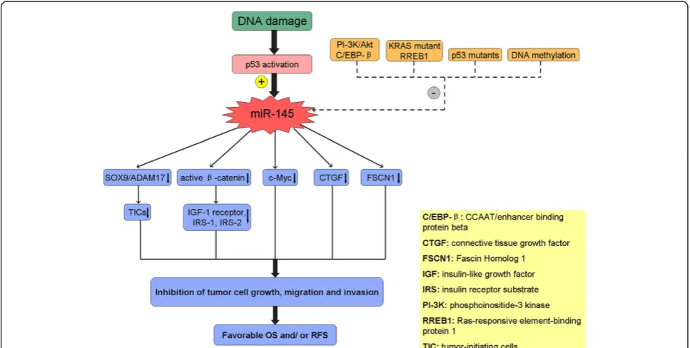
The anti-tumor effects of miR-145 with various mech-anisms have been demonstrated by abundant clinical and basic studies [8-11]. In hepatocellular carcinoma (HCC), miR-145 was found to target a number of genes along the signaling pathway of insulin-like growth factor (IGF), including IGF-1 receptor, insulin receptor substrate-1 (IRS-1), and IRS-2, all of which are directly downregulated
by miR-145 [8]. Law et al. also confirmed that miR-145
modulates the IGF signaling pathway by reducing its down-stream mediator, the active β-catenin [9]. In addition, p53 * Correspondence:yj197912@163.com
†Equal contributors
1
Department of Urology, First Affiliated Hospital of Nanjing Medical University, Nanjing 210029, China
Full list of author information is available at the end of the article
can induce miR-145 by binding directly to its promoter, after which miR-145 can silence c-Myc, demonstrating the role of miR-145 in p53-mediated c-Myc repression [10]. In head and neck squamous cell carcinoma (SCC), miR-145 also can target the SOX9/ADAM17 axis to in-hibit tumor-initiating cells and IL-6-mediated paracrine effects [11] (Figure 1).
As a tumor suppressor, miR-145 has been reported to have downregulated expression in various cancer tissues, although the targets of miR-145 have not been defini-tively identified [8-11]. A number of studies have shown significant associations between low miR-145 expression and poor cancer prognosis, but other studies did not find any significant association, and still others showed a negative correlation [12,13] that might cast doubt on the anti-oncogenic role of miR-145. However, in spite of these contradictory results, miR-145 is still an attractive tumor biomarker with considerable prognostic value, and deserves to be further investigated. Therefore, we conduc-ted a meta-analysis to clarify the preliminary predictive value of miR-145 in tumor prognoses.
Methods
Search strategy, eligibility criteria, and data extraction
We performed this meta-analysis following the guide-lines of the Meta-analysis of Observational Studies in Epidemiology group (MOOSE) [14]. Because newly pub-lished original works may get overlooked, we searched PubMed, EMBASE, and Web of Science up to March 2014 to identify relevant studies. Several combinations
of the following keywords were simultaneously applied: ‘cancer’,‘carcinoma’, ‘neoplasm’, ‘tumour’, ‘tumor’,‘ micro-RNA-145’, ‘microrna-145’, ‘miRNA-145’, ‘miR-145’, ‘ sur-vival’, ‘recurrence’, ‘relapse’, ‘metastasis’, and ‘prognosis’. Studies were considered eligible for further evaluation when they met the following criteria: studies that 1) fo-cused on patients with any malignant neoplasmI and 2) investigated the association between miR-145 expression and prognosis outcomes. In addition, the bibliographies of all eligible studies were reviewed for additional relevant publications to supplement our literature search. When studies deriving from the same series of study subjects were reported in multiple publications, only the most recent and the most complete studies were used for the meta-analysis.
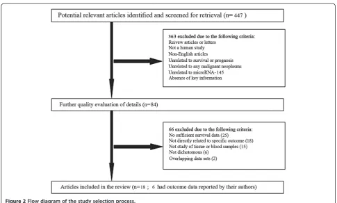
To fit the eligible criteria, selected studies had to be published in English, had to focus on human malignant tumors, and had to have performed stratified analyses on patient prognoses using the dichotomous expression levels of miR-145. Studies lacking the key data of hazard ratios (HRs) or confidence intervals (CIs), without survival curves, were not analyzed. A flow diagram of the study selection process is presented in Figure 2.
Extracted data elements included the following: 1) first author name and publication year; 2) characteristics of the studied population, including patient nationality, eth-nicity, disease, pathological type, and sample category; 3) detection method and cut-off definition; 4) follow-up time; and 5) HRs of elevated miR-145 expression for overall survival (OS), recurrence-free survival (RFS),
free survival (DFS), metastasis-free survival (MFS), and progression-free survival (PFS), along with their 95% CIs
and P values. If HRs and 95% CIs were not directly
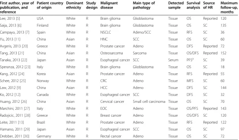
reported in publications, the total numbers of cases and deaths in each group were extracted to calculate HRs [15]. If only Kaplan-Meier curves were available, data were extracted from graphical survival plots to extrapolate HRs and 95% CIs, using previously described methods [16,17]. All the aforementioned data are comprehensively detailed in Table 1 and Table 2.
Statistical analysis
The aggregation of HRs and 95% CIs were calculated following Tierney method [17]. Forest plots were used to estimate the effect of miR-145 expression on patient sur-vival and disease progress. Heterogeneity test for pooled HRs was verified by Cochran Q-test and Higgins I-squared statistic (I2). Heterogeneity was considered statistically sig-nificant atP< 0.1 or if the percentage of I2was greater than 50%; if so, the random-effects model (DerSimonian and Laird method) was applied, otherwise, the fixed-effects model (Mantel-Haenszel test) was used [18]. In addition, we also executed stratified analyses to minimize the influ-ence of heterogeneity by classifying analyzed studies into subgroups based on similar characteristics. Publication bias was estimated by Egger linear regression test with a funnel plot [19]. AllP values were two-sided and a P< 0.05 was considered statistically significant. All statistical analyses
were conducted with Stata® (v11; StataCorp LP, College Station, TX, USA).
Results
Summary of analyzed studies
In total, 447 studies focusing on the relationship between miR-145 and cancer were identified from an initial online literature search, and 363 studies were excluded by man-ual screening of titles and abstracts. The full text of the remaining 84 studies was further evaluated, and finally, 18 studies [5-7,9,11-13,20-30] were considered eligible for this meta-analysis (Table 1). The selection process and ex-cluding reasons of candidate studies are shown in detail in Figure 2.
and PFS. All of the analyzed studies were retrospective, and maximal follow-up time ranged from 18 to 152 months. The main characteristics of analyzed studies are systematic-ally listed in Table 1 and Table 2.
Overall survival associated with miR-145 expression
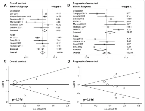
Ten articles analyzed OS, and seven of these showed statistical significance (Table 2). Significant heterogeneity between studies was observed (P< 0.001, I2= 83.9%), thus a random-effects model was applied to estimate a pooled HR along with 95% CI. Our results showed that upre-gulated miR-145 expression was a significant predictor of favorable OS (pooled HR = 0.47, 95% CI 0.31 to 0.72) (Table 3; Figure 3A).
Furthermore, we performed stratified analyses by clas-sifying studies into subgroups of ethnicity and main pathologic type (Table 3; Figure 3A; Figure 4A). First, six studies in the White subgroup displayed a better OS associated with elevated miR-145 expression (pooled HR = 0.67, 95% CI 0.47 to 0.95) by a random-effects
model (P= 0.032, I2= 59.1%). The other four studies
in Asians also showed that high miR-145 expression was significantly associated with a favorable OS (pooled HR = 0.35, 95% CI 0.19 to 0.64) using a random model
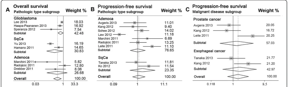
(P = 0.025, I2= 67.9%). Second, eight studies were divided into three main pathologic subgroups of adenocarcinoma, SCC, and glioblastoma. High miR-145 expression was found to be significantly associated with favorable OS in both SCC (pooled HR = 0.34, 95% CI 0.13 to 0.93) and glioblastoma (pooled HR = 0.72, 95% CI 0.52 to 0.99). No significant association was found for the adenocarcinoma subgroup (pooled HR = 0.47, 95% CI 0.14 to 1.61).
Tumor progression associated with miR-145 expression
We analyzed tumor progression by combining disease recurrence and metastasis. Eleven studies were included in PFS analysis, and five showed statistical significance (Table 2). A random-effects model was applied to calcu-late the pooled HR along with 95% CI for the significant heterogeneity (P < 0.001, I2= 72.9%), but failed to show any statistical significance (pooled HR = 0.87, 95% CI 0.51 to 1.47) (Table 3).
Stratified analyses displayed that high miR-145 expres-sion was a significantly favorable prediction for tumor progression in Asian subgroup of four studies (pooled HR = 0.43, 95% CI 0.21 to 0.89) by a random-effects model (P = 0.090, I2= 53.8%), but failed to show a signifi-cant association between miR-145 expression and tumor Table 1 Main characteristics of studies included in the meta-analysis
First author, year of
Lee, 2013 [5] USA White R Brain glioma Glioblastoma Tissue OS Reported 120
Saija, 2013 [6] Finland White R Brain glioma Glioblastoma Tissue OS SC 135
Campayo, 2013 [7] Spain White R NSCLC Adeno/SCC Tissue RFS SC 36
Yu, 2013 [11] China Asian R HNC SCC Tissue OS SC 60
Avgeris, 2013 [20] Greece White R Prostate cancer Adeno Tissue DFS Reported 72
Tang, 2013 [21] China Asian R Osteosarcoma Sarcoma Tissue OS/DFS Reported 152
Tanaka, 2013 [22] Japan Asian R Esophageal cancer SCC Serum PFSa SC 39
Speranza, 2012 [23] Italy White R Brain glioma Glioblastoma Tissue OS SC 18
Kang, 2012 [24] Korea Asian R Prostate cancer Adeno Tissue RFS Reported 55
Schee, 2012 [25] Norway White R CRC Adeno Tissue MFS SC 60
Law, 2012 [9] China Asian R HCC Adeno Tissue DFS SC 144
Ko, 2012 [12] Canada White R Esophageal cancer SCC Tissue DFS SC 32
Huang, 2012 [26] China Asian R Cervical cancer Small cell carcinoma Tissue OS SC 70
Marchini, 2011 [27] Italy White R EOC Adeno Tissue OS/PFS Reported 143
Radojicic, 2011 [28] Greece White R Breast cancer Adeno Tissue OS/DFS SC 120
Leite, 2011 [13] Brazil White R Prostate cancer Adeno Tissue RFS Reported 122
Hamano, 2011 [29] Japan Asian R Esophageal cancer SCC Tissue OS SC 97
Drebber, 2011 [30] Germany White R Rectal cancer Adeno Tissue OS SC 72
Adeno, adenocarcinoma; CRC, colorectal cancer; DFS, disease-free surviva; EOC, epithelial ovarian cancer; HCC, hepatocellular carcinoma; HNC, head and neck cancer; HR, hazard ratio; MFS, metastasis-free survival; NSCLC, non-small cell lung cancer; OS, overall survival; P, prospective; PFS, progression-free survival; R, retrospective; RCC, renal cell carcinoma; RFS, relapse-free survival; SC, survival curve; SCC, squamous cell carcinoma.
a
Table 2 Patient survival or disease progression associated with miR-145 expression in analyzed studies
First author, year of publication, and reference
Assay method
Cut-off value
Cases, n OS PFSa
High expression Low expression HR (95% CI) P HR (95% CI) P
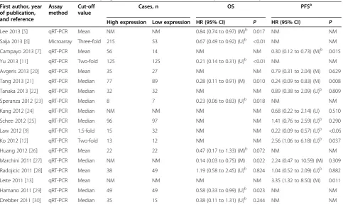
Lee 2013 [5] qRT-PCR Mean NM NM 0.84 (0.74 to 0.97) (M)b 0.017 NM NM
Saija 2013 [6] Microarray Three-fold 215 53 0.67 (0.49 to 0.92) (U)b <0.01 NM NM
Campayo 2013 [7] qRT-PCR Mean 56 14 NM NM 0.30 (0.12 to 0.73) (M)b 0.015
Yu 2013 [11] qRT-PCR Two-fold 125 125 0.21 (0.14 to 0.31) (U)b <0.01 NM NM
Avgeris 2013 [20] qRT-PCR Mean 35 27 NM NM 0.79 (0.31 to 2.04) (M) 0.629
Tang 2013 [21] qRT-PCR Median 77 89 0.28 (0.11 to 0.91) (M) 0.010 0.24 (0.09 to 0.83) (M) 0.008
Tanaka 2013 [22] qRT-PCR Median 32 32 NM NM 0.89 (0.38 to 2.09) (U)b 0.809
Speranza 2012 [23] qRT-PCR Median 8 7 0.23 (0.06 to 0.83) (U)b 0.018 NM NM
Kang 2012 [24] qRT-PCR Median NM NM NM NM 0.68 (0.22 to 2.14) (U) 0.510
Schee 2012 [25] qRT-PCR Median 96 97 NM NM 1.41 (0.76 to 2.59) (U)b 0.290
Law 2012 [9] qRT-PCR 1.5-fold 15 32 NM NM 0.22 (0.09 to 0.57) (U)b <0.05
Ko 2012 [12] qRT-PCR Two-fold 13 12 NM NM 2.56 (1.06 to 6.18) (U)b 0.037
Huang 2012 [26] qRT-PCR Mean 22 22 0.47 (0.17 to 1.33) (M)b 0.072 NM NM
Marchini 2011 [27] qRT-PCR Median NM NM 0.14 (0.03 to 0.75) (M) 0.022 2.24 (0.47 to 10.59) (M) 0.309
Radojicic 2011 [28] qRT-PCR Mean 38 49 1.19 (0.58 to 2.45) (U)b 0.824 1.04 (0.52 to 2.09) (U)b 0.882
Leite 2011 [13] qRT-PCR Mean NM NM NM NM 3.35 (1.32 to 8.50) (M) 0.011
Hamano 2011 [29] qRT-PCR Median 49 49 0.58 (0.33 to 0.99) (U)b 0.023 NM NM
Drebber 2011 [30] qRT-PCR Median 35 15 0.38 (0.11 to 1.31) (U)b 0.244 NM NM
CI, confidence interval; DFS, disease-free survival; HR, hazard ratio; M, multivariate analysis; MFS, metastasis-free survival; NM, not mentioned; OS, overall survival; PFS, progression-free survival; qRT-PCR, quantitative real-time PCR; RFS, relapse-free survival; U, univariate analysis.
a
PFS included any of the following: DFS, MFS or RFS.
b
HR and 95% CI calculated by survival curve.
Table 3 Patient survival or disease progression by total and stratified analyses
Subgroup OS PFSa
n HR (95% CI) Pvalue n HR (95% CI) Pvalue
Total 10 0.47 (0.31 to 0.72)b <0.001 11 0.87 (0.51 to 1.47)b 0.596
Subtotal by ethnicity
White 6 0.67 (0.47 to 0.95)b 0.026 7 1.27 (0.71 to 2.26)b 0.420
Asian 4 0.35 (0.19 to 0.64)b 0.001 4 0.43 (0.21 to 0.89)b 0.024
Subtotal by disease
Brain glioma 3 0.72 (0.52 to 0.99)b 0.045 – – –
Prostate cancer – – – 3 1.25 (0.45 to 3.48)b 0.671
Esophageal cancer – – – 2 1.50 (0.53 to 4.22)b 0.443
Subtotal by main pathology
Adeno 3 0.47 (0.14 to 1.61)b 0.228 7 1.01 (0.55 to 1.89)b 0.965
SCC 2 0.34 (0.13 to 0.93)b 0.035 2 1.50 (0.53 to 4.22)b 0.443
Glioblastoma 3 0.72 (0.52 to 0.99)b 0.045 – – –
OS, overall survival; PFS, progression-free survival including any of relapse-free survival (RFS), disease-free survival (DFS), and metastasis-free survival (MFS); N, number of studies; HR, hazard ratio; CI, confidence interval; SqCa, squamous carcinoma; Adeno, adenocarcinoma.
a
PFS included any of the following: DFS, MFS or RFS.
b
The HRs and 95% CIs of the analyzed studies were pooled by the random-effects model if thePvalue for heterogeneity was less than 0.10 or I2
progression in White subgroup of seven studies (pooled HR = 1.27, 95% CI 0.71 to 2.26) (Table 3; Figure 3B). In addition, no significant relevance was observed in sub-groups of PCa (pooled HR = 1.25, 95% CI 0.45 to 3.48), adenocarcinoma (pooled HR = 1.01, 95% CI 0.55 to 1.89), and squamous carcinoma (pooled HR = 1.50, 95% CI 0.53 to 4.22) (Table 3; Figure 4B; Figure 4C).
Publication bias
Publication bias of total analyses for patient survival and tumor progression was evaluated by funnel plots and Egger tests. As expected, the funnel plots were symmet-rical and thePvalues of the Egger test were 0.076 for OS and 0.566 for PFS, suggesting the absence of significant publication bias (Figure 3C, D).
Discussion
Aberrant expression of miRNAs has been found in vari-ous diseases including human carcinomas, and specific miRNAs have been shown to play crucial roles in the biological behaviors of initiation, progression, migration, and invasion of tumors [31-35]. Compared with mRNAs and proteins, miRNAs are more stable and not easily degraded. They can be detected accurately by qRT-PCR in both fresh and formalin-fixed tissues [36], and can also be quantified in serum, urine, or saliva samples [37]. Therefore, miRNAs are considered promising tumor biomarkers for early diagnosis and accurate progno-sis, as well as potential targets for clinical treatment [1,3,22,36,38,39].
survival and prognostic outcomes of patients with cancer [5-9,11,21,27,29]. Lee et al. reported that low miR-145 expression in glial tumors predicted poor prognosis, and upregulated miR-145 significantly decreased the migration and invasion of glioma cells by targeting connective tissue growth factor [5]. Campayoet al. found that low miR-145 expression independently predicted a shorter time to relapse in patients with NSCLC [7]. In addition, down-regulated miR-145 expression was found in PCa tissue compared with benign prostatic hyperplasia tissue, and this was correlated with higher Gleason score, advanced clinical stage, larger tumor diameter, and higher levels of prostate-specific antigen. The loss of the anti-oncogenic miR-145 may result in higher risk of biochemical recur-rence, shorter DFS, and worse OS in patients with PCa [20]. In the present meta-analysis, we gathered the avail-able evidence from all relevant studies to evaluate the prognostic values of miR-145 for malignant neoplasms. Our results demonstrated that high miR-145 expression was significantly correlated with favorable OS in overall analyses (P< 0.001), but did not exhibit an obvious association with tumor progression (P= 0.596) (Table 3). These inconsistent outcomes might hint at dissimilar potential mechanisms that affect patient survival or tumor progression.
Studies on miRNA expression profiles have indicated that downregulation of miR-145 is a common event in malignant disease [5,21,26]. However, little is known about why miR-145 is often downregulated in tumors. Recent studies show several underlying mechanisms playing key roles in regulating miR-145 expression, espe-cially in relation to p53, the central tumor suppressor. Suh et al. found that downregulation of miR-145 was mediated through DNA methylation and p53 mutation pathways [40], which frequently occur in various malignant
tumors. p53 can enhance post-transcriptional maturation of miR-145 in response to DNA damage, and transcrip-tionally inactive p53 mutants result in attenuation of miRNA biological processing activity and predict worse prognosis in patients with low miR-145 expression [41]. Loss of miR-145 expression is also observed frequently in
KRAS-mutated pancreatic cancer, and the downregulation
of miR-145 requires Ras-responsive element-binding pro-tein (RREB1) to repress its promoter [42]. In addition,
Sachdeva et al. suggested that a regulatory system of
miR-145 involving the Akt and CCAAT/enhancer binding protein beta (C/EBP-β) may contribute to the downregula-tion of miR-145 in cancer cells [43] (Figure 1).
Based on the respective results of the analyzed studies, we found that genetic background, pathology, or disease type seemed to have specific effects on the association of miR-145 expression and patient prognosis. In addition, both of the overall analyses for patient survival and dis-ease progression presented significant heterogeneity. All of these data demonstrated that the pooled results of over-all analyses are crude and cannot give accurate values of miR-145 for prognosis. Stratified analyses based on ethnic affiliation, pathology, or disease categories should be carried out to minimize the impact of heterogeneity.
considerable impact on the prognostic role of miR-145. High expression of miR-145 seemed to be predict favor-able OS in patients with SCC or glioblastoma but not adenocarcinoma, but these results need to be confirmed by further research.
These results indicate that miR-145 is a promising bio-marker to predict prognosis for patients with cancers. However, the conclusion is not sufficiently persuasive, and needs to be further refined for several reasons. First, no independent studies for black were included in our analysis, and this omission might hinder comprehensive investigation. Second, the number of analyzed studies was not adequate, which weakens the reliability of our results and hindered the execution of some subgroup analyses. Furthermore, all the studies included in our meta-analysis were retrospective, and no prospective studies were available, which also weakens the values of pooled results. Third, although there was no evidence of obvious publication bias in this meta-analysis, there is a possibility of language bias because only studies published in English were included. In addition, the tendency for authors and journals to publish only studies with positive results may also be a source of bias. Fourth, the expression of miR-145 was detected by tissue samples in all of the analyzed articles except one, which used serum. Different results might be obtained for detection of miR-145 in peripheral blood samples. Furthermore, as a cancer bio-marker, detection of miR-145 ub serum samples is more convenient, faster and more acceptable for patients to dynamically monitor their prognosis and therapeutic effects through their lifetime. Therefore, the association between patient prognosis and serum expression of miR-145 should be further investigated. Fifth, it remains unknown whether miR-145 should be used as an inde-pendent biomarker or as part of a combination of sev-eral biomarkers for predicting tumor prognosis. Using
Cox proportional regression analysis, Avgeris et al.
demonstrated that patients with PCa who had lower miR-145 expression exhibited a significantly higher risk for disease recurrence. Furthermore, this unfavorable prognosis, associated with low miR-145 expression, was independent of patient Gleason score, clinical stage, PSA levels, and age [20]. Lawet al. also reported that miR-145 independently coordinated the regulation of many components along the IGF pathway via its multigene tar-gets, and was an independent prognostic predictor [9]. By contrast, Huanget al. claimed that the downregulation of a combination of six miRNAs including miR-145 was signifi-cantly correlated with advanced stage, lymph node metas-tasis, and poor prognosis in small cell carcinoma of the cervix [26]. Liuet al. also reported that Fascin Homolog 1 (FSCN1) could be co-regulated by miR-43 and miR-145, and suggested the combination of miR-143 and miR-145 as a potential biomarker for the prognosis of esophageal
cancer [47]. The combined application of miR-145 with other miRNAs might confer more specificity for pre-dicting patient prognosis in various malignant diseases, but this hypothesis needs to be proved by more clinical research.
Conclusions
Our meta-analysis results indicate that high miR-145 expression is more suitable as a biomarker to predict favorable patient survival rather than to predict tumor progression, especially for SCC and glioblastoma in Asians. Given the current insufficient evidence, further inves-tigations and more studies are needed to focus on the relationship between miR-145 expression and patient prognosis.
Abbreviations
CI:Confidence interval; DFS: Disease-free survival; HCC: Hepatocellular carcinoma; HNC: Head and neck cancer; HR: Hazard ratio; I2: Higgins I-squared
statistic; MFS: Metastasis-free survival; miR-145: MicroRNA-145; miRNA: MicroRNA; NSCLC: Non-small cell lung cancer; OS: Overall survival; PCa: Prostate cancer; PFS: Progression-free survival; qRT-PCR: Quantitative reverse transcription PCR; RFS: Recurrence-free survival.
Competing interests
The authors declare that they have no conflict of interests.
Authors’contributions
JY and JL conceived and designed the study; JC and CC collected the data; YX and XS checked and verified the data input by JC and CC; JZ and JC performed statistical analyses; JY and JZ drafted the manuscript; and JY and JL revised the manuscript. All authors have read and approved the final version.
Author details
1Department of Urology, First Affiliated Hospital of Nanjing Medical
University, Nanjing 210029, China.2Department of General Surgery, Nanjing Hospital Affiliated to Nanjing Medical University, Nanjing, China.3State Key
Laboratory of Oral Diseases, West China School of Stomatology, Sichuan University, Chengdu, China.4Institute of Stomatology, Department of Oral
and Maxillofacial Surgery, School of Stomatology, Nanjing Medical University, Nanjing, China.
Received: 22 March 2014 Accepted: 20 July 2014 Published: 9 August 2014
References
1. Karakatsanis A, Papaconstantinou I, Gazouli M, Lyberopoulou A, Polymeneas G, Voros D:Expression of microRNAs, miR-21, miR-31, miR-122, miR-145, miR-146a, miR-200c, miR-221, miR-222, and miR-223 in patients with hepatocellular carcinoma or intrahepatic cholangiocarcinoma and its prognostic significance.Mol Carcinog2013,52(4):297–303.
2. Lu J, Getz G, Miska EA, Alvarez-Saavedra E, Lamb J, Peck D, Sweet-Cordero A, Ebert BL, Mak RH, Ferrando AA, Downing JR, Jacks T, Horvitz HR, Golub TR:microRNA expression profiles classify human cancers.Nature2005, 435(7043):834–838.
3. Wang QZ, Xu W, Habib N, Xu R:Potential uses of microRNA in lung cancer diagnosis, prognosis, and therapy.Curr Cancer Drug Targets2009, 9(4):572–594.
4. Rong M, Chen G, Dang Y:Increased MiR-221 expression in hepatocellular carcinoma tissues and its role in enhancing cell growth and inhibiting apoptosis in vitro.BMC Cancer2013,13:21.
5. Lee HK, Bier A, Cazacu S, Finniss S, Xiang C, Twito H, Poisson LM, Mikkelsen T, Slavin S, Jacoby E, Yalon M, Toren A, Rempel SA, Brodie C:microRNA-145 is downregulated in glial tumors and regulates glioma cell migration by targeting connective tissue growth factor.PLoS One2013,8(2):e54652. 6. Haapa-Paananen S, Chen P, Hellström K, Kohonen P, Hautaniemi S,
identifies MicroRNAs essential for glioma proliferation.PLoS One2013, 8(4):e60930.
7. Campayo M, Navarro A, Viñolas N, Diaz T, Tejero R, Gimferrer JM, Molins L, Cabanas ML, Ramirez J, Monzo M, Marrades R:Low miR-145 and high miR-367 are associated with unfavourable prognosis in resected nonsmall cell lung cancer.Eur Respir J2013,41(5):1172–1178.
8. Shi B, Sepp-Lorenzino M, Prisco M, Linsley P, de Angelis T, Baserga R:Micro RNA 145 targets the insulin receptor substrate-1 and inhibits the growth of colon cancer cells.J Biol Chem2007,282:32582–32590.
9. Law PT, Ching AK, Chan AW, Wong QW, Wong CK, To KF, Wong N:miR-145 modulates multiple components of the insulin-like growth factor pathway in hepatocellular carcinoma.Carcinogenesis2012,33(6):1134–1141. 10. Sachdeva M, Zhu S, Wu F, Wu H, Walia V, Kumar S, Elble R, Watabe K, Mo
YY:p53 represses c-Myc through induction of the tumor suppressor miR-145.Proc Natl Acad Sci U S A2009,106(9):3207–3212.
11. Yu CC, Tsai LL, Wang ML, Yu CH, Lo WL, Chang YC, Chiou GY, Chou MY, Chiou SH:miR145 targets the SOX9/ADAM17 axis to inhibit tumor-initiating cells and IL-6-mediated paracrine effects in head and neck cancer.Cancer Res
2013,73(11):3425–3440.
12. Ko MA, Zehong G, Virtanen C, Guindi M, Waddell TK, Keshavjee S, Darling GE:microRNA expression profiling of esophageal cancer before and after induction chemoradiotherapy.Ann Thorac Surg2012,94(4):1094–1102. discussion 1102–3.
13. Leite KR, Tomiyama A, Reis ST, Sousa-Canavez JM, Sañudo A, Dall'Oglio MF, Camara-Lopes LH, Srougi M:microRNA-100 expression is independently related to biochemical recurrence of prostate cancer.J Urol2011, 185(3):1118–1122.
14. Stroup DF, Berlin JA, Morton SC, Olkin I, Williamson GD, Rennie D, Moher D, Becker BJ, Sipe TA, Thacker SB:Meta-analysis of observational studies in epidemiology: a proposal for reporting. meta-analysis Of Observational Studies in Epidemiology (MOOSE) group.JAMA2000,283:2008–2012. 15. Parmar MK, Torri V, Stewart L:Extracting summary statistics to perform
meta-analyses of the published literature for survival endpoints.
Stat Med1998,17(24):2815–2834.
16. Williamson PR, Smith CT, Hutton JL, Marson AG:Aggregate data meta-analysis with time-to-event outcomes.Stat Med2002,21(22):3337–3351.
17. Tierney JF, Stewart LA, Ghersi D, Burdett S, Sydes MR:Practical methods for incorporating summary time-to-event data into metaanalysis.Trials2007, 8:16.
18. DerSimonian R, Laird N:Meta-analysis in clinical trials.Control Clin Trials
1986,7(3):177–188.
19. Egger M, Davey Smith G, Schneider M, Minder C:Bias in meta-analysis detected by a simple, graphical test.BMJ1997,315(7109):629–634. 20. Avgeris M, Stravodimos K, Fragoulis EG, Scorilas A:The loss of the
tumour-suppressor miR-145 results in the shorter disease-free survival of prostate cancer patients.Br J Cancer2013,108(12):2573–2581.
21. Tang M, Lin L, Cai H, Tang J, Zhou Z:MicroRNA-145 downregulation associates with advanced tumor progression and poor prognosis in patientssuffering osteosarcoma.Onco Targets Ther2013,6:833–838. 22. Tanaka K, Miyata H, Yamasaki M, Sugimura K, Takahashi T, Kurokawa Y,
Nakajima K, Takiguchi S, Mori M, Doki Y:Circulating miR-200c levels significantly predict response to chemotherapy and prognosis of patients undergoing neoadjuvant chemotherapy for esophageal cancer.
Ann Surg Oncol2013,20(Suppl 3):S607–S615.
23. Speranza MC, Frattini V, Pisati F, Kapetis D, Porrati P, Eoli M, Pellegatta S, Finocchiaro G:NEDD9, a novel target of miR-145, increases the invasiveness of glioblastoma.Oncotarget2012,3(7):723–734. 24. Kang SG, Ha YR, Kim SJ, Kang SH, Park HS, Lee JG, Cheon J, Kim CH:Do
microRNA 96, 145 and 221 expressions really aid in the prognosis of prostate carcinoma?Asian J Androl2012,14(5):752–757.
25. Schee K, Boye K, Abrahamsen TW, Fodstad Ø, Flatmark K:Clinical relevance of microRNA miR-21, miR-31, miR-92a, miR-101, miR-106a and miR-145 in colorectal cancer.BMC Cancer2012,12:505.
26. Huang L, Lin JX, Yu YH, Zhang MY, Wang HY, Zheng M:Downregulation of six microRNAs is associated with advanced stage, lymph node metastasis and poor prognosis in small cell carcinoma of the cervix.
PLoS One2012,7(3):e33762.
27. Marchini S, Cavalieri D, Fruscio R, Calura E, Garavaglia D, Fuso Nerini I, Mangioni C, Cattoretti G, Clivio L, Beltrame L, Katsaros D, Scarampi L, Menato G, Perego P, Chiorino G, Buda A, Romualdi C, D'Incalci M: Association between miR-200c and the survival of patients with stage I
epithelial ovarian cancer: a retrospective study of two independent tumour tissue collections.Lancet Oncol2011,12(3):273–285. 28. Radojicic J, Zaravinos A, Vrekoussis T, Kafousi M, Spandidos DA,
Stathopoulos EN:MicroRNA expression analysis in triple-negative (ER, PR and Her2/neu) breast cancer.Cell Cycle2011,10(3):507–517. 29. Hamano R, Miyata H, Yamasaki M, Kurokawa Y, Hara J, Moon JH, Nakajima K,
Takiguchi S, Fujiwara Y, Mori M, Doki Y:Overexpression of miR-200c induces chemoresistance in esophageal cancers mediated through activation of the Akt signaling pathway.Clin Cancer Res2011,17(9):3029–3038. 30. Drebber U, Lay M, Wedemeyer I, Vallböhmer D, Bollschweiler E, Brabender J,
Mönig SP, Hölscher AH, Dienes HP, Odenthal M:Altered levels of the onco-microRNA 21 and the tumor-supressor microRNAs 143 and 145 in advanced rectal cancer indicate successful neoadjuvant chemoradiotherapy.
Int J Oncol2011,39(2):409–415.
31. Bai Y, Liao H, Liu T, Zeng X, Xiao F, Luo L, Guo H, Guo L:MiR-296-3p regulates cell growth and multi-drug resistance of human glioblastoma by targeting ether-à-go-go (EAG1).Eur J Cancer2013,49(3):710–724. 32. Dudda JC, Salaun B, Ji Y, Palmer DC, Monnot GC, Merck E, Boudousquie C,
Utzschneider DT, Escobar TM, Perret R, Muljo SA, Hebeisen M, Rufer N, Zehn D, Donda A, Restifo NP, Held W, Gattinoni L, Romero P:MicroRNA-155 is required for effector CD8 (+) T cell responses to virus infection and cancer.
Immunity2013,38(4):742–753.
33. Manavalan TT, Teng Y, Litchfield LM, Muluhngwi P, Al-Rayyan N, Klinge CM: Reduced expression of miR-200 family members contributes to antiestrogen resistance in LY2 human breast cancer cells.PLoS One2013, 8(4):e62334.
34. Fu G, BrkićJ, Hayder H, Peng C:MicroRNAs in human placental development and pregnancy complications.Int J Mol Sci2013, 14(3):5519–5544.
35. DeCastro AJ, Dunphy KA, Hutchinson J, Balboni AL, Cherukuri P, Jerry DJ, DiRenzo J:miR203 mediates subversion of stem cell properties during mammary epithelial differentiation via repression ofΔNP63αand promotes mesenchymal-to-epithelial transition.Cell Death Dis2013, 4:e514.
36. Ferracin M, Veronese A, Negrini M:Micromarkers: miRNAs in cancer diagnosis and prognosis.Expert Rev Mol Diagn2010,10:297–308. 37. Kim DJ, Linnstaedt S, Palma J, Park JC, Ntrivalas E, Kwak-Kim JY,
Gilman-Sachs A, Beaman K, Hastings ML, Martin JN, Duelli DM:Plasma components affect accuracy of circulating cancer-related microRNA quantitation.J Mol Diagn2012,14(1):71–80.
38. Landi MT, Zhao Y, Rotunno M, Koshiol J, Liu H, Bergen AW, Rubagotti M, Goldstein AM, Linnoila I, Marincola FM, Tucker MA, Bertazzi PA, Pesatori AC, Caporaso NE, McShane LM, Wang E:MicroRNA expression differentiates histology and predicts survival of lung cancer.Clin Cancer Res2010, 16:430–441.
39. Yu SL, Chen HY, Chang GC, Chen CY, Chen HW, Singh S, Cheng CL, Yu CJ, Lee YC, Chen HS, Su TJ, Chiang CC, Li HN, Hong QS, Su HY, Chen CC, Chen WJ, Liu CC, Chan WK, Chen WJ, Li KC, Chen JJ, Yang PC:MicroRNA signature predicts survival and relapse in lung cancer.Cancer Cell2008,13:48–57.
40. Suh SO, Chen Y, Zaman MS, Hirata H, Yamamura S, Shahryari V, Liu J, Tabatabai ZL, Kakar S, Deng G, Tanaka Y, Dahiya R:MicroRNA-145 is regulated by DNA methylation and p53 gene mutation in prostate cancer.Carcinogenesis2011,32:772–778.
41. Suzuki HI, Yamagata K, Sugimoto K, Iwamoto T, Kato S, Miyazono K: Modulation of microRNA processing by p53.Nature2009, 460:529–533.
42. Kent OA, Chivukula RR, Mullendore M, Wentzel EA, Feldmann G, Lee KH, Liu S, Leach SD, Maitra A, Mendell JT:Repression of the miR-143/145 cluster by oncogenic Ras initiates a tumor-promoting feed-forward pathway.Genes Dev2010,24(24):2754–2759.
43. Sachdeva M, Liu Q, Cao J, Lu Z, Mo YY:Negative regulation of miR-145 by C/EBP-βthrough the Akt pathway in cancer cells.Nucleic Acids Res2012, 40(14):6683–6692.
44. Yazici H, Zipprich J, Peng T, Akisik EZ, Tigli H, Isin M, Akisik EE, Terry MB, Senie RT, Li L, Peng M, Liu Z, Dalay N, Santella RM:Investigation of the miR16-1 (C > T) + 7 substitution in seven different types of cancer from three ethnic groups.J Oncol2009,2009:827532.
45. Bovell LC, Shanmugam C, Putcha BD, Katkoori VR, Zhang B, Bae S, Singh KP, Grizzle WE, Manne U:The prognostic value of microRNAs varies with patient race/ethnicity and stage of colorectal cancer.Clin Cancer Res
46. Huang RS, Gamazon ER, Ziliak D, Wen Y, Im HK, Zhang W, Wing C, Duan S, Bleibel WK, Cox NJ, Dolan ME:Population differences in microRNA expression and biological implications.RNA Biol2011, 8(4):692–701.
47. Liu R, Liao J, Yang M, Sheng J, Yang H, Wang Y, Pan E, Guo W, Pu Y, Kim SJ, Yin L:The cluster of miR-143 and miR-145 affects the risk for esophageal squamous cell carcinoma through co-regulating fascin homolog 1.
PLoS One2012,7(3):e33987.
doi:10.1186/1477-7819-12-254
Cite this article as:Yanget al.:Prognostic role of microRNA-145 in various human malignant neoplasms: a meta-analysis of 18 related studies.World Journal of Surgical Oncology201412:254.
Submit your next manuscript to BioMed Central and take full advantage of:
• Convenient online submission
• Thorough peer review
• No space constraints or color figure charges
• Immediate publication on acceptance
• Inclusion in PubMed, CAS, Scopus and Google Scholar
• Research which is freely available for redistribution