1071-412X/03/$08.00⫹0 DOI: 10.1128/CDLI.10.4.696–701.2003
Copyright © 2003, American Society for Microbiology. All Rights Reserved.
Use of Mouse Models To Evaluate the Persistence, Safety, and
Immune Modulation Capacities of Lactic Acid Bacteria
Sonia Pavan,
1Pierre Desreumaux,
2and Annick Mercenier
1*
Bacte´riologie des Ecosyste`mes, Institut Pasteur de Lille,1and Equipe propre INSERM 0114
Physiopathologie des Maladies Inflammatoires Intestinales, CHU Lille,2Lille, France
Received 16 May 2002/Returned for modification 8 February 2003/Accepted 26 April 2003
Recent clinical and experimental observations showed that specific probiotic microorganisms may provide therapeutic benefits in inflammatory bowel disease. However, a rigorous screening for new candidate probiotic strains with optimized therapeutic properties necessitates also determining possible adverse interactions with the host, particularly in individuals who are not healthy. We have evaluated the persistence of strains of lactic acid bacteria (LAB) in the digestive tracts of mice, their immunomodulation capacity, and their safety in healthy animals and in a colitis model. Following daily administration of 109
CFU of viable LAB orally, intragastrically, or intrarectally, the animals’ feces were examined for bacterial excretion and cytokines were quantified in intestinal samples by quantitative reverse transcription-PCR. The level of bacterial translocation was assessed in healthy mice and in mice suffering from colitis induced by 2,4,6-trinitrobenzene sulfonic acid (TNBS). Irrespective of the route of administration, the potential probiotic strain Lactobacillus plantarum
NCIMB8826 was found to persist for up to 10 days in the digestive tracts of mice. This strain did not induce detrimental effects in healthy or in TNBS-treated animals, as was reflected by the absence of weight loss, intestinal inflammation, modification of cytokine levels in the ileum and colon (healthy mice), and bacterial dissemination (healthy and colitic animals). Moreover, the translocation of endogenous microflora to the mesenteric lymph nodes and spleen was greatly reduced in the TNBS-treated mice after administration of LAB. This property, together with the strain’s persistence capacity and innocuousness renders L. plantarum
NCIMB8826 an attractive candidate as a probiotic to be used in the prevention or treatment of chronic inflammation.
Inflammatory bowel disease (IBD), including Crohn’s dis-ease and ulcerative colitis, is a significant health care problem of unknown etiology which affects 0.5% of the population in northern Europe. It is a chronic immune-mediated disease in which endogenous bacteria are thought to play an important role, as suggested by numerous clinical observations and ex-perimental studies, summarized in recent reviews (21, 41). Notably, most of the models in which animals develop spon-taneous or chemically induced inflammatory colitis are influ-enced by the bacterial flora present in the intestinal lumen. Despite the fact that our current understanding of the com-plexity of the intestinal flora is very limited, many studies have shown that not all bacterial species have equal activities in promoting or reducing intestinal inflammation (15, 35–38, 46). This evidence has led to a new concept in the therapy of IBD based on probiotic preparations which usually contain lacto-bacilli, bifidobacteria, orEscherichia colistrains (6, 12, 18, 20, 25, 39, 45). However, many of these therapeutic trials make limited allowances for (i) optimal frequency and route of pro-biotic administration, (ii) rational selection of bacterial strains, (iii) possible adverse effects, and (iv) in vivo immune effects of probiotics on the intestinal mucosa.
Hence, the selection of new probiotic strains should take into account the above-mentioned points. To optimize the
dose and frequency of administration, the persistence of the bacteria in the gastrointestinal tract (GIT) should be investi-gated. At present, these data are available only for a very few strains used in oral therapeutic trials. In animal models most data are obtained from intragastric gavages. Other modes of administration, such as the oral and rectal routes, also deserve to be evaluated in mice, as they may be more relevant to the treatment of certain pathologies.
The influence of probiotic strains on the immune system is often evaluated through in vitro or ex vivo measurements of cytokine or immunoglobulin production, T- or B-cell prolifer-ation, or nitric oxide induction (14). The physiological rele-vance of these observations remains unclear because they show very little correlation with in vivo and clinical studies. More-over, the techniques used for cytokine quantification (reverse transcriptase PCR, immunohistochemical analysis, or enzyme-linked immunosorbent assay) differ greatly between studies and may be the cause of sometimes contradictory conclusions. Among the possible adverse events of regular probiotic feed-ing, the risk of bacterial translocation (BT; i.e., the passage of viable indigenous bacteria from the GIT to the mesenteric lymph nodes [MLN] and other extraintestinal sites) (5) should be evaluated carefully. In a healthy host, BT is a highly regu-lated event which occurs continuously at a low rate. When the integrity of the intestinal barrier is disturbed or when the immune system is not able to confine an infection, pathogenic bacteria can reach the bloodstream and cause septicemia (4). Consequently, it appears necessary to verify the safety of pro-biotic strains by assessing their potential to translocate, not
* Corresponding author. Present address: Functional Microbiology, Department of Nutrition and Health, Nestle´ Research Center, Vers-chez-les-Blanc, 1000 Lausanne 26, Switzerland. Phone: 41 21 785 84 66. Fax: 41 21 785 85 49. E-mail: [email protected].
696
on August 17, 2020 by guest
http://cvi.asm.org/
only in healthy individuals but also in individuals with injured intestinal mucosae.
Our laboratory selected the human isolate Lactobacillus plantarumNCIMB8826 as a promising probiotic strain based, on the one hand, on its technological properties and, on the other hand, on its ability to survive passage through the human stomach (47) and to induce secretion of anti-inflammatory cytokines by peripheral blood mononuclear cells (32). In this paper, we evaluated the persistence of strain NCIMB8826 af-ter oral, intragastric, or intrarectal administration to mice and compared its persistence to that of other lactic acid bacteria (LAB), including the reference probiotic strainLactobacillus salivariusUCC118. This strain is indeed well documented for its survival and persistence in mouse and human GITs, its influence on cytokine expression, and its therapeutic effects in animal models of colitis and in patients with IBD (12, 13, 34). Furthermore, we compared L. plantarum NCIMB8826 and Lactococcus lactisMG1363 (a dairy starter derivative) to de-termine their influence on cytokine levels in the intestinal mucosa. Finally, we evaluated the potential risk of BT in a model that uses 2,4,6-trinitrobenzene sulfonic acid (TNBS) to induce colitis (22, 33) by using TNBS doses that very severely damaged (histological score, 6) the intestinal barrier.
MATERIALS AND METHODS
Bacteria and growth conditions.ThreeLactobacillusstrains and one Lacto-coccusstrain were used in this study.L.plantarumNCIMB8826 (National Col-lection of Industrial and Marine Bacteria, Aberdeen, United Kingdom) was isolated from human saliva.L. plantarum256 (provided by P. Pouwels, TNO, Leiden, The Netherlands) is a silage isolate (S. Ahrne´, personal communication), andL.salivariusspp.salivariusUCC118 (provided by J. K. Collins, University College Cork, Cork, Ireland) was isolated from the human intestine.Lactococcus lactisMG1363 (17) is a nonpathogenic strain derived from a cheese starter. In order to monitor the bacteria after their administration to the mice, the strains were electrotransformed with a plasmid conferring resistance to chlorampheni-col or erythromycin. For chloramphenichlorampheni-col resistance, pTG2247 (23) was used with theL. plantarumstrains and pNZ8020 (7) was used with theL. salivarius strain, and for erythromycin resistance, pTREX (50) was used with the Lacto-coccus lactisstrain. Lactobacilli were grown at 37°C in MRS medium (Difco, Detroit, Mich.) supplemented with 10g of chloramphenicol (Sigma, St. Louis, Mo.) per ml under limited aeration.Lactococcus lactiswas grown at 30°C in M17 medium (Difco) supplemented with 0.5% glucose (GM17) and 5g of erythro-mycin (Sigma) per ml under limited aerobic conditions. It was verified that culturing the strains in vitro without antibiotic selection pressure did not lead to plasmid loss for at least a few generations.
Preparation of the bacterial strains and administration to mice.The strains were grown to an A600 of 1 to 2 (exponential growth phase), harvested by centrifugation at 3,000⫻gfor 10 min, washed with phosphate-buffered saline (PBS), and resuspended at the appropriate cell concentration either in 0.2 M NaHCO3buffer containing 2% glucose or in PBS. Bacterial suspensions were prepared daily. For oral administration, mice received 109
CFU resuspended in bicarbonate buffer by mouth (25l) or intragastrically (100 l). Intrarectal administration of 109
CFU resuspended in PBS (100l) was performed with mice anaesthetized with 3 mg of ketamine (Imalgene 1000; Me´rial, Lyon, France), 46.7g of diazepam (Valium; Roche Diagnostics), and 15g of atro-pine (Aguettant Laboratory, Lyon, France), using a 3.5 F catheter (EO 3416-1; Biotrol, Chelles, France) inserted 4 cm proximal to the anus.
Study design.Animal experiments were performed in an accredited establish-ment (number A59107; animal facility of the Institut Pasteur de Lille) according to French governmental guidelines (number 86/609/CEE). Adult male BALB/c mice were purchased from Iffa Credo (Saint Germain sur l’Arbresle, France) and kept under filter top hoods. The mice had free access to tap water and standard mouse chow.
Persistence of LAB in the GITs of mice.Groups of four animals received a single dose per day of lactobacilli or lactococci for three consecutive days by the oral, intragastric, or intrarectal route. Fecal samples were collected daily, resus-pended at 100 mg of feces/ml in one-fourth-strength Ringer’s diluent, and
me-chanically homogenized. Dilutions were plated on the selected media described above and incubated at 37 or 30°C for 2 days before enumeration. The experi-ment was repeated three times for each strain and for each mode of adminis-tration tested.
Effects of LAB on the immune response.Groups of eight to nine mice were givenL. plantarumNCIMB8826,Lactococcus lactisMG1363, or carbonate buffer once a day for 4 days and were sacrificed on day 5 by cervical dislocation. Total RNA was extracted from ileal and colonic samples kept in TriReagent (Eurome-dex, Souffelweyersheim, France), and reverse transcribed into cDNA as de-scribed previously (9). Quantification of-actin, tumor necrosis factor alpha (TNF-␣), gamma interferon (IFN-␥), interleukin-4 (IL-4), and IL-10 cDNA was performed by PCR on 1g of cDNA using the appropriate competitors and primers (11). Quantification of the amplified cDNA was performed by electro-phoresis in a 3% agarose gel by means of an image analyzer (Gel Analyst; Clara Vision, Orsa, France). Results of the cytokine measurements were expressed as cytokine cDNA per-actin cDNA in picograms per milliliter.
Safety assessment of LAB in mice.The risk of BT was determined for healthy mice and mice with TNBS-induced colitis. This colitis model was used, as it results in severe colonic inflammation similar to that described for patients with IBD (31). For both experiments, groups of five to nine mice received buffer (negative control),L. plantarumNCIMB8826, orLactococcus lactisMG1363 orally once a day for 4 days. Colitis was induced in the experimental group of mice on day 5 by intrarectal administration of TNBS (150 mg/kg of body weight; Fluka, Saint Quentin Fallavier, France) mixed with an equal volume of ethanol, as previously described (9). Control mice received an intrarectal administration of 50% ethanol. Healthy mice were sacrificed on day 5, and mice with TNBS-induced colitis were sacrificed on day 7. The MLN and spleen were aseptically removed and immediately placed in one-fourth-strength Ringer’s diluent. The samples were mechanically homogenized, and cultures for both aerobic and anaerobic bacteria were performed on Columbia blood agar supplemented with 0.05% cystein hydrochloride and 0.5% glucose. Anaerobic conditions were ob-tained in hermetic jars with Anaerogen (Oxoid, Basingstoke, United Kingdom). Selective enumeration of L. plantarum NCIMB8826 or Lactococcus lactis MG1363 was performed by plating the organ suspensions on MRS-chloramphen-icol or GM17-erythromycin agar, respectively. Whenever the situation war-ranted, the mice were weighed daily and their appearance was evaluated accord-ing to an activity score (51) rangaccord-ing from 1 to 3 (1, lazy, slow movement; 2, intermediate level of activity; 3, active movement or searching). The colon and the small intestine were macroscopically and histologically scored as previously described (9), the histological scoring being performed in a blind manner. We used both the scoring described by Wallace et al. (49), which reflects the level of inflammation and the extent of ulceration (score range, 0 to 10), and the criteria described by Ameho et al. (2), which take into account the degree of the inflammatory infiltrate and cell alterations (score range, 0 to 6). Quantification of myeloperoxidase (MPO) in ileal and colonic samples was performed by immu-noblotting as described previously (9).
Statistics.The comparisons were analyzed by the nonparametric Kruskal-Wallis one-way analysis of variance test or by the Mann-Whitney U test. Differ-ences were judged to be statistically significant when thePvalue was⬍0.05.
RESULTS
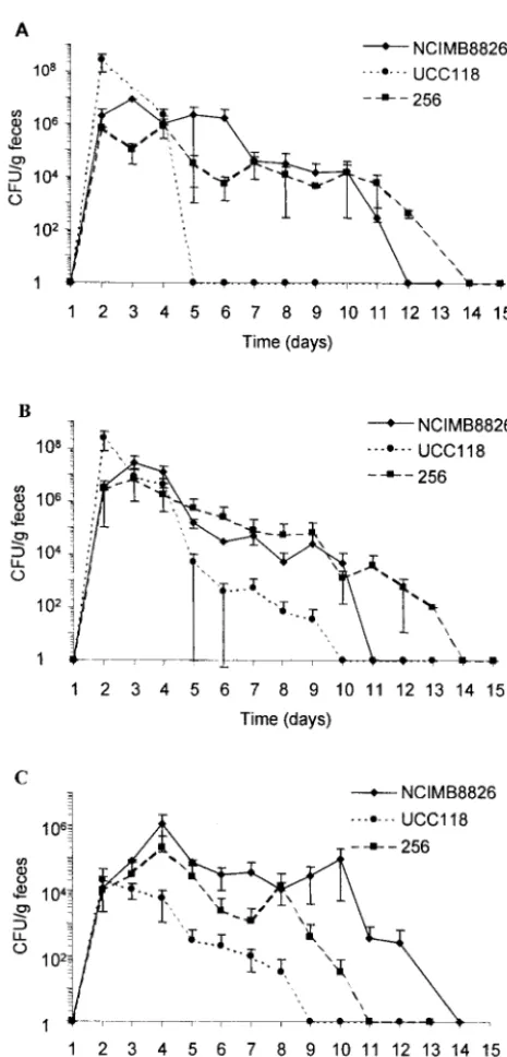
Persistence of LAB in the mouse intestine.Four strains of LAB were evaluated for their ability to persist in the GITs of mice after 3 days of oral, intragastric, or rectal administration. After the oral and intragastric administrations (Fig. 1A and B, respectively), the twoL. plantarumstrains were able to persist for 7 to 10 days after the last dose (day 3), with bacterial levels ranging from 104to 106CFU/g of feces during 1 week. After
intragastric administration,L. salivariusUCC118 was detected in feces at lower counts and for fewer days thanL. plantarum. This difference was even more pronounced after oral admin-istration, asL. salivariuswas recovered from the feces only 1 day after the last administration. The three Lactobacillus strains given by the intrarectal route were able to persist for several days (Fig. 1C). It is noteworthy that L. plantarum, especially strain NCIMB8826, was still found in the feces 10 days after the last administration and maintained itself at levels of approximately 105CFU/g of feces from days 4 to 10.
on August 17, 2020 by guest
http://cvi.asm.org/
Growth of the mouse endogenous intestinal flora (enterobac-teria and streptococci) on GM17-erythromycin as well as on other selective lactococcal media prevented us from obtaining precise persistence data for Lactococcus lactis. However, colonization studies from Gruzza et al. indicate thatLactococcus lactisonly passively transits through the digestive tracts of mice colonized with a human microflora (19). In view of the duration and high level of persistence of L. plantarum NCIMB8826 in mice, this strain was selected for the next experiments. Oral administration was retained, as it is less stress-inducing for the mice than the
intragastric route and as it is similar to natural food intake. More-over, this mode of administration corresponds to the one used in human clinical trials.Lactococcus lactisMG1363 was also chosen as a prototype of a noncolonizing strain (19, 47) that exhibits in vitro immunomodulation properties that are quite different from those ofL. plantarum(32).
Cytokine expression in the intestinal mucosa.We quantified TNF-␣, IFN-␥, IL-4, and IL-10 mRNA in the ilea and colons of mice receiving a single daily dose of L. plantarum NCIMB8826 or Lactococcus lactisfor four consecutive days (Fig. 2). Among the four cytokines, TNF-␣(levels 10- to 100-fold higher than those of the other cytokines) was dominant in healthy mice. IFN-␥was found in larger amounts in the colon than in the ileum, while IL-4 and IL-10 were more predomi-nant in the ileum than in the colon. However, only IL-4 levels appeared significantly higher in the ileum than in the colon (P
⫽0.009). This result indicates that the Th1/Th2 balance may differ between these two subcompartments. As shown in Fig. 2, no significant variation in the cDNA levels of TNF-␣, IL-4, or IL-10 was observed after oral administration of LAB. Only the level of IFN-␥was significantly increased in the ilea of mice
FIG. 1. Fecal counts ofL. plantarumNCIMB8826,L. plantarum256, andL. salivariusUCC118 given orally (A), intragastrically (B), or intrar-ectally (C) for three consecutive days (days 1 to 3). Data represented (numbers of CFU/gram of feces) are the arithmetic means⫾standard errors of the means of results from three separate experiments.
FIG. 2. Cytokine mRNA levels in the ilea (A) and the colons (B) of mice after oral administration for 4 days of carbonate buffer,L. plan-tarumNCIMB8826, orLactococcus lactisMG1363. Data correspond to the means of results from eight samples and are representative of two separate experiments.
on August 17, 2020 by guest
http://cvi.asm.org/
receiving Lactococcus lactis. However, the cDNA amounts were detected in only four of the eight samples and were so close to the detection limit of the method (0.2 pg/PCR) that the physiological significance of this increase in the level of IFN-␥ can be questioned. When heat-killed L. plantarum NCIMB8826 orLactococcus lactis MG1363 was given to the mice, the levels of the four cytokines remained identical to those observed with a buffer or live bacteria (data not shown).
Effects of repeated oral administration of LAB in healthy and TNBS-treated mice. Oral administrations of 109 CFU of L.
plantarum NCIMB8826 or Lactococcus lactis MG1363 were performed daily for 4 days, and different parameters were monitored to evaluate the potential adverse effect of repeated bacterial ingestion (Table 1). No variation in mouse activity and weight was observed in comparison to the activity and weight of the buffer control group. No sign of macroscopic or histological inflammation was detected (Wallace and Ameho scores of 0) either in the ileum or in the colon. MPO amounts, which reflect the levels of neutrophil recruitment in tissues, remained undetectable by Western blot analysis in all mice. The MLN and spleen cultures were negative for bothL. plan-tarumNCIMB8826 andLactococcus lactisMG1363, indicating that repeated ingestion of high doses of either strain did not induce abnormal BT or dissemination in healthy mice.
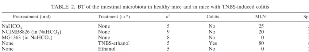
The BT of the global endogenous flora was also determined for healthy mice and for mice with TNBS-induced colitis. Rectal administration of TNBS resulted in increased BT in the MLN and spleen, as only 20% of the organs remained free of bacteria in these mice, whereas 50 to 75% of the organs remained sterile in healthy animals (Table 2). Bacteria were then enumerated in each positive organ (Fig. 3). In healthy mice receiving buffer, ethanol, L. plantarum NCIMB8826, orLactococcus lactis MG1363, low numbers of bacteria were found, reflecting the normal basal translocation level. In TNBS-treated mice the level of transloca-tion was much higher (107-fold increase). Interestingly, the total
number of intestinal bacteria that translocated was greatly re-duced in the TNBS-treated mice that preventively received L. plantarumNCIMB8826 (105-fold decrease) orLactococcus lactis
MG1363 (103-fold decrease) compared to numbers in the
TNBS-treated mice that received no preventive dose of bacteria. As observed with healthy mice, the orally givenLactobacillusor Lac-tococcus strains were not found in the MLN or spleens of the TNBS-treated mice in spite of the dramatic increase in the level of translocation of the endogenous microflora.
DISCUSSION
According to the current definition (40), a probiotic strain is expected to transiently persist in the GIT of the host and to reach high numbers of viable cells in the targeted part of the gut. Persistence is thought to be dependent on the mode of administration and on the intrinsic properties of the bacterial strain, including resistance to gastric acid and bile and adhe-sion to the intestinal epithelium or mucus (44). The pharma-cokinetic properties of potential probiotic strains should de-termine at which dose and for how long the strains have to be administered to the host. This determination, in turn, will allow correlation of the observed biological effect with the presence of the probiotic strain under study.
In this work we first compared the levels of persistence of four LAB strains administered to mice by different routes (oral, intragastric, and rectal). In addition to L. plantarum NCIMB8826 andLactococcus lactisMG1363, we chose to in-clude the well-described probiotic strainL. salivariusUCC118 as well as anotherL. plantarumstrain of nonhuman origin (L. plantarum256). Irrespective of the route of administration, the L. plantarumstrains were detected for longer periods than was L. salivarius UCC118 in the feces of mice. Interestingly, the best-surviving strain in our mouse experiments also performed very well in human feeding trials. Vesa et al. have shown that
TABLE 1. Effect on mice of oral administration of NaHCO3buffer,L. plantarumNCIMB8826, orLactococcus lactisMG1363a
Buffer or strain
No. of mice
Activity score
% Variation in wt (day 5⫺day 1)
Macroscopic score
Histological score
NCIMB8826 or MG1363 translocation
or disseminationb
OD with MPO
NaHCO3 8 3⫾0 ⫹1.7⫾1.9 0 0 ND 0
NC1MB8826 9 3⫾0 ⫹1.1⫾2.2 0 0 No 0
MG1363 8 3⫾0 ⫹6.4⫾7.9 0 0 No 0
aValues are means⫾standard errors of the means. OD, optical density; ND, not determined. bDetection level,ⱖ100 CFU/organ.
TABLE 2. BT of the intestinal microbiota in healthy mice and in mice with TNBS-induced colitis
Pretreatment (oral) Treatment (i.r.a) nb Colitis MLNc Spleenc
NaHCO3 None 5 No 25 50
NCIMB8826 (in NaHCO3) None 9 No 20 33
MG1363 (in NaHCO3) None 8 No 0 37
None TNBS-ethanol 5 Yes 80 80
None Ethanol 5 No 0 25
NCIMB8826 (in NaHCO3) TNBS-ethanol 8 Yes 75 62
MG1363 (in NaHCO3) TNBS-ethanol 8 Yes 86 43
a
i.r., intrarectal administration.
b
n, number of animals per group.
c
Data are the percentages of organs positive for bacterial cultures (detection limit,ⱖ100 CFU/organ).
on August 17, 2020 by guest
http://cvi.asm.org/
L. plantarum NCIMB8826 reached the human ileum with a survival rate of 7%, which was far superior to the survival rate of the other tested LAB (47). We also evaluated the intrarectal route as it might allow an increase in the concentrations of probiotics in the colon, which may be advantageous for specific applications, such as the treatment of pouchitis. Indeed, Mad-sen et al. reported that rectal administration ofLactobacillus reuteri in IL-10 gene-deficient mice prevents colitis and nor-malizes the colonicLactobacilluslevels (28). To the best of our knowledge, our study was the first to analyze the persistence rate of LAB delivered by the rectal route. The three Lactoba-cillus strains were found to establish themselves, though at variable levels. Again,L. plantarumNCIMB8826 was found in feces for especially long periods.
Previous work in the laboratory demonstrated that heat-inactivated L. plantarumNCIMB8826 andLactococcus lactis MG1363 differ substantially in their capacities to modulate cytokine production by human peripheral blood mononuclear cells. While the former leads to strong IL-10 production, the latter is characterized by the stimulation of TNF-␣, IFN-␥, and IL-12 (32). In the present study, we verified whether substan-tial in vivo cytokine modulation could also be measured after mice were fed withL. plantarumNCIMB8826 orLactococcus lactisMG1363. For this purpose, we used quantitative reverse transcriptase PCR rather than other in vitro or ex vivo tech-niques (for example, enzyme-linked immunosorbent assay), as this method was successfully employed in previous studies to measure mRNA expression directly in the intestinal mucosa (8–11). No major immunomodulatory effects were induced in the ileal and colonic mucosae of mice after the administration ofL. plantarumorLactococcus lactis. As an additional control, we administered heat-killedL. plantarumorLactococcus lactis to mice to verify whether the cytokine stimulation profile ob-served in vitro could be reproduced in vivo. This was not found to be the case. Nevertheless, these results were coherent with the data reported by Maassen et al. which show thatL. plan-tarum NCIMB8826 is a weaker cytokine inducer than other Lactobacillusspecies (27). In the human feeding trial described by Dunne et al., the systemic cytokine levels remained un-changed also after the consumption of L. salivariusUCC118 (12). It might be argued that in healthy individuals, probiotic strains should not definitely or significantly affect the natural
cytokine balance as this might, for example, increase the risk of uncontrolled inflammation. All together, these data indicate that immunomodulatory effects observed in vitro need to be corroborated by animal studies.
Finally, we examined the safety of the two LAB strains since a probiotic should, above all else, be innocuous even when the integrity of the host intestinal barrier is partly impaired. Even though dietary LAB benefit from the GRAS (generally recog-nized as safe) status, some reports have identifiedLactobacillus species associated with bacteremia or endocarditis (1, 3, 16, 24, 30, 48). We therefore chose to examine the rates of BT in a TNBS-induced acute colitis model that leads to important le-sions and necrosis in the mouse GIT. This model displays immunological features common to the chronic stage of Crohn’s disease. It is characterized by an inflammation of the colon marked by a transmural cellular infiltration with T cells and macrophages in a pattern similar to that described for the human situation (33, 42). It is noteworthy that we purposely used high doses of TNBS (150 mg/kg) in order to exacerbate the risk of BT, yet these experimental conditions are less ap-propriate for evaluating the capacity of probiotic LAB strains to prevent the onset of colitis. Significantly, in healthy as well as in TNBS-treated mice, feeding liveL. plantarumor Lacto-coccus lactisinduced no detrimental effects and no abnormal translocation of the administered bacteria. On the contrary, prefeeding TNBS-treated mice withL. plantarumNCIMB8826 and, to a lesser extent (2-log difference), with Lactococcus lactisMG1363 significantly reduced the BT of the global in-testinal microflora. Other studies have reported the preventive effect ofLactobacillus rhamnosusGG (26),Lactobacillus reuteri R2LC andL. plantarumDSM9843 (29), and Bifidobacterium longum (43) on BT, but the studies were performed mainly with healthy mice. In the present study, we showed that even when the intestinal mucosa was severely injured,L. plantarum andLactococcus lactisexerted a beneficial effect which might be critical for the restoration of the intestinal homeostasis. Under our experimental conditions, this effect did not seem to be mediated by strong in vivo cytokine modifications. At present, it is difficult to assess whether the greater efficacy ofL. plantarumthan that ofLactococcus lactisis linked to its supe-rior persistence capacity rather than to its intrinsic immuno-modulatory properties.
ACKNOWLEDGMENTS
This work was supported by Rhodia Food S.A.S., France, FEDER funds, and Institut Pasteur de Lille funding. This work was also sup-ported by a grant from the EUQLK1-2000-00146 research program.
We acknowledge P. Pouwels and J. K. Collins for providing L. plantarum 256 and L. salivarius UCC118 strains, respectively. We warmly thank B. Pot for critical reading of the manuscript.
REFERENCES
1. Aguirre, M., and M. D. Collins.1993. Lactic acid bacteria and human clinical infection. J. Appl. Bacteriol.75:95–107.
2. Ameho, C. K., A. A. Adjei, E. K. Harrison, K. Takeshita, T. Morioka, Y. Arakaki, E. Ito, I. Suzuki, A. D. Kulkarni, A. Kawajiri, and S. Yamamoto.
1997. Prophylactic effect of dietary glutamine supplementation on interleu-kin 8 and tumour necrosis factor alpha production in trinitrobenzene sul-phonic acid induced colitis. Gut41:487–493.
3. Bayer, A. S., A. W. Chow, D. Betts, and L. B. Guze.1978. Lactobacillemia— report of nine cases. Important clinical and therapeutic considerations. Am. J. Med.64:808–813.
4. Berg, R. D.1999. Bacterial translocation from the gastrointestinal tract. Adv. Exp. Med. Biol.473:11–30.
FIG. 3. BT of the intestinal microflora in MLN and spleen. Mice received carbonate,L. plantarumNCIMB8826, orLactococcus lactis
MG1363 orally and were treated or not with TNBS-ethanol or ethanol (EtOH) by intrarectal administration. Data correspond to mean num-bers of CFU per positive organ⫾standard errors of the means.
on August 17, 2020 by guest
http://cvi.asm.org/
5. Berg, R. D., and A. W. Garlington.1979. Translocation of certain indigenous bacteria from the gastrointestinal tract to the mesenteric lymph nodes and other organs in a gnotobiotic mouse model. Infect. Immun.23:403–411. 6. Campieri, M., and P. Gionchetti.1999. Probiotics in inflammatory bowel
disease: new insight to pathogenesis or a possible therapeutic alternative? Gastroenterology116:1246–1249.
7. de Ruyter, P. G. G. A., O. P. Kuipers, and W. M. de Vos.1996. Controlled gene expression systems forLactococcus lactiswith the food-grade inducer nisin. Appl. Environ. Microbiol.62:3662–3667.
8. Desreumaux, P., E. Brandt, L. Gambiez, D. Emilie, K. Geboes, O. Klein, N. Ectors, A. Cortot, M. Capron, and J. F. Colombel.1997. Distinct cytokine patterns in early and chronic ileal lesions of Crohn’s disease. Gastroenter-ology113:118–126.
9. Desreumaux, P., L. Dubuquoy, S. Nutten, M. Peuchmaur, W. Englaro, K. Schoonjans, B. Derijard, B. Desvergne, W. Wahli, P. Chambon, M. D. Lei-bowitz, J. F. Colombel, and J. Auwerx.2001. Attenuation of colon inflam-mation through activators of the retinoid X receptor (RXR)/peroxisome proliferator-activated receptor gamma (PPARgamma) heterodimer. A basis for new therapeutic strategies. J. Exp. Med.193:827–838.
10. Desreumaux, P., O. Ernst, K. Geboes, L. Gambiez, D. Berrebi, H. Muller-Alouf, S. Hafraoui, D. Emilie, N. Ectors, M. Peuchmaur, A. Cortot, M. Capron, J. Auwerx, and J. F. Colombel.1999. Inflammatory alterations in mesenteric adipose tissue in Crohn’s disease. Gastroenterology117:73–81. 11. Dombrowicz, D., S. Nutten, P. Desreumaux, C. Neut, G. Torpier, M. Peeters,
J. F. Colombel, and M. Capron.2001. Role of the high affinity immunoglob-ulin E receptor in bacterial translocation and intestinal inflammation. J. Exp. Med.193:25–34.
12. Dunne, C., L. Murphy, S. Flynn, L. O’Mahony, S. O’Halloran, M. Feeney, D. Morrissey, G. Thornton, G. Fitzgerald, C. Daly, B. Kiely, E. M. Quigley, G. C. O’Sullivan, F. Shanahan, and J. K. Collins.1999. Probiotics: from myth to reality. Demonstration of functionality in animal models of disease and in human clinical trials. Antonie Leeuwenhoek76:279–292.
13. Dunne, C., L. O’Mahony, L. Murphy, G. Thornton, D. Morrissey, S. O’Halloran, M. Feeney, S. Flynn, G. Fitzgerald, C. Daly, B. Kiely, G. C. O’Sullivan, F. Shanahan, and J. K. Collins.2001. In vitro selection criteria for probiotic bacteria of human origin: correlation with in vivo findings. Am. J. Clin. Nutr.73:386S-392S.
14. Erickson, K. L., and N. E. Hubbard.2000. Probiotic immunomodulation in health and disease. J. Nutr.130:403S-409S.
15. Garcia-Lafuente, A., M. Antolin, F. Guarner, E. Crespo, A. Salas, P. Forcada, M. Laguarda, J. Gavalda, J. A. Baena, J. Vilaseca, and J. R. Malagelada.1997. Incrimination of anaerobic bacteria in the induction of experimental colitis. Am. J. Physiol.272:G10–G15.
16. Gasser, F.1994. Safety of lactic acid bacteria and their occurrence in human clinical infections. Bull. Inst. Pasteur92:45–67.
17. Gasson, M. J.1983. Plasmid complements ofStreptococcus lactisNCDO 712 and other lactic streptococci after protoplast-induced curing. J. Bacteriol.
154:1–9.
18. Gionchetti, P., F. Rizzello, A. Venturi, P. Brigidi, D. Matteuzzi, G. Bazzocchi, G. Poggioli, M. Miglioli, and M. Campieri.2000. Oral bacteriotherapy as maintenance treatment in patients with chronic pouchitis: a double-blind, placebo-controlled trial. Gastroenterology119:305–309.
19. Gruzza, M., M. Fons, M. F. Ouriet, Y. Duval-Iflah, and R. Ducluzeau.1994. Study of gene transfer in vitro and in the digestive tract of gnotobiotic mice from Lactococcus lactis strains to various strains belonging to human intes-tinal flora. Microb. Releases2:183–189.
20. Guslandi, M., G. Mezzi, M. Sorghi, and P. A. Testoni.2000. Saccharomyces boulardii in maintenance treatment of Crohn’s disease. Dig. Dis. Sci.45:
1462–1464.
21. Hendrickson, B. A., R. Gokhale, and J. H. Cho.2002. Clinical aspects and pathophysiology of inflammatory bowel disease. Clin. Microbiol. Rev.15:
79–94.
22. Hoffmann, P., J. M. Zeeh, J. Lakshmanan, V. S. Wu, F. Procaccino, M. Reinshagen, J. A. McRoberts, and V. E. Eysselein.1997. Increased expres-sion of transforming growth factor alpha precursors in acute experimental colitis in rats. Gut41:195–202.
23. Hols, P., P. Slos, P. Dutot, J. Reymund, P. Chabot, B. Delplace, J. Delcour, and A. Mercenier.1997. Efficient secretion of the model antigen M6-gp41E in Lactobacillus plantarum NCIMB 8826. Microbiology143:2733–2741. 24. Husni, R. N., S. M. Gordon, J. A. Washington, and D. L. Longworth.1997.
Lactobacillus bacteremia and endocarditis: review of 45 cases. Clin. Infect. Dis.25:1048–1055.
25. Kruis, W., E. Schutz, P. Fric, B. Fixa, G. Judmaier, and M. Stolte.1997. Double-blind comparison of an oral Escherichia coli preparation and me-salazine in maintaining remission of ulcerative colitis. Aliment. Pharmacol. Ther.11:853–858.
26. Lee, D. J., R. A. Drongowski, A. G. Coran, and C. M. Harmon.2000. Evaluation of probiotic treatment in a neonatal animal model. Pediatr. Surg. Int.16:237–242.
27. Maassen, C. B., C. van Holten-Neelen, F. Balk, M. J. den Bak-Glashouwer, R. J. Leer, J. D. Laman, W. J. Boersma, and E. Claassen.2000.
Strain-dependent induction of cytokine profiles in the gut by orally administered Lactobacillus strains. Vaccine18:2613–2623.
28. Madsen, K. L., J. S. Doyle, L. D. Jewell, M. M. Tavernini, and R. N. Fedorak.
1999. Lactobacillus species prevents colitis in interleukin 10 gene-deficient mice. Gastroenterology116:1107–1114.
29. Mao, Y., S. Nobaek, B. Kasravi, D. Adawi, U. Stenram, G. Molin, and B. Jeppsson.1996. The effects of Lactobacillus strains and oat fiber on meth-otrexate-induced enterocolitis in rats. Gastroenterology111:334–344. 30. Maskell, R., and L. Pead.1992. 4-Fluoroquinolones and Lactobacillus spp as
emerging pathogens. Lancet339:929.
31. Morris, G. P., P. L. Beck, M. S. Herridge, W. T. Depew, M. R. Szewczuk, and J. L. Wallace.1989. Hapten-induced model of chronic inflammation and ulceration in the rat colon. Gastroenterology96:795–803.
32. Mu¨ller-Alouf, H., C. Grangette, D. Goudercourt, N. Reveneau, and A. Mer-cenier.1999. Comparative cytokine inducing pattern of lactic acid bacteria used for mucosal vaccine development. Immunol. Lett.69:33.
33. Neurath, M. F., I. Fuss, B. L. Kelsall, E. Stuber, and W. Strober.1995. Antibodies to interleukin 12 abrogate established experimental colitis in mice. J. Exp. Med.182:1281–1290.
34. O’Mahony, L., M. Feeney, S. O’Halloran, L. Murphy, B. Kiely, J. Fitzgibbon, G. Lee, G. O’Sullivan, F. Shanahan, and J. K. Collins.2001. Probiotic impact on microbial flora, inflammation and tumour development in IL-10 knockout mice. Aliment. Pharmacol. Ther.15:1219–1225.
35. Rath, H. C., H. H. Herfarth, J. S. Ikeda, W. B. Grenther, T. E. Hamm, Jr., E. Balish, J. D. Taurog, R. E. Hammer, K. H. Wilson, and R. B. Sartor.1996. Normal luminal bacteria, especially Bacteroides species, mediate chronic colitis, gastritis, and arthritis in HLA-B27/human2microglobulin trans-genic rats. J. Clin. Investig.98:945–953.
36. Rath, H. C., J. S. Ikeda, H. J. Linde, J. Scholmerich, K. H. Wilson, and R. B. Sartor.1999. Varying cecal bacterial loads influences colitis and gastritis in HLA-B27 transgenic rats. Gastroenterology116:310–319.
37. Rath, H. C., M. Schultz, R. Freitag, L. A. Dieleman, F. Li, H.-J. Linde, J. Scholmerich, and R. B. Sartor.2001. Different subsets of enteric bacteria induce and perpetuate experimental colitis in rats and mice. Infect. Immun.
69:2277–2285.
38. Rath, H. C., K. H. Wilson, and R. B. Sartor.1999. Differential induction of colitis and gastritis in HLA-B27 transgenic rats selectively colonized with Bacteroides vulgatusorEscherichia coli.Infect. Immun.67:2969–2974. 39. Rembacken, B. J., A. M. Snelling, P. M. Hawkey, D. M. Chalmers, and A. T.
Axon.1999. Non-pathogenic Escherichia coli versus mesalazine for the treat-ment of ulcerative colitis: a randomised trial. Lancet354:635–639. 40. Schrezenmeir, J., and M. de Vrese.2001. Probiotics, prebiotics, and
synbi-otics—approaching a definition. Am. J. Clin. Nutr.73:361S-364S. 41. Shanahan, F.2002. Crohn’s disease. Lancet359:62–69.
42. Strober, W., B. R. Ludviksson, and I. J. Fuss.1998. The pathogenesis of mucosal inflammation in murine models of inflammatory bowel disease and Crohn disease. Ann. Intern. Med.128:848–856.
43. Suzuki, T., K. Itoh, T. Kaneko, and H. Suzuki.1997. Inhibition of bacterial translocation from the gastrointestinal tract of mice by oral administration of a culture condensate of Bifidobacterium longum. J. Vet. Med. Sci.59:665– 669.
44. Tuomola, E., R. Crittenden, M. Playne, E. Isolauri, and S. Salminen.2001. Quality assurance criteria for probiotic bacteria. Am. J. Clin. Nutr.73: 393S-398S.
45. Venturi, A., P. Gionchetti, F. Rizzello, R. Johansson, E. Zucconi, P. Brigidi, D. Matteuzzi, and M. Campieri.1999. Impact on the composition of the faecal flora by a new probiotic preparation: preliminary data on maintenance treatment of patients with ulcerative colitis. Aliment. Pharmacol. Ther.13:
1103–1108.
46. Verdu, E. F., P. Bercik, B. Cukrowska, M. A. Farre-Castany, H. Bouzourene, E. Saraga, A. L. Blum, I. Corthesy-Theulaz, H. Tlaskalova-Hogenova, and P. Michetti.2000. Oral administration of antigens from intestinal flora anaer-obic bacteria reduces the severity of experimental acute colitis in BALB/c mice. Clin. Exp. Immunol.120:46–50.
47. Vesa, T., P. Pochart, and P. Marteau.2000. Pharmacokinetics of Lactoba-cillus plantarum NCIMB 8826, LactobaLactoba-cillus fermentum KLD, and Lacto-coccus lactis MG 1363 in the human gastrointestinal tract. Aliment. Phar-macol. Ther.14:823–828.
48. Wagner, R., and E. Balish.1998. Potential hazards of probiotic bacteria for immunodeficient patients. Bull. Inst. Pasteur96:165–170.
49. Wallace, J. L., W. K. MacNaughton, G. P. Morris, and P. L. Beck.1989. Inhibition of leukotriene synthesis markedly accelerates healing in a rat model of inflammatory bowel disease. Gastroenterology96:29–36. 50. Wells, J., and K. Schofield.1996. Cloning and expression vectors for
lacto-cocci, p. 37–62.InT. F. Bozoglu and R. Bibek (ed.), Lactic acid bacteria: current advances in metabolism, genetics, and applications. NATO ASI Series H, vol. H 98. Springer-Verlag, Heidelberg, Germany.
51. Zhou, J. S., Q. Shu, K. J. Rutherfurd, J. Prasad, P. K. Gopal, and H. S. Gill.
2000. Acute oral toxicity and bacterial translocation studies on potentially probiotic strains of lactic acid bacteria. Food Chem. Toxicol.38:153–161.