1
BIOCOMPATIBILITY OF 3D PRINTED METHACRYLATE FOR HEARING DEVICES
Frank Alifui-Segbaya 1* and Roy George 2
1 School of Dentistry and Oral Health, Griffith Health, Gold Coast campus, Griffith
University, QLD 4222, Australia. Email: [email protected]
2 School of Dentistry and Oral Health, Griffith Health, Griffith University, Australia.
Email [email protected]
* Corresponding Author
2
ABSTRACT
The capacity of 3D printing (3DP) technologies to initiate speedy polymerization of solvent free
resins accounts for their utility in the manufacturing of medical devices. Nonetheless,
independent biological evaluation of 3D printed materials is recommended due to the unique
parameters of the manufacturing process, which can influence their physical, chemical, and
biological properties. In this study, E-Shell 450 clear methacrylate indicated for 3DP of hearing
devices was examined for biological safety using zebrafish bioassays adapted to OECD fish
embryo test. In addition, the proprietary material was characterized for composition using
headspace gas-chromatography mass spectrometry (GC-MS). To initiate the biological test,
newly fertilized zebrafish eggs were cultured on non-treated and ethanol-treated methacrylates
in glass petri dishes with ultrapure water, incubated at 28.5°C and assessed for developmental
endpoints of toxicity at 24h interval until 96h. Toxicological data indicate non-treated
methacrylate is extremely toxic in zebrafish bioassays whereas ethanol-treated counterpart
showed a relative lower toxicity possibly due to ethanoic-aqueous interactions as observed by
GC-MS. With the current influx of 3D printing materials, users are urged to exercise caution.
Operators must also take cognizance of the potential toxicity of the chemicals used in 3DP and
implement safety measures to limit their exposure.
Keywords
3D printing; biocompatibility; hearing devices; methacrylates; zebrafish embryo model; digital
3
1 INTRODUCTION
The recent hype surrounding 3D printing (3DP) attests to its growing popularity in almost every
manufacturing sector including medicine, architecture, sports, aerospace and automotive
engineering and contemporary arts [1]. 3DP comprise a host of processes and technologies that
offer a diverse spectrum of capabilities for the manufacturing of end-use products and devices in
different materials [2]. The digital manufacturing process simply involves feeding a virtual
model (usually ‘STL’ file) into a designated 3D printer to build parts in successive layers until
the desired 3D part is completed. Despite the potentials of 3DP offering significant benefits in
terms of speed [3], independent biological evaluation of manufactured devices is highly
recommended [4,5] due to the unique parameters of the manufacturing process, which can
influence their physical, chemical, and biological properties [6]. In this study, E-Shell 450 clear
methacrylate indicated for hearing devices is examined for biological safety using zebrafish
bioassays adapted to the Organization for Economic Cooperation and Development (OECD)
fish embryo test [7]. The zebrafish is considered an excellent model for developmental toxicity,
offering economy and ease of quantifying multiple toxicity endpoints [8] as well as fulfilling the
pertinent aim to replace, reduce, or refine the use of animals for the purposes of research or
hazard identification [9,10]. Representative materials were built with digital light processing
(DLP) technology [11]. DLP is similar to stereolithography (SL) in that both are vat
photopolymerization processes that require washing built parts in organic solvents to remove any
wet resin remnants, followed by postcuring to harden them. However, DLP uses a more
conventional light source such as an arc lamp, with a liquid crystal display panel or a deformable
mirror device, which is applied to the entire surface of the vat of resin in a single pass, relatively
making it faster than SL [2]. To extrapolate toxicity effects to residual monomer and degradation
products [12] that may be present in the proprietary material, it was characterized for chemical
4
2 MATERIALS AND METHODS
EnvisionTec GmbH (Brüsseler Str. 51, 45968 Gladbeck, Germany) supplied 60x3 mm
disk-shaped samples that were built from E-Shell 450 clear resin. Postcuring (2 x 100 flashes) of
samples was completed by the manufacturer in Otoflash G171 (NK-Optik GmbH, Isarstr. 2,
D-82065 Baierbrunn, Germany). Table 1 shows the manufacturing parameters, hazardous
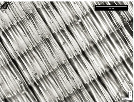
composition [13] and physical properties [14] of photocured E-Shell 450 clear (Figure 1).
To initiate the biological test, newly fertilized (1.5-hour postfertilized) zebrafish eggs (n=20)
obtained from FishCore (Australian Regenerative Medicine Institute, Monash University) were
cultured on non-treated (‘as-received’) and ethanol-treated [6] samples in glass petri dishes using
ultrapure water (3cm2:1mL ratio) [15] as test medium. The bioassays were incubated at 28.5 °C in Heracell CO2 incubator (Thermo Fisher Scientific Inc.) and assessed for developmental
endpoints [7,16,17] shown in Table 2 at 24h interval until 96h using Olympus MVX10 Research
Macro Zoom Microscope, Olympus DP 72 digital colour microscope camera and cellSens
imaging software (Olympus Soft Imaging Solutions GmbH). As per OECD test guidelines, fish
is considered dead if one of the lethal endpoints is present. Ethical approval (MARP/2015/094)
5
Table 1. Manufacturing parameters, composition and physical properties of E-Shell 450 methacrylate
MANUFACTURING PARAMETERS HAZARDOUS INGREDIENT(S) W/W % PHYSICAL PROPERTIES
Photocured samples were built with
Perfactory DDP 4M 3D printer.
Manufacturing parameters are z-height:
67.98mm, voxel: 100m and light power:
180 Mw/dm2).
60-80% Proprietary methacrylate oligomers;
15-30% Proprietary methacrylate monomers;
1-2% diphenyl 2,4,6-trimethylbenzoyl.
Flexural strength: 60-80 MPa;
Flexural modulus: 1200-1500 MPa;
Elongation at break: 2-4%;
Tensile strength: 40-48 MPa;
Tensile modulus: 2150-3250 MPa;
Izod impact: 30 J/m;
HDT: 75° at 1.82 MPa;
Hardness, D Scale: 82-85;
6
Figure 1.Surface topography of E-Shell 450 clear. Imaging was carried out with Olympus AX70
Fluorescence Microscope; Monochrome FViewII Peltier cooled digital camera (Olympus,
7
Table 2. Biomarkers of lethality, sublethality and teratogenicity assessed at 24h interval until
96h
24H 48H 72H 96H
LETHAL ENDPOINTS
Coagulation
.
.
.
.
Lack of somite formation
.
.
.
.
Non-detachment of tail-bud
.
.
.
.
Lack of heart-beat
.
.
.
.
SUBLETHAL DEVELOPMENTAL ENDPOINTS
Development of eyes
.
.
.
.
Spontaneous movement
.
.
.
.
Hypopigmentation
.
.
.
Formation of edemata
.
.
.
ENDPOINTS OF TERATOGENICITY
Spinal curvature and malformation of tail
.
.
.
.
Yolk deformation
.
.
.
.
8
3 RESULTS
After 24h, non-treated methacrylate induced ≈70% embryo death or lethality while surviving
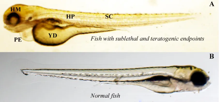
embryos (Fig. 2) were largely unhealthy, hence test was discontinued. Although ethanol-treated
methacrylate recorded only 5% mortality after 24h, additional 50% with cumulative
hypopigmentation, pericardial edema, yolk sac resorption delay and hypoactive behavior were
observed in surviving fish by 96h (Fig. 3). Average growth length in surviving fish after 96h
was 3241.30µm compared to 3590.33µm in controls. At the end of the test, fish were euthanized
in 0.4 % anaesthetic tricaine mesylate solution.
Figure 2. Toxicity effects induced by non-treated methacrylate in zebrafish embryo bioassays
after 24h (A): CE coagulated embryo, DE dead embryo, MF malformed embryo without somite
9
Figure 3. Fish in toxic (A) and control (B) bioassays after 96h. Note the phenotype differences
in unhealthy zebrafish (A): HM Head malformation HP Hypopigmentation SC Spinal curvature
PE Pericardial edemata YD Yolk sac resorption delay, as compared to normal zebrafish (B).
3.1 Qualitative gas chromatography-mass spectrometry
Since the toxicological effects observed in bioassays are likely due to residual monomer and
degradation products, the photocured materials were examined for chemical composition using
headspace GC-MS. Prior to analysis, they were frozen in liquid nitrogen at -196°C and ground
into powder before tested in Shimadzu TQ8040 GC-MS/MS (Shimadzu Corporation, Japan). GC
column is Agilent J&W DB5-MS 30m 0.25mm ID 0.25um film thickness. Test parameters were,
column oven temperature at 40.0 °C, injection temperature at 250 °C, column flow rate at 1.16
mL/min, split ratio of 5.0 and a total run time of 15 minutes. GC-MS data shown in Table 3
shows 16 chemical compounds that reduced to 5 with the applied ethanol treatment. For
10
Table 3. Chemical composition of non-treated and ethanol-treated photocured E-Shell 450 methacrylates
NON-TREATED E-SHELL 450 METHACRYLATE ETHANOL-TREATED E-SHELL 450 METHACRYLATE
2-Hydroxyethyl methacrylate Propylene glycol methyl ether
Propylene glycol methyl ether m-Xylene
Octyl Acrylate Methyl 3-methoxy-2-methylpropanoate
2-Propyl-1-pentanol Toluene
Benzaldehyde Undecane
Tetrahydrofufuryl Butyrate
Cyclohexanone
Cyclomethicone 5
Cyclomethicone 6
2,6,11-Trimethyldodecane
Ethylbenzene
N-[1-(4-Hydroxy-5-hydroxymethyltetrahydrofuran-2-yl)-4-oxo-
Texanol
Methyl 3-methoxy-2-methylpropanoate
Toluene
11
4 DISCUSSION
Experimental results in this study indicate non-treated methacrylate is extremely toxic in
zebrafish bioassay. Ethanol-treated methacrylate, on the other hand, induced a relatively lower
lethality but surviving fish showed cumulative sublethal and teratogenic effects. The improved
biological performance is likely due to induced swelling in polymeric chains, which allowed
chemical compounds to diffuse in the ultrapure water [18] used to rinse the samples.
Acrylics are probably the most versatile family of monomers that can be used to prepare
polymers with rigid, flexible, ionic, nonionic, hydrophobic, or hydrophilic properties [19]. They
are preferred in free-radical synthesis of plastics because of the high reactivity of the acrylate
double bond. Under intense ultraviolet illumination, crosslinking polymerization of liquid resins
proceeds extensively within a fraction of a second to generate a 3D polymer network [20]. As
per standards definitions [21], E-Shell 450 is a surface device hence does not require stringent
biological evaluation compared to methacrylates for intraoral devices. Nonetheless, uncured
methacrylate monomers can be absorbed through the skin [22] and cause allergic reactions (e.g.
dermatitis) [23,24]. In addition, the toxicity of methacrylate esters is theorized to involve
alkylation of critical cellular nucleophiles via Michael addition [25]. Some of the developmental
endpoints observed in toxic bioassays are comparable to those reported in animal studies that
linked methacrylic esters to embryonic fetal toxicity, teratogenicity [22] and cardiovascular
function [26,27]. The observed chemical compounds are also used in industrial applications and
can be toxic to humans if present in threshold dose. For instance, cyclomethicone is used in
cosmetic and personal products [28], 2-hydroxyethyl methacrylate for desensitizing teeth,
benzaldehyde for pharmaceutical products and texanol as fuel additives [29]. It is worth
emphasizing that, inhalation toxicity [30] may result from exposure to liquid photopolymer
12
outcomes, resins should possess high curing rate, good storage stability, low toxicity, low
viscosity, and display adequate mechanical properties after photocuring [31]. Resins with
relatively low viscosity are likely to produce rapid polymerisation yielding crosslinked polymers
with properties suited to the demands imposed by the target application [32]. Some 3D printers
have in-built heating mechanism for achieving the desired resin viscosity [33] but often not
applicable to third-party materials. In general, the mechanical properties of photocured materials
depend primarily on the chemical structure, functionality and concentration of the various
constituents of the resin, and the degree of cure [34].
5 Conclusion
With the current influx of 3D printers and materials [35], it is imperative that the biological
performance of 3D printed medical devices is not overlooked. Users are advised to exercise
caution and if necessary demand approved certification for the materials. Since 3DP is not a
“one-stop” manufacturing process, operators should take cognizance of the potential toxicity of
the chemicals used and implement safety measures to limit their exposure. The limitations of the
study lies in the extrapolation of the toxicological data to human responses hence quantitative
analysis of the observed compounds and their throughput in zebrafish bioassays are
recommended for further study.
6 ACKNOWLEDGEMENTS
The authors are grateful to EnvisionTec GmbH (Germany) for supplying E-Shell 450 samples
for the study. We are grateful to Professor GJ Lieschke (ARMI, Monash University, Melbourne)
for supporting the study with zebrafish resources and expertise, Ms. Sony Varma (ARMI,
Monash University, Melbourne) for helping with the biological test, and Mr. Adam White and
Mr. Jasper Bowman (School of Natural Sciences, Griffith University) for helping with GC-MS
13
7 CONFLICT OF INTEREST STATEMENT
No competing financial interests exist.
8 REFERENCES
1. Wohlers, T.; Gornet, T. History of additive manufacturing. 2014; p 34.
2. 3D Printing Industry. 3d printing basics: The free beginner’s guide.
http://3dprintingindustry.com/wp-content/uploads/2014/07/3D-Printing-Guide.pdf
(accessed on 16 January 2016),
3. Alifui-Segbaya, F.; Williams, R.J.; George, R. Additive manufacturing: A novel
method for fabricating cobalt-chromium removable partial denture frameworks.
European Journal of Prosthodontics and Restorative Dentistry 2017, 25, 73-78.
4. Stratasys. Safety data sheet (med610).
http://www.stratasys.com/materials/material-safety-data-sheets/polyjet/~/media/394dd5015bb6473496a19ea837881c25 (accessed on
30 November 2016),
5. Stratasys. Printing bio-compatible parts on polyjet™ 3d printers with med620.
http://usglobalimages.stratasys.com/Main/Files/SDS/MED620_Usage_Terms_1116.pdf
?v=636160085940019299 (accessed on 18 January 2016),
6. Alifui-Segbaya, F.; Varma, S.; Lieschke, G.J.; George, R. Biocompatibility of
photopolymers in 3d printing. 3d Print Addit Manuf 2017, 4, 185-191.
7. Organisation for Economic Co-operation and Development. Test no. 236: Fish embryo
acute toxicity (fet) test. OECD Publishing: Paris, 2013; pp 1-22.
8. Lieschke, G.J.; Currie, P.D. Animal models of human disease: Zebrafish swim into
view. Nat Rev Genet 2007, 8, 353-367.
9. International Organization for Standardization. Biological evaluation of medical
devices - part 2: Animal welfare requirements. International Organization for
Standardization: Geneva, 2006.
10. European Union. Directive 2010/63/eu of the european parliament and of the council of
22 september 2010 on the protection of animals used for scientific purposes. Official
Journal of the European Union 2010, 53, 33-79.
11. EnvisionTec GmBH. Advanced dlp for superior 3d printing.
14
12. Araújo, P.H.H.; Sayer, C.; Giudici, R.; Poço, J.G.R. Techniques for reducing residual
monomer content in polymers: A review. Polymer Engineering & Science 2002, 42,
1442-1468.
13. EnvisionTec GmbH. E-shell 450: Safety data sheet.
https://envisiontec.com/wp-content/uploads/2016/11/MK-SDS-EShell450Series-V20161114-FN-EN.pdf (accessed
on 21 May 2018),
14. EnvisionTec GmbH. E-shell® 450 series.
https://envisiontec.com/3d-printing-materials/perfactory-materials/e-shell-450-series/ (accessed on 22 July 2017),
15. International Organization for Standardization. Iso 10993-5, biological evaluation of
medical devices - part 5: Tests for in vitro cytotoxicity. International Organization for
Standardization: Geneva, 2009.
16. Nagel, R. Dart: The embryo test with the zebrafish danio rerio - a general model in
ecotoxicology and toxicology. Altex-Altern Tierexp 2002, 19, 38-48.
17. Lammer, E.; Carr, G.J.; Wendler, K.; Rawlings, J.M.; Belanger, S.E.; Braunbeck, T. Is
the fish embryo toxicity test (fet) with the zebrafish (danio rerio) a potential alternative
for the fish acute toxicity test? Comparative Biochemistry and Physiology Part C:
Toxicology & Pharmacology 2009, 149, 196-209.
18. Neves, C.B.; Lopes, L.P.; Ferrao, H.F.; Miranda, J.P.; Castro, M.F.; Bettencourt, A.F.
Ethanol postpolymerization treatment for improving the biocompatibility of acrylic
reline resins. BioMed research international 2013, 2013, 485246.
19. Merck. Acrylic monomers.
https://www.sigmaaldrich.com/materials-science/material-science-products.html?TablePage=16397884 (accessed on 3 December 2017),
20. Decker, C. Photoinitiated crosslinking polymerisation. Progress in Polymer Science
1996, 21, 593-650.
21. International Organization for Standardization. Iso 10993-1: Biological evaluation of
medical devices -part 1: Evaluation and testing within a risk management process.
International Organization for Standardization: Geneva, 2009.
22. Autian, J. Structure-toxicity relationships of acrylic monomers. Environmental Health
Perspectives 1975, 11, 141-152.
23. Geurtsen, W. Polymethylmethacrylate resins. In Biocompatibility of dental materials,
15
24. Kanerva, L.; Estiander, T.; Jolanki, R.; Tarvainijn, K. Occupational allergic contact
dermatitis caused by exposure to acrylates during work with dental prostheses. Contact
Dermatitis 1993, 28, 268-275.
25. McCarthy, T.J.; Witz, G. Structure-activity relationships in the hydrolysis of acrylate
and methacrylate esters by carboxylesterase in vitro. Toxicology 1997, 116, 153-158.
26. Mir, G.N.; Lawrence, W.H.; Autian, J. Toxicological and pharmacological actions of
methacrylate monomers i: Effects on isolated, perfused rabbit heart. Journal of
Pharmaceutical Sciences 1973, 62, 778-782.
27. Mir, G.N.; Lawrence, W.H.; Autian, J. Toxicological and pharmacological actions of
methacrylate monomers iii: Effects on respiratory and cardiovascular functions of
anesthetized dogs. Journal of Pharmaceutical Sciences 1974, 63, 376-381.
28. DrugBank. Cyclomethicone 5. https://www.drugbank.ca/drugs/DB11244 (accessed on
18 June 2018),
29. National Center for Biotechnology Information. Pubchem compound database.
https://pubchem.ncbi.nlm.nih.gov/ (accessed on 19 June 2018),
30. Azimi, P.; Zhao, D.; Pouzet, C.; Crain, N.E.; Stephens, B. Emissions of ultrafine
particles and volatile organic compounds from commercially available desktop
three-dimensional printers with multiple filaments. Environmental Science & Technology
2016, 50, 1260-1268.
31. Li, J.; Cui, Y.; Qin, K.; Yu, J.; Guo, C.; Yang, J.; Zhang, C.; Jiang, D.; Wang, X.
Synthesis and properties of a low-viscosity uv-curable oligomer for three-dimensional
printing. Polymer Bulletin 2016, 73, 571-585.
32. Vitale, A.; Cabral, J.T. Frontal conversion and uniformity in 3d printing by
photopolymerisation. Materials 2016, 9, 760.
33. Formlabs. Expanding 3d printing technologies to high-volume applications and beyond.
Mary Ann Liebert, Inc. publishers: pp 1-53.
34. Decker, C. Kinetic study and new applications of uv radiation curing. Macromolecular
Rapid Communications 2002, 23, 1067-1093.