ISSN 2286-4822
www.euacademic.org DRJI Value: 5.9 (B+)
Study of serum levels of Melatonin, Paraoxonase,
Oxidative stress in Iraqi patients with Acute
Myocardial Infarction
ENTEDHAR RIFAAT SARHAT
Ph.D. Clinical Biochemistry, Dentistry College University of Tikrit, Iraq
THURAIA R. SARHAT
B.Sc. of Science, Iraq
DINA N.TAWFEEQ
M.B.Ch.B. College of Medicine, University of Tikrit, Iraq
Abstract:
Background: Acute myocardial infarction continues to be a major health concern. It contributes to morbidity and may end fatally. The aim of the study was to evaluate melatonin, paraoxonase levels and correlate these parameters to oxidative stress and lipid profile in AMI.
Methodology: A melatonin, paraoxonase (PON), malondialdehyde (MDA), Nitric oxide (NO), superoxide dismutase (SOD), and glutathione peroxidase (GPx) were analyzed in 60 subjects, 30 AMI patients and 30 age/sex-matched controls.
Results: The current study reported that the patients with AMI had significantly lower level of melatonin, PON, SOD, TAC, NO and HDL and higher level of MDA, GPx, cholesterol, and TG levels than the healthy controls. The positive correlation was found between melatonin with paraoxonase, SOD, GPx, NO, TAS, HDL, and glucose, also there was positive correlation between paraoxonase with MDA, SOD, GPx, HDL.
independent of other traditional cardiovascular risk factors, Melatonin may provide a novel therapeutic target.
Key words: Myocardial infarction, Melatonin, paraoxonase, Nitric oxide.
INTRODUCTION
Acute myocardial infarction (AMI) is defined as a part of acute coronary syndrome characterized by a typical clinical syndrome consisting of chest pain, dyspnea with rise and fall in troponin or creatine kinase–MB to values greater than 99% of a normal reference population with 7.2 million deaths and 12.2 % of total
deaths, coronary heart disease (CHD) is a worldwide disease (1).
Melatonin (N-acetyl-5-methoxytryptamine), a tryptophan derivative secreted by the pineal gland, is a highly evolutionarily conserved molecule – present virtually in all organisms(2). Evidence from the last ten years suggests that
melatonin may influence the cardiovascular system in
humans(3). Furthermore, exogenous melatonin has induced
several hemodynamic effects in healthy men and women(4).
Serum paraoxonase (PON1) EC 3.1.8.1 an
arylesterasesynthesised in the liver. HDL-C associated enzyme which is responsible for the antioxidant properties of HDL. This enzyme plays an important role in preventing LDL–C oxidation, it is considered to protect against the development of coronary heart disease. Knowledge about the link between paraoxonase activity and atherosclerosis comes largely from the biological rather than epidemiological studies as there is evidence that peroxidation of LDL-C is an important factor for atherosclerosis (5). About 25% of cell death in cardiomyocytes
Oxidative stress is the state of imbalance between the reactive oxygen species (ROS) and the ability of a biological system to detoxify readily the reactive intermediates. Development of oxidative stress because of free oxygen radical generation has been implicated in the pathogenesis of many diseases including Parkinson's disease, Alzheimer's disease, atherosclerosis, heart
failure, myocardial infarction and even cancer(6).
Malondialdehyde (MDA), a carbonile group produced during lipid peroxidation, is used widely in determining oxidative
stress(7). Progression of atherosclerosis is correlated with
oxidative stress and can be followed up by MDA measurements
(8).
The key antioxidant enzymes, including superoxide
dismutase (SOD), catalase (CAT) and glutathione peroxidase (GPx), provide a defense system against oxidative stress by removing the OFR, thus protecting cells from oxidative damage
(9). However, the endogenous antioxidant activity is severely
damaged after ischemia-reperfusion which makes the myocardium extremely vulnerable to OFR. Moreover, exogenous SOD and CAT cannot be delivered into living cells because of the poor permeability and selectivity of the cell membrane, which has limited its usage in protecting
cells/tissues from oxidative stress damage (10). Nitric oxide (NO),
which is produced by the action of endothelial nitric oxide synthase (eNOS) in the vascular endothelium, is a potent antiatherosclerotic factor. Together, NO and the potent vasoconstrictor endothelin-1 (ET- 1) act as a pair of mutually constraining factors to play a key role in the regulation of vascular tone. An imbalance in either of these opposing factors can result in disease development(11).
SUBJECTS AND METHODS
A total of 30 patients (aged 40-60 years) with AMI who were admitted to Tikrit Teaching Hospital / Tikrit / Iraq were enrolled to this cross-sectional, case-controlled study. The diagnosis of AMI was established according to the presence of two of the following criteria: i-prolonged chest pain compatible with AMI, ii-typical electrocardiogram changes, iii-raising of cardiac enzymes. A second control group was comprised of 30 sex- and age-matched subjects with similar geographic and socioeconomic background without AMI. Exclusion criteria were valvular heart disease, surgery, trauma within the prior month, cardiomyopathy, liver disease, renal failure, malignant diseases, other inflammatory disease (such as septicemia and pneumonia) and oral anticoagulant therapy.
From each patient and control, five (5) ml venous blood samples were aspirated from a suitable vein. Samples were collected between (8-9 A.M.) after 12 hours fast. Blood samples were divided into two parts, three ml transferred to a plain tube for (lipid Profile and troponin .The remaining of blood was transferred to another sterile plain tubes for storage. The non-heparinized blood in the plain tubes were left to clot and then centrifuged at 4000 rpm for 5 minutes to separate the serum and dispensed into tightly closed tubes in 1.0 ml aliquot, and stored at -20 C° until assayed. Serum melatonin was measured by using the commercial enzyme-linked immunosorbent assay (ELISA) kits from United States Biological-Company.
Malondialdehyde was estimated by the thiobarbituric
acid assay method of Beuge and Aust(12). Glutathione
peroxidase was measured by using commercially available kits according to the manufacturer’s protocol (Nanjing Jiancheng
Bioengineering Institute, Nanjing, China)(13). Superoxide
utilizing sodium cyanide as peroxidase inhibiter (14). Serum NO
was assayed by the method of Najwa K. Cortas and Nabil W. Wakid by camium reduction method and color complex
produced was measured at540nm(15). Serum total antioxidant
capacity was determined using Randox total antioxidant status Kit (Randox).
STATISTICAL ANALYSIS
Statistical analysis was performed using SPSS-21 (Statistical Packages for Social Sciences- version 21). All results are presented as mean ± SD. Student's t-test was used for the analysis of data. Values were considered to be significant at P < 0.05.
RESULTS
The study group included 30 patients of acute myocardial infarction with a mean age of 57.46 ± 10.95. The control group consisted of 30 healthy individuals with a mean age of 51. ± 10.47.
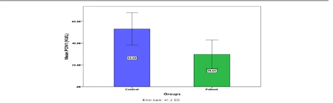
The results showed highly significant decrease in the levels of melatonin ,PON, SOD ,TAC, and NO in MI group versus the control group (5.4020±0.19422 vs 10.4857±0.24614 pg/ml; P<0.0001), (29.6±1.20465vs53±1.35504 KU/L; P<0.0001),
(5.01±.29736 vs 6.24±.32004 U/mL; P< 0.007),
(0.8233±.03920vs1.16±.05741 mmol/L; P< 0.0001),
(37.8±1.15264 vs 51.32±.93204umol/L; P< 0.0001) respectively, while there was highly significant increase in the levels of GPx32.09±1.00487 in MI patients when compared to the controls12±.65379U/mL; P< 0.0001.
±.07196 mmol/l; P < 0.0001) ,(5.7225±0.15404 vs4.7452±0.10583 mmol/l; P< 0.0001 ) respectively. High-density lipoprotein, and creatinine levels were significantly lower in the cases than the controls (1.0853±0.02270 vs 1.6133±0.03577 mmol/l; p< 0.0001),
(1.0432±.09976 vs0.8737±.01821 mg/dl; P < 0.0001)
respectively.
The positive correlation was found between melatonin with paraoxonase, SOD, GPx, NO, TAS, HDL, and glucose.(r=703, 0.5, 0.892, 563, 864, 0.909, 0.630) respectively. The results showed that there was positive correlation between paraoxonase with MDA, SOD, GPx, HDL (r= 0.608, 0.763, 0.5, 0.646) respectively.
Table 1. Baseline biochemical parameters of MI and control groups Mean± Std. Error Mean
Parameter Control group MI group p value
Melatonin (pg/mL) 10.4857±.24614 5.4020±.19422 P<0.0001 Paraoxonase (KU/L) 53±1.35504 29.6±1.20465 P<0.0001
SOD(U/mL) 6.24±.32004 5.01±.29736 P< 0.007
GPx (U/mL) 12±.65379 32.09±1.00487 P< 0.0001 Nitric Oxide (umol/L) 51.32±.93204 37.8±1.15264 P< 0.0001 TAC(mmol/L) 1.16±.05741 0.8233±.03920 P< 0.0001
MDA(µmol) 3.71±0.6 9.34±1.8 p<0.001
Total cholesterol(mmol/l) 4.0483± 0.12095 5.7483±0.14793 P< 0.0001 TG (mmol/l) 1.4150 ±.07196 2.3172±.11091 P < 0.0001 HDL-C(mmol/l) 1.6133±0.03577 1.0853±0.02270 p < 0.0001 Glucose(mmol/l) 4.7452±0.10583 5.7225±0.15404 P< 0.0001 Creatinine (mg/dl) 0.8737±..01722 1.0432±0.01821 P< 0.0001
Figure (1):-The level of SerumPON in MI group and the control group
DISCUSSION
Despite the finding that circulating melatonin levels are
reduced in patients with myocardial infarction (16), experimental
myocardial infarction was shown to increase circulating melatonin levels, followed by enhancement of MI receptors
expression(17). The importance of these receptors in
cardioprotection was further supported by the observation that luzindole, a melatonin receptor antagonist, was able to suppress the cardioprotection induced by melatonin (18). These
events may eventually affect the probability of the MPTP
opening (19). Petrosillo and coworkers (20) reported that
melatonin protected hearts against reperfusion injury by inhibiting the opening of the MPTP probably via prevention of mitochondrial cardiolipin peroxidation.
The antioxidant actions of melatonin are well
established (21-23). Several reports indicated that melatonin
protects the heart directly via its free radical scavenging actions and indirectly via its stimulatory effects on antioxidant capacity (24,25). Indeed, melatonin was able to neutralize a
number of toxic reactants including reactive oxygen species and free radicals induced by myocardial IRI (25,26). For example, in
an in vivo rat model of Myocardial Ischaemia and reperfusion injury, melatonin significantly increased GSH levels and reduced the MDA of the heart tissues after reperfusion (27).
studies where it was shown that melatonin also suppressed superoxidase (˙O2) production, reduced myeloperoxidase (MPO) activity and increased SOD, reduced hydroxyl radical (OH˙), as
well as total ROS generation)(28,29). Interestingly, these
antioxidant properties were also confirmed in rat models of
coronary artery ligation-induced ischaemic heart failure (30), and
isoproterenol induced MI (31). Melatonin does not undergo an
enzymatic pathway of reduction after oxidation, but binds irreversibly to free radicals, and these compounds are removed by the kidneys. Not only melatonin but also precursors of its synthesis and products of its metabolism (eg, tryptophan, serotonin, 6-sulphatoxymelatonin) are able to reduce oxidative
reactions (29,32). examined the role of MPO activity in
melatonin-induced cardioprotection. They found that N(omega)-nitro-L-arginine-methyl ester( L-NAME) (a NOS inhibitor) treatment innonischemic animals increased blood pressure and lipidperoxidation and depressed glutathione (GSH) levels in themyocardial tissue when compared to the non-L-NAMEtreatedanimals.
Paraoxonase1 is tightly bound with HDL particles in
serum. It may play an important role in cardioprotection, as it prevents LDL cholesterol oxidation, metabolizes oxidized LDL-C, and interferes the macrophage uptake of LDL particles (33).
In present study, we found decreased levels of PON in AMI as compared to controls. Decreased PON1 activities might be due to enhanced protection 26 from free radical damage in AMI
during the ischaemic process (34). Few studies reported that
OFRs generation in AMI patients is substantiated by
measuring PON1 activity(35). The decrease in PON1 activity
diseases. Recent studies showed that PON1 playsimportant role in detoxification of harmful compound homocysteine thiolactone (HTL) synthesized in altered homocysteine metabolism. It has been reported that HTL served as substrate for PON1. Low activities of PON1 in present study could due to increased utilization of PON1 to detoxify HTL (36).
Nitric oxide has various physiological properties
including vasodilatation, inhibition of platelet aggregation,
neutrophil adhesion, scavenging superoxide (O -2) radical and
inhibition of xanthine oxidase activity(37). The production of
nitric oxide is decrease in response to oxidative stress; this may be one of the possible causes of complications associated with
dyslipidemia(38). It has been shown that L-arginine serves as a
precursor for the synthesis of nitric oxide. Decline nitric oxide may be due to either deficiency of L-Arginine (Arg) or a deficiency of some cofactors required for nitric oxide
production(39). Melatonin incapacitates NO and prevents NO
induced apoptosis and induction of inducible NO-synthase melatonin may potentiate the effects of NOS augmentation
during beta-adrenergic stimulation(40). Evidence exists for
beta-adrenergic mechanisms in the control of NO generation in cardiomyocytes; for example, isoproterenol has been
demonstrated to upregulate NOS expression (41). And to activate
eNOS via Giα, causing a rise in cyclic adenosine Monophosphate(cGMP) (42). In a study by Genade et al. (43).
Nitric oxide is a potent stimulus for the expression of SOD. SOD is an important antioxidant enzyme having an antitoxic effect against super oxide anion. The over-expression of SOD might be an adaptive response, and it results in increased
dismutation of superoxide to hydrogen peroxide (45). This study
showed that SOD activity were significantly decreased in AMI patients. The results of this study are Similar to Kumar and
Das (46). while in agreement with those reported by Kayyum et
al.(47). SOD is an important enzymatic antioxidant which is
positioned in the arterial wall where NO may be inactivated by O2ˉ.Hence, pathological states like atherosclerosis which results in NO depletion may be associated with a fall in extracellular SOD.
In their study, TAC levels were also significantly lower in AMI patients Fazendas et al.,(48). Reported that TAC levels
decreased in young survivors of acute Myocardial Infarction,
Yegin et al.,(49). Also showed reduction in the level of TAC. Our
findings are in consistent with them, and a decreased TAC level might be associated with an enhanced protective mechanism to oxidative stress in AMI.
In AMI patients, we found significantly higher levels of GPx in patients group. GPX, a selenium containing enzyme, is
an important antioxidant enzyme of erythrocytes (50,51). GPX
plays a significant role in the peroxyl scavenging mechanism, and in maintaining functional integration of the cell membranes(52).
In the present study, MDA levels have been increased
superoxide radicals formed which can stimulate Haber-Weiss reaction for further generation of ROS, initiating lipid peroxidation (53). This is similar to work of Dubois Rande et
al.(54) and McMurray.(55). who showed a decrease in antioxidant
enzyme activities and increase in lipid peroxidation products (MDA, TBARS) in patients with unstable angina and chronic
heart failure. Belch et al(56). showed that progression of
atherosclerosis is correlated with oxidative stress and can be followed up by MDA measurements. Results of the studies of
Pezeshkian et al.(57). Showed that, MDA levels increased
significantly in heart diseases.
Fasting levels of triglycerides, LDL-C and total
cholesterol in patients of AMI were significantly higher as compared to those in controls whereas the levels of serum HDL- cholesterol was significantly lower as compared to that in controls. In a prospective cardiovascular Munster study, elevated TG has been found to be significant and independent risk factor for major coronary events even after adjustment for LDL-C and HDL-C levels and other risk factors (58). Assessing
the lipid ratio in a normal individual as it is one of the atherogenic factors for development of myocardial infarction
and other coronary complications. Lehto et al.(59). Have
demonstrated that there was a direct correlation between the incidence of acute myocardial infarction and plasma lipid abnormalities. Abdulla(58). Has found that significant increase
in lipid and lipoprotein, total cholesterol and LDL-C in the sera, which showed severity of clinical symptoms of endothelial symptoms. According to him, abnormal lipid profile along with other crucial factors in the cascade leading to ischemic damage in the cardium.
The physiological function of PON seems to be a degrade specific oxidized cholesteryl ester and oxidized phospholipids in
lipoproteins and cell membranes(60). The lower activity of PON1
LDL from oxidation, to participate in reverse cholesterol transport pathway and to inhibit monocytes-endothelial cell interaction. All these appear to be important in the
inflammatory response in artery that promotes
atherogenesis(61). Andan Akey et al.(61). Have found that no
significant correlation in between PON1 activity and other metabolic parameter like HDL-C, TG, Insulin in the CAD and
metabolic syndrome .Amur Ayab et al.(62). Have established that
sustained myocardial infarction did not show markedly decreased HDL-C concentration but PON1 activity and PON
concentration were profoundly decreased Kumar et al(46). Have
studied serum PON1 activity in normolipidimic patients with acute myocardial infarction. According to them, no correlation was observed between PON1 activity and HDL-C in acute myocardial infarction which suggested that decreased PON 1 activity could be oxidative stress in acute myocardial infarction
(58).
CONCLUSION:
This study shows the low serum paraoxonase activity may be an independent risk factor for AMI furthermore it can be used as primitive marker of progression of atherosclerosis and AMI, Serum PON may play an important role in the instability of atherosclerotic plaque, Melatonin may provide a novel therapeutic target. Depression of antioxidant system in these patients confirms this conclusion.
REFERENCES
1. Venkaramana G, Krishnamurthy V. and
Cerluplasmin In Acute Myocardial Infarction . Int J Pharm Bio Sci 2012,3(3): 456 – 461.
2. Debasri Mukherjee, Arnab K. Ghosh, Anjalibasu,
SantanuDatta, Sanjib K. Pattari, Arunbandyopadhyay and DebasishBandyopadhyay. Beneficial Role of
Melatonin in the Complete Recovery From
IIsoproterenol-Induced Cardiac Injury in Rat. . Int J Pharm Pharm Sci,2013; 5(2): 561-569.
3. Sewerynek E. Melatonin and the cardiovascular system.
NeuroEndocrinolLett. 2002; 23:79–83.
4. Ehud Grossman,MosheLaudon,NavaZisapel. Effect of
melatonin on nocturnal blood pressure:meta-analysis of randomized controlled trials. Vascular Health and Risk Management 2011:7 577–584
5. Sangita M. Patil, Mangesh P. Banker, Ramchandra K.
Padalkar, AbhijitP.Pathak, ShitalGhodke, Anjali S. Phatake, Sonali S. Bhagat, Rahul A. Ghone. Study of Paraoxonase 1 (PON1) Activity as an Independent Risk Factor in Coronary Artery Disease. National Journal of Laboratory Medicine. 2013, 2(2): 1-6
6. Zorawar SINGH , Indrakaran P. KARTHIGESU ,
Pramjit SINGH, Rupinder KAUR. Use of
Malondialdehyde as a Biomarker for Assessing Oxidative Stress in Different Disease Pathologies: a Review. Iranian J Publ Health, 2014 ; 43 ,(3), 7-16
7. Soydinç S, Çelik A, Demiryürek S. The relationship
between oxidative stress, nitric oxide, and coronary artery disease. Eur J Gen Med 2007; 4:62-66.
8. Mogadam RAP, Nemati A, Baghi AN. Serum MDA as a
Diagnostic`s Biomarker in Stable Coronary Heart
Disease. Research Journal of Bio-logical
Sciences.2008;3:206-210.
9. Guang-Qing Huang,, Jia-Ning Wang1,, Jun-Ming Tang,
Kong , Yong-Zhang Huang , Yong Liu and Shi-You Chen. The combined transduction of copper, zincsuperoxide dismutase and catalase mediated by cell-penetrating peptide, PEP-1, to protect myocardium from ischemia-reperfusion injury. Journal of Translational Medicine 2011, 9:73
10.Vijayasarathy K, K Shanthi Naidu, BKS Sastry,
Melatonin metabolite 6- Sulfatoxymelatonin, Cu/Zn
superoxide dismutase, oxidized LDL and
malondialdehyde in unstable angina. Int J Cardiol. 2010,144: 315–317 .
11.ZyangJl L, Swi and Kong J S. Relationship between
Endothelial Nitric Oxide Synthase, Insulin Resistance and Macrovascular Disease in Patients with Acute Myocardial Infarction. The Journal of International Medical Research. 2012; 40: 687 – 693.
12.Roeschlau P., Bernt E, and Gruber W.A., Estimation of
serum cholesterol; clinical biochemistry. 1974, 12:226
.
13.Faribanks, V. F., Klee, G. G. Biochemical aspects of
hematology. IN: Tietzs Textbook of clinical chemistry (Burtis CA, Ashood ER, Eds), WB Saunders, Philadelphia, 1999:188-2106.
14.Aebihuge, A. methods in enzymatic analysis.1974;
12:561.
15.Cortas NK, Wakid NW. Determination of inorganic
nitrate inserum and urine by a kinetic cadmimum method. ClinChem1990; 36(8):1440-1443.
16.Dominguez-Rodriguez A, Abreu-Gonzalez P, Garcia MJ,
Sanchez J, Marrero F, deArmas-Trujillo D. Decreased
nocturnal melatonin levels during acute
myocardialinfarction. J Pineal Res 2002;33:248-52.
17.Sallinen P, Manttari S, Leskinen H, Ilves M, Vakkuri O,
the synthesis, concentration and receptor expression ofendogenous melatonin. J Pineal Res 2007;42:254-60.
18.Lochner A, Genade S, Davids A, Ytrehus K, Moolman
JA. Short- and long-term effects of melatonin on myocardial post-ischemic recovery. J Pineal Res 2006;40:56-63.
19.Petrosillo G, Colantuono G, Moro N, Ruggiero FM,
Tiravanti E, Di Venosa N, et al.Melatonin protects against heart ischemia-reperfusion injury by inhibiting mitochondrial permeability transition pore opening. Am J Physiol Heart CircPhysiol 2009a;297:1487-93.
20.Petrosillo G, Moro N, Ruggiero FM, Paradies G.
Melatonin inhibits cardiolipin peroxidation in
mitochondria and prevents the mitochondrial
permeability transition and cytochrome c release. Free RadicBiol Med 2009b; 47:969-74.
21.Reiter RJ, Tan DX, Jou MJ, Korkmaz A, Manchester LC,
Paredes SD. Biogenic amines in the reduction of oxidative stress: melatonin and its metabolites. NeuroEndocrinolLett 2008;29:391-8.
22.Korkmaz A, Reiter RJ, Topal T, Manchester LC, Oter S,
Tan DX. Melatonin: an established antioxidant worthy of use in clinical trials. Mol Med 2009a;15:43-50.
23.Bonnefont-Rousselot D, Collin F. Melatonin: Action as
antioxidant and potentialapplications in human disease and aging. Toxicology 2010;278:55-67.
24.Patel V, Upaganlawar A, Zalawadia R, Balaraman R.
Cardioprotective effect of melatonin against
isoproterenol induced myocardial infarction in rats: A biochemical,electrocardiographic and histoarchitectural evaluation. Eur J Pharmacol 2010;644:160-8.
25.Reiter RJ, Tan DX, Paredes SD, Fuentes-Broto L.
26.Reiter RJ, Tan DX. Melatonin: a novel protective agent against oxidative injury of theischemic/reperfused heart. Cardiovasc Res 2003;58:10-9.
27.Sahna E, Parlakpinar H, Turkoz Y, Acet A. Protective
effects of melatonin on myocardial ischemia/reperfusion induced infarct size and oxidative changes. Physiol Res 2005;54:491-5.
28.Ceyran H, Narin F, Narin N, Akgun H, Ceyran AB,
Ozturk F, et al. The effect of high dose melatonin on cardiac ischemia- reperfusion Injury. Yonsei Med J 2008;49:735-41.
29.Sahna E, Deniz E, Bay-Karabulut A, Burma O.
Melatonin protects myocardium
fromischemia-reperfusion injury in hypertensive rats: role of
myeloperoxidase activity. ClinExpHypertens
2008;30:673-81.
30.Sehirli AO, Koyun D, Tetik S, Ozsavci D, Yiginer O,
Cetinel S, et al. Melatonin protects against ischemic heart failure in rats. J Pineal Res 2013;55:138-48.
31.Mukherjee D, Roy SG, Bandyopadhyay A,
Chattopadhyay A, Basu A, Mitra E, et al.Melatonin protects against isoproterenol-induced myocardial injury in the rat: antioxidative mechanisms. J Pineal Res 2010;48:251-62.
32.SMozaffari, S. Hasani-Ranjbar, M. Abdollah I, The
mechanisms of positive effects of melatonin in dyslipidemia: a systematic review of animal and human studies. Int J Pharmacol, 8, 2012, 496–509.
33.Mackness M I , Mackness B, Durrington P N.
Paraoxonase and coronary heart disease.
AtherosclerSuppl 2002; 3: 49-55.
34.Patch B, Jerudi M O, O'Neil PG. Human superoxide
35.Jaouad L, Milochevitch C, Khali A. Paraoxonase1 activity is reduced during HDL oxidation and is an indicator of HDL antioxidant capacity. Free Radic Res 2003; 37: 77-83.
36.Jakuboski H. The pathophysiological hypothesis of
homocysteine thiolactone-mediated vascular disease. Journal of physiological and pharmacology 2008; 59(9):155-167.
37.Tripathi P, Singh TP, Chandra M, Mishra M K.
L-Arginine is effective in scavenging oxygen derived free radicals in the patients of ischemic heart diseases. Int J Biol Res 2010;1(3):15-19.
38.Zweier J L, Hassan M A Talukder. The role of oxidants
and free radicals in reperfusion injury. Cardiovascular research 2006; 70: 181-190.
39.Gamal El-din Harisa. L-Arginine ameliorates
aryesterase/paraoxonase -1 in hypercholesterolemic rats.Asian Journal of Biochemistry 2011; 6(3): 263-272.
40.Yang Yang, Yang Sun, Wei Yi1, Yue Li,
ChongxiFan,ZhenlongXin, ShuaiJiang,Shouyin Di, Yan Qu, Russel J. Reiter and Dinghua Yi1. A review of melatonin as a suitable antioxidant against myocardial ischemia–reperfusion injury and clinical heart diseases. J. Pineal Res. 2014; 57:357–366
41.Buchwalowib IB, Schulze W, Karczewski P et al.
Cellular control of nitric oxide synthase expression and activity in rat cardiomyocytes. Antioxid Redox Signal 2004; 6:345–352.
42.Balligand JL. Regulation of cardiac b-adrenergic
response by nitric oxide. Cardiovasc Res 1999; 43:607– 620.
43.Gendes S, Genis A, Yttrehus K et al. Melatonin
ischaemia/reperfusion injury: role of its anti-adrenergic actions. J Pineal Res.2008; 45:449–458.
44.Massion PB, Pelat M, Belege C et al. Regulation of the
mammalian heart function by nitric oxide. Comp BiochemPhysiol A Mol IntegrPhysiol 2005; 142:144–150.
45.Khaki-khatibi F, Yaghoubi A.R, Rahbani N.M. Study of
antioxidant enzymes, lipid peroxidation, lipid profile and immunologic factor in coronary artery disease in East Azarbijan. Int J Med Biomed Res 2012;1(2):147-152.
46.A. Kumar, R. Sivakanesan. Serum lipid profile
abnormality in predicting the risk of myocardial infarction in elderly normolipidaemic patients in South Asia: A case controlled study. The Internet Journal of Alternative Medicine. 2009; 6(2):1-5.
47.Kayyum-Shaikh A, Suryakar AN. Oxidative stress and
antioxidant status before and after supplementation of A-Z antioxidant tablets in coronary artery disease. Biom Res 2009;20:136-140.
48.Fazendas P, Joao IF, Llobet S. Plasma total antioxidant
status in young survivors of myocardial infarction. Rev Port Card 2000;19:463-467.
49.Yegin A, Yegin H, Aliciguzel Y. Erythrocyte Selenium
glutathione peroxide activity is lower in patients with Coronary atherosclerosis. Jpn Heart Jr 1997;38:793-79.
50.Mutlu-Turkoglu U, Akalin Z, Ilhan E. Increased plasma
malondialdehyde and protein carbonyl levels and
lymphocyte DNA damage in patients with
angiographically defined coronary artery disease. ClinBiochem 2005;38:1059-65.
51.Serdar Z, Aslan K, Dirican M. Lipid and protein
52.Vishnu-Priya V, Surapaneni KM. Erythrocyte lipid peroxidation, glutathione, ascorbic acid, vitamin E,antioxidant enzymes and serum homocysteine levels in patients with coronary artery disease. J ClinDiag Res 2008;2:1180-1185.
53.Pasupathi P, Rao Y Y, Farook J, Saravanan G,
Bakthavathsalam G. Oxidative stress and cardiac biomarkers in patients with acute myocardial infarction. European Journal of Scientific Research 2009; 27(2): 275-285.
54.Dubois-Rande JL, Artigou JY, Darmon JY, Habbal R,
Manuel C, Tayarani I, et al. Oxidative stress in patients with unstable angina. Eur Heart J 1994; 15(2): 179-83.
55.McMurray J. Evidence of oxidative stress in chronic
heart failure in humans. Eur Heart J 1993; 14 (11): 1493-7.
56.Belch JJ, Mackay IR, Hill A, Jennings P, McCollum P.
Oxidative stress is present in atherosclerotic peripheral arterial disease and further in-creased by diabetes mellitus. IntAngiol. 1995;14:385-388.
57.Pezeshkian M, Nouri M, Zahraei M, Afrasiabi A, Abadi
NA. Study of MDA, antioxidant vitamins, lipoproteins serum levels and anthropome-try parameters in coronary artery disease patients. Medical Journal of Islamic Academy of Sciences. 2001;14:5-8.
58.Abdullah MR. Comparative study of serum lactic acid,
lactate Dehydrogenase and lipid profile in ischemic heart disease patients and healthy control.Ibn Al-Haitham J for Pure Sci. 2010;23(1):
59.Lehto S, Palomaki P, Miettimen H, et al. Serum
60.JordiCamps,JuditMarsillach, Jorge Joven. Measurement of serum paraoxonase – 1 activity in the evaluation of liver function. World J Gastroenterol. 2009;15(16): 1929-33.
61.Adnan BurakAkcay, AhmetCamseri, TurkayOzcan,
DilekCicek.NecdetAkkus, Sabriseyis, BurakCimen,
BarisCelebi, obenDoven,GokhenCin. The relationship between paraoxonase_1 activity and coronary artery disease in patients with metabolic syndrome. TrrkkardiyoldemArs-Arch turkSocCardiol. 2011;39 (5): 371-77.
62.AamirAyub, Michael Mackness, Sharon
Arrol,BhartiMackness, Jeetesh Patel and Paul N. Serum
paraoxonase after myocardial infarction.