Scholarship@Western
Scholarship@Western
Electronic Thesis and Dissertation Repository
7-31-2013 12:00 AM
Spinal Cord Control of Ejaculatory Reflexes in Male Rats
Spinal Cord Control of Ejaculatory Reflexes in Male Rats
Natalie Kozyrev
The University of Western Ontario
Supervisor
Dr. Lique M. Coolen
The University of Western Ontario
Graduate Program in Anatomy and Cell Biology
A thesis submitted in partial fulfillment of the requirements for the degree in Doctor of Philosophy
© Natalie Kozyrev 2013
Follow this and additional works at: https://ir.lib.uwo.ca/etd
Part of the Neuroscience and Neurobiology Commons, and the Physiology Commons
Recommended Citation Recommended Citation
Kozyrev, Natalie, "Spinal Cord Control of Ejaculatory Reflexes in Male Rats" (2013). Electronic Thesis and Dissertation Repository. 1596.
https://ir.lib.uwo.ca/etd/1596
This Dissertation/Thesis is brought to you for free and open access by Scholarship@Western. It has been accepted for inclusion in Electronic Thesis and Dissertation Repository by an authorized administrator of
(Thesis format: Integrated Article)
by
Natalie Kozyrev
Graduate Program in Anatomy and Cell Biology
A thesis submitted in partial fulfillment of the requirements for the degree of
Doctor of Philosophy
The School of Graduate and Postdoctoral Studies The University of Western Ontario
London, Ontario, Canada
ii
ABSTRACT
Ejaculatory dysfunction impacts large numbers of men of all ages and around the world. In
addition, a great majority of men with chronic spinal cord injury (SCI) experience ejaculatory
dysfunction, which negatively impacts the quality of life of these individuals and their partners.
SCI men emphasize the significance of regaining sexual function as their main goal. Currently,
there is a marked absence of literature reporting the alterations to sexual function and ejaculation
in particular in animal models of chronic SCI. In addition, there are many unanswered questions
pertaining to the spinal cord control of ejaculation in healthy, intact men. It is known that
ejaculation is controlled by a population of lumbar spinothalamic (LSt) cells in the lumbar spinal
cord through their direct projections to preganglionic autonomic and motor neurons in the
lumbosacral spinal cord. It is hypothesized that LSt cells control ejaculatory reflexes through the
release of their neuropeptides galanin, cholecystokinin (CCK), gastrin-releasing peptide (GRP),
and enkephalin onto receptors in autonomic and motor areas of the lumbosacral spinal cord. This
hypothesis was tested in this thesis utilizing a paradigm in anesthetized and spinalized male rats,
with stimulation of the sensory inputs via the dorsal penile nerve. Consistent with the hypothesis,
mu and delta opioid receptor, galanin, CCK, and GRP receptor activation in LSt target areas in
the lumbosacral spinal cord was demonstrated to be critical for ejaculatory reflexes. Next, the
hypothesis that intrathecal infusions of the LSt neuropeptides can improve ejaculatory reflexes in
male rats with chronic SCI was tested. Results indicated that intrathecal infusions of GRP and
the mu opioid receptor agonist DAMGO improved ejaculatory reflexes in male rats with chronic
iii
ejaculatory function in male rats with chronic spinal cord injury was tested. Indeed, systemic
infusions of 7-OH-DPAT greatly improved ejaculatory reflexes in SCI males. Together, the
studies in this thesis further clarified the mechanisms involved in the spinal cord control of
ejaculation in male rats and represent an initial but pivotal first step towards the recovery of
ejaculatory function after chronic spinal cord injury.
Key Words: ejaculation, spinal ejaculation generator, chronic spinal cord injury, lumbosacral
spinal cord, spinal cord contusion, galanin, cholecystokinin, enkephalin, gastrin-releasing
peptide, dopamine D3 receptor.
iv
Co-Authorship Statement
Chapter 2 entitled “Activation of gastrin-releasing peptide receptors in the lumbosacral spinal
cord is required for ejaculation in male rats”, chapter 3 entitled “Activation of mu and delta
opioid receptors in the lumbosacral spinal cord is required for ejaculatory reflexes in male rats”,
chapter 4 entitled “Activation of galanin and cholecystokinin receptors in the lumbosacral spinal
cord is required for ejaculatory reflexes in male rats”, chapter 5 entitled “Activation of mu opioid
and GRP receptors in the lumbosacral spinal cord can improve ejaculatory reflexes in male rats
with chronic contusion spinal cord injury” and chapter 6 entitled “Systemic infusions of
7-OH-DPAT rescued ejaculatory reflexes in male rats with chronic contusion spinal cord injury” were
written by Natalie Kozyrev with inputs by Dr. Lique M. Coolen. Experimental procedures and
data analysis were performed by Natalie Kozyrev and intellectual input was provided by Lique
v
ACKNOWLEDGEMENTS
First of all, I would like to thank my supervisor, Dr. Lique Coolen, for her patience, hard work
and dedication in training and mentoring me along the way to becoming a scientist. I am grateful
for all that I have learned from Lique as these skills and knowledge will be invaluable to me in
all my future endeavors. I would also like to thank members of my advisory committee, Drs.
Michael Lehman, Arthur Brown, and Michael Poulter, for their advice and support throughout
the duration of my degree. In addition, I would like to thank former and current members of the
Coolen and Lehman laboratories for being such exceptional people to work with and spend time
with in and outside of the lab. In particular, I wish to thank Drs. Karla Frohmader, Cleusa De
Oliveira, Ian Webb and Michael Staudt for their help and advice with experiments and for all the
training and support I received. Finally, I would like to thank my family and close friends for all
vi
Table of Contents
SPINAL CORD CONTROL OF EJACULATORY REFLEXES IN MALE RATS ... i
ABSTRACT ... ii
Co-Authorship Statement ... iv
Table of Contents ... vi
List of Tables ... xii
List of Figures ... xiii
List of Appendices ... xvii
List of abbreviations ... xviii
Chapter 1: Introduction ... 1
1.1 Introduction ... 2
1.2 Ejaculation ... 2
1.3 The Spinal Ejaculation Generator ... 3
1.3.1 LSt Cells Form an Integral Component of the Spinal Ejaculation Generator ... 4
1.3.2 Spinal Inputs to the Ejaculation Generator ... 6
1.3.3 Outputs of the Spinal Ejaculation Generator ... 7
1.4 Supraspinal Sites Regulating Ejaculation ... 9
1.4.1 The Medial Preoptic Area (MPOA) ... 9
1.4.2 Nucleus of the paragigantocellularis (NPGi) ... 10
1.4.3 The paraventricular nucleus of the hypothalamus (PVN) ... 12
1.5 Supraspinal network displaying neural activation specifically with ejaculation ...13
1.6 LSt-SPFp pathway ... 14
1.7 Summary: Spinal cord control of ejaculation ... 15
vii
1.9 Summary: Effects of spinal cord injury on ejaculation ... 20
1.10Animal models of chronic spinal cord injury ... 21
1.11Thesis rationale and objectives ... 22
Chapter 2: Activation of gastrin-releasing peptide receptors in the lumbosacral spinal cord is required for ejaculation in male rats1 ... 24
2.1 Introduction ... 25
2.2 Materials and Methods ... 29
2.2.1 Animals ... 29
2.2.2 Experimental designs ... 29
2.2.3 BCM EMG analysis ... 32
2.2.4 Immunohistochemistry experiments:... 33
2.2.5 Immunohistochemistry ... 35
2.2.6 Data Analysis:... 36
2.3 Results ... 38
2.3.1 GRP expression in LSt cells and axons ... 39
2.3.2 GRP receptor antagonist blocked ejaculatory reflexes ... 41
2.3.3 RC-3095 did not affect DPN stimulation-Induced pERK in LSt cells ... 45
2.3.4 GRP20-29 agonist facilitated BCM activity... 46
2.3.5 GRP mediates SVP Increases following DPN stimulation... 51
2.4 Discussion... 56
Chapter 3: Activation of mu and delta opioid receptors in the lumbosacral spinal cord is required for ejaculatory reflexes in male rats ... 62
3.1 Introduction ... 63
3.2 Materials and Methods ... 66
3.2.1 Animals ... 66
viii
3.2.3 BCM EMG and SVP Analysis ... 69
3.2.4 Immunohistochemistry Experiments ... 71
3.2.5 Immunohistochemistry: Galanin/pERK dual fluorescence ... 72
3.2.6 Data Analysis ... 73
3.3 Results ... 73
3.3.1 Mu Opioid Receptor Antagonist CTOP Suppressed Ejaculatory Reflexes73 3.3.2 Delta Opioid Receptor Antagonist TIPP Suppressed Ejaculatory Reflexes78 3.3.3 CTOP and TIPP did not affect DPN Stimulation-Induced pERK in LSt Cells ...81
3.3.4 Mu Opioid Receptor Agonist DAMGO Triggered and Facilitated Ejaculatory Reflex... 82
3.3.5 Delta Opioid Receptor Agonist Deltorphin Facilitated Ejaculatory reflexes ...86
3.4 Discussion... 89
Chapter 4: Activation of galanin and cholecystokinin receptors in the lumbosacral spinal cord is required for ejaculatory reflexes in male rats ... 94
4.1 Introduction ... 95
4.2 Materials and Methods ... 98
4.2.1 Animals ... 98
4.2.2 Experimental Designs ... 99
4.2.3 BCM EMG and SVP Analysis ... 102
4.2.4 Immunohistochemistry Experiments ... 103
4.2.5 Immunohistochemistry: Galanin/pERK dual fluorescence ... 104
4.2.6 Data Analysis ... 105
4.3 Results ... 106
4.3.1 Galantide Suppressed Ejaculatory Reflexes ... 106
ix
4.3.3 Galantide and Proglumide Did Not Prevent DPN Stimulation-Induced pERK
in LSt Cells ... 114
4.3.4 Galanin Facilitated BCM Events, Bursts and SVPs ... 114
4.3.5 Cholecystokinin (CCK) Facilitated BCM Events, Bursts and SVPs ... 119
4.4 Discussion... 121
Chapter 5: Activation of mu opioid and GRP receptors in the lumbosacral spinal cord can improve ejaculatory reflexes in male rats with chronic contusion spinal cord injury ... 129
5.1 Introduction ... 130
5.2 Materials and Methods ... 133
5.2.1 Animals ... 133
5.2.2 Spinal Cord Surgeries ... 133
5.2.3 Locomotor testing ... 134
5.3 Experimental Designs ... 135
5.3.1 Intrathecal infusions of neuropeptides... 135
5.3.2 Intrathecal infusions of a cocktail of neuropeptides ... 139
5.3.3 BCM EMG and SVP Analysis ... 139
5.3.4 Statistical analyses ... 140
5.4 Results ... 140
5.4.1 Locomotor activity was impaired in SCI male rats ... 140
5.4.2 Experiment 1: Ejaculatory reflexes were absent in SCI males ... 142
5.4.3 Infusions of GRP and DAMGO in sham and SCI male rats ... 148
5.4.4 Infusions of GRP triggered and facilitated ejaculatory reflexes in male SCI rats ... 149
5.4.5 Infusions of DAMGO triggered ejaculatory reflexes in male SCI rats ... 154
x
5.4.7 Experiment 3: Cocktail infusions triggered ejaculatory reflexes in a subset of
SCI males ... 164
5.5 Discussion... 166
Chapter 6: Systemic infusions of 7-OH-DPAT rescued ejaculatory reflexes in male rats with chronic contusion spinal cord injury ... 170
6.1 Introduction ... 171
6.2 Materials and Methods ... 174
6.2.1 Animals ... 174
6.2.2 Spinal Cord Surgeries ... 174
6.2.3 Locomotor testing ... 175
6.3 Experimental Designs ... 175
6.3.1 General Methods: ... 175
6.3.2 BCM EMG and SVP Analysis ... 177
6.3.3 Experiment 1: 7OHDPAT or saline infusions after acute spinal transection ...178
6.3.4 Experiment 2: 7OHDPAT infusions before and after acute spinal transection ..180
6.4 Results ... 181
6.4.1 Locomotor activity was impaired in SCI male rats ... 181
6.4.2 7OHDPAT infusions improved ejaculatory reflexes in chronic SCI male rats after acute spinal transection... 183
6.4.3 7OHDPAT double infusion experiment: Results ... 190
6.5 Discussion... 193
Chapter 7: General Discussion ... 199
7.1 Summary of Results and Conclusions ... 200
7.1.1 Chapter 2: Activation of gastrin-releasing peptide receptors in the lumbosacral spinal is required for ejaculation in male rats ... 201
xi
7.1.3 Chapter 4: Activation of Galanin and CCK Receptors is Required for
Ejaculatory Reflexes in Male Rats ... 203
7.1.4 Chapter 5: Activation of Mu opioid and GRP receptors in the lumbosacral spinal cord can improve ejaculatory reflexes in male rats with chronic contusion SCI ... 204
7.1.5 Chapter 6: Systemic Infusions of 7-OH-DPAT rescued ejaculatory reflexes in male rats with chronic contusion SCI ... 205
7.2 Future Directions ... 207
References ... 208
Appendix A: Animal protocol approval ... 225
Appendix B: License Agreement ... 227
xii
List of Tables
Table 1 Numbers of SVP increases and BCM bursts following infusions of saline, RC-3095 and
GRP in male rats. ... 53
Table 2 Initial 60 Hz DPN stimulation facilitates BCM events, bursts and SVP increases in sham
xiii
List of Figures
Figure 1 Schematic diagram of the spinal ejaculation generator ... 26
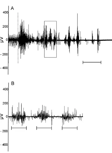
Figure 2 Example of characteristic BCM EMG induced by DPN stimulation. ... 31
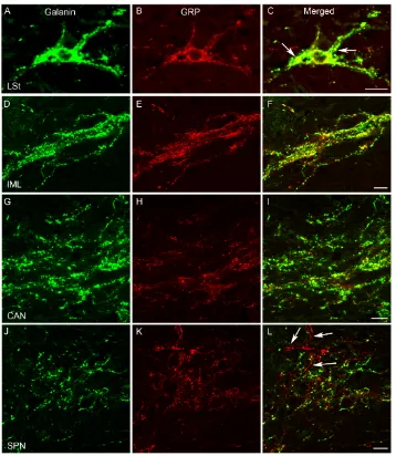
Figure 3 The co-expression of galanin-ir and GRP-ir in LSt cells, IML, CAN and SPN of the male rat... 40
Figure 4 Effects of GRP receptor antagonist RC-3095 infusion on the numbers of BCM events and bursts in male rats... 43
Figure 5 Infusions of RC-3095 prevent DPN stimulation-induced bursting of the BCM in male rats. ... 44
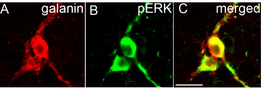
Figure 6 Expression of pERK in LSt cells induced by DPN stimulation ... 45
Figure 7 Effects of GRP infusions on the numbers of BCM events and bursts in male rats. ... 48
Figure 8 Effects of GRP infusions on BCM activity in male rats. ... 50
Figure 9 SVP and concurrent BCM activity induced by DPN stimulation ... 52
Figure 10 BCM activity and concurrent increases in SVP triggered by infusions of GRP in a male rat... 55
Figure 11 Effects of CTOP infusions on the numbers of BCM events and bursts induced by DPN stimulation in male rats. ... 75
Figure 12 Effects of CTOP on the numbers of BCM bursts and SVP increases induced by DPN stimulation in male rats. ... 77
xiv
Figure 14 CTOP infusions did not prevent the activation of LSt cells induced by DPN
stimulation in male rats. ... 82
Figure 15 Effects of DAMGO infusions on the numbers of BCM events, bursts and SVP
increases in male rats. ... 84
Figure 16 Effects of Deltorphin II on the numbers of BCM events, bursts and SVP increases in
male rats. ... 88
Figure 17 Effects of galantide on numbers of BCM events and bursts in male rats. ... 108
Figure 18 Effects of galantide on the numbers of BCM bursts and SVP increases in male rats. 110
Figure 19 Effects of proglumide on the numbers of BCM events, bursts and SVP increases in
male rats. ... 112
Figure 20 Effects of galanin on numbers of BCM events and bursts in male rats ... 116
Figure 21 Effects of galanin on numbers of BCM bursts and SVP increases in male rats. ... 118
Figure 22 Effects of CCK on the numbers of BCM events, bursts and SVP increases in male rats.
... 120
Figure 23 Timeline of experimental manipulations. ... 138
Figure 24 Locomotor activity in sham and SCI male rats 2, 3 and 4 weeks after surgery. ... 142
Figure 25 Effects of DAMGO and GRP infusions on the numbers of BCM events in sham and
SCI male rats. ... 144
Figure 26 Effects of GRP and DAMGO infusions on the numbers of BCM bursts in sham and
SCI male rats. ... 146
Figure 27 Effects of GRP and DAMGO infusions on the numbers of SVP increases in sham and
xv
Figure 28 Representative BCM EMG and SVP recording traces showing SVP increases and
concurrent BCM activity induced by intrathecal infusions of GRP and DAMGO in sham and SCI
male rats. ... 151
Figure 29 Representative BCM EMG and SVP recording traces depicting SVP and concurrent
BCM EMG activity triggered by subthreshold (5 Hz) and threshold (30 Hz) levels of DPN
stimulation following infusions of saline and GRP in sham and SCI male rats ... 153
Figure 30 Effects of galanin and deltorphin II infusions on the numbers of BCM events in sham
and SCI male rats. ... 156
Figure 31 Effects of galanin and deltorphin infusions on the numbers of BCM bursts in sham and
SCI male rats ... 158
Figure 32 Effects of galanin and deltorphin infusions on the numbers of SVP increases in sham
and SCI male rats ... 160
Figure 33 Representative BCM EMG and SVP recording traces depicting SVP and concurrent
BCM EMG activity triggered by subthreshold (5 Hz) DPN stimulation following infusions of
saline, galanin and deltorphin in sham and SCI male rats. ... 162
Figure 34 Representative BCM EMG and SVP recording traces depicting SVP and concurrent
BCM EMG activity induced by threshold (30 Hz) DPN stimulation following infusions of saline,
galanin and deltorphin in sham and SCI male rats. ... 163
Figure 35 Effects of cocktail infusion on BCM EMG and SVP in sham and SCI male rats ... 165
Figure 36 7-OH-DPAT experiments: Timeline of experimental manipulations. ... 179
Figure 37 7-OH-DPAT experiments: Locomotor activity in sham and SCI male rats 2, 3, and 4
weeks after surgery. ... 182
Figure 38 Single-infusion 7-OH-DPAT experiment: Effects of 30 and 60 Hz DPN stimulations
xvi
Figure 39 Single-infusion 7-OH-DPAT experiment: Effects of subcutaneous 7-OH-DPAT
infusions on the numbers of BCM events, bursts and SVP increases in sham and SCI male rats
... 188
Figure 40 Double-infusion 7-OH-DPAT experiment: Effects of 7-OH-DPAT infusions on the
numbers of BCM events, bursts and SVP increases in spinally-intact and transected sham and
SCI male rats. ... 193
xvii
List of Appendices
Appendix A: Animal Protocol Approval ... 225
xviii
List of abbreviations
ABC, avidin-biotin-horseradish peroxidase complex
BCM, muscle of the bulbocavernosus
BNSTpm, posteromedial bed nucleus of the stria terminalis
CAN, central autonomic nucleus
CCK, cholecystokinin
DMSO, dimethyl sulfoxide
DOR, delta opioid receptor
DPN, dorsal penile nerve
DRG, dorsal root ganglion
EMG, electromyography
ERK, extracellular-regulated protein kinase
GRP, gastrin-releasing peptide
GIRK, G-protein-coupled inwardly-rectifying K+
IML, intermediolateral cell column
LSt, lumbar spinothalamic
MAP, mitogen-activated protein
MEK, mitogen-activated protein extracellular signal-regulated kinase
xix
MPOA, medial preoptic area
NPGi, nucleus of the paragigantocellularis
PB, phosphate buffer
PBS, phosphate buffered saline
pERK, phosphorylated MAP kinase
PVN, paraventricular nucleus of the hypothalamus
SCI, spinal cord injury
SNB, sacral nucleus of the bulbocavernosus
SPN, sacral parasympathetic nucleus
SV, seminal vesicle
Chapter 1: Introduction
Spinal cord control of ejaculatory reflexes in male rats: Effects of
1.1Introduction
Male sexual dysfunction, including erectile and ejaculatory dysfunction, commonly occurs in
adult men of all ages and around the world. While the mechanisms of erectile dysfunction are
well-known and various effective treatment options are currently available, the mechanisms
underlying the control of ejaculation remain largely unknown. Premature ejaculation is one of
the most frequently reported ejaculatory dysfunctions and at present there are no effective
treatment options available (Vale, 1999). Ejaculation is also severely altered in men with chronic
spinal cord injury (SCI) (Brackett et al., 1998). However, among paraplegic men, the recovery of
normal sexual function is the highest priority and is even ranked above recovery of locomotor
function (Anderson, 2004). The current review will summarize literature describing the central
control of ejaculation in humans and animal models. In addition, the effects of chronic SCI on
ejaculation are reviewed. Finally, potential treatments geared toward the recovery of normal
sexual function in men suffering from premature ejaculation and in men with chronic SCI will be
reviewed.
1.2 Ejaculation
Male sexual behavior is highly complex and consists of several components including the pursuit
and investigation of the female, mounts and intromissions finally culminating in ejaculation, a
process that is closely associated with orgasm in men (Meisel R, 1994) and reward in male rats
contents from the urethral meatus (Marberger, 1974; McKenna, 1999) and is composed of two
separate components, emission and expulsion. Emission refers to the secretion of seminal
contents from the accessory sex glands, including the vas deferens, seminal vesicles and prostate
(Wang et al., 1991) and the closure of the bladder neck and external uretheral sphincter to avert
retrograde ejaculation (Newman et al., 1982). Expulsion refers to the coordinated, rhythmic
contractions of the striated perineal muscles and the bulbocavernosus muscle (BCM) in
particular, as well as the smooth muscles of the urethra, which forcefully expel semen from the
urethral meatus (Breedlove and Arnold, 1980, 1981; Breedlove et al., 1983).
1.3 The Spinal Ejaculation Generator
It has been known for some time from studies of chronic SCI patients and animal models that
ejaculation is a reflex and the central mechanisms that regulate this reflex are localized to the
lumbosacral spinal cord (Comarr, 1970; Comarr and Gunderson, 1975; McKenna, 1997;
McKenna, 1999). Previous studies have shown that vibratory penile stimulation is sufficient to
trigger ejaculatory reflexes in men with complete spinal cord injuries above the 10th thoracic
spinal segment (Brackett et al., 1998; Brackett, 1999; Sonksen and Ohl, 2002). Similarly,
stimulation of the dorsal penile nerve (DPN), the sensory branch of the pudendal nerve, triggers
ejaculatory reflexes in male rats with complete spinal cord transection (Staudt et al., 2010;
Staudt, 2011; Kozyrev et al., 2012). The fact that ejaculatory reflexes persist in spinalized men
and male rats (Comarr, 1970; Comarr and Gunderson, 1975; Pescatori et al., 1993; Brackett et
with supraspinal regions suggests that ejaculation is regulated by a control center in the
lumbosacral spinal cord. This control center is known as the spinal pattern generator (McKenna
et al., 1991; Carro-Juarez and Rodriguez-Manzo, 2007), spinal pacemaker (Sachs and Garinello,
1979), or spinal ejaculation generator (Marberger, 1974; Marson and McKenna, 1994) and it
controls ejaculation in two main ways. First, it coordinates sympathetic, parasympathetic and
motor outflow to trigger the two components of ejaculation: emission and expulsion. Secondly,
the spinal ejaculation generator integrates this autonomic and motor outflow with sensory inputs
necessary to induce ejaculation which are conveyed during sexual activity (Coolen et al., 2004;
Allard et al., 2005; Coolen, 2005; Young et al., 2009).
1.3.1 LSt Cells Form an Integral Component of the Spinal Ejaculation Generator
About a decade ago, substantial progress was made in identifying a population of interneurons in
the lumbosacral spinal cord that form a critical component of the spinal ejaculation generator
(Truitt and Coolen, 2002). These interneurons are located in lamina 10 around the central canal
and the medial portion of lamina 7 in lumbar levels (L3 – L4) of the spinal cord and have direct
projections to the parvocellular subparafascicular thalamic nucleus (SPFp) (Ju et al., 1987;
Coolen et al., 2003; Truitt et al., 2003). Moreover, these lumbar spinothalamic (LSt) cells, named
according to their anatomical position in the spinal cord and thalamic projections, are
sexually-dimorphic and show neural activation, visualized with Fos, specifically following ejaculation in
male but not in female rats (Truitt and Coolen, 2002; Coolen et al., 2003; Truitt et al., 2003). In
addition, LSt cells co-express several neuropeptides including galanin, cholecystokinin (CCK),
Newton, 1999; Newton and Phan, 2006; Sakamoto et al., 2008; Kozyrev et al., 2012) and project
their axons to autonomic preganglionic neurons (Xu et al., 2006) and pudendal motoneurons
(Newton, 1993; Newton and Phan, 2006) in the thoracolumbar and lumbosacral spinal cord.
Galanin, a marker of LSt cells, is expressed in axons projecting to preganglionic sympathetic and
parasympathetic neurons as well as pudendal motor neurons (Newton, 1993; Truitt and Coolen,
2002). Neural activation of LSt cells, based on Fos and galanin co-expression, is specifically
related to the onset of ejaculation (Truitt et al., 2003; Coolen et al., 2004). Targeted lesions of the
LSt cells with intraspinal injections of a neurotoxin SSP-Saporin abolished ejaculation in
copulating male rats but spared all other components of male rat sexual behavior including
pursuit and investigation of the female, mounts and intromissions (Truitt and Coolen, 2002).
Furthermore, targeted lesions of the LSt cells with the same neurotoxin prevented rhythmic
contractions of the BCM following DPN, urethral or pharmacological stimulation with the
dopamine D3 receptor agonist 7-OH-DPAT, in anesthetized and spinalized male rats (Staudt,
2011). In addition, in male rats with LSt lesions, galanin-immunoreactive fibers normally
projecting to autonomic nuclei in the lumbosacral spinal cord were absent, confirming that LSt
cells are required for these functional projections (Truitt and Coolen, 2002). Together these
studies demonstrated that LSt cells are essential for ejaculation in male rats and that LSt cells
regulate ejaculation through intraspinal connections to preganglionic autonomic and motor
neurons in the lumbosacral spinal cord (Truitt and Coolen, 2002; Truitt et al., 2003; Staudt,
2011). Thus, LSt cells are a population of interneurons in the lumbar spinal cord which comprise
a pivotal component of the spinal ejaculation generator and transform sensory inputs during
1.3.2 Spinal Inputs to the Ejaculation Generator
During sexual activity, the DPN, the sensory branch of the pudendal nerve, plays a central role in
the transmission of sensory inputs to the spinal ejaculation generator required to trigger
ejaculation (Marberger, 1974; Hull et al., 2002). Indeed, electrical stimulation of the DPN
triggers rhythmic contractions of the BCM similar to those observed in copulating male rats
(Pescatori et al., 1993). Furthermore, both DPN stimulation in anesthetized and spinalized male
rats and ejaculation in mating males triggers phosphorylated ERK (pERK) and phosphorylated
NMDA receptor subunit 1 (pNR1) in LSt cells (Staudt et al., 2010, 2011). Moreover, injections
of antagonists for ERK and NMDA receptors blocked DPN stimulation-induced BCM bursting
in male rats (Staudt et al., 2010, 2011). Pudendal nerve terminals are found in close proximity to
the LSt cells in the lumbosacral spinal cord and are mainly localized to medial dorsal horn and
dorsal gray commissure (DGC) (McKenna and Nadelhaft, 1986; Ueyama et al., 1987).
Presumably, these sensory signals are relayed to the LSt cells through intraspinal connections
between the LSt cells and the dorsal horn and DGC. However, precisely how this transfer occurs
is currently unknown as there are no known direct projections from the dorsal horn and DGC to
the LSt cells. These sensory inputs are conveyed by the DPN during sexual activity prior to
ejaculation and activate the LSt cells to induce ejaculation and may include visceral sensory,
somatosensory, proprioceptive or noxious inputs (Carro-Juarez and Rodriguez-Manzo, 2005).
Some sensory inputs related to the summation of sexual activity may also be conveyed by the
hypogastric and pelvic nerves (McKenna, 2001; Coolen et al., 2004; Coolen, 2005). Pelvic nerve
afferents terminate in the vicinity of pudendal nerve afferent terminals in the lumbosacral spinal
conveys sensory stimuli associated with mating to supraspinal regions including the central
tegmental field and lesions of the pelvic nerve dramatically reduce neural activation of the
central tegmental field induced by mating (Wersinger et al., 1993). Unlike the pudendal and
pelvic nerves, the hypogastric nerve terminates in the dorsal horn of the thoracolumbar spinal
cord, in segments T13 – L2 in male rats (De Groat W, 1990). Nevertheless, electrical stimulation
of the intermesenteric nerve which contains fibers of the hypogastric nerve triggered seminal
plug expulsion in anesthetized rats (Bernabe et al., 2007). In men, the penis is innervated by a
large number of sensory fibers which include mainly small, unmyelinated Aδ and C fibers
terminating in free nerve endings (Halata and Munger, 1986). These sensory fibers of the penis
can detect temperature, pain and mechanical stimuli including touch, pressure and stretch
(Kitchell et al., 1982). In addition, the penis exclusively contains a genital end bulb which is a
type of encapsulated nerve ending that may regulate sensory responses to stimulation of the glans
(Coolen et al., 2004). Furthermore, the sensitivity of penile receptors is greatly enhanced by
penile erection (Johnson, 1988). In summary, penile receptors convey sensory information
regarding temperature, touch, stretch, pressure and pain via the DPN as well as the pelvic and
hypogastric nerves to the lower thoracic, lumbar and sacral spinal cord. These sensory inputs
ultimately converge in the spinal ejaculation generator and trigger the activation of LSt cells,
which in turn triggers ejaculation.
1.3.3 Outputs of the Spinal Ejaculation Generator
During the emission phase of ejaculation, autonomic nuclei in the thoracolumbar and
viscera including the seminal vesicles, ductus deferens and prostate(Giuliano and Clement,
2005). The intermediolateral cell column (IML) and dorsal central autonomic nucleus (DCN)
located in the thoracolumbar spinal cord in spinal segments (T13 – L2) contain sympathetic
preganglionic neurons that send sympathetic ouflow mainly through the hypogastric and pelvic
nerves to the visceral organs (Hancock and Peveto, 1979a; Nadelhaft and McKenna, 1987; Baron
and Janig, 1991; Giuliano et al., 1997). Among the two populations of sympathetic preganglionic
neurons, it is those of the DCN which send projections to the prevertebral ganglia and are
implicated in the control of the visceral organs while IML neurons are thought to be involved
mainly in the control of cardiovascular function (Janig and McLachlan, 1987). Parasympathetic
preganglionic neurons are located in the sacral parasympathetic nucleus (SPN) in spinal
segments (S2-S5) which innervate the pelvic organs including the prostate (Orr and Marson,
1998), urethra (Vizzard et al., 1995) and penis (Marson and Carson 3rd, 1999). In summary, the
emission phase of ejaculation is under the control of both the sympathetic and parasympathetic
preganglionic cell populations as are many of the physiological events leading up to emission,
including contractions of the ductus deferens (Kolbeck and Steers, 1992; Kihara and de Groat,
1997) and the discharge of prostatic contents into the urethra (Wang et al., 1991). The expulsion
phase of ejaculation is under the control of pudendal motoneurons in the lumbosacral spinal cord
(Coolen et al., 2004). These pudendal motoneurons are localized to the spinal nucleus of the
bulbocavernosus (SNB) as well as the dorsolateral (DL) nucleus in male rats (Schroder, 1980;
McKenna and Nadelhaft, 1986; Ueyama et al., 1987), and to Onuf’s nucleus in men (Schroder,
1985; Thor et al., 1989). Motoneurons in these nuclei innervate the bulbospongiosus and
ischiocavernosus striated perineal muscles and the external urethral and anal sphincters.
urethral meatus (Bohlen et al., 1980; Gerstenberg et al., 1990; Holmes et al., 1991; Holmes and
Sachs, 1991). Notably, the striated perineal muscle contractions occur concurrently with the
pleasurable orgasmic sensations experienced at ejaculation (Gerstenberg et al., 1990).
1.4Supraspinal Sites Regulating Ejaculation
Several supraspinal regions in the brain and brainstem provide descending modulation of the
spinal ejaculation generator. These supraspinal regions exert excitatory and inhibitory influences
over the spinal ejaculation generator to regulate ejaculation.
1.4.1 The Medial Preoptic Area (MPOA)
One such region is the medial preoptic area (MPOA) of the hypothalamus (Pehek et al., 1989;
Markowski et al., 1994; Hull et al., 1997; Hull et al., 2004), a central area in regulating male
sexual behavior (Hull et al., 2004). The MPOA exerts its excitatory effects on ejaculation
through dopamine D2 (Hull et al., 1989; Hull et al., 1992) and D3 receptors (Kitrey et al., 2007)
and electrical stimulation of the MPOA triggers contractions of the striated penile muscles in
anesthetized male rats (Sato and Christ, 2000). Moreover, the activation of D3 receptors has been
recently shown to be critical for ejaculation in male rats (Clement et al., 2007; Kitrey et al.,
2007; Clement et al., 2009b). Since there are no known direct projections from the MPOA to the
connections with other brain regions involved in the control of ejaculation, including the nucleus
paragigantocellularis (NPGi)(Marson et al., 1992; Marson and McKenna, 1992; Yells et al.,
1992; Murphy et al., 1999) and the paraventricular nucleus of the hypothalamus (PVN) (Marson
and McKenna, 1994).
1.4.2 Nucleus of the paragigantocellularis (NPGi)
Neurons of the NPGi in the ventral medulla are well-known for their inhibitory effects of
ejaculation in male rats (Marson et al., 1992; Marson and McKenna, 1992; Yells et al., 1992). It
is currently suggested that this descending inhibition is serotonergic in nature and is mediated by
5-hydroxytryptamine (5-HT)(Marson and McKenna, 1992). Bilateral lesions of the NPGi
enhanced sexual performance in sexually-naïve male rats, by reducing the frequency of mounts,
intromission and latency to ejaculation. In addition, males with such lesions demonstrated
increased latency to copulate to exhaustion and dramatically augmented numbers of ejaculations
prior to the onset of sexual exhaustion (Yells et al., 1992). Furthermore, bilateral lesions of the
NPGi markedly reduced the latency to the onset of ex copula reflexes (Marson et al., 1992).
In addition, 50% of anesthetized male rats with intact spinal cords displayed the urethrogenital
reflex which can normally be elicited only in animals with completely transected spinal cords,
following the removal of descending inhibition (Marson et al., 1992). NPGi neurons have direct
projections to the ventral horn in the vicinity of pudendal motor neurons in the lumbosacral
spinal cord (Marson and McKenna, 1992). Moreover, intrathecal infusions of 5-HT prevented
of the urethra while infusions of the 5-HT antagonist abolished this inhibitory effect (Marson and
McKenna, 1992). This finding supports the notion that 5-HT exerts descending inhibitory
influences on ejaculation. However, conflicting evidence exists for the inhibitory effects of
serotonin on ejaculation. It has been demonstrated that the serotonin 1A receptor agonist
8-OH-DPAT inhibits emissions elicited in the ex copula paradigm (Lee et al., 1990). In contrast, other
studies demonstrated enhanced effects of 8-OH-DPAT on ejaculation in copulating animals
(Haensel et al., 1991; Fernandez-Guasti et al., 1992; Coolen et al., 1996; Coolen et al., 1997b). In
addition, another study found a facilitative effect of 8-OH-DPAT on urethral stimulation-induced
rhythmic bursting patterns of the BCM, indicative of the expulsion phase of ejaculation
(Carro-Juarez and Rodriguez-Manzo, 2001). One proposed explanation for the discrepancies is that the
two components of ejaculation: emission and expulsion can occur independently of one another
(Staudt, 2011). Indeed, the seminal vesicles are not required for the contractions of striated
perineal muscles in copulating male rats (Cukierski et al., 1991). Similarly, the emission of
seminal fluids into the urethra or urethral stimulation is not required for the expulsion reflex in
male rats (Holmes and Sachs, 1991). Therefore, the inhibitory effects of 8-OH-DPAT on
emission can exist alongside enhanced effects of expulsion in mating animals. In summary,
descending projections of the NPGi exert inhibitory influences on ejaculatory reflexes in the ex
copula paradigm and on ejaculatory behavior in copulating male rats by acting on motor neurons
in the ventral horn of the lumbosacral spinal cord. Moreover, bilateral lesions of the NPGi
facilitated ejaculatory behavior in mating animals and ejaculatory reflexes in anesthetized rats,
providing further evidence of the inhibitory effects of NPGi on ejaculation. However, due to
conflicting results, it is currently unclear whether this inhibition of ejaculation is mediated by
1.4.3 The paraventricular nucleus of the hypothalamus (PVN)
The PVN is another brain region which provides descending excitatory modulation of
ejaculation. Axons of PVN neurons terminate in the lumbosacral spinal cord and release the
neurotransmitter oxytocin (Wagner and Clemens, 1991, 1993). Specifically, PVN neurons
project to the pudendal motor neurons in the lumbar spinal cord around segments L5 – L6 in the
region of the SNB (Wagner and Clemens, 1991). Moreover, neurophysin (the product of
oxytocin and vasopressin) containing fibers and putative terminals were observed in close
contact to SNB motor neurons(Wagner and Clemens, 1993). Lesions of the PVN resulted in an
absence of neurophysin-containing fibers in the pudendal motor neurons, indicating that these
fibers originated in the PVN (Wagner and Clemens, 1993). Additional support for the role of the
PVN comes from a recent report that oxytocin receptors in the brain regulate ejaculation
triggered by infusions of the D3 agonist 7-OH-DPAT (Clement et al., 2008). Specifically, i.c.v.
infusions of an oxytocin antagonist resulted in a dose-dependent blockade of sexual responses
elicited by 7-OH-DPAT, including changes in seminal vesicle pressure (SVP), intracavernosal
pressure (ICP) and rhythmic bursting of the BCM (Clement et al., 2008). In addition, intrathecal
administration of the oxytocin antagonist to the L6 lumbar segment significantly diminished
ejaculation and the duration of BCM bursting but had no effect on ICP responses (Clement et al.,
2008). From these results, the authors concluded that while brain oxytocin receptors mediate
ejaculatory responses triggered by infusions of 7-OH-DPAT, spinal oxytocin receptors only
control the occurrence of ejaculation. These findings provide further support to the idea that
pudendal motor neurons in the lumbosacral spinal cord to modulate ejaculation in male rats
(Clement et al., 2008). Furthermore, the levels of oxytocin in blood plasma are substantially
enhanced in male rats (Stoneham et al., 1985), rabbits (Todd and Lightman, 1986) and men
(Murphy et al., 1987) following ejaculation. PVN lesions do not prevent ejaculation but reduce
the amount of seminal contents ejected in copulating male rats suggesting that the PVN is critical
for the emission component of ejaculation (Ackerman et al., 1997).
1.5Supraspinal network displaying neural activation specifically with ejaculation
It has been demonstrated that certain regions of the posterior thalamus, MPOA, bed nucleus of
the stria terminals (BNST) and medial amygdala (MEA) express Fos, a marker of neural
activation, following various components of sexual behavior in male rats (Coolen et al., 2004). In
addition, certain subdivisions of these regions express Fos specifically following ejaculation and
not following other components of male rat sexual behavior including intromissions and mounts
(Coolen et al., 2004). These regions include subdivisions of the posterior thalamus, BNST and
MEA. In particular, these include the medial subregion of the parvocellular subparafascicular
thalamic nucleus (SPFp), the lateral zone of the posterior medial MEA, the posterior medial
BNST and the posterodorsal preoptic nucleus (PDPn) (Baum and Everitt, 1992; Coolen et al.,
1996, 1997a). Although all of these regions show neural activation with ejaculation, most of
these, in particular the posterior medial MEA and PDPn, are not critical for the display of
ejaculatory behavior (Coolen et al., 2004). In contrast, lesions of the SPFp, including the zona
incerta abolished ejaculation in male rats (Maillard and Edwards, 1991). However, when lesions
results indicate that the SPFp is involved in the relay of sensory signals associated with
ejaculation but is not involved in inducing ejaculation in male rats (Coolen et al., 2004).
1.6 LSt-SPFp pathway
It has long been unknown precisely how sensory signals associated with ejaculation are
conveyed to supraspinal regions. Several years ago, a candidate pathway was identified which
transmits sensory information associated with ejaculation from the sexual organs to the thalamus.
Specifically, the DPN transmits sensory signals from the periphery to the LSt cells and from
there these signals are relayed to the medial SPFp in the thalamus by way of the spinothalamic
pathway (Ju et al., 1987; Coolen et al., 2003). The LSt – medial SPFp pathway is involved in the
transmission and processing of sensory information associated with ejaculation (Truitt et al.,
2003). In support of this hypothesis, the SPFp and the LSt cells demonstrate neural activation
specifically following ejaculation (Truitt et al., 2003). Neural activation, visualized with Fos
expression, is not observed following other aspects of male sexual behavior including mounts
and intromissions. Moreover, several neuropeptides are expressed in the LSt – medial SPFp
pathway including galanin, CCK and enkephalin (Ju et al., 1987; Nicholas et al., 1999; Coolen et
al., 2003). Of these neuropeptides, galanin infusions in the medial SFPp have been shown to
suppress sexual behavior in male rats, indicating that endogenous galanin is released in the
medial SFPp following ejaculation and acts to suppress subsequent mating for a short duration
during the post-ejaculatory interval or for a longer duration once sexual satiety is reached
1.7Summary: Spinal cord control of ejaculation
In summary, ejaculation is controlled by the LSt cells in the lumbosacral spinal cord
which form a critical component of the spinal ejaculation generator. Sensory signals from the
periphery are conveyed by the DPN to activate the LSt cells and trigger ejaculation. In turn, the
LSt cells convert these sensory signals into secretory or motor outputs to induce the emission and
expulsion components of ejaculation. Outputs of the LSt cells include sympathetic and
parasympathetic preganglionic neurons in the IML, CAN and SPN respectively as well as motor
neurons in the SNB. Both sympathetic and parasympathetic preganglionic neurons are involved
in initiating the emission component of ejaculation by triggering the secretion of seminal fluids
into the prostatic urethra, closure of the bladder neck and contractions of the seminal vesicles,
prostate and vas deferens. Expulsion is controlled by motor neurons which induce contractions of
the striated perineal muscle thereby eliciting the ejection of seminal contents from the urethral
meatus. In addition, supraspinal regions in the brain and brainstem provide excitatory and
inhibitory descending modulation of ejaculation. Excitatory input is provided by the MPOA and
PVN while inhibitory modulation originates in the NPGi in the ventral medulla. Finally, sensory
signals associated with ejaculation are conveyed from the LSt cells to the medial SPFp in the
posterior thalamus through the spinothalamic pathway. Neural activation of the SPFp specifically
following ejaculation or infusions of galanin inhibit sexual behavior in male rats. In addition, the
medial SPFp has a number of connections with other brain regions including the BNST, MPOA
specifically with ejaculation. Therefore, the medial SPFp likely exerts inhibition of mating
through its connections with these brain regions.
1.8 Ejaculation is severely impaired following chronic spinal cord injury
Traumatic spinal cord injury (SCI) radically alters erectile and ejaculatory function in men. In
particular, SCI is associated with severely disrupted ejaculation. Of men with SCI, 0% to 55%
(with a median of 15%) can achieve ejaculation with a partner during sexual intercourse or
during masturbation (Biering-Sorensen and Sonksen, 2001). Moreover, the majority of SCI men
can only ejaculate with the aid of intense penile vibratory stimulation or electroejaculation and
both of these procedures are effective first-line treatment of ejaculatory disorders in SCI men
(Soler and Previnaire, 2011). Complete transection of the spinal cord results in a loss of
sensation and voluntary muscle control below the level of the lesion in the spinal cord. Even
though SCI men can ejaculate through intense vibratory stimulation and electroejaculation, the
ejaculation is sometimes retrograde, suggesting that ejaculatory dysfunction in SCI men may be
due to the lack of coordination between sympathetic, parasympathetic and motor neurons (Soler
and Previnaire, 2011).
It is known that fewer than 10% of men with complete SCI are able to ejaculate with
sexual intercourse or genital stimulation (Bors, 1960; Comarr, 1970). Preliminary studies of men
with SCI determined that vibratory penile stimulation is an effective means to trigger ejaculation
in patients with spinal cord lesions above the 12th thoracic segment (Brindley, 1984). More
thoracic and 2nd lumbar segments (T10 – L2) as well as an intact sacral spinal cord is necessary
to elicit ejaculation (Soler et al., 2008). Therefore, the preservation of intact preganglionic
autonomic and motor neurons is necessary in order to integrate the autonomic and somatic
outflow required to trigger ejaculation with sensory inputs conveyed by the DPN during sexual
intercourse or genital stimulation. In addition, these regions of the spinal cord must be intact to
coordinate the autonomic and motor outflow to trigger the two phases of ejaculation: emission
and expulsion.
Among available treatments for ejaculatory dysfunction following SCI, midodrine
appears to have promising results. Midodrine, an alpha1-adrenergic agonist, is predominantly
used as a remedy for neurogenic orthostatic hypotension (Soler et al., 2008), is a vasoconstrictor
of venous capacitance vessels as well as arterial resistance and has been shown to be effective in
the treatment of anejaculation in men with SCI (Staerman et al., 2001; Soler et al., 2007;
Courtois et al., 2008). In addition to enhancing ejaculation and orgasmic sensations in men after
SCI, midodrine use also has a low reported incidence of autonomic dysreflexia, a dangerous
condition which frequently accompanies ejaculation and orgasm in SCI patients (Sheel et al.,
2005; Courtois et al., 2008). The dramatic increase in blood pressure and heart rate immediately
prior to ejaculation typically return to normal within minutes following ejaculation in healthy
able-bodied men (Littler et al., 1974; Nemec et al., 1976; Kruger et al., 1998). However, in men
with SCI, these considerable increases in blood pressure and heart rate do not return to baseline
levels for several hours, and sometimes for days following ejaculation (Sheel et al., 2005).
Autonomic dysreflexia is a cardiovascular deficit associated with SCI and occurs due to a
dysregulation of the sympathetic nervous system following SCI (Sheel et al., 2005). Autonomic
spinal cord, at or above T6 and involves a sudden, extreme rise in blood pressure, headache,
perspiration, bradycardia and anxiety (Karlsson, 1999; Teasell et al., 2000).
Intense penile vibratory stimulation is considerably more effective than sexual
intercourse in eliciting ejaculation in SCI with 65% of SCI men reaching ejaculation by these
means compared to 5 – 15 % achieving ejaculation through sexual intercourse (Soler et al., 2007,
2008). During penile vibratory stimulation, there is a strong activation of the autonomic nervous
system in response to intense stimulation of the DPN (Soler et al., 2007, 2008). Moreover,
vibratory stimulation can be used as a home treatment to enhance bladder capacity and alleviate
spasticity in SCI men (Biering-Sorensen et al., 2005). In addition, penile vibratory stimulation
can be utilized to facilitate ejaculatory reflexes in SCI men and with repeated use can improve
ejaculatory function and responsiveness during sexual intercourse (Elliott, 2002; Soler et al.,
2008). Patients with upper motor neuron lesions (above T10) are often able to ejaculate and
orgasm when penile vibratory stimulation is combined with midodrine (Soler et al., 2007, 2008).
Indeed, penile vibratory stimulation in conjunction with midodrine elicited ejaculation in 84% of
men with complete SCI and of these, 59% were able to reach orgasm as well (Soler et al., 2008).
However, vibratory stimulation is ineffective in SCI men with injuries to the T11-S4 spinal
segments, since this method depends on intact preganglionic autonomic neurons in the
lumbosacral spinal cord (Brackett et al., 2010). Penile vibratory stimulation triggers ejaculation
through intense stimulation of the DPN and subsequent powerful activation of the autonomic
nervous system. Because the spinal cord must be intact between segments T11-S4 for vibratory
stimulation to be effective, this suggests the presence of a spinal ejaculation generator in humans,
which is localized to the lumbosacral spinal cord. While the existence of the spinal ejaculation
men and whether it is located in the lumbosacral spinal cord. However, studies in SCI men
indicate that if a spinal ejaculation generator indeed exists in men, it is localized to the
lumbosacral spinal cord, to coordinate the autonomic and motor outflow required for ejaculation
and to integrate this outflow with sensory signals from the sexual organs during sexual activity.
Electroejaculation is utilized as a treatment for ejaculatory dysfunction in SCI men in
whom penile vibratory stimulation is ineffective in eliciting ejaculation or who have spinal cord
lesions caudal to T11 which disrupt the ejaculatory reflex arc (Brackett et al., 2010). The
electroejaculation protocol involves inserting an electrical probe into the rectum such that the
probe is adjacent to the rectal wall in close proximity to the seminal vesicles and prostate gland
(Soler and Previnaire, 2011). Next, electrical stimulation of the prostate and seminal vesicles is
administered in waves, with increasing voltage as the procedure progresses until ejaculation
ensues. One disadvantage of electroejaculation is the considerable discomfort experienced by
patients undergoing the procedure. These patients tend to have some residual sensation following
the SCI and an invasive procedure like electroejaculation can produce painful sensations
requiring the administration of spinal anesthetics (Halstead et al., 1987; Bennett et al., 1988;
Biering-Sorensen and Sonksen, 2001). Because electroejaculation is a procedure which is
invasive, requires the use of medical equipment and may require the use of spinal anesthesia, it
cannot be used as a home treatment for anejaculation in men with SCI. Another disadvantage of
electroejaculation is that it often results in retrograde ejaculation with very low antegrade sperm
motility and motile sperm counts compared to a procedure such as penile vibratory stimulation
(Halstead et al., 1987; Bennett et al., 1988; Ohl et al., 1996; Ohl et al., 1997). Therefore, penile
vibratory stimulation is a more suitable procedure for sperm retrieval for reproductive purposes
1996; Ohl et al., 1997). One significant advantage of electroejaculation is that it has a success
rate of nearly 100% in triggering ejaculation in men with SCI that do not respond to genital
stimulation or penile vibratory stimulation. Moreover, the effectiveness of electroejaculation
does not depend on the level of spinal cord lesion or on an intact ejaculatory reflex arc (T11-S4)
(Halstead et al., 1987; Bennett et al., 1988).
1.9Summary: Effects of spinal cord injury on ejaculation
In summary, ejaculation is severely impaired and a greater amount and intensity of
stimulation may be necessary to trigger ejaculation in men with SCI (Elliott, 2002). One
possibility is that ejaculatory function is dramatically diminished following chronic SCI due to
the loss of supraspinal influences on the ejaculatory reflex arc in the lower thoracic, lumbar and
sacral spinal cord (Soler and Previnaire, 2011). Intense penile vibratory stimulation is a
promising treatment of ejaculatory dysfunction in SCI men (Sonksen et al., 1994; Ohl et al.,
1997; Brackett et al., 1998) which induces intense vibratory stimulation of the DPN and as a
result triggers powerful activation of autonomic nuclei in the lumbosacral spinal cord (Soler et
al., 2007). It is not clear whether penile vibratory stimulation also triggers activation of motor
neurons in Onuf’s nucleus, in the sacral spinal cord. In male rats, stimulation of the DPN triggers
rhythmic bursting of the perineal muscles, including the BCM, which are under the control of
motor neurons in the SNB in the lumbosacral spinal cord (Pescatori et al., 1993; McKenna, 1999;
Staudt et al., 2010; Kozyrev et al., 2012; Kozyrev, 2013c, 2013b). Moreover, rhythmic bursting
stimulation likely triggers the activation of autonomic as well as motor neurons in the
lumbosacral spinal cord, to induce the emission and expulsion components of ejaculation in SCI
men. The DPN is required for ejaculation induced by penile vibratory stimulaiton since local
anesthesia of the DPN prevents ejaculation in SCI men (Wieder et al., 2000). Penile vibratory
stimulation in combination with midodrine is even more effective and can restore ejaculation and
orgasmic sensations in up to 80% of SCI men (Soler et al., 2007, 2008). In addition, SCI men
who do not respond to vibratory stimulation alone are more likely to ejaculate and experience
orgasmic sensations when this type of stimulation is supplemented with midodrine (Soler et al.,
2007, 2008). Interestingly, repeated sessions of penile vibratory stimulation can reduce the
threshold necessary to trigger ejaculation in SCI men (Elliott, 2002) suggesting that recurrent
DPN stimulation can improve the conductance of sensory signals required to activate autonomic
and motor nuclei in the lumbosacral spinal cord and induce ejaculation. Therefore, repeated
sessions of penile vibratory stimulation can be useful in treating ejaculatory dysfunction and may
be easily administered at home (Elliott, 2002).
1.10 Animal models of chronic spinal cord injury
Animal models, by imitating the human experience, can be used to study the effects of SCI on
ejaculation. Currently, there are three validated models of human SCI that are frequently utilized
in animal studies. The first model, complete spinal transection, results in the complete
denervation of the spinal cord from supraspinal regions and is a model of complete spinal cord
injury (Wrathall, 1992; Jacob et al., 2001; Young, 2002; Gris et al., 2003; Jacob et al., 2003;
Onifer et al., 2007). The second model is the clip compression model in which an aneurysm clip
clasped around the cord for one minute to induce trauma to the spinal cord. The last method,
contusion injury of the spinal cord, is performed using a spinal cord impactor, such as the Infinite
Horizon IH0400 Impactor (200 kdyne, Precision Systems and Instrumentation). The clip
compression model and the contusion injury models are considered to be models of partial SCI
and are validated models of SCI in humans (Wrathall, 1992; Young, 2002; Onifer et al., 2007).
1.11 Thesis rationale and objectives
Given that men with SCI emphasize the importance of regaining sexual function as their primary
goal (Anderson, 2004), it is astounding to observe the absence of literature describing the
changes to sexual function and ejaculation in particular in animal models of chronic SCI. In
addition, many more questions remain regarding the spinal cord control of ejaculation in intact,
healthy men. Although some progress has been made in the spinal cord research of ejaculatory
reflexes, the literature is still lacking and treatment options for ejaculatory dysfunction are
unsatisfactory. However, recent advances in science have generated insight into the spinal cord
control of ejaculation. It is currently known that ejaculation is under the control of LSt cells in
the lumbosacral spinal cord (Truitt and Coolen, 2002; Truitt et al., 2003; Staudt, 2011). Targeted
lesions of the LSt cells abolish ejaculation and ejaculatory reflexes via their direct projections to
preganglionic sympathetic neurons in the IML and CAN and to preganglioninc parasympathetic
neurons in the SPN as well as motor neurons in the SNB (Truitt and Coolen, 2002; Staudt, 2011).
My overall hypothesis is that LSt cells control ejaculatory reflexes via release of their
motorneurons. In these LSt target areas, neuropeptide receptor activation is therefore critical in
triggering ejaculation. This hypothesis is tested in Chapters 2, 3, and 4 utilizing the DPN
stimulation model described above in anesthetized, spinalized male rats. In Chapter 5, I will
determine the effects of chronic spinal injury using a contusion paradigm on ejaculatory reflexes.
Moreover, I will next test the hypothesis that administration of the LSt neuropeptides can
alleviate the detrimental effects of chronic spinal cord injury in male rats. Finally, in Chapter 6, I
will test the hypothesis that D3 receptor agonist will improve ejaculatory function in male rats
with chronic spinal cord injury. The D3 receptor agonist 7-OH-DPAT has been demonstrated to
facilitate ejaculation in spinal intact and acutely spinalized rats (Ahlenius and Larsson, 1995;
Clement et al., 2007), but the effect of this agonist on impaired ejaculatory function in rats with
spinal cord injury has not been determined.
Together, the studies in this thesis further elucidate the mechanisms by which the spinal
ejaculation generator controls ejaculatory function in male rats. Moreover, these studies will
form a first, but critical step towards an improvement of ejaculatory dysfunction following
Chapter 2: Activation of gastrin-releasing peptide
receptors in the lumbosacral spinal cord is required for
ejaculation in male rats
11
2.1 Introduction
Male sexual behavior is a complex and rewarding behavior that ultimately results in
ejaculation (Hull et al., 2002). Ejaculation is defined as the expulsion of seminal fluid from the
urethral meatus (Marberger, 1974; McKenna, 1999) and comprises two phases: emission and
expulsion. Emission refers to the secretion of seminal fluids from the accessory sex glands,
contraction of the ductus deferens, and closure of the bladder neck and external urethral
sphincter. Expulsion refers to the forceful ejection of semen which is produced by the rhythmic
contractions of the striated perineal and smooth muscles of the urethra, and the bulbocavernosus
muscle (BCM) in particular (Bohlen et al., 1980; Gerstenberg et al., 1990; Holmes et al., 1991;
Holmes and Sachs, 1991; Pescatori et al., 1993).
It is well known that ejaculation is a reflex and that the central components that regulate
the ejaculatory reflex are located in the lumbosacral spinal cord (Griffith et al., 1973; Pescatori et
al., 1993; McKenna, 1999) (Figure 1). Indeed, ejaculatory reflexes remain intact in animals and
men with complete spinal cord transection (Griffith et al., 1973; Pescatori et al., 1993; McKenna,
1999). Thus, ejaculation is mediated by a spinal control center, referred to as a spinal ejaculation
generator (Marberger, 1974; McKenna, 1999; Carro-Juarez and Rodriguez-Manzo, 2007), spinal
pattern generator (McKenna et al., 1991), or spinal pacemaker (Sachs and Garinello, 1979). This
spinal ejaculation generator is suggested to coordinate sympathetic, parasympathetic, and motor
outflow to induce the emission and expulsion phases of ejaculation. In addition, the spinal
ejaculation generator is hypothesized to integrate this autonomic and motor outflow with sensory
Figure 1 Schematic diagram of the spinal ejaculation generator
Lumbar spinothalamic (LSt) cell
activity via the dorsal nerve of the penis (DPN), triggering activation of the LSt cells. LSt cells
send axonal projections to preganglionic sympathetic neurons in the central autonomic nucleus
(CAN) and intermediolateral cell column (IML), to preganglionic parasympathetic neurons in the
sacral parasympathetic nucleus (SPN), and to pudendal motorneurons in the sacral nucleus of the
bulbocavernosus (SNB). It is hypothesized that the neuropeptides ex
termimals of the LSt cells trigger ejaculatory reflexes by acting in the LSt target
The spinal ejaculation generator is composed of a population of interneurons in the
central gray of lumbar levels L3
hypothesized to convert sensory signals into motor or secretory outputs
population of interneurons is located in lamina 10 and the medial portion of lamina 7 of lumbar Schematic diagram of the spinal ejaculation generator
Lumbar spinothalamic (LSt) cells receive sensory inputs related to the summation of sexual
activity via the dorsal nerve of the penis (DPN), triggering activation of the LSt cells. LSt cells
send axonal projections to preganglionic sympathetic neurons in the central autonomic nucleus
AN) and intermediolateral cell column (IML), to preganglionic parasympathetic neurons in the
sacral parasympathetic nucleus (SPN), and to pudendal motorneurons in the sacral nucleus of the
bulbocavernosus (SNB). It is hypothesized that the neuropeptides expressed in the axon
termimals of the LSt cells trigger ejaculatory reflexes by acting in the LSt target
The spinal ejaculation generator is composed of a population of interneurons in the
central gray of lumbar levels L3-4 that play a pivotal role in the control of ejaculation, and are
hypothesized to convert sensory signals into motor or secretory outputs (Truitt et al., 2003)
population of interneurons is located in lamina 10 and the medial portion of lamina 7 of lumbar s receive sensory inputs related to the summation of sexual
activity via the dorsal nerve of the penis (DPN), triggering activation of the LSt cells. LSt cells
send axonal projections to preganglionic sympathetic neurons in the central autonomic nucleus
AN) and intermediolateral cell column (IML), to preganglionic parasympathetic neurons in the
sacral parasympathetic nucleus (SPN), and to pudendal motorneurons in the sacral nucleus of the
pressed in the axon
areas.
The spinal ejaculation generator is composed of a population of interneurons in the
in the control of ejaculation, and are
(Truitt et al., 2003). This