P35
Percutaneous Nephrolithotomy in the Prone-Flexed Position: Is There an Optimal Access?
R. John D’ A. Honey, Joshua D. Wiesenthal, Daniela Ghiculete, Steven Pace, Kenneth T. Pace
University of Toronto, Toronto, ON, Canada
Background: Percutaneous nephrolithotomy (PCNL) is the gold standard
therapy for large renal calculi. The optimal puncture site for collecting system access remains controversial, with many suggesting increased mor-bidity from supracostal puncture. We report a retrospective review of 318 consecutive PCNL cases over a sixty-month time period performed by two surgeons in a teaching environment.
Methods: Perioperative data was collected on 318 consecutive PCNL
cases performed for intra-renal nephrolithiasis from May 2004 to June 2009; patient anatomic anomalies, calyceal diverticuli, endopyelotomy, antegrade ureteroscopy, and patients failing to return for follow-up were excluded. Patient demographics, stone, operative, post-operative and fol-low-up data were collected. Complications were assessed using the Clavien system and stone outcomes were assessed with KUB x-rays and CT scans. Successful treatment outcome was defined as stone free or sand-like (1mm or less) particles on CT scan.
Results: 318 patients undergoing PCNL in the prone-flexed position (57.9%
male) with a mean age of 52.9 years (SD 14.4) and mean BMI 27.8 kg/m2 (SD 6.0) were analyzed. Sixteen (5.4%) of the tracts were above the 11th rib, 120 (40.8%) were above the 12thrib and 158 (53.7%) were infra-costal. Multiple tracts were used in 16 (5%) of patients. There were no significant differences between patients undergoing supracostal versus infracostal puncture with respect to side, stone area, number of tracts, number of stones, or the presence of staghorn or struvite calculi, although there was a trend to larger stones and more staghorn calculi in the supra-costal group. Success rates in the suprasupra-costal group (89.8%) were equiv-alent to the infracostal group (94.1%). Complication rates across groups were low, with no significant difference in complications between the supracostal and infracostal puncture groups, respectively, across Clavien grade I (12 vs. 1), grade II (4 vs. 11), grade IIIa (4 vs. 0), grade IIIb (2 vs 0), and grade IVa (1 vs 2), p = 0.067. All four (2.6%) Clavien grade IIIa complications in the supracostal group were pleural complications requir-ing chest drain insertion. No patients required a blood transfusion or angioembolisation.
Conclusion: When indicated, supracostal access can provide excellent
outcomes, particularly for patients with complex stone disease while obvi-ating the need for multiple tracts or retreatment. Although complications, particularly pleural, occur more frequently in those patients undergoing supracostal puncture, the incidence is low and acceptable considering the significant advantages in this patient population.
P36
Effective Radiation Exposure in follow-up of patients with Nephrolithiasis
Nader Fahmy, Sero Andonian
McGill University, Montréal, QC, Canada
Background: Patients known to have previous nephrolithiasis are
regu-larly followed in Stone Clinic with frequent imaging to rule out recur-rences. Recently, there has been increasing awareness and concerns among both patients and health care professionals regarding long term risks from radiation exposure in follow-up of patients with benign disease. The aim
of the present study was to quantify the yearly effective radiation doses associated with the follow-up of patients with nephrolithiasis in Stone Clinic at a single institution.
Methods: Retrospective chart review of 56 patients attending the Stone
Clinic at a single academic centre between January and September 2009 was performed. Number and modality of diagnostic imaging studies in the previous 2 years were collected. Effective radiation exposure doses (reported in mSV) were calculated from the Dose Length Product values reported with each CT scan performed since November 2007. Radiation exposure from Kidney Ureter Bladder Plain Films (KUBs), Intravenous Pyelograms (IVPs) and fluoroscopic examinations were estimated from previous published data.
Results: A total of 38 males and 18 females with a mean age of 49 years
(range: 21-78 years) were included in this study. 137 KUBs, 9 IVPs, 47 fluoroscopic examinations and 73 CTs were reviewed. Median yearly cal-culated effective radiation exposure dose was 36.87mSV (Mean: 33.86, range: 1.7 - 54.59 mSV) in 2008 and 21.64 mSV (mean 22.42, range: 1.4-77.27 mSV) in 2009. A total of 7 (12.5%) patients received a calcu-lated effective radiation exposure dose >50 mSV per year. Mean effective radiation exposure dose was significantly higher in 2008 when compared to 2009, but did not correlate with stone location, age or sex of the patient.
Conclusions: Currently there are no guidelines on imaging modality or
frequency in follow-up of patients with nephrolithiasis. A significant por-tion of patients are receiving effective radiapor-tion doses that exceed the cur-rent occupational radiation hazard limits. Therefore, urologists should be cognizant of the radiation exposure of patients when ordering imaging studies for follow-up of patients with benign disease.
P37
Predictors of Outcome of Percutaneous Nephrolithotomy
Khaled Shahrour, Jeffrey Tomaszewski, Tara Ortiz, Kevan Sternberg, Li Wang, Marc Smaldone, Stephen Jackman, Timothy Averch
University of Pittsburgh, Pittsburgh, PA, USA
Background: Percutaneous nephrolithotomy (PCNL) is the treatment of
choice in patients with large stone burden or when other modalities of treatment are not successful. The stone-free rates after PCNL have ranged from 70% to 85%. At the same time, the use of second look nephroscopy is assumed to increase stone-free rate. However, the predictors of having residual stones post PCNL have not been studied previously. Our objec-tive is to evaluate the preoperaobjec-tive and perioperaobjec-tive factors associated with residual stones after PCNL. Our hypothesis is the use of second look nephroscopy is associated with improved outcome.
Methods: Data were extracted from an institutional review board-approved
retrospective review of 230 procedures performed at our institution between January 2000 and December 2008. PCNL was performed by two endourol-ogists with similar training and experience. Second look nephroscopy was done in patients who were not stone-free after the initial procedure. Outcome was defined as stone-free status after initial PCNL or second look nephroscopy while overall stone-free status is after subsequent addi-tional procedures. Stone-free status was defined based on complete absence of stones on subsequent radiological imaging. The assessed parameters were age, sex, body mass index (BMI), stone size, number, and location, history of genitourinary anomaly or surgery, staghorn stones, prior sur-gery for the same stone, access location, and use of specific lithotripters. A sub-analysis was performed in patients with residual stones after the initial procedure to evaluate the role of second look nephroscopy. Univariate
and multivariate analyses were performed using Pearson Chi-square, two-tailed t, and Fisher’s Exact tests. All variables were considered sta-tistically significant at p < 0.05
Results: Mean age and BMI were 54.15 years and 29.59 kg/ m2 respec-tively. Univariate analysis revealed that stone diameter (p < 0.0001), complete staghorn stones (p < 0.001), and upper pole stones (p < 0.0001) were associated with having more residual stones while prior extracor-poreal shockwave lithotripsy (p < 0.037) and holmium laser use (p < 0.01) were associated with improved stone-free status. In a sub-analysis of patients who had residual stones after the initial procedure, second look nephroscopy was associated with improved overall stone-free status (p < 0.019) in comparison to patients who did not have a second-look.
Conclusions: Preoperative and perioperative parameters can predict
outcome of PCNL. Second look nephroscopy is associated with improved stone clearance. These results indicate that further studies with larger sample size are needed to construct preoperative prediction models.
P38
Single Institution Review of the Incidence of Perinephric Hematoma after Shock Wave Lithotripsy
Rasmus Leistner, Carlos E. Mendez-Probst, Linda Nott, Petar Erdeljan, Sumit Dave, Hassan Razvi
Schulich School of Medicine and Dentistry, The University of Western Ontario, London, ON, Canada
Background: Perinephric hematoma formation is a potentially serious
complication of extracorporeal shock wave lithotripsy (SWL), with a reported incidence of between 0.2% and 30%. The aim of this study was to determine the incidence of and evaluate the risk factors for the development of clinically apparent post SWL renal hematoma with the latest generation shock wave lithotripter.
Methods: From April 2006 to September 2008, 3351 SWL treatments
were performed using the Storz Modulith SLX-F2. Data was collected prospectively for patient age, body mass index, gender, stone size, stone location, number of shock waves, energy level, shock frequency, med-ications and the existence of hypertension and/or diabetes mellitus. A case-control analysis was then conducted to compare risk factors.
Results: Following SWL treatment, 12 patients developed clinically
appar-ent renal hematomas for an overall incidence of 0.3%. All patiappar-ents were male and 8 (66%) of the affected patients had known hypertension. Pre-operative use of drugs with antiplatelet effect was associated with a greater risk of perinephric hematoma (p = 0.047).
Conclusions: The incidence of clinical apparent hematomas following
SWL with the SLX-F2 was 0.3%. Statistical significant risk factors for perinephric hematoma included male gender and use of drugs with antiplatelet effect.
P39
Intermediate-Sized Urolithiasis: The Equivalency of Treatment Modalities
Joshua D. Wiesenthal, Daniela Ghiculete, R. John D’ A. Honey, Kenneth T. Pace
University of Toronto, Toronto, ON, Canada
Background: Shock wave lithotripsy (SWL) is considered a standard
treatment for upper tract stones less than 10 mm in diameter, whereas stones larger than 20 mm are best treated by percutaneous nephrolitho-tomy (PCNL). The treatment of stones between these sizes remains con-troversial. We review our modern series of SWL, RIRS and PCNL out-comes for intermediate-sized upper tract calculi.
Methods: Data from patients treated with SWL, RIRS and PCNL from
June 2005 to June 2009 were reviewed. Analysis was restricted to those patients with a pre-treatment non-contrast CT scan conducted at our centre demonstrating an upper tract calculus measuring an area between 100 and 300 mm2. Demographic, stone, patient, treatment and follow-up data were collected from a prospective database and review of CT and KUB imaging performed by two independent urologists and one radiologist. Data was analyzed with Chi square analysis and ANOVA where appropriate.
Results: 137 patients were referred with non-staghorn calculi with an
area between 100-300 mm2. Across groups, there were 89 males (65.0%), 61 right-sided stones (44.5%), and an overall mean age of 53.1 years (SD 14.2) and BMI of 29.0 kg/m2(SD 6.6). 53 (38.7%) patients were treated with SWL, while 41 (29.9%) and 43 (31.4%) underwent RIRS and PCNL, respectively. Mean stone area was higher in the PCNL group at 211.1 mm2(SD 56.8), compared with 172.6 mm2(SD 58.2) for the SWL group and 162.9 mm2(SD 54.9) for the RIRS group (p < 0.001). Stone density, measured by Hounsfield units (HU) on CT, were signifi-cantly higher for SWL patients (1008 HU, SD 244) versus 786 HU (SD 289) for RIRS and 837 HU (SD 326), p = 0.002. Single treatment suc-cess rates were significantly better for PCNL at 95.3%, versus 87.8% for RIRS and 60.4% for SWL, p < 0.001. However, when up to two SWL treatments were administered, the success rate improved to 79.2%, thus removing any significant difference between the success of the three treatment modalities (p = 0.66). Auxiliary treatments were more com-mon after SWL (42.3%) vs. 9.8% and 7.0% in the RIRS and PCNL groups, respectively.
Conclusions: Although success rates are significantly higher with single
treatment PCNL and RIRS when compared to SWL, when allowing for up to two SWL treatments there was no significant difference between treatment modalities. Thus, SWL is a reasonably successful treatment alternative for patients not fit for or not wishing a general anesthetic, provided they accept a higher number of treatments.
P40
Ambulatory percutaneous nephrolithotomy: Initial series
Walid Shahrour, Sero Andonian
McGill University, Montréal, QC, Canada
Introduction: Percutaneous nephrolithotomy (PCNL) is the gold
stan-dard for management of large renal stones. Traditionally, patients are admitted post-operatively with a large-bore nephrostomy tube, which is removed after a normal nephrostogram on post-operative day two. Although tubeless PCNL has been described previously, there have been no reports of ambulatory tubeless PCNL. The aim of the present study was to assess the safety and feasibility of ambulatory tubeless PCNL. Here the initial 10 patients are presented.
Methods: The initial series of 10 patients undergoing ambulatory
tube-less PCNL was included in the present study. Patient information includ-ing age, sex, fluoroscopy time, operatinclud-ing room time, stone size (usinclud-ing largest diameter), and Hounsfield Units (HU) were collected prospec-tively and analyzed retrospecprospec-tively. Furthermore, number of needle punc-tures, number of tracts, and stone free status were ascertained. Amount of narcotic administered in the recovery room (mg morphine equiva-lents), amount of time spent in recovery room (minutes), amount of nar-cotics used at home, and complications were recorded and documented. Criteria for same day discharges were: single tract, stone free status, adequate pain control, and satisfactory post-operative chest X ray and CBC. All patients had antegrade double J stents placed intra-operatively. Male patients were discharged home with Foley catheter. Follow-up office visit was done on post operatively 2 days for trial of void and removal of the flank dressing. Double J stents were removed a week later cystoscopically.
Results: Out of the 10 patients undergoing ambulatory PCNL, 2 had
estab-lished nephrostomy tracts. The rest of the 8 patients had nephrostomy tract established intra-operatively by the urologist. The median operating and fluoroscopy times were 83.5 and 4.45 minutes, respectively. The median stone diameter was 20 mm with a median of 800 HU. Patients spent a median of 240 minutes in the recovery room and received a median of 19.25 mg of morphine equivalents. There were no intra-oper-ative complications and none of the patients required transfusions. There were two post-operative complications. The first was a Deep Vein Thrombosis requiring anticoagulation. The second was re-admission for multi-resistant E. Coli UTI requiring intravenous antibiotics.
Conclusion: In highly-selected patients, ambulatory tubeless PCNL is
P41
Radiation Exposure During Percutaneous Nephrolithotomy in a Contemporary Series
Walid Shahrour, Sero Andonian, Konrad Szymanski McGill University, Montréal, QC, Canada
Introduction: Minimizing radiation exposure during urologic
proce-dures has gained importance recently. Traditionally, percutaneous nephrolithotomy (PCNL) has been associated with the highest radiation exposure in endourologic procedures. Therefore, the aim of the present study was to document radiation exposure during a contemporary series of PCNL and to determine factors influencing fluoroscopy time.
Methods: All patients with large renal stones presenting for PCNL between
July 31, 2009 and November 20th, 2009 were included in the study. Patient information including age, sex, fluoroscopy time (seconds), oper-ating room time (minutes), stone size (using largest diameter in mm), and stone density measured in Hounsfield Units (HU) were collected prospectively and analyzed retrospectively. Linear regression was used to determine whether fluoroscopy time varied with length of surgery, stone size and stone density.
Results: There were a total of 19 patients with a median age of 51yrs
old (14 males and 5 females). Median OR time was 85 minutes and the median fluoroscopy time was 317 s (5.28 minutes) (range 127-720 s). Median stone size was 25 mm with a median of 800 HU. Length of flu-oroscopy was independent of the length of surgery (p > 0.05) and stone size (p > 0.05). There was a trend towards less fluoroscopy time with denser stones (-19s/100HU). However, this did not reach statistical sig-nificance (p = 0.06). This could be due to the small sample size.
Conclusions: Urologists should be cognizant of radiation exposure
dur-ing PCNL. More patients are needed to verify whether fluoroscopy times decrease with higher stone densities.
P42
Tibial Nerve Neuromodulation of Bladder Activity in Cats
Mang L. Chen,1Bing Shen,2Jicheng Wang,1Hailong Liu,1James Roppolo,2 William de Groat,2Changfeng Tai1
1University of Pittsburgh Medical Center, Pittsburgh, PA, USA; 2University of Pittsburgh Department of Pharmacology, Pittsburgh, PA, USA
Background: Posterior tibial nerve stimulation (PTNS) is a relatively new
approach used to treat medically refractory overactive bladder. However, its efficacy and poststimulation effects are not yet fully recognized. The goals of this study were to therefore test the efficacy of different stimu-lation parameters and to determine the post-stimustimu-lation effects of PTNS.
Methods: Ten females cats anesthetized with alpha-chloralose
under-went PTNS via a nerve cuff electrode. Initially, the bladder was infused with saline via a urethral catheter to a volume about 100-120% of blad-der capacity in orblad-der to induce rhythmic bladblad-der contractions. Short duration (2-3 minutes) PTNS of 5 Hz or 30 Hz at various voltages was then applied to suppress the isovolumetric bladder contractions. After the effective stimulation parameters were determined, PTNS (5Hz or 30 Hz) was applied for 30 minutes to inhibit the rhythmic bladder contrac-tions. CMG was then performed 5 times in the ensuing 1-1.5 hours with-out PTNS in order to determine the post-stimulatory effect on the mic-turition reflex. Finally, PTNS was applied during CMG to determine its maximal inhibitory effect on bladder activity.
Results: PTNS at both 5 Hz and 30 Hz significantly (p < 0.05) inhibited
iso-volumetric bladder contractions at a stimulation intensity 2-3 times that of the threshold (T) voltage—the minimum voltage required for inducing toe movement at 5 Hz. After continuous 30 minute PTNS, bladder capacity was significantly (p < 0.05) increased to 134±2.1% (30 Hz) and 137.8 ± 1.2% (5 Hz) of the control capacity. There was no significant difference between 5 Hz and 30 Hz stimulation. In the post-stimulation period, PTNS at 5 Hz further increased bladder capacity to 177 ± 10% of control capacity.
Conclusions: This study demonstrated that PTNS could significantly
inhibit bladder activity at both low (5 Hz) and high (30 Hz) stimulation frequencies. Furthermore, continuous 30 minute PTNS could induce a post-stimulation inhibitory effect on the bladder lasting 1-1.5 hours, supporting the clinical observation of PTNS having a long-lasting inhibitory effect on bladder overactivity.
P43
WITHDRAWN
P44
Standard of Care Outcomes using Mesh Products (“Perigee” and “Apogee”) for Treating Female Pelvic Prolapse in 170 patients - Single Surgeon Experience
Erin Bowser, 1Ratna Ganabathi,2Kumaresan Ganabathi1
1Clarion Hospital, Clarion, PA, USA; 2Lake Erie Osteopathic College of Medicine, Erie, PA, USA
Background: The POWER Registry was established as an observational
registry measuring standard of care practices assessing safety and effi-cacy of type 1 mesh products to treat female pelvic prolapse, across dif-ferent specialties with sites from USA and New Zealand. This report is from one local center with one surgeon that participated in this registry.
Methods: Eligible patients are adult females with at least one AMS
prod-uct implanted for prolapse repair and no other restrictions on prodprod-uct type. Prolapse was quantified via Baden-Walker measurement in this center. The serial follow-up period was for two years.
Results: Patients were enrolled from May 2005 and the study was
com-pleted in August 2009. During this period 170 patients were enrolled in this center. The median procedure time was 78 min (53-123 IQR). Areas of prolapse repair included cystocele (89%), rectocele (49%), vault (45%), and enterocele (6%). The rates of concomitant hysterec-tomy and incontinence repair were 52% and 90%, respectively. More than 95% percent of patients were Caucasian. Mean patient age was 60 years and mean gravidity was 2.8 with 1.33 standard deviation (range 0-8). The mean intra-operative complication rate was 1.2%. Most patients (83%) were free of prolapse device-related events. Conversely 17% of patients experienced at least one prolapse device-related event and one with bladder perforation during insertion requiring closure as a serious event, but with no subsequent issues. In another, the insertion needle was noted in the bladder, and after repositioning, did not requir blad-der repair. The most frequent event, mesh extrusion through the vaginal mucosa, occurred in 6.5% of all patients. All of them were satisfactorily managed with estrogen cream and/or trimming and secondary closure. Mesh erosion involving the urethra/bladder did not occur. Incontinece issues were noted in 3.5% of patients. Other events included healing issues, pain, granuloma formation (4%) , and dehiscence (2.4%).
In 63 patients who completed 19-24 months follow-up, the anterior prolapse significantly improved at least one grade (ΔBW Cystocele -1.94, p < 0.001). Similar improvement was seen in the posterior area (ΔBW Rectocele -1.12, p < 0.001, ΔBW Vault -1.69, p < 0.001, and ΔBW Enterocele -0.13 p < 0.001).
Conclusions: Standard of care outcomes using minimally invasive, type 1
mesh for treating prolapse exhibited a good safety profile with low com-plication rates and good efficacy. The most common prolapse device-related complication was mesh extrusion through the vaginal mucosa that was satisfactorily managed with no subsequent or long-standing issues.
P45
Pelvic Floor Reconstruction with Customized Mesh
Kenneth K. Hu, Julian Tolentino, Marydonna Ravasio, John Glaser, Aaron Schlott
Butler Memorial Hospital, Butler, PA, USA
Background: Pelvic floor reconstruction with mesh graft is a currently
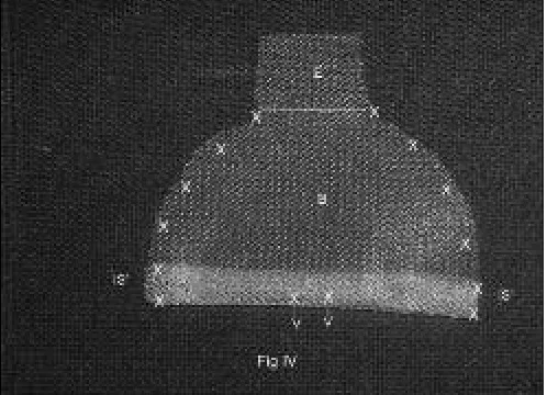
Methods: Twenty femal patients with vault prolapse and grade III or IV
cystocele were studies past 2-3 years. A 3D pelvic CT scan with cys-togram is performed. The pelvic size is measured by Vitea 2,3D pro-cessing software (Fig. 1). A 15x15 cm polypropylene mesh (pore size 1121,w-66 c-90) is tailoring according the above measurement (Fig. 2). The mesh is anchoring into the sacrospinous ligament and each side of Arch Tendinous Pelvic Fascia (ATPF) by Capio devic. The normal pos-terior urethrovesical angle is also resorted by anchoring the mesh to the insertion area of ATPF to the pelvic ramus. The mesh also sutured to peri-urethral ligament to correct hypermobility of the urethra.
Results: All twenty patients have not recurrent proplase. No erosion or
extrusion of mesh are observed. Pelvic muscular pain occured in 4 patients and usually resolved in three months. Two patients developed ureteral obstruction from the procedure and had lysis procedure. Ureteral obstruction can be prevented with ureteral catheter placement prior to the procedure.
Conclusions: The pelvic floor reconstruction with individual customized
mesh graft can provided a better result than the one-size-fit mesh graft. More study is needed.
P46
A Trial of Bilateral Percutaneous Nephrostomy Tube Placement in Refractory Non-ulcerative Interstitial Cystitis Patients Prior to Simple Urinary Diversion
Mini Varghese, Robert D. Mayer
University of Rochester, Rochester, NY, USA
Background: Urinary diversion (with or without cystectomy) as a
treat-ment modality for end-stage classic ulcerative interstitial cystitis (IC) has been reported to be beneficial. Diversion with cystectomy for non-ulcer-ative IC fails to benefit the majority of patients and can add significant morbidity. Recent reports suggest that simple urinary diversion without cystectomy could benefit non-ulcerative IC patients. Simple diversion could improve IC symptoms by preventing bladder stretching and remov-ing chemical irritation from urine. We offered a trial of bilateral percuta-neous nephrostomy tubes with Fogarty ureteral occlusion balloons to four patients with refractory non-ulcerative IC to determine if their symptoms would significantly decline enough to be offered a simple diversion.
Methods: Four female patients, who fulfilled the National Institute of
Arthritis, Diabetes, Digestive, and Kidney Diseases criteria for non-ulcer-ative IC and who had failed previous medical and intravesical treatments, underwent placement of bilateral 8 French percutaneous nephrostomies with 4 French Fogarty ureteral occlusion balloons. Pre- and post-proce-dure pain scores were reviewed for each patient. Three patients had sig-nificant reduction of pain scores and subsequently underwent ileal con-duit urinary diversion (2 open, 1 robotic assisted laparoscopic approach).
Results: Mean duration of disease at time of nephrostomy placement
was 7 years (range 5-11). Mean duration of nephrostomy placement was 27 days (range 9-50). Mean decrease in pain scores at time of nephros-tomy removal was 5.3 (range 3-8). Mean time between nephrosnephros-tomy placement and ileal conduit urinary diversion was 154 days (range 103-227). Mean hospital length of stay after diversion was 18 days (range 6-29). At six month postoperative visits, patients were re-evaluated. One patient (age = 63) had complete resolution of pelvic symptoms with pain score = 0. One patient (age = 32) went back to her pre-nephros-tomy pain score of 9; her pain is exacerbated by bowel movements and she continued on extensive narcotic regimen and intravesical therapy. One patient (age = 27) intermittently has pre-nephrostomy pain scores but at six month visit was using significantly less narcotics for symptom control and was overall pleased with surgical outcome.
Conclusions: Patients who suffer from refractory non-ulcerative IC
pres-ent a treatmpres-ent challenge for urologists. Therapy with irreversible uri-nary diversion can be beneficial but long term success is not easily pre-dicted by a preoperative trial of bilateral nephrostomy tubes with ureteral occlusion balloon catheters . Our limited series suggest that further cri-teria may need to be fulfilled prior to a patient being eligible for simple diversion.
P47
Modified Scrotal (Bianchi) Mid-Raphe Single-Incision Orchidopexy for Palpable Undescended Testis
Jonathan Cloutier, Katherine Moore, Vincent Fradet, Stéphane Bolduc Université Laval, Québec, QC, Canada
Background: To compare the results of a low trans-scrotal mid-raphe
orchidopexy in patients with palpable undescended testes (UDT), to a high-scrotal incision (Bianchi) and to the conventional inguinal approach.
Methods: We used a retrospective cohort study design. All
orchidopex-ies performed between 2003 and 2009 with a minimum of 3-month fol-low-up were included. All palpable UDT that could be brought down manually into the upper third of the scrotum under general anaesthesia, were then reviewed (group 1: high-scrotal incision, group 2: low-scro-tal incision) and compared to the inguinal two-incision technique (group 3). We excluded cases who had undergone previous inguinal surgery or with concomitant surgeries. We comprehensively reviewed the charts and focused on the following outcomes: operative time, success as defined by mid or lower scrotal position of the testicle, and complications at 6-12-weeks and 1-year after surgery.
Fig. 1.P45.
Results: A total of 286 orchidopexies were performed in 214 patients
with palpable UDT. In group 1, a high-scrotal incision was performed in 44 patients for 60 UDT (success 59/60, 98%) with one recurrence. A modification to the technique was adopted and since 2005, patients in group 2 had a trans-scrotal orchidopexy through a single low-scrotal incision on the median raphe. It was performed in 81 patients for 125 UDT. All testes except 1 (99%) were located in a good position within the scrotum. In group 3, a standard inguinal two-incision orchidopexy was performed in 89 patients for 101 UDT (success 100%). The mean operative time for unilateral UDT was significantly shorter for the low trans-scrotal orchidopexy (mean 28 min vs. mean 37 min; p < 0.001) than for the inguinal orchidopexy but equivalent to a high scrotal sion (27 min; p = 0.59). One patient approached by high-scrotal inci-sion required converinci-sion to a traditional inguinal approach. All patent processes vaginalis were ligated, regardless of their size. In all 160 chil-dren followed at 1 year, no long term atrophy or secondary reascent were observed. Postoperative complications included transient postop-erative scrotal hematoma in a single patient from group 1 and 2 wound infections in group 3.
Conclusion: Low trans-scrotal mid-raphe orchidopexy appears to be an
excellent alternative to the high-scrotal incision or the standard inguinal orchidopexy for low palpable UDT especially for bilateral cases. Scrotal orchidopexy is simple, safe, and effective in selected cases.
P48
Renal Perfusion Pump vs. Cold Storage for Donation after Cardiac Death Kidneys: A Systematic Review
Thomas B. McGregor, Varunkumar Bathini, Vivian McAlister, Patrick Luke, Alp Sener
University of Western Ontario, London, ON, Canada
Introduction: The use of a renal perfusion pump vs. cold storage for the
preservation of kidneys obtained from donors after cardiac death (DCD) is controversial. Herein, we examine the impact of using renal perfu-sion pumps on delayed graft function and graft survival using a system-atic review.
Methods: The PUBMED database was searched and reference lists from
1960 to 2009 were consulted. Randomized controlled trials as well as prospective and retrospective cohort studies that compared delayed graft function and graft survival were included. Studies excluded from review were unable to discriminate between DCD and neurologically deceased donor (NDD) deaths. Eleven studies that followed a total of 3377 participants qualified for review. Statistical analysis was carried out using the random effects criteria and odds ratios calculated using the Cochrane database software.
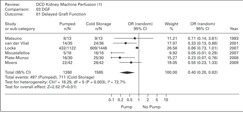
Results: We found that perfusion pumped kidneys from DCD donors
had reduced DGF rates vs. kidneys that were placed in cold storage (Fig. 1) (p = 0.008; odds ratio 0.4, CI 0.20-0.82). In addition, graft func-tion at 1 year post-transplantafunc-tion favored perfusion pumped kidneys (p = 0.04, odds ratio 1.96, CI 1.02-3.77). No differences in primary non-function rates between groups were noted.
Conclusion: This review supports the use of perfusion pumps for DCD
kidney transplantation with respect to delayed graft function rates and graft survival.
Review: DCD Kidney Machine Perfusion (1) Comparison: 03 DGF
Outcome: 01 Delayed Graft Function
Study Pumped Cold Storage OR (random) Weight OR (random)
or sub-category n/N n/N 95% CI % 95% CI Year
Matsuno 8/13 9/13 11.21 0.71 (0.14, 3.61) 1993
van der Vliet 14/35 24/36 17.97 0.33 (0.13, 0.88) 2001 Locke 432/1122 609/1446 26.58 0.86 (0.73, 1.01) 2007 Moustafellos 5/18 16/18 9.92 0.05 (0.01, 0.29) 2007 Plate-Munoz 16/30 25/30 15.27 0.23 (0.07, 0.76) 2008
Moers 22/42 28/42 19.05 0.55 (0.23, 1.33) 2009
Total (95% CI) 1260 1585 100.00 0.40 (0.20, 0.82) Total events: 497 (Pumped), 711 (Cold Storage)
Test for heterogeneity: Chi2 = 18.29, df = 5 (P = 0.003), l2 = 72.7%
Test for overall effect: Z=2.52 (P=0.01)
0.1 0.2 0.5 1 2 5 10
Pump No Pump