Developmental Expression Pattern
Isolation, genomic structure and developmental expression of
Fgf8 in the short-tailed fruit bat, Carollia perspicillata
CHRIS J. CRETEKOS
1, JIAN-MIN DENG
1, ERIC D. GREEN
2, NISC COMPARATIVE SEQUENCING PROGRAM
2,#,
JOHN J. RASWEILER
3and RICHARD R. BEHRINGER
1,*
1Department of Molecular Genetics, University of Texas M.D. Anderson Cancer Center, Houston, Texas, 2NISC Comparative Sequencing Program,Genome Technology Branch and NIH Intramural Sequencing Center (NISC), National Human Genome Research Institute, Bethesda, Maryland and 3Department of Obstetrics and Gynecology, State University of New York Downstate Medical Center, Brooklyn,
New York, USA
ABSTRACT Fibroblast growth factor-8 (Fgf8) encodes a secreted protein which was initially identified as the factor responsible for androgen-dependant growth of mouse mammary carci-noma cells (Tanaka et al., 1992). Fgf8 has been subsequently implicated in the patterning and growth of the gastrulating embryo, paraxial mesoderm (somites), limbs, craniofacial tissues, central nervous system and other organ systems during the development of several vertebrate model animals. Consistent with these findings, Fgf8 is expressed in a complex and dynamic pattern during vertebrate embryogenesis. Here we report the isolation and characterization of a bat (Carollia perspicillata) Fgf8 orthologue. Compared with those of other model vertebrates, Carollia Fgf8 is conserved with respect to genomic structure, sequence and many domains of developmental expression pattern. Interestingly, the expression domain marking the apical ectodermal ridge of the developing limb shows a striking difference compared to that of mouse, consistent with evolutionary diversification of bat limb morphology.
KEY WORDS: Chiroptera, Carollia perspicillata, Fgf8, genomic structure, gene expression
The fibroblast growth factors (FGFs) are a family of related polypeptides that act to regulate growth, motility and differentia-tion of a diverse variety of cell-types and tissues. In mammals, 22 members of the FGF gene family (Fgf1-Fgf23) have been de-scribed to date (Ornitz and Itoh, 2001). The FGF gene family encodes proteins that share an internal core region containing 28 conserved and 6 identical amino acids, but vary in both sequence and size of their N- and C- termini. Most FGFs encode secreted ligands that signal to target cells through a family of high-affinity receptor tyrosine kinases, which act primarily through a con-served MAP Kinase-dependant signal transduction pathway (Eswarakumar et al., 2005).
Fgf8 is expressed in a complex and dynamic pattern during embryogenesis and regulates growth and patterning of the brain, limbs, heart, kidney, ear, nose and eye (Boulet et al., 2004, Crossley et al., 1996a, Crossley et al., 1996b, Kawauchi et al., 2005, Lewandoski et al., 2000, Meyers et al., 1998, Perantoni et al., 2005, Reifers et al., 1998, Reifers et al., 2000, Shanmugalingam et al., 2000, Storm et al., 2006, Sun et al., 1999, Trumpp et al., 1999). Fgf8 activity is further regulated by alternative splicing and
*Address correspondence to: Richard R. Behringer. Department of Molecular Genetics, University of Texas, M.D. Anderson Cancer Center, 1515 Holcombe Blvd., Houston, Texas 77030, USA. Fax: +1-713-834-6339. e-mail: rrb@mdanderson.org
# Note: NISC comparative sequencing program refers to a group author. The staff of the NISC comparative sequencing program are acknowledged for their construction of the Carollia genomic BAC library and sequencing of the Fgf8-containing BAC insert.
0214-6282/2007/$30.00 © UBC Press
Printed in Spain
www.intjdevbiol.com
has been shown to be alternatively spliced in human, mouse, chick and Xenopus (Crossley and Martin, 1995, Fletcher et al., 2006, Gemel et al., 1996, Ghosh et al., 1996, Haworth et al., 2005, MacArthur et al., 1995). The alternatively spliced mRNAs give rise to protein isoforms, differing in the length and/or sequence of the N terminus, several of which are known to have distinct activities in vivo (Blunt et al., 1997, Fletcher et al., 2006, MacArthur et al., 1995, Olsen et al., 2006, Song et al., 2000).
1a 1b 1c 1d 2 3 CACGTGCAGGTCCTGGCCAACAAGCGCATCAACGCCATGGCAGAGGACGGCGACCCCTTCG Human CACGTGCAGGTCCTGGCCAACAAGCGCATCAACGCCATGGCAGAAGACGGAGACCCCTTCG Mouse
GCTGTTGCACTTGCTGGTTCTCTGCCTCCACGCCCAGGTAAGGCGCACTGCGCGGAGGCG Bat GCTGTTGCACTTGCTGGTCCTCTGCCTCCAAGCCCAG Human GCTGTTGCACTTGCTGGTTCTCTGCCTCCAAGCCCAGGTAAGGAGCGCTGCGCAGAAGCG Mouse
^
GGGGCCGGGGGCCGGGGGCCGGCGGGGAAGGCCGGCTGGCCAAGTCGGGCAGGGGCACCA Bat GGGGCCGGGCGCGGGGAACC---CAGCTGACACTCTCGGGCAGGGACACGA Mouse
GGACCGACGCTGCGGCCAGCGCGGCAGGGTTGGAAGGAACCTTAGAAACCCAACCCCGAA Bat GGACCGACCCTTCGGCCAGCGCAGCAGGGCTGGAAAGAACTTTACAAATCCAGCCCCAAA Mouse
CTATCCCGAGTCGGGAGCTAAGGAACAGAGAGGTAGCGCCCAGCACTAGGTCACACAGCGA Bat CTACCCCGAGGAGGGATCTAAGGAACAGAGAGACAGTGTCCTGCCTAAAGTCACACAGCGA Mouse
GAAGGCCCGGGCGGGGGGCCTGCGCCGGGCAGGGAGCTGGCTTCCCTGTTCCGAGCTGGC Bat GAAGGCCCGGGCAGGGGCCCTGCGCTGGGCAGGGAGCTCGCTTCCCTGTTCCGGGCTGGC Human GAAGGCCCGGGCGGGGGGCCTGCGCTGGGCAGGGAGCCCACTTCCCTGCTCCGAGCTGGC Mouse
CGCGAGTCCCAGGGTGTTTCCCAACAG Bat CGGGAGCCCCAGGGTGTCTCCCAACAG Human CGGGAGCCCCAGGGTGTTTCCCAACAG Mouse
CCAAGCTCATTGTGGAGACAGATACGTTTGGGAGCCGGGTTCGAGTTCGTGGAGCTGAGA Bat CAAAGCTCATCGTGGAGACGGACACCTTTGGAAGCAGAGTTCGAGTCCGAGGAGCCGAGA Human CGAAGCTCATTGTGGAGACCGATACTTTTGGAAGCAGAGTCCGAGTTCGCGGCGCAGAGA Mouse
CAGGCCTCTACATCTGCATGAACAAGAAGGGGAAGCTGATTGCCAAG Bat CGGGCCTCTACATCTGCATGAACAAGAAGGGGAAGCTGATCGCCAAG Human CAGGTCTCTACATCTGCATGAACAAGAAGGGGAAGCTAATTGCCAAG Mouse
AGCAACGGCAAAGGCAAGGATTGTGTCTTCACGGAGATCGTGCTGGAGAACAACTACACG Bat AGCAACGGCAAAGGCAAGGACTGCGTCTTCACGGAGATTGTGCTGGAGAACAACTACACAHuman AGCAACGGCAAAGGCAAGGACTGCGTATTCACAGAGATCGTGCTGGAGAACAACTACACG Mouse
GCCCTGCAGAACGCCAAGTACGAGGGCTGGTACATGGCCTTCACCCGCAAGGGCCGGCCABat GCGCTGCAGAATGCCAAGTACGAGGGCTGGTACATGGCCTTCACCCGCAAGGGCCGGCCC Human GCGCTGCAGAACGCCAAGTACGAGGGCTGGTACATGGCCTTTACCCGCAAGGGCCGGCCC Mouse
CGCAAGGGCTCCAAGACGCGGCAGCACCAGCGCGAGGTCCACTTCATGAAGCGGCTGCCC Bat CGCAAGGGCTCCAAGACGCGGCAGCACCAGCGTGAGGTCCACTTCATGAAGCGGCTGCCC Human CGCAAGGGCTCCAAGACGCGCCAGCATCAGCGCGAGGTGCACTTCATGAAGCGCCTGCCGMouse
CGTGGCCACCACACCACCGAGCAGAGCCTGCGGTTCGAGTTCCTCAACTACCCGCCCTTC Bat
MGSPRSALSCL LLHLLVLCLHAQVRRTARRRGPGAGGRRGRPAGQVGQGHQDRRCGQRGRBat MGSPRSALSCL LLHLLVLCLQAQVRRGARRRGPGAPV. Human MGSPRSALSCL LLHLLVLCLQAQVRSAAQKRGPGAGN----PADTLGQGHEDRPFGQRSRMouse
1a 1b
VGRNLRNPTPNYPESGAKEQRGSAQH. EGPGGGPAPGRELASLFRAGRESQGVSQQ Bat EGPGRGPALGRELASLFRAGREPQGVSQQ Human AGKNFTNPAPNYPEEGSKEQRDSVLPKVTQR EGPGGGPALGREPTSLLRAGREPQGVSQQ Mouse
long form of mouse 1b 1c 1c
VTVQSSPNFTQHVREQSLVTDQLSRRLIRTYQLYSRTSGKHVQVLANKRINAMAEDGDPF Bat
Many bat species are also unusual in that they navigate in darkness using echolocation. Echolocation in bats likely required modification of facial structures to emit focused ultrasonic sounds, modification of ears to receive sound reflections and modification of neural auditory processing centers to translate aural input into
spatial information. Since Fgf8 has been shown to be an important regulator of limb, craniofacial, ear and CNS development as well as a useful molecular marker for all of these tissues, we chose to identify and analyze the Fgf8 gene of our bat model Carollia perspicillata (henceforth Carollia).
As a first step in isolating the bat Fgf8 orthologue, we PCR amplified a Carollia embryo cDNA template using a set of primers designed against regions of near identity among mammalian Fgf8 cDNA sequence alignments. One primer pair, FGF8-F3 and -R1, gave a robust product at the expected size of ~480 bp. Sequence analysis of the cloned product, CpFgf8F3R, revealed 94.6% and 94.2% identity to the corresponding region of human and mouse Fgf8 cDNAs, respectively. This fragment corresponds to portions
A
B
C
of mouse and human Fgf8 exons 1d, 2 and 3 which are common to all known mRNA isoforms (Crossley and Martin, 1995, Ghosh et al., 1996, MacArthur et al., 1995).
A probe generated from this cloned fragment was used to screen Carollia cDNA library and a Carollia genomic BAC library. Partial Fgf8 cDNAs were isolated from the former, whose 5’ ends were within the sequence contained in CpFgf8F3R1 but extended 3’ an additional ~300 bp to a poly-A tail. Five Fgf8-containing BAC clones were identified from the latter and one clone was selected for sequencing (106,727 bp insert, Genbank accession # AC147851). Comparisons with available vertebrate genomes revealed that in addition to Fgf8, the BAC insert also contained two syntenic loci, NPM3 and MGEA5. These comparisons were also used to determine the structure of the bat Fgf8 coding region and to predict exon–intron junctions (Fig. 1).
Like those of other mammals, the bat Fgf8 locus consists of 6 exons, designated exon 1a-d, 2 and 3 (Fig. 1A, B, C). Alignment of the DNA sequence of the predicted bat exons with those of human and mouse revealed a high degree of sequence conser-vation, 95% and 94% identity to human and mouse, respectively, for all of the potential coding sequences combined (Fig. 1C). The 3’ untranslated region (UTR) sequence obtained from Carollia Fgf8 partial cDNA clones shows 73% and 68% identity to human and mouse 3’ UTRs, respectively and alignment of the Carollia genomic sequence immediately upstream of exon 1a with human and mousenFgf8 5’ UTRs reveals 76% and 69% identity, respec-tively (data not shown).
Predicted exons 1a, 1d, 2 and 3 of Carollia Fgf8 encode amino acids that are 100% identical to the corresponding human and mouse proteins (Fig. 1D). Carollia exon 1c shows 3 differences in 29 amino acids encoded compared to human exon 1c and 5 differences compared to mouse exon 1c (Fig. 1D). Carollia and human exon 1b show a marked divergence compared to the corresponding mouse exon. Mouse exon 1b splices to down-stream exons from two potential slice donors (indicated by arrow-heads below the DNA sequence in Fig. 1B). If the first conserved splice donor in Carollia Fgf8 is utilized, 11 of 12 amino acids are identical to human or mouse, but if the second splice donor is used the reading frame is blocked by an in-frame stop codon (boxed in Fig. 1B, C) and no functional FGF8 protein would be produced (Fig. 1D). In human FGF8, the reading frame of the longer alternative form of exon 1b is similarly blocked by in-frame stop codons (Gemel et al., 1996). This data, combined with a lack of human cDNAs containing the long form of exon 1b, led to the
conclusion that the human FGF8 isoforms corresponding to mouse Fgf8c, d, g and h are not made (Gemel et al., 1996, Ghosh et al., 1996). In addition to the in-frame stop codon, the Carollia genomic sequence that corresponds to the longer form of mouse exon 1b is markedly less conserved than the rest of the gene, showing only 61% identity to mouse at the amino acid level, compared to ~98% amino acid identity for remaining coding sequence. It therefore seems likely that Carollia, like human, lacks the 4 potential Fgf8 isoforms that include the longer form of exon 1b (dashed lines below the exons in Fig. 1A).
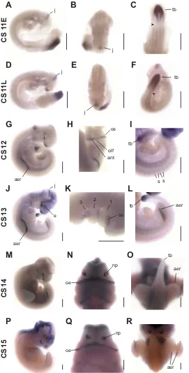
Fgf8 expression was examined in Carollia stage (CS) 11–15 embryos (Figs. 2, 3). The CS11 bat embryo is morphologically equivalent to a ~ 9 day (E9.0) mouse embryo. During the course of CS11, the developing bat embryo goes from 13 to 20 pair of trunk somites, the ventricles of the brain expand while the brain bends ventrally at the level of the midbrain to form the cranial flexure and the first two pharyngeal arches form (Cretekos et al., 2005). The CS15 bat embryo is morphologically equivalent to a ~ E11.5 mouse embryo. During the course of CS15, the forelimb and hindlimb footplates form sequentially and the distal parts of the right and left first pharyngeal arch fuse at the midline to form the upper and lower jaws (Cretekos et al., 2005).
At CS11, Fgf8 mRNA is detected primarily in a posterior domain of the main body axis and in the anterior neural tube. In the posterior region, Fgf8 is expressed in a graded fashion extending from the tailbud to an area about 2 somite-widths posterior to the nascent somite (nascent somite indicated by arrowhead in Fig. 2C, F). Expression in this domain is detected at least through late CS13 (Fig 2I, L). We have shown previously that the process of somitogenesis continues until 40 pairs of somites have formed at CS14 (Cretekos et al., 2005), thus downregulation of expression in this domain is concurrent with the end of somite formation. Fgf8 is not detected in newly-formed somites, but once the somites have differentiated Fgf8 mRNA is detected in narrow stripes of lateral myotome cells located at the anterior and posterior margins of each somite (Fig. 2D, I).
Fgf8 expression was detected in four distinct domains in the developing anterior neural tube. From anterior to posterior, the first is at the anterior end, the second is in the region of the developing eyes, the third is along the ventral midline of the of the fore- and mid-brain and the last is in the border between the prospective midbrain and hindbrain (Fig. 2A, B, D, E, H, J). This latter expression domain is restricted to a narrow band of express-ing cells in the region referred to as the midbrain-hindbrain
e
In addition to the expression domains in the developing brain, Fgf8 mRNA is also detected in several other regions of the head. Fgf8 is expressed in the pharyngeal arches, which give rise to many craniofacial tissues including jaws, teeth, tongue and outer ears. Fgf8 expression is detected in the lateral regions of the pharyngeal arches, along the grooves that separate each arch (Fig. 2K). High level expression is found in the oral ectoderm of the first pharyngeal arch (Fig. 2H, K, N, Q; Fig. 3 G, H). Fgf8 expression was also detected in the ectoderm covering the anterior forebrain in the region of the prospective olfactory placodes by late CS11 (Fig. 2D). By CS14, expression in this region is localized in the ectoderm of the nasal processes bracketing the nasal pits (Fig. 2N, Q; Fig. 3 G, H). At morphologi-cally similar stages of mouse development, Fgf8 expression in this region is continuous around the nasal pits (Bachler and Neubuser, 2001), whereas in Carollia we see separate medial and lateral domains (Fig. 3 G, H).
The appearance of the forelimb buds in a developing Carollia embryo marks the onset of CS12 and hindlimb buds appear one stage later (Cretekos et al., 2005). Fgf8 is detected in the presumptive apical ectodermal ridge (AER) of the forelimb buds during early CS12 and in the presumptive AER of the hindlimb buds in early CS13 (Fig. 2G, J, L). Expression in the AER persists until at least late CS15 (Fig. 2R). The AER has long been known to be a source of signals required for proximal-distal growth of the limb bud and Fgf8 expression in the AER has been shown to be required, along with Fgf4, for survival and proliferation of the underlying mesoderm during limb bud outgrowth (Lewandoski et al., 2000; Moon and Capecchi, 2000; Boulet et al., 2004). The Fgf8 expression domain in the bat forelimb AER appears rela-tively wide in the dorsal-ventral axis compared to that of mouse Fgf8 at comparable stages (Fig. 3A, B). Transverse sections through forelimb buds of bat embryos at CS12 and 14 and mouse embryos at E9.5 and 10.5 reveal that the AER expression domain in bat is 2.7 times wider in CS12 bat embryos compared to E9.5 mouse embryos and 3 times wider in CS14 bat vs. E10.5 mouse (Fig. 3C, D, E, F and data not shown). The overall size of junction or isthmus. This region has been shown to be an
important organizing center for brain pattering and Fgf8 expres-sion is known to be critical for this function (Olsen et al., 2006, Reifers et al., 1998, Storm et al., 2006). Expression of Fgf8 in the midbrain-hindbrain junction was detected until at least CS14 (Fig. 2J and data not shown).
bat and mouse forelimb buds at CS12/E9.5 is similar, while at CS14/E10.5 the bat forelimb bud is somewhat larger in all dimen-sions. A novel domain of Fgf8 expression in the interdigital mesen-chyme between forelimb digits 2 - 5 during later stages of Carollia development has been previously described and been proposed to play a role in the persistence of these tissues for the formation of the chiropatagium (handwing) wing membrane (Weatherbee et al., 2006).
Experimental Procedures
Identification of a Carollia Fgf8 probe
A probe for Carollia Fgf8 was generated by PCR using primers FGF8-F3 (5’ATG TGA GGG AGC AGA GCC TG 3’) and FGF8-R1 (5’ GGT AGT TGA GGA ACT CGA AGC 3’), designed from regions of near identity in cDNA sequence alignments of human, mouse, rat and cow Fgf8. The 484 bp product was cloned into EcoRV-digested and T-tailed pBluescriptII KS (Stratagene) and confirmed by sequencing (CpFgf8F3R1, Genbank ac-cession # EF035451).
Isolation of a Carollia Fgf8 partial cDNA
The above primer pair was also used to screen a fractionated Carollia embryo cDNA library. This library (CPMGE) was generated in λ-ZAPII (Stratagene) from oligo-dT primed cDNA prepared from poly-A selected RNA purified from 5 whole Carollia (CS14, 14L, 15, 16 and 17) embryos. The primary library was titered at 7.9 X 10⁶ pfu/µg λ-arms. Two million
recombinants of CPMGE were simultaneously amplified and fractionated into 94 sublibraries (CPMGE A1 - H10), each initiated with ~20,000 independent recombinant clones. Phagemid DNA was prepared from each sublibrary and arrayed into 96-well microtiter plates, leaving 2 wells open for positive and negative control DNA, such that the library can be initially screened by 96-well PCR. Sublibrary CPMGE D8 produced an ~480 bp product when amplified using FGF8F3/R1 primer pair. This sublibrary was plated and screened by plaque hybridization using a radiolabeled probe prepared from CpFgf8F3R1. Two hybridizing phagemid were plaque purified, “popped-out” into plasmid clones using eXassist helper phage (Stratagene) and sequenced (Genbank accession #s EF035452 and EF035453)
Isolation and sequencing of the Carollia Fgf8 genomic locus The CpFgf8F3R1 radiolabeled probe was also used to screen an arrayed Carollia genomic BAC library (E.D.G., unpublished). 5 indepen-dent hybridizing BAC clones were confirmed by PCR with the FGF8DF3/R1 primer pair and subjected to insert end sequencing. BAC-end sequences were aligned to the human genome using BLAST (www.ncbi.nlm.nih.gov) and BAC clone 3O14 was selected for shotgun sequencing by the NIH Intramural Sequencing Center (www.nisc.nih.gov) (106,727 bp insert, Genbank accession # AC147851).
In situ hybridization and histology
Whole-mount in situ hybridization (ISH) was performed as described previously (Chen et al., 2005) on Carollia embryos with the following modifications: Carollia embryos were treated with proteinase K (Roche) 2 min for CS11, 3 min for CS11L, 4 min for CS12, 6 min for CS13, 15 min for CS14 and 20 min for CS15. An antisense riboprobe against Carollia Fgf8 was generated by digesting CpFgf8F3R1 with EcoRI followed by transcrip-tion with T7 RNA polymerase in the presence of digoxigenin-11-UTP (Roche). This probe was used at a final concentration of 0.5 - 1 µg/ml. Hybridization and post-hybridization washes were carried out at 65 - 70° C. The Carollia embryos used in this study were collected from a wild population on the island of Trinidad, as previously described (Chen et al., 2005, Cretekos et al., 2005) and staged according to Cretekos et al. (2005). Whole-mount in situ hybridization (ISH) on mouse embryos was performed as described previously (Wilkinson, 1992), using an antisense riboprobe
ne
Fig. 3. Detailed analysis of Fgf8 expression.(A,B,C) Fgf8in situ of Carollia forelimb buds; (D,E,F) comparable views of Fgf8 in situ of mouse forelimb buds at similar stages. (A,D) Edge-on views of bat CS14 (A) and mouse E10.5 (D) distal forelimb buds, anterior is to the top and scalebar = 0.5 mm. (B,E) Transverse sections through the central region of bat CS12 (B) and mouse E9.5 (E) forelimb buds, dorsal is to the top and anterior is into the page, scalebar = 50 µm. (C,F) Transverse sections through the distal tip of bat CS14 (C) and mouse E10.5 (F) forelimb buds, dorsal is to the top and anterior is into the page; scalebar, 50 µm. Measured width of the AER is shown in panels (B, C, E and F). (G,H) Face-on view (G) and mFace-ontage image of frFace-ontal sectiFace-ons (H), showing Fgf8 expression in craniofacial tissues at CS14. Anterior is to the top, bp, branchial pouch; g, glossopharyngeal arch; h, hyoid arch; ma, mandible; mx, maxilla; ne, nasal epithelium; oe, oral ectoderm; * indicates a probe trapping artifact in the forebrain ventricle. Scalebar, 0.5 mm in panel (G) and 200 µm in panel (H).
transcribed from a mouse Fgf8 cDNA template (provided by R. L. Johnson). Some specimens were embedded and processed for cryostat sectioning after whole-mount ISH (Nagy et al., 2003).
Acknowledgements
of Life Sciences, University of the West Indies, Trinidad and particularly Indira Omah-Maharaj, for generous assistance and the use of departmental facilities. We are grateful to the Wildlife Section, Forestry Division, Agriculture Land and Marine Resources of the Republic of Trinidad and Tobago for providing collecting and export permits. These studies were supported by National Science Foundation grant IBN 0220458 and endowments from the Barnts Family and Ben F. Love to R.R.B. C.J.C. was supported by NIH training grants CA09299 and HD07325. DNA sequencing and veterinary resources were supported by the NIH Cancer Center Support (Core) Grant CA16672. This work was supported in part by the Intramural Research Program of the National Human Genome Research Institute, NIH.
References
BACHLER, M. and NEUBUSER, A. (2001). Expression of members of the fgf family and their receptors during midfacial development. Mech. Dev. 100: 313-6.
BEHRINGER, R.R., EAKIN, G.S. and RENFREE, M.B. (2006). Mammalian diversity: Gametes, embryos and reproduction. Reprod. Fert. Dev. 18: 99-107.
BLUNT, A.G., LAWSHE, A., CUNNINGHAM, M.L., SETO, M.L., ORNITZ, D.M. and MACARTHUR, C.A. (1997). Overlapping expression and redundant activation of mesenchymal fibroblast growth factor (fgf) receptors by alternatively spliced fgf-8 ligands. J. Biol. Chem. 272: 3733-8.
BOULET, A.M., MOON, A.M., ARENKIEL, B.R. and CAPECCHI, M.R. (2004). The roles of fgf4 and fgf8 in limb bud initiation and outgrowth. Dev. Biol. 273: 361-72.
CARROLL, S.B., GRENIER, J. K. & WEATHERBEE, S. D. (2004). From DNA to
diversity: Molecular genetics and the evolution of animal design. Blackwell Science,
Malden, Massachusetts.
CHEN, C.H., CRETEKOS, C.J., RASWEILER, J.J. 4th and BEHRINGER, R.R. (2005). Hoxd13 expression in the developing limbs of the short-tailed fruit bat, carollia perspicillata. Evo. Develop. 7: 130-41.
CRETEKOS, C.J., WEATHERBEE, S.D., CHEN, C.H., BADWAIK, N.K., NISWANDER, L., BEHRINGER, R.R. and RASWEILER, J.J. 4th (2005). Embryonic staging system for the short-tailed fruit bat, carollia perspicillata, a model organism for the mammalian order chiroptera, based upon timed pregnancies in captive-bred animals. Dev. Dynam. 233: 721-38.
CROSSLEY, P.H. and MARTIN, G.R. (1995). The mouse fgf8 gene encodes a family of polypeptides and is expressed in regions that direct outgrowth and patterning in the developing embryo. Development 121: 439-51.
CROSSLEY, P.H., MARTINEZ, S. and MARTIN, G.R. (1996a). Midbrain development induced by fgf8 in the chick embryo. Nature 380: 66-8.
CROSSLEY, P.H., MINOWADA, G., MACARTHUR, C.A. and MARTIN, G.R. (1996b). Roles for fgf8 in the induction, initiation and maintenance of chick limb development.
Cell 84: 127-36.
EAKIN, G.S. and BEHRINGER, R.R. (2004). Diversity of germ layer and axis formation among mammals. Semin. Cell & Dev. Biol. 15: 619-29.
ESWARAKUMAR, V.P., LAX, I. and SCHLESSINGER, J. (2005). Cellular signaling by fibroblast growth factor receptors. Cytokine & Growth Factor Reviews 16: 139-49.
FLETCHER, R.B., BAKER, J.C. and HARLAND, R.M. (2006). Fgf8 spliceforms mediate early mesoderm and posterior neural tissue formation in xenopus. Development 133: 1703-14.
GEMEL, J., GORRY, M., EHRLICH, G.D. and MACARTHUR, C.A. (1996). Structure and sequence of human fgf8. Genomics 35: 253-7.
GHOSH, A.K., SHANKAR, D.B., SHACKLEFORD, G.M., WU, K., T’ANG, A., MILLER, G.J., ZHENG, J. and ROY-BURMAN, P. (1996). Molecular cloning and character-ization of human fgf8 alternative messenger rna forms. Cell Growth & Differentiation 7: 1425-34.
HAWORTH, K.E., HEALY, C. and SHARPE, P.T. (2005). Characterisation of the genomic structure of chick fgf8. DNA Sequence 16: 180-6.
KAWAUCHI, S., SHOU, J., SANTOS, R., HEBERT, J.M., MCCONNELL, S.K., MA-SON, I. and CALOF, A.L. (2005). Fgf8 expression defines a morphogenetic center required for olfactory neurogenesis and nasal cavity development in the mouse.
Development 132: 5211-23.
LEWANDOSKI, M., SUN, X. and MARTIN, G.R. (2000). Fgf8 signalling from the aer is essential for normal limb development. Nat. Genetics 26: 460-3.
MACARTHUR, C.A., LAWSHE, A., XU, J., SANTOS-OCAMPO, S., HEIKINHEIMO, M., CHELLAIAH, A.T. and ORNITZ, D.M. (1995). Fgf-8 isoforms activate receptor splice forms that are expressed in mesenchymal regions of mouse development.
Development 121: 3603-13.
MEYERS, E.N., LEWANDOSKI, M. and MARTIN, G.R. (1998). An fgf8 mutant allelic series generated by cre- and flp-mediated recombination. Nat. Genetics 18: 136-41.
MOON, A.M. and CAPECCHI, M.R. (2000). Fgf8 is required for outgrowth and patterning of the limbs. Nat Genet. 26: 455-9.
NAGY, A., GERTSENSTEIN, M., VINTERSTEN, K. and BEHRINGER, R. (2003).
Manipulating the mouse embryo: A laboratory manual. Cold Spring Harbor
Labo-ratory Press, Cold Spring Harbor, New York.
NEUWEILER, G. (2000). The biology of bats. Oxford University Press, New York.
NOWAK, R. (1999). Walker’s mammals of the world. The Johns Hopkins University Press, Baltimore.
OLSEN, S.K., LI, J.Y., BROMLEIGH, C., ELISEENKOVA, A.V., IBRAHIMI, O.A., LAO, Z., ZHANG, F., LINHARDT, R.J., JOYNER, A.L. and MOHAMMADI, M. (2006). Structural basis by which alternative splicing modulates the organizer activity of fgf8 in the brain. Genes Dev. 20: 185-98.
ORNITZ, D.M. and ITOH, N. (2001). Fibroblast growth factors. Genome Biology 2: REVIEWS3005.
PERANTONI, A.O., TIMOFEEVA, O., NAILLAT, F., RICHMAN, C., PAJNI-UNDERWOOD, S., WILSON, C., VAINIO, S., DOVE, L.F. and LEWANDOSKI, M. (2005). Inactivation of fgf8 in early mesoderm reveals an essential role in kidney development. Development 132: 3859-71.
REIFERS, F., BOHLI, H., WALSH, E.C., CROSSLEY, P.H., STAINIER, D.Y. and BRAND, M. (1998). Fgf8 is mutated in zebrafish acerebellar (ace) mutants and is required for maintenance of midbrain-hindbrain boundary development and somi-togenesis. Development 125: 2381-95.
REIFERS, F., WALSH, E.C., LEGER, S., STAINIER, D.Y. and BRAND, M. (2000). Induction and differentiation of the zebrafish heart requires fibroblast growth factor 8 (fgf8/acerebellar). Development 127: 225-35.
SHANMUGALINGAM, S., HOUART, C., PICKER, A., REIFERS, F., MACDONALD, R., BARTH, A., GRIFFIN, K., BRAND, M. and WILSON, S.W. (2000). Ace/fgf8 is required for forebrain commissure formation and patterning of the telencephalon.
Development 127: 2549-61.
SIMMONS, N. (2001). Reassessing bat diversity: How many species are there in the world? Bat Res News 179-180.
SONG, Z., POWELL, W.C., KASAHARA, N., VAN BOKHOVEN, A., MILLER, G.J. and ROY-BURMAN, P. (2000). The effect of fibroblast growth factor 8, isoform b, on the biology of prostate carcinoma cells and their interaction with stromal cells. Cancer
Res. 60: 6730-6.
STORM, E.E., GAREL, S., BORELLO, U., HEBERT, J.M., MARTINEZ, S., MCCONNELL, S.K., MARTIN, G.R. and RUBENSTEIN, J.L. (2006). Dose-dependent functions of fgf8 in regulating telencephalic patterning centers. Development 133: 1831-44.
SUN, X., MEYERS, E.N., LEWANDOSKI, M. and MARTIN, G.R. (1999). Targeted disruption of fgf8 causes failure of cell migration in the gastrulating mouse embryo.
Genes Dev. 13: 1834-46.
TANAKA, A., MIYAMOTO, K., MINAMINO, N., TAKEDA, M., SATO, B., MATSUO, H. and MATSUMOTO, K. (1992). Cloning and characterization of an androgen-induced growth factor essential for the androgen-dependent growth of mouse mammary carcinoma cells. Proc. Natl. Acad. Sci. USA 89: 8928-32.
TRUMPP, A., DEPEW, M.J., RUBENSTEIN, J.L., BISHOP, J.M. and MARTIN, G.R. (1999). Cre-mediated gene inactivation demonstrates that fgf8 is required for cell survival and patterning of the first branchial arch. Genes Dev. 13: 3136-48.
WEATHERBEE, S.D., BEHRINGER, R.R., RASWEILER, J.J. 4th and NISWANDER, L.A. (2006) Interdigital webbing retention in bat wings illustrates genetic changes underlying amniote limb diversification.Proc Natl Acad Sci USA. 103(41):15103-7.
WILKINSON, D.G. (1992). In situ hybridization: A practical approach. Oxford University Press, New York.
Related, previously published Int. J. Dev. Biol. articles
See our recent Special Issue on Invasion in Cancer & Embryonic Development at: http://www.ijdb.ehu.es/web/contents.php?vol=48&issue=5-6
Interplay between FGF10 and Notch signalling is required for the self-renewal of pancreatic progenitors Francisco Miralles, Luciane Lamotte, Dominique Couton and Rajiv L. Joshi
Int. J. Dev. Biol. (2006) 50: 17-26
Isthmus organizer and regionalization of the mesencephalon and metencephalon Harukazu Nakamura and Yuji Watanabe
Int. J. Dev. Biol. (2005) 49: 231-235
Effects of FGF9 on embryonic Sertoli cell proliferation and testicular cord formation in the mouse Louise Willerton, Robert A. Smith, David Russell and Sarah Mackay
Int. J. Dev. Biol. (2004) 48: 637-643
A history of mammalian embryological research. H Alexandre
Int. J. Dev. Biol. (2001) 45: 457-467
Co-expression pattern analysis of Fgf4, Fgf8 and Shh gene expression at diverse signalling centers during mouse development.
D Bueno and J K Heath
The International Journal of Developmental Biology
Editorial Office, Uni. of the Basque Country Dept. Cell Biology and Histology Faculty of Medicine, E-48940 Leioa Vizcaya, SPAIN
ORDER BY
Web:
http://www.intjdevbiol.com
E-mail: ijdb@ehu.es
(include the information indicated above)FAX:
+34-94-601-3266
POST: to the address shown to the right
ORDER FORM
I would like to order ____ cop(y/ies) of the Int. J. Dev. Biol. Special Issue “Invasion in Cancer & Embryonic Development” (Vol. 48, Nos. 5/6) at US$ 90 or Euro 70 per copy (including post and packaging). Total to be charged: __________US$ / Euro (please specify currency)
Preface
by Marc Mareel and Juan Aréchaga
ONCOGENES, TUMOR SUPPRESSOR GENES & GENETIC REGULATION
Molecular cell biology and cancer metastasis. An interview with Garth Nicolson
by Marc Mareel
Transcriptional regulation of cadherins during development and carcinogenesis
by Héctor Peinado, Francisco Portillo and Amparo Cano
APC dosage effects in tumorigenesis and stem cell differentiation
by Claudia Gaspar and Riccardo Fodde
ACTIVITIES OF INVASIVE CELLS
Discovery and characterization of the cadherin family of cell adhesion molecules. An interview with Masatoshi Takeichi
by Douglas Sipp
Cadherin-mediated cell-cell adhesion and tissue segregation in relation to malignancy
by Ramsey A. Foty and Malcolm S. Steinberg
Matrix metalloproteinases in cancer: from new functions to improved inhibition strategies
by Alicia R. Folgueras, Alberto M. Pendás, Luis M. Sánchez and Carlos López-Otín
Cytoskeletal mechanisms responsible for invasive migration of neoplastic cells
by Jury M. Vasiliev
Collective cell migration in morphogenesis and cancer
by Peter Friedl, Yael Hegerfeldt and Miriam Tusch
Invasive growth: a genetic program
by Alessandra Gentile and Paolo M. Comoglio
INVASION SIGNALING PATHWAYS
From here to there; a life based on migration. An interview with Isaiah J. Fidler
by Ian R. Hart
N-cadherin in the spotlight of cell-cell adhesion, differentiation, embryogenesis, invasion and signalling
by Lara D.M. Derycke and Marc E. Bracke
The Wnt connection to tumorigenesis
by Jürgen Behrens and Barbara Lustig
The chemokine network in cancer - much more than directing cell movement
by Hagen Kulbe, Neil R. Levinson, Fran Balkwill and Julia L. Wilson
PARTICIPATION OF THE HOST
Brain tumour development and invasion. An interview with Ole Didrik Laerum
by Dieter F. Hülser
The stroma reaction myofibroblast:
a key player in the control of tumor cell behavior
by Alexis Desmoulière, Christelle Guyot and Giulio Gabbiani
The countercurrent principle in invasion and metastasis of cancer cells. Recent insights on the roles of chemokines
by Ghislain Opdenakker and Jo Van Damme
NORMAL EMBRYONIC INVADERS & THEIR MALIGNANT COUNTERPARTS
The migration and differentiation of a chemist entangled in developmental and cancer biology. An interview with Jean-Paul Thiery
by Fred T. Bosman
Primordial germ cell migration
by Kathleen Molyneaux and Christopher Wylie
Germinal tumor invasion and the role of the testicular stroma
by Alejandro Díez-Torre, Unai Silván, Olivier De Wever, Erik Bruyneel, Marc Mareel and Juan Aréchaga
METHODOLOGY
Tumor invasion and metastasis: getting more basic to come closer to the patient. An interview with Lance A. Liotta
by Vincent Castronovo
The "chemoinvasion assay": a tool to study tumor and endothelial cell invasion of basement membranes
by Adriana Albini, Roberto Benelli, Douglas M. Noonan and Claudio Brigati
Novel technologies and recent advances in metastasis research
by Ivana Crnic and Gerhard Christofori
THERAPEUTIC ASPECTS
Parallels in invasion and angiogenesis provide pivotal points for therapeutic intervention
by Suzanne A. Eccles
Pain control by ionizing radiation of bone metastasis
by Luc A.M.-L. Vakaet and Tom Boterberg
Vol. 48, Nos. 5/6
"I do not understand my images and anyone is free to understand them as he wishes. I have only tried to depict my own dreams..."
J.M. Folon. This neo-surrealist painting entitled "Invasion" by
Jean-Michel Folon (1934, Brussels, Belgium), with its swirling components against a stable background can be considered as a representation of avid cells breaking through the basement membrane to invade the adjacent stroma. With kind permission from J.-M. Folon (2004)