Renovation of the egg extracellular matrix at fertilization
JULIAN L. WONG and GARY M. WESSEL*
Department of Molecular Biology, Cellular Biology, and Biochemistry, Brown University, Providence, RI, USA
ABSTRACT Extracellular matrices are essential for cell survival and function. This is especially relevant for eggs, which establish a physical barrier at fertilization to protect a new embryo from additional sperm and pathogens. Formation of an extracellular matrix is most dramatic in sea urchins, in which fertilization was first observed in animals with the "sudden appearance of a perfectly transparent envelope" (A. Derbès, 1847). The process of assembling this extracellular "envelope" has been a topic of intense study ever since. Here we integrate the cellular and molecular events necessary to form this fertilization envelope within the first few minutes of a new embryo’s life.
KEY WORDS: hydrogen peroxide, peroxidase, transglutaminase, protease, cortical granule, dual oxidase
Introduction
Most cells within an animal body are in contact with an extracellular matrix. This heterogeneous environment partici-pates in signaling to and from the cell, functions as adhesive substrates for movement or tension, and protects the cell. No-where is the function of protection more evident than at fertiliza-tion, when the egg modifies its extracellular matrix to block supernumerary sperm from reaching the egg cell surface. Despite the diversity in morphology, molecular composition, and mecha-nism of renovation following fertilization across species, the ultimate role of these extracellular matrices is highly conserved: eggs with aberrant matrices are subject to multiple sperm fusions, leading to irregular cell division and eventual embryonic death (comparatively reviewed in Wong and Wessel, 2006a).
The sea urchin fertilization envelope is a classic representative of the protective function of an extracellular matrix. It serves as the first indication of sperm-mediated egg activation, described by Derbès as a “circular line” encompassing the egg following fertilization (Derbès, 1847; Briggs and Wessel, 2006). Immedi-ately after its formation, this fertilization envelope is further challenged in the open ocean by thousands of sperm, microbes, and high shear force. Thus, the formation and stability of this matrix is essential for the survival of the early sea urchin embryo. The sea urchin egg has selected for an extracellular matrix with several critical properties that ensure its timely formation, includ-ing: 1) Speed of modification. The sea urchin egg extracellular matrix is called the vitelline layer, a nascent scaffold used to assemble the final product, the fertilization envelope. The
fertili-BIOLOGY
www.intjdevbiol.com
*Address correspondence to: Gary M. Wessel. Department of Molecular Biology, Cellular Biology, and Biochemistry, Box G-L173, Brown University, Providence, RI 02912, USA. Fax: +1-401-863-2421. e-mail: rhet@brown.edu
Published online: 4 July 2008
0214-6282/2008/$35.00
© UBC Press Printed in Spain
embryo from this shell for further development (Mozingo et al., 1993; Lepage and Gache, 1989, 1990; Nomura et al., 1997). 4) Filtration. The fertilization envelope establishes a diffusion limit of approximately 40 kDa (Wong and Wessel, 2008; Kay and Shapiro, 1985). (By comparison, the kidney glomerulus, the major blood filtration organ of vertebrates, achieves a filtration limit of approxi-mately 69 kDa – the molecular mass and globular radius of serum albumin – through a dynamic interface established by extracellu-lar matrix proteins and celluextracellu-lar remodeling (Patari-Sampo et al., 2006; Chang et al., 1975)). Given the environment in which the sea urchin embryo develops, this limited permeability may be important for establishing an embryonic microenvironment free of microbial toxins or harmful pathogens. This review will emphasize both the classical studies and recent molecular details regarding the fertilization envelope that help us understand the extracellular architecture that enhances the reproductive success of the sea urchin.
Construction of the Fertilization Envelope
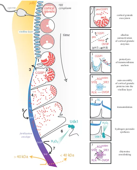
The fertilization envelope is constructed from two protein populations (Fig. 1). The unfertilized egg has a nascent extracel-lular matrix referred to as the vitelline layer, upon which other proteins are added during a secretory process that occurs after fertilization. Of the estimated twenty-five major glycoproteins in the sea urchin vitelline layer (Gache et al., 1983; Niman et al., 1984; Longo, 1981), two are known and, consistent with historical biochemical data, are sensitive to disulfide reducing agents (Aketa and Tsuzuki, 1968; Chandler and Heuser, 1980; Tegner and Epel, 1976; Epel et al., 1970; Carroll et al., 1977). The two vitelline layer proteins identified are enriched in CUB domains (originally found in complement factors Clr and Cls, urchin EGF, and bone morphogenetic protein 1), motifs of an anti-parallel β -strand fold that are stabilized by up to four positionally conserved disulfide bonds (Bork and Beckmann, 1993; Romero et al., 1997; Varela et al., 1997). These domains have been shown to homo-or heterodimerize to fhomo-orm carbohydrate-binding pockets homo-or pro-tein-interactive surfaces, and are found in extracellular proteins throughout animal phylogeny (Romero et al., 1997; Varela et al., 1997).
The two CUB-containing proteins within the vitelline layer include p160, a 160-kDa post protein clustered at the tips of microvilli (Haley and Wessel, 2004), and rendezvin120, the egg
extracellular matrix splice-variant of the oocyte-specific rendezvin gene (Wong and Wessel, 2006b) [see Box 1 below]. p160 con-tains 5 CUB domains and anchors the vitelline layer to the egg plasma membrane. Cleavage of this linker protein must occur in order for the vitelline layer scaffold to separate from the egg surface (Haley and Wessel, 2004). The protease responsible for this cleavage appears to be CGSP1, the serine protease that resides within the cortical granules (Haley and Wessel, 1999, 2004). Unlike the proteolyzed fragments of p160, rendezvin120 is
retained in the modified egg ECM after it lifts off the egg surface (Wong and Wessel, 2006b), serving as a core scaffold that organizes fertilization envelope assembly (Carroll et al., 1986; Ruiz-Bravo et al., 1986; Kay and Shapiro, 1985). It too has several CUB domains, and we hypothesize that it associates directly with p160 to remain anchored to the egg surface. This binding may occur directly through the CUB domains, forming heterodimers, or indirectly, by a lectin function of CUB dimers for binding to other glycoproteins (Romero et al., 1997; Varela et al., 1997; Bork and Beckmann, 1993). Different from most extracellular matrices, the vitelline layer of the egg is synthesized with the anticipated function of separating from the plasma membrane. Thus, the points of anchorage appear to be limited to enable rapid separa-tion of the vitelline layer scaffold from the egg surface at fertiliza-tion.
The second major contribution in construction of the fertiliza-tion envelope is the contents of the cortical granules. Cortical granules are secretory vesicles synthesized during oogenesis and released following gamete fusion (reviewed in Wessel et al., 2001). These oocyte- and egg-specific organelles number up to 15,000 per sea urchin egg (Laidlaw and Wessel, 1994), and are enriched within the distal region of the egg’s cortex, subjacent to the plasma membrane. Sea urchin cortical granule membranes are hemifused to the plasma membrane, making them primed for content release upon exposure to an appropriate concentration of their calcium trigger (Wong et al., 2007). The shear number of granules per egg and their rapid secretion en masse following fertilization implies that cortical granule contents can significantly alter the local extracellular environment upon exocytosis, easily transforming a sperm-competent egg vitelline layer into a physical barrier against additional sperm.
The major mass of protein released from the sea urchin cortical granules is non-enzymatic and contributes significantly to the permanent block to polyspermy. Of the 12 different proteins secreted by the cortical granules (Wessel et al., 2001), the six major proteins visible by Coomassie staining are directly incorpo-rated into the fertilization envelope (Wong and Wessel, 2004). Five genes encode these cortical granule proteins, including proteoliaisin (Somers et al., 1989; Somers and Shapiro, 1991), sfe1 (Wessel et al., 2000; Laidlaw and Wessel, 1994), sfe9 (Wessel, 1995; Laidlaw and Wessel, 1994), rendezvin (Wong and Wessel, 2006b) [see Box 1 below], and the enzyme ovoperoxidase (LaFleur et al., 1998; Nomura and Suzuki, 1995; Nomura et al., 1999). These six proteins rapidly self-assemble within the vitelline layer scaffold to form the fertilization envelope (Fig. 1). These high affinity interactions are likely due to the tandem arrangement of common protein-binding domains in all these proteins: proteoliaisin, SFE1, and SFE9 are abundant in low density lipoprotein receptor type A (LDLrA) repeats – containing up to 28 tandem LDLrA repeats, in some orthologs (Wessel et al., 2000; Wessel, 1995; Wong and Wessel, 2004) whereas the cortical granule members of the rendezvin family are, like their vitelline layer sibling
BOX 1: The rendezvin protein family has a complex series of processing and distribution events. The full-length rdz transcript is alternatively spliced into at least three forms, with the most abundant pool of mRNA encoding the parent of cortical granule-destined rendezvin60 and
rendezvin90 (Wong and Wessel, 2006b). Two significantly less-abundant transcripts are also created, encoding the vitelline layer-destined
rendezvin120. During posttranslational processing, S. purpuratus rendezvin60 is cleaved from its carboxy-terminal partners (cortical granule
rendezvin90 or vitelline layer rendezvin120). Differential trafficking of each variant follows secretory paths for their respective destinations.
CGSP1
Udx1
< 40 kDa
> 40 kDa
jelly
egg
cytoplasm
sperm
cortical
granule
H
2O
2time
vitelline layer
fertilization envelope
Y=Y
Q=K TG
OVOP
2
3
4
5
6
1
cortical granule exocytosis
alkaline autoactivation of cortical granule
enzymes
2
CGSP1 pro
(pH 5 pH 8)
OVOP ovop
transamidation
5
p
R-Q=K-R
TG
proteolysis of transmembrane
anchors
3
p 160
CGSP1
p160
hydrogen peroxide synthesis
Udx1 H2O2
6
dityrosine crosslinking
7
OVOP RDZ60&90
SFE1
PLN RDZ120 SFE9
1 proCGSP1 ovop
auto-assembly of cortical granule
proteins into the vitelline layer
4
OVOP RDZ60&90
SFE1
PLN
RDZ120
SFE9
7
rendezvin120, abundant in CUB domains (Wong and Wessel,
2006b) [see Box 1]. Although CUB domains and LDLrA repeats are common throughout the animal kingdom where they function in protein–protein binding, the fertilization envelope provides the first direct evidence that proteins with these two modules interact in a specific, high-affinity manner (Song et al., 2006).
Proteolysis is one enzymatic activity considered to be con-served in the block to polyspermy (Carroll and Epel, 1975b; 1975a; Runnstrom, 1966; Vacquier et al., 1973). Many functions have been ascribed to a cortical granule-derived protease, includ-ing removal of the sperm receptor, modification of the vitelline layer, and even egg activation (Carroll and Epel, 1975a; Carroll and Jaffe, 1995; Runnstrom, 1966; Vacquier et al., 1973) – yet it appears that only one enzyme is responsible for all activities. The cortical granule serine protease (CGSP1) is the only protease activity detected from sea urchin cortical granules (Carroll and Epel, 1975a; Haley and Wessel, 1999). Furthermore, CGSP1 activity at the egg surface appears to be selective, suggesting that it has specific roles and/or regulators that are not required at any other time during development (Haley and Wessel, 1999). Of the 200-300 estimated proteins at the egg cell surface, this protease cleaves and liberates peptides from approximately 15% of the total population (Carroll and Epel, 1981; Shapiro, 1975). Cleaving the vitelline post protein p160, for example, separates 85, 60, and 35 kDa peptides of the ectodomain from its transmembrane domain (Haley and Wessel, 2004), thus permitting the physical detachment of the vitelline layer from the egg surface during the formation of the fertilization envelope (Kay and Shapiro, 1985).
The sea urchin fertilization envelope is also mechanically transformed from a flexible network of glycoproteins into a hard-ened shell. This physical transformation is a consequence of enzymatic activity including transglutaminase and peroxidase. Transglutaminase catalyzes isopeptide amide bonds between glutamine and lysine, thereby fusing adjacent proteins to one another (Lorand and Graham, 2003; Battaglia and Shapiro, 1988; Nemes et al., 2005). This extended family of calcium-dependent enzymes generates intermolecular bonds through a cysteine-protease-like catalytic mechanism (reviewed in Lorand and Gra-ham, 2003; Nemes et al., 2005). A zymogenic form of transglutaminase appears to reside at the sea urchin egg surface and is activated within 1 minute following cortical granule exocy-tosis (Battaglia and Shapiro, 1988). This activity generates isopeptide bonds necessary for stabilizing the initial fertilization envelope assembly (Battaglia and Shapiro, 1988; Kay and Shapiro, 1985; Cariello et al., 1994). Curiously, transglutaminase activity requires cortical granule exocytosis, suggesting that its zymogen is activated by CGSP1 proteolysis – a similar phenomenon for the activation of homologs such as transglutaminase type 3 and plasma factor XIIIa (Lorand and Graham, 2003; Nemes et al., 2005).
While transglutaminase activity is necessary for fertilization envelope resilience (Cariello et al., 1994), matrix rigidity is prima-rily established by peroxidase activity (Showman and Foerder, 1979; Wong and Wessel, 2008). Peroxidases catalyze the forma-tion of dityrosine bonds between adjacent proteins through a free-radical intermediate that originates from hydrogen peroxide (Gross, 1959). The hydrogen peroxide is synthesized by members of a specific family of enzymes e.g. the Nox family (Lambeth, 2002; Lambeth et al., 2007). The source of the sea urchin hydrogen
peroxide is a single enzyme member of the dual oxidase (Duox) family of enzymes. This sea urchin dual oxidase Udx1 is present at the egg cell surface and is activated by calcium fluxes within the cytoplasm as a result of sperm activation (Wong et al., 2004). Duoxes are transmembrane enzymes with a peroxidase (hydro-gen peroxide consumer) domain at their amino termini, and an NADPH-dependent oxidase domain (hydrogen peroxide genera-tor) at their carboxy-termini (Lambeth et al., 2007; Lambeth, 2002). The function of the amino terminal peroxidase domain is unclear, but its similarity to catalases suggests that it may be important for neutralizing stray hydrogen peroxide before the reactive molecule can damage the embryo (Wong et al., 2004). The NADPH oxidase domain utilizes the cofactor NADPH as an electron donor, and is activated by calcium, presumably through the EF-hands located in the cytoplasm between the peroxidase and oxidase domains (Lambeth et al., 2007; Wong et al., 2004). While steric conformational changes that occur following calcium binding are hypothesized to enable enzyme activation, exactly how calcium regulates this or any other Duox is unknown.
In the sea urchin egg, the cortical granule-derived ovoperoxidase consumes the hydrogen peroxide generated by Udx1 in order to establish mechanical and chemical resilience in the sea urchin fertilization envelope (Showman and Foerder, 1979; Foerder and Shapiro, 1977; Hall, 1978; Bryan, 1970). This myeloperoxidase-like family of enzymes is specifically transcribed in oocytes, and packaged into cortical granules (LaFleur et al., 1998; Nomura and Suzuki, 1995). When released from the cortical granule lumen, ovoperoxidase is drawn away from the egg surface by the tether-ing protein, proteoliaisin, that keeps the enzyme associated with the elevating vitelline layer, thereby restricting its cross-linking activity to the ECM undergoing modification (Somers et al., 1989; Mozingo et al., 1994).
The chemistry of the peroxidase-mediated dityrosine crosslinking process was recently exploited to identify ovoperoxidase protein substrates within the fertilization envelope (Wong and Wessel, 2008). A fluorophore-conjugated tyramide molecule from the tyramide signal amplification mechanism (Bobrow et al., 1989) was used to compete with the formation of endogenous dityrosine crosslinks, thereby covalently labeling the protein substrate and enabling the proteins of the fertilization envelope to remain separable by SDS-PAGE. These targets were then identified by immunoblotting and tandem mass spectrom-etry. Surprisingly, these results show that the free-radical crosslinking mechanism in the sea urchin egg extracellular is remarkably selective and is capable of distinguishing target proteins derived from the same gene family. All of the LDLrA-containing proteins (proteoliaisin, SFE1, SFE9) are cross-linked to the vitelline layer rendezvin120 whereas the cortical
granule-derived rendezvin60 and rendezvin90 remain free, both intra- and
inter-molecularly (Wong and Wessel, 2008). The relationship between binding affinity and crosslinking of cortical granule pro-teins with rendezvin120 is consistent with the high percentage of
inter-actions dictate selectivity of crosslinking. Although the binding interactions between the different proteins that constitute the fertilization envelope are known, we do not yet know if the molecular structure of this matrix can distinguish between these two models describing how the specificity of free radical cross-linking is achieved.
Implications
At the level of the single cell, the paradoxical relationship between conservation of function and sequence radiation is best exemplified by the constituents of the physical block to polyspermy (Wong and Wessel, 2006a). Yet the implications for the simplicity of interacting partners and biochemical modifications that occur to this extracellar matrix extend well into embryogenesis and adult tissue. For example, the more high-affinity interacting domains a protein retains, the more freedom a structurally unrelated domain in that same protein would have to diverge without losing affinity for its original binding partner. Thus, minor alterations to an otherwise conserved protein foundation can greatly expand the functionality and flexibility of an extracellular matrix, possibly resulting in diversification and specialization of the tissues that rely on these evolving matrices. Some cases where this can be seen in action include protein superfamilies such as laminin, fibronectin, and collagen, whose members are used throughout the animal kingdom in matrices as diverse as the basal lamina to tendons and ligaments.
References
AKETA, K., AND TSUZUKI, H. (1968) Sperm-binding capacity of the S-S reduced
protein of the vitelline membrane of the sea urchin egg. Exp Cell Res
50:675-676.
BATTAGLIA, D.E., AND SHAPIRO, B.M. (1988) Hierarchies of protein cross-linking in the extracellular matrix: involvement of an egg surface transglutaminase in
early stages of fertilization envelope assembly. J Cell Biol 107:2447-2454.
BOBROW, M.N., HARRIS, T.D., SHAUGHNESSY, K.J., AND LITT, G.J. (1989) Catalyzed reporter deposition, a novel method of signal amplification.
Applica-tion to immunoassays. J Immunol Methods 125:279-285.
BORK, P., AND BECKMANN, G. (1993) The CUB domain. A widespread module
in developmentally regulated proteins. J Mol Biol 231:539-545.
BRIGGS, E., AND WESSEL, G.M. (2006) In the beginning.animal fertilization and
sea urchin development. Dev Biol 300:15-26.
BRYAN, J. (1970) On the reconstitution of the crystalline components of the sea
urchin fertilization membrane. J Cell Biol 45:606-614.
CARIELLO, L., ZANETTI, L., AND LORAND, L. (1994) Effects of inhibiting
transglutaminase during egg fertilization and development. Biochem Biophys
Res Commun 205:565-569.
CARROLL, D.J., ACEVEDO-DUNCAN, M., JUSTICE, R.W., AND SANTIAGO, L. (1986) Structure, assembly and function of the surface envelope (fertilization
envelope) from eggs of the sea urchins, Strongylocentrotus purpuratus. Adv
Exp Med Biol 207:261-291.
CARROLL, D.J., AND JAFFE, L.A. (1995) Proteases stimulate fertilization-like
responses in starfish eggs. Dev Biol 170:690-700.
CARROLL, E.J., JR., BYRD, E.W., AND EPEL, D. (1977) A novel procedure for obtaining denuded sea urchin eggs and observations on the role of the vitelline
layer in sperm reception and egg activation. Exp Cell Res 108:365-374.
CARROLL, E.J., JR., AND EPEL, D. (1975a) Elevation and hardening of the fertilization membrane in sea urchin eggs. Role of the soluble fertilization
product. Exp Cell Res 90:429-432.
CARROLL, E.J., JR., AND EPEL, D. (1975b) Isolation and biological activity of the
proteases released by sea urchin eggs following fertilization. Dev Biol 44:22-32.
CARROLL, E.J., JR., AND EPEL, D. (1981) Reevaluation of cell surface protein release at fertilization and its role in regulation of sea urchin egg protein
synthesis. Dev Biol 87:374-378.
CHANDLER, D.E., AND HEUSER, J. (1980) The vitelline layer of the sea urchin egg and its modification during fertilization. A freeze-fracture study using
quick-freezing and deep-etching. J Cell Biol 84:618-632.
CHANG, R.L., UEKI, I.F., TROY, J.L., DEEN, W.M., ROBERTSON, C.R., AND BRENNER, B.M. (1975) Permselectivity of the glomerular capillary wall to
macromolecules. II. Experimental studies in rats using neutral dextran. Biophys
J 15:887-906.
DERBÈS, A. (1847) Observations sur le mecanisme et les phenomenes qui
accompagnent la formation de l’embryon chezl’oursin comestible. Ann Sci Nat
Zool 8:80-98.
EPEL, D., WEAVER, A.M., AND MAZIA, D. (1970) Methods for revoval of the vitelline membrane of sea urchin eggs. I. Use of dithiothreitol (Cleland Reagent).
Exp Cell Res 61:64-68.
FOERDER, C.A., AND SHAPIRO, B.M. (1977) Release of ovoperoxidase from sea
urchin eggs hardens the fertilization membrane with tyrosine crosslinks. Proc
Natl Acad Sci USA 74:4214-4218.
GACHE, C., NIMAN, H.L., AND VACQUIER, V.D. (1983) Monoclonal antibodies to the sea urchin egg vitelline layer inhibit fertilization by blocking sperm adhesion.
Exp Cell Res 147:75-84.
GROSS, A.J. (1959) The oxidation of tyramine, tyrosine, and related compounds by
peroxidase. J Biol Chem 234:1611-1614.
HALEY, S.A., AND WESSEL, G.M. (1999) The cortical granule serine protease CGSP1 of the sea urchin, Strongylocentrotus purpuratus, is autocatalytic and
contains a low-density lipoprotein receptor-like domain. Dev Biol 211:1-10.
HALEY, S.A., AND WESSEL, G.M. (2004) Proteolytic cleavage of the cell surface protein p160 is required for detachment of the fertilization envelope in the sea
urchin. Dev Biol 272:191-202.
HALL, H.G. (1978) Hardening of the sea urchin fertilization envelope by
peroxidase-catalyzed phenolic coupling of tyrosines. Cell 15:343-355.
HARVEY, E.N. (1909) The mechanism of membrane formation and other early changes in developing sea urchins’ eggs as bearing on the problem of artificial
parthenogenesis. J Exp Zool 8:355-376.
JUST, E.E. (1939) The Biology of the Cell Surface. P. Blackiston’s Song &
Company, Philadelphia, PA.
KAY, E.S., AND SHAPIRO, B.M. (1985) The formation of the fertilization membrane of the sea urchin egg. In: Metz, C.B., and Monroy, A. (eds) Biology of fertilization.
Academic Press Inc., Orlando, FL, pp. 45-80.
LAFLEUR, G.J., JR., HORIUCHI, Y., AND WESSEL, G.M. (1998) Sea urchin ovoperoxidase: oocyte-specific member of a heme-dependent peroxidase
superfamily that functions in the block to polyspermy. Mech Dev 70:77-89.
LAIDLAW, M., AND WESSEL, G.M. (1994) Cortical granule biogenesis is active
throughout oogenesis in sea urchins. Development 120:1325-1333.
LAMBETH, J.D. (2002) Nox/Duox family of nicotinamide adenine dinucleotide
(phosphate) oxidases. Curr Opin Hematol 9:11-17.
LAMBETH, J.D., KAWAHARA, T., AND DIEBOLD, B. (2007) Regulation of Nox and
Duox enzymatic activity and expression. Free Radic Biol Med 43:319-331.
LEPAGE, T., AND GACHE, C. (1989) Purification and characterization of the sea
urchin embryo hatching enzyme. J Biol Chem 264:4787-4793.
LEPAGE, T., AND GACHE, C. (1990) Early expression of a collagenase-like
hatching enzyme gene in the sea urchin embryo. Embo J 9:3003-3012.
LONGO, F.J. (1981) Effects of concanavalin A on the fertilization of sea urchin eggs.
Dev Biol 82:197-202.
LORAND, L., AND GRAHAM, R.M. (2003) Transglutaminases: crosslinking
en-zymes with pleiotropic functions. Nat Rev Mol Cell Biol 4:140-156.
MOZINGO, N.M., HOLLAR, L.R., AND CHANDLER, D.E. (1993) Degradation of an extracellular matrix: sea urchin hatching enzyme removes cortical
granule-derived proteins from the fertilization envelope. J Cell Sci 104 (Pt 3):929-938.
MOZINGO, N.M., SOMERS, C.E., AND CHANDLER, D.E. (1994) Ultrastructure of the proteoliaisin-ovoperoxidase complex and its spatial organization within the
Strongylocentrotus purpuratus fertilization envelope. J Cell Sci 107 (Pt
10):2769-2777.
Structure-function relationships of transglutaminases—a contemporary view. Prog Exp Tumor Res 38:19-36.
NIMAN, H.L., HOUGH-EVANS, B.R., VACQUIER, V.D., BRITTEN, R.J., LERNER, R.A., AND DAVIDSON, E.H. (1984) Proteins of the sea urchin egg vitelline layer.
Dev Biol 102:390-401.
NOMURA, K., HOSHINO, K., AND SUZUKI, N. (1999) The primary and higher order structures of sea urchin ovoperoxidase as determined by cDNA cloning and
predicted by homology modeling. Arch Biochem Biophys 367:173-184.
NOMURA, K., SHIMIZU, T., KINOH, H., SENDAI, Y., INOMATA, M., AND SUZUKI, N. (1997) Sea urchin hatching enzyme (envelysin): cDNA cloning and
depriva-tion of protein substrate specificity by autolytic degradadepriva-tion. Biochemistry
36:7225-7238.
NOMURA, K., AND SUZUKI, N. (1995) Sea urchin ovoperoxidase: solubilization and isolation from the fertilization envelope, some structural and functional
properties, and degradation by hatching enzyme. Arch Biochem Biophys
319:525-534.
PATARI-SAMPO, A., IHALMO, P., AND HOLTHOFER, H. (2006) Molecular basis of the glomerular filtration: nephrin and the emerging protein complex at the
podocyte slit diaphragm. Ann Med 38:483-492.
ROMERO, A., ROMAO, M.J., VARELA, P.F., KOLLN, I., DIAS, J.M., CARVALHO, A.L., SANZ, L., TOPFER-PETERSEN, E., AND CALVETE, J.J. (1997) The
crystal structures of two spermadhesins reveal the CUB domain fold. Nat Struct
Biol 4:783-788.
RUIZ-BRAVO, N., ROSSIGNOL, D.P., DECKER, G., ROSENBERG, L.I., AND LENNARZ, W. (1986) Characterization of the Strongylocentrotus purpuratus
egg cell surface receptor for sperm. Adv Exp Med Biol 207:293-313.
RUNNSTROM, J. (1966) The vitelline membrane and cortical particles in sea urchin
eggs and their function in maturation and fertilization. Adv Morphog 5:221-325.
SCHUEL, H., KELLY, J.W., BERGER, E.R., AND WILSON, W.L. (1974) Sulfated acid mucopolysaccharides in the cortical granules of eggs. Effects of quaternary
ammonium salts on fertilization. Exp Cell Res 88:24-30.
SHAPIRO, B.M. (1975) Limited proteolysis of some egg surface components is an early event following fertilization of the sea urchin, Strongylocentrotus purpuratus.
Dev Biol 46:88-102.
SHOWMAN, R.M., AND FOERDER, C.A. (1979) Removal of the fertilization
membrane of sea urchin embryos employing aminotriazole. Exp Cell Res
120:253-255.
SOMERS, C.E., BATTAGLIA, D.E., AND SHAPIRO, B.M. (1989) Localization and developmental fate of ovoperoxidase and proteoliaisin, two proteins involved in
fertilization envelope assembly. Dev Biol 131:226-235.
SOMERS, C.E., AND SHAPIRO, B.M. (1991) Functional domains of proteoliaisin,
the adhesive protein that orchestrates fertilization envelope assembly. J Biol
Chem 266:16870-16875.
SONG, J., L, WONG, J., L, AND WESSEL, G., M, (2006) Oogenesis: Single cell
development and differentiation. Dev Biol 300:385-405.
TEGNER, M.J., AND EPEL, D. (1976) Scanning electron microscope studies of sea
urchin fertilization. I. Eggs with vitelline layers. J Exp Zool 197:31-57.
VACQUIER, V.D., TEGNER, M.J., AND EPEL, D. (1973) Protease released from sea urchin eggs at fertilization alters the vitelline layer and aids in preventing
polyspermy. Exp Cell Res 80:111-119.
VARELA, P.F., ROMERO, A., SANZ, L., ROMAO, M.J., TOPFER-PETERSEN, E., AND CALVETE, J.J. (1997) The 2.4 A resolution crystal structure of boar seminal plasma PSP-I/PSP-II: a zona pellucida-binding glycoprotein heterodimer
of the spermadhesin family built by a CUB domain architecture. J Mol Biol
274:635-649.
WESSEL, G.M. (1995) A protein of the sea urchin cortical granules is targeted to
the fertilization envelope and contains an LDL-receptor-like motif. Dev Biol
167:388-397.
WESSEL, G.M., BROOKS, J.M., GREEN, E., HALEY, S., VORONINA, E., WONG, J., ZAYDFUDIM, V., AND CONNER, S. (2001) The biology of cortical granules.
Int Rev Cytol 209:117-206.
WESSEL, G.M., CONNER, S., LAIDLAW, M., HARRISON, J., AND LAFLEUR, G.J., JR. (2000) SFE1, a constituent of the fertilization envelope in the sea urchin is made by oocytes and contains low-density lipoprotein-receptor-like
repeats. Biol Reprod 63:1706-1712.
WONG, J., L, AND WESSEL, G., M, (2008) Free-radical crosslinking of specific proteins alters the function of the egg extracellular matrix at fertilization.
Development 135: 431-440.
WONG, J.L., CRÉTON, R., AND WESSEL, G.M. (2004) The oxidative burst at
fertilization is dependent upon activation of the dual oxidase Udx1. Dev Cell
7:801-814.
WONG, J.L., KOPPEL, D.E., COWAN, A.E., AND WESSEL, G.M. (2007)
Mem-brane hemifusion is a stable intermediate of exocytosis. Dev Cell 12:653-659.
WONG, J.L., AND WESSEL, G.M. (2004) Major components of a sea urchin block
to polyspermy are structurally and functionally conserved. Evol Dev 6:134-153.
WONG, J.L., AND WESSEL, G.M. (2006a) Defending the zygote: Search for the
ancestral animal block to polyspermy. Curr Top Dev Biol 72:1-151.
WONG, J.L., AND WESSEL, G.M. (2006b) Rendezvin: An essential gene encoding independent, differentially-secreted egg proteins that organize the fertilization
Related, previously published Int. J. Dev. Biol. articles
See our recent Special Issue Developmental Biology in Poland edited by Tarkowski, Maleszewski and Kloc at: http://www.ijdb.ehu.es/web/contents.php?vol=52&issue=2-3
See our recent Special Issue Ear Development edited by Fernando Giraldez and Bernd Fritzsch at: http://www.ijdb.ehu.es/web/contents.php?vol=51&issue=6-7
Mammalian fertilization: the egg's multifunctional zona pellucida Paul M. Wassarman and Eveline S. Litscher
Int. J. Dev. Biol. (2008) Vol. 52 (doi: 10.1387/ijdb.072524pw)
2006 ISI **Impact Factor = 3.577**
Allorecognition mechanisms during ascidian fertilization
Yoshito Harada and Hitoshi Sawada
Int. J. Dev. Biol. (2008) Vol. 52: (doi: 10.1387/ijdb.072544yh)
Distinct mechanisms underlie sperm-induced and protease-induced oolemma block to sperm penetration.
Sebastian Komorowski, Katarzyna Szczepanska and Marek Maleszewski Int. J. Dev. Biol. (2003) 47: 65-69
Sperm-egg interaction at fertilization: glycans as recognition signals.
F Rosati, A Capone, C D Giovampaola, C Brettoni and R Focarelli Int. J. Dev. Biol. (2000) 44: 609-618
Calcium in sea urchin egg during fertilization.